Article

Strong Photoluminescence Enhancement from Bilayer Molybdenum Disulfide via the Combination of UV Irradiation and Superacid Molecular Treatment
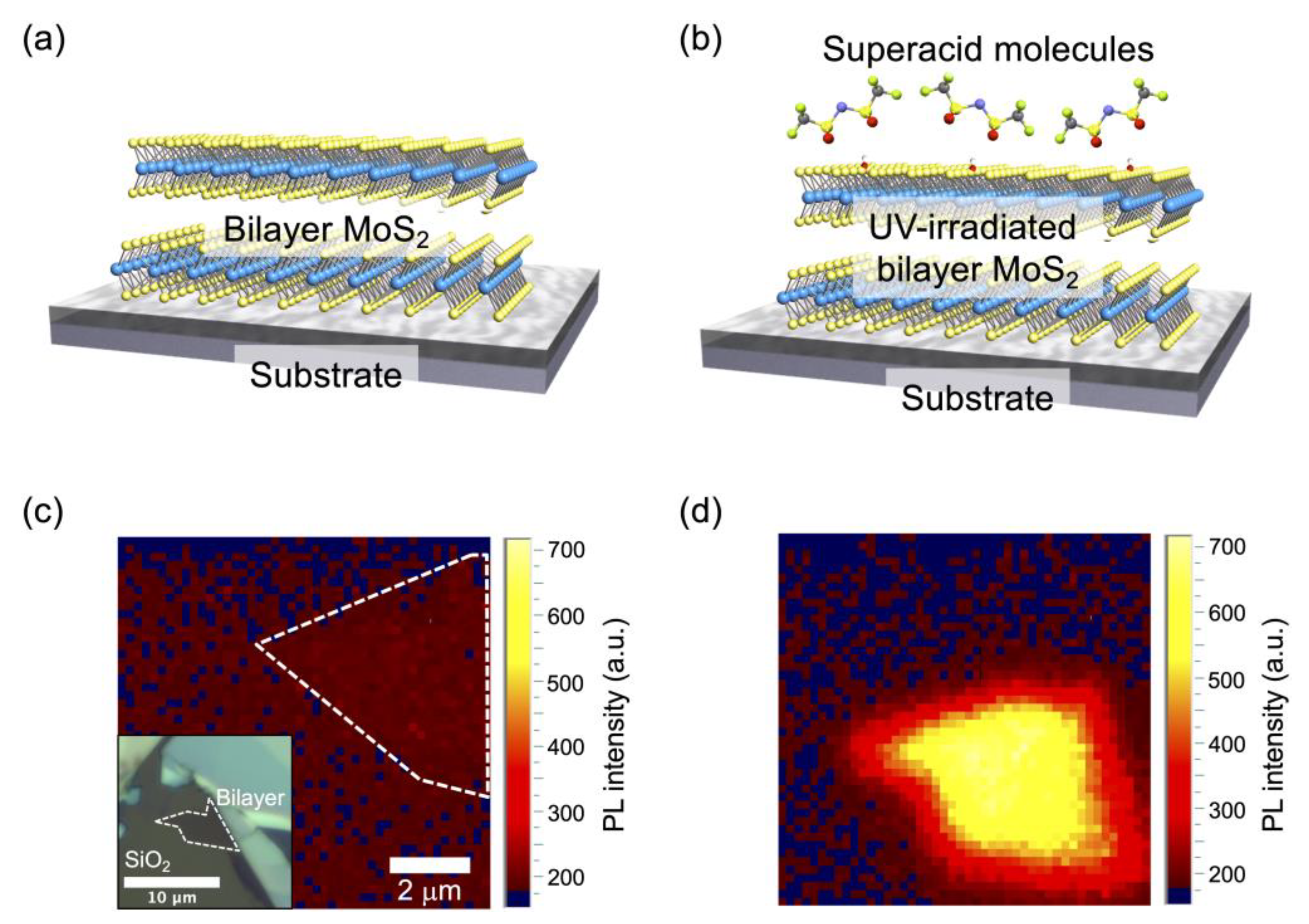
Department of Physics and Electronics, Osaka Prefecture University, 1-1 Gakuen-cho, Naka-ku, Sakai-shi, Osaka 599-8531, Japan
*
Author to whom correspondence should be addressed.
Appl. Sci. 2021, 11(8), 3530; https://doi.org/10.3390/app11083530
Submission received: 4 March 2021 / Revised: 1 April 2021 / Accepted: 7 April 2021 / Published: 15 April 2021

Abstract

:
A direct band gap nature in semiconducting materials makes them useful for optical devices due to the strong absorption of photons and their luminescence properties. Monolayer transition metal dichalcogenides (TMDCs) have received significant attention as direct band gap semiconductors and a platform for optical applications and physics. However, bilayer or thicker layered samples exhibit an indirect band gap. Here, we propose a method that converts the indirect band gap nature of bilayer MoS2, one of the representative TMDCs, to a direct band gap nature and enhances the photoluminescence (PL) intensity of bilayer MoS2 dramatically. The procedure combines UV irradiation with superacid molecular treatment on bilayer MoS2. UV irradiation induces the conversion of the PL property with an indirect band gap to a direct band gap situation in bilayer MoS2 when the interaction between the top and bottom layers is weakened by a sort of misalignment between them. Furthermore, the additional post-superacid treatment dramatically enhances the PL intensity of bilayer MoS2 by a factor of 700×. However, this procedure is not effective for a conventional bilayer sample, which shows no PL enhancement. From these results, the separated top layer would show a strong PL from the superacid treatment. The monolayer-like top layer is physically separated from the substrate by the intermediate bottom MoS2 layer, and this situation would be preferable for achieving a strong PL intensity. This finding will be useful for controlling the optoelectronic properties of thick TMDCs and the demonstration of high-performance optoelectronic devices.

1. Introduction

Transition metal dichalcogenides (TMDCs) with a formula of MX2 (M: Mo and W; X: S, Se, and Te) have garnered attention as atomically thin optoelectronic materials [1,2,3,4]. Various optical devices have been demonstrated, including phototransistors [5,6], photodetectors [7,8], valley-dependent optoelectronics [9,10,11], light-emitting diodes [12,13], and photovoltaics [14,15,16]. So far, monolayer (ML) TMDCs have been widely studied for application in optoelectronic devices because of their direct band gap nature at K and K’ points in the Brillouin zone in the ML [17]. The layer-dependent electronic structure of TMDCs is well known, and the bilayer and thicker layered TMDCs show an indirect band gap nature [17,18]. The Г point in the Brillouin zone increases with increasing the thickness, and both the conduction minimum and the K-Г points decrease with increasing the number of layers [17,18]. Photoluminescence (PL) shows clearly the difference in the electronic structure between ML and bilayer TMDCs [18]. In the case of a representative TMDC, MoS2, the ML shows a single PL spectrum around 1.9 eV corresponding to the direct band gap, while the bilayer shows two main peaks around 1.6 eV and 1.9 eV for the coexistence of indirect and direct band gaps, respectively [18]. The PL intensity shown in the bilayer is usually very low compared with ML because of the dominant indirect band gap character.
Recently, chemical treatment on ML TMDCs has been reported, and a large enhancement of the PL intensity was demonstrated [19,20,21,22,23]. In the case of ML MoS2, treatment by oxidative chemicals such as tetracyanoquinodimethane [19] and quinone [20] derivatives, acids such as sulfuric acid [24], and hydrogen peroxide [25] show a large enhancement of the PL intensity. One of the strongest modulating agents of PL intensity in ML MoS2 is a superacid molecule, bis(trifluoromethane)sulfonimide (TFSI), for both mechanically exfoliated and chemical-vapor-deposited ML flakes [26,27,28,29,30]. The PL enhancement mechanism is still under debate, but the existence of TFSI molecules on ML TMDC would be essential for achieving the strong PL intensity. Recently, we reported the consistent substantial PL enhancement of ML MoS2 via a combination of TFSI treatment and additional UV irradiation [31]. In this work, we found an unusually strong PL enhancement from bilayer MoS2 by combining the TFSI treatment with UV irradiation (Figure 1 and Figure 2). The PL enhancement is over several hundred and some of them over 700× from the as-exfoliated bilayer flakes, and the shape of the PL spectrum dramatically changes to a sort of direct band gap. The plausible mechanism would be that the separation into a monolayer-like state would happen under the process. Furthermore, the indirect-to-direct band gap transition was only observed at the specific bilayer sample which would be like twisted bilayers. This work would facilitate controlling the interlayer interaction in TMDCs for modulating electronic structures to achieve atomically thin optoelectronic devices and physics platforms.

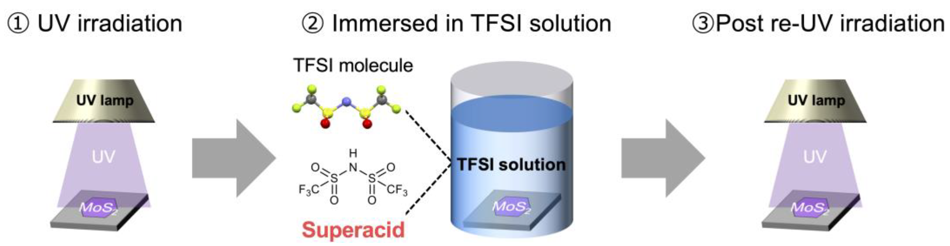
2. Materials and Methods

Bulk MoS2 was purchased from Structure Probe, Inc. (SPI Supplies). The bilayer flakes were prepared on Si wafer with 260 nm thermally grown SiO2 (Silicon Valley Microelectronics, Inc.) via the mechanical exfoliation process. A weight (~3.0 kg cm−2) was used for the exfoliation process (Figure 1, Figure 3, Figure 4 and Figure 5), pressing it over an exfoliation tape. In the case shown in Figure 6, the pressing weight was not applied. The TFSI molecule was purchased from Sigma-Aldrich, and acetonitrile (Kanto Chemical Co., Inc. Japan) was used as the solvent for the TFSI treatment. The TFSI solution (2 mg mL−1) was prepared under an Ar atmosphere in a glove box (UNICO LTd. Japan, UL-1000A), and the TFSI treatment procedure was carried out in an ambient environment. The illustrative image of the process is shown in Figure 2. The UV irradiation process was performed for 5 min each (Figure 2, Figure 3, Figure 4 and Figure 5). The exfoliated samples on the Si wafer were immersed in the solution at room temperature for 10 min in the normal treatment process (Figure 4). In the case shown in Figure 6, the total UV irradiation times were 15 min (5 min × 3) for the initial UV irradiation procedure and 15 min (5 min × 3) for the last repeated UV irradiation process to sufficiently modulate the bilayer sample. The lamp house (high-pressure mercury lamp, 170 mW cm−2) was placed 2 cm (~4 cm from the lamp) above the Si wafer. PL spectra were measured using a LabRAM HR800 equipped with an EMCCD camera (HORIBA Scientific, Japan). The wavelength was 532 nm for all measurements. The power of the incident beam was ~2.7 W cm−2 for measurements of PL and Raman spectra in Figure 6b and 27 W cm−2 for other Raman spectra and PL mapping in Figure 1. Atomic force microscope (AFM) images were obtained using the dynamic force mode in SII instruments.

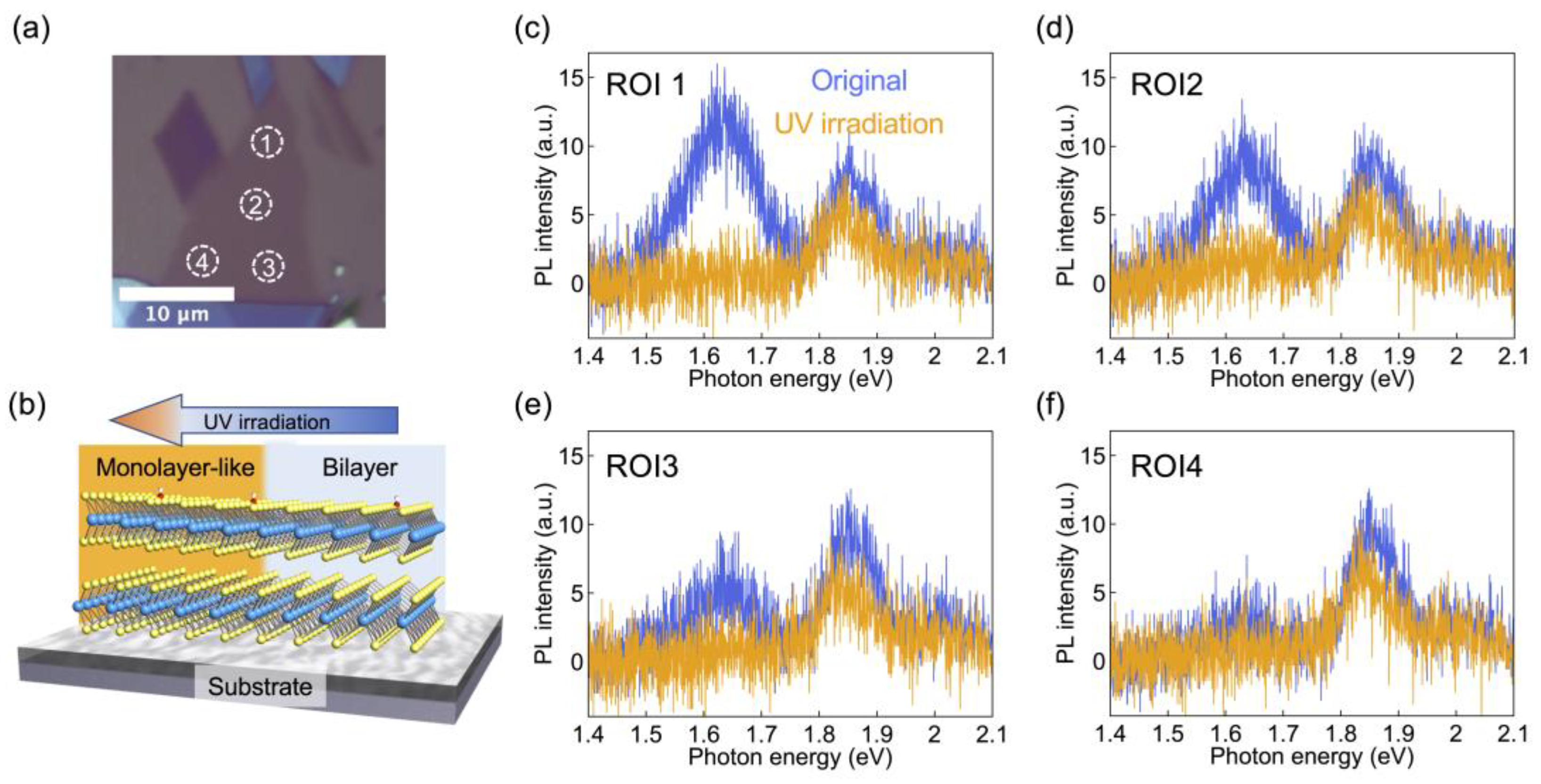
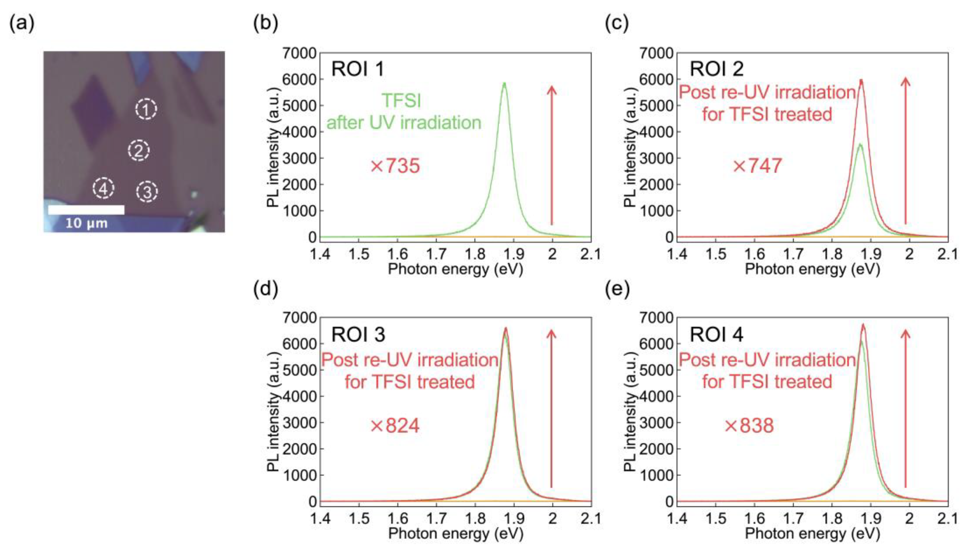
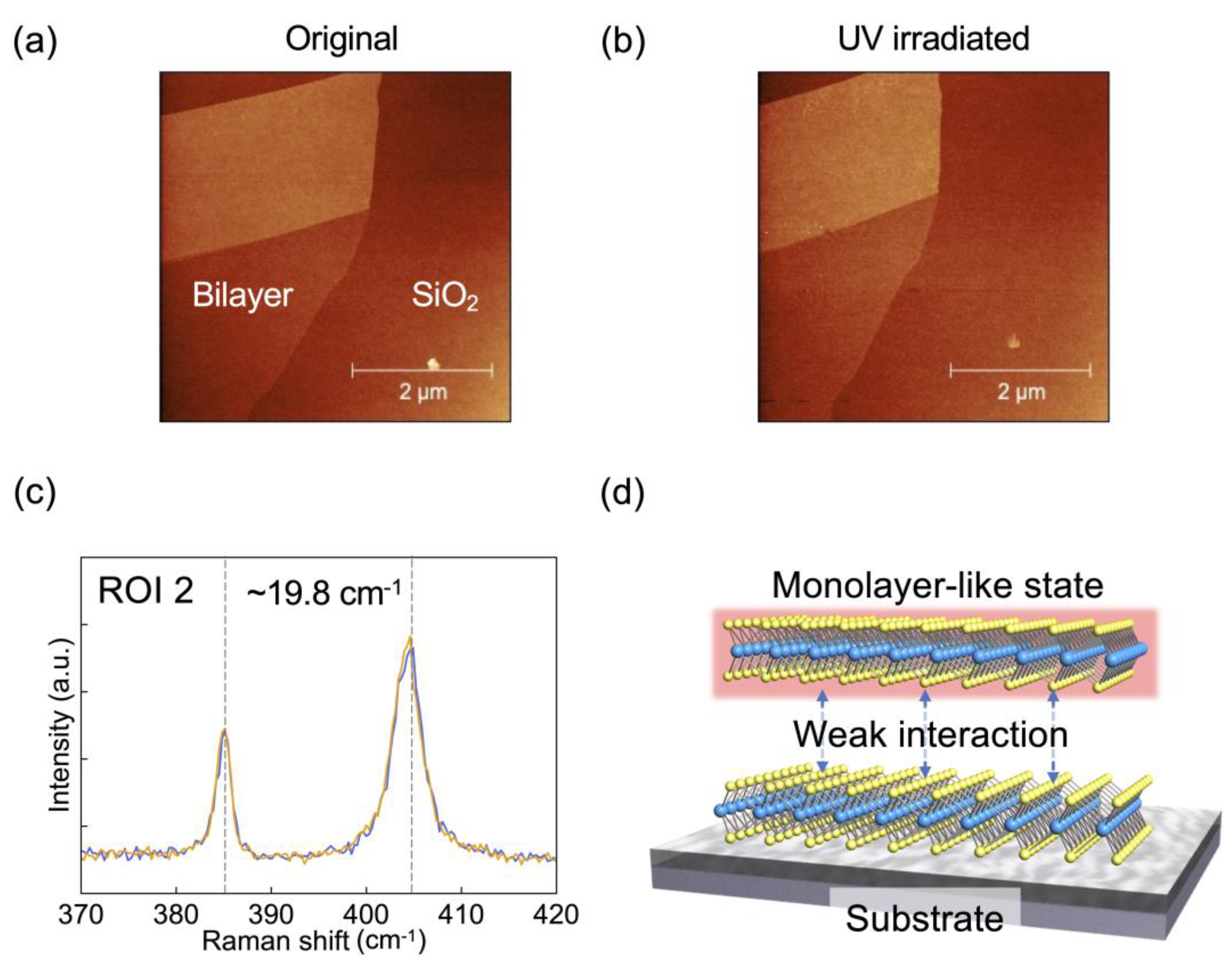
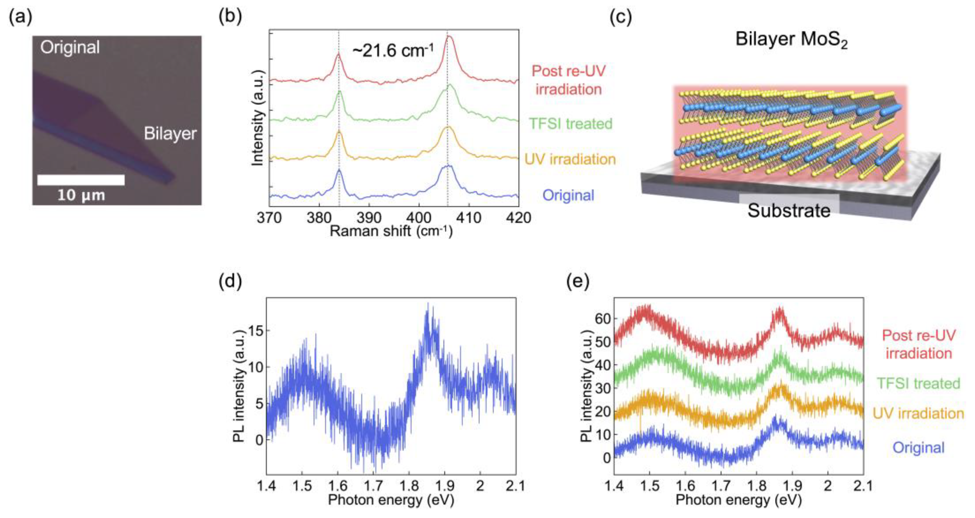
3. Results and Discussion

The sample targeted in this research is illustrated in Figure 1a,b. The bilayer MoS2 flakes were prepared on the Si wafer covered with 260 nm SiO2 via an exfoliation process. The flake was exfoliated with pressing the weight as shown in the experimental section. The TFSI molecular treatment was conducted along with the previously reported process; details are the combination of the UV irradiation and immersion into the TFSI acetonitrile solution [31]. Figure 1c,d show PL mapping images for the as-exfoliated and the processed bilayer MoS2, respectively. The bilayer MoS2 shows a considerable PL enhancement, more than 100× around 1.9 eV, corresponding to a plausible direct band gap nature as seen in monolayers, via the TFSI molecular treatment with UV irradiation.
We examined each process for the PL enhancement of the bilayer MoS2. The UV irradiation process was carried out under an ambient condition, which may generate OH radicals on the surface of MoS2 (Figure 3). In the previous work, an electronic structure of ML MoS2 was maintained under UV irradiation, even while some chemical reactions happened on the surface [31,32]. Here, a laterally large (>10 μm) bilayer MoS2 was applied to the UV irradiation process for 5 min; as a result, the whole area, regions of interest (ROIs) 1–4 in Figure 3, showed a more direct band gap nature with a single peak around 1.86 eV. The clear signal around 1.64 eV, corresponding to an indirect band gap, was diminished via the UV irradiation. According to the above result, UV irradiation would induce the separation of the top and bottom layers to achieve a ML-like state (Figure 3b).
We examined the ML-like sample with UV irradiation by further applying the TFSI treatment (Figure 4). This treatment changed the PL signal dramatically over the whole area (Figure 4b–e). All regions (ROIs 1–4) showed a direct band gap with a single PL signal around 1.88 eV by the TFSI treatment. The factor of the PL enhancement was over 700× from the as-exfoliated bilayer flakes. Therefore, the combination of UV irradiation and the TFSI treatment would be useful for modulating the optical property of bilayer MoS2 to have a strong PL intensity like that of the TFSI-treated ML samples reported previously.
The supplemental UV irradiation was applied to the TFSI-treated bilayer MoS2 but was found not to be so effective (re-UV irradiation for TFSI treated in Figure 4). This indicates that the combination of the first UV irradiation and the TFSI treatment process would achieve saturation of the PL enhancement. In the previous report, the UV irradiation was applied after the TFSI treatment and was effective in enhancing the PL intensity for ML MoS2 flakes [31]. We confirmed the same procedure as in the previous report for bilayer MoS2; a bilayer MoS2 was treated first with TFSI solution, then by UV irradiation (Figure S1). The TFSI treatment itself changed the bilayer-like nature to a ML-like nature, but the PL intensity was still poor. The subsequent UV irradiation strongly enhanced the PL intensity, and the factor of the enhancement was several hundred times with a single signal around 1.9 eV. Therefore, the sequence of the TFSI treatment and the UV irradiation does not matter for achieving the PL enhancement; rather, the combination of both processes is the essence of obtaining the strong PL in the bilayer MoS2 flakes.
Figure 5 shows atomic force microscope (AFM) images for the as-exfoliated (original) and UV-irradiated bilayer MoS2. The morphologies of bilayer MoS2 maintain the structure via the UV irradiation process. Raman spectra for the sample maintain typical E2g and A1g modes for lateral and vertical vibration signals of MoS2. However, the peak separation of the two signals is less than 20 cm−1. In previous work, it was found that the peak separation of E2g and A1g modes is strongly dependent on the interlayer stacking of the bilayer, and the peak separation reduces if the bilayer is twisted [33,34,35]. We hypothesize that the bilayer sample might twist under the exfoliation process, because we pressed the exfoliation tape onto a substrate with a weight (~3.0 kg cm−2) when exfoliating the flake. MoS2 is well known as a lubricant, and sliding between layers would happen with low friction [36]. This twisting would require more study in order to completely understand the situation. Importantly, the Raman signal is maintained after the UV irradiation process over the bilayer flake (Figure 5c and Figure S2), even though the character of the PL spectra converted from an indirect to a direct band gap nature, as shown in Figure 3.
The unusual event shown in the indirect-to-direct band gap conversion for the bilayer MoS2 would be explained by the interlayer separation between the top and bottom layers (Figure 5d). Previously, a systematic study of twisted bilayer MoS2 showed different PL and Raman signals [33,34,35]. By twisting the top and bottom layers, the peak separation of E2g and A1g reduces, as observed in our case. In addition, the peak position of the PL signal corresponding to the indirect band gap upshifts about 0.1 eV. Furthermore, the interlayer separation is also dependent on the interlayer stacking of the bilayer. If the bilayer is twisted, weak interaction between the top and bottom layers is expected. This scenario requires more study, but it would be a plausible explanation for the strong PL intensity from the bilayer MoS2. Another probable scenario to explain our observations would be etching the top layer and thinning it to a ML of MoS2 via UV irradiation, because a laser-thinning process has been reported to reduce the thickness of MoS2 [37,38,39]. However, the morphology and color of the sample are maintained in the process, as shown in Figure 5. It also would be hard to imagine the uniform etching of the whole region of the flake; therefore, we think the structure is maintained in the process.
To further confirm the process with a conventional, non-twisted bilayer MoS2. We prepared bilayer MoS2 without using the weight simply by contacting bulk MoS2 with a Si/SiO2 wafer and exfoliating a bilayer flake (Figure 6a). The Raman spectrum showed E2g and A1g signals with a peak separation of 21.6 cm−1, corresponding to a typical (non-twisted) bilayer MoS2 (Figure 6b,c) [19,40,41]. The PL measurement for the bilayer MoS2 showed an indirect band gap around 1.5 eV and a direct band gap nature around 1.86 eV, respectively. The indirect band gap obtained without the weight showed a downshift from the bilayer signal exfoliated via pressing with the weight. These PL and Raman tendencies indicate a non-twisted bilayer MoS2 via the exfoliation process without pressing with the weight. Then, the non-twisted bilayer flake was applied with the whole process of UV irradiation, TFSI treatment, and supplemental UV irradiation. Raman spectra showed almost no change in the process, even though the signal of the A1g showed a bit of an upshift because of the surface oxidation by the TFSI treatment. The PL signal also maintained the structure during the whole process. Therefore, the conventional, non-twisted bilayer flake did not show the conversion to a direct band gap. As in the above considerations, the observed direct band gap conversion in the bilayer MoS2 would be a sort of separation of the top and bottom layers. One explanation is that the conversion would happen in the twisted bilayer case because of the weak interaction between the top and bottom layers, and it facilitates the separation to the ML-like situation by the external perturbations.

4. Conclusions

We demonstrated a substantial PL enhancement in the bilayer MoS2. The original bilayer MoS2 showed a weak PL intensity with the signal of the indirect band gap. A key finding in this paper is that by both UV irradiation and TFSI treatment, the bilayer MoS2 demonstrates a ML-like PL with a single signal at the direct band gap. In addition, the combined process induces more than several hundred times PL enhancement from the bilayer MoS2 compared to the as-exfoliated bilayer flakes. The enhancement mechanism requires more study, but it would involve the separation of the top and bottom layers in the bilayer MoS2. This situation would be due to a sort of misalignment of the top and bottom layers caused by pressing the weight and sliding each layer in the exfoliation process. If the bilayer is twisted, the interlayer separation between the top and bottom layers would happen easily, which would lead to an indirect-to-direct band gap conversion via the process. These phenomena are also supported by treating a conventional bilayer MoS2 flake; there is no PL enhancement in the conventional case. This technique is useful for preparing high-optical-quality MoS2 materials and devices. Since the sample is bilayer, the top layer is separated from the substrate; the bottom MoS2 layer would serve as an interface layer between the top layer with a strong PL and the substrate. This structure would be effective in removing substrate effects. This work would open another strategy to obtain a strong PL intensity from 2D materials and be useful in optical device applications.

Supplementary Materials

The following are available online at https://www.mdpi.com/article/10.3390/app11083530/s1, Figure S1: PL enhancement via the process of TFSI treatment followed by UV irradiation for the bilayer MoS2 and Figure S2: Raman spectra for the UV-irradiated bilayer MoS2.

Author Contributions

Conducted all experiments, Y.Y.; designed the experiments and wrote the manuscript, Y.Y. and D.K. All authors discussed continuously and contributed to the writing of the manuscript. All authors have read and agreed to the published version of the manuscript.

Funding

This research was funded by JSPS KAKENHI (20H02574), the Research Foundation for Opto-Science and Technology, the Konica Minolta Science and Technology Foundation, and the Sumitomo Foundation.

Institutional Review Board Statement

This project does not include biological items requiring ethical approval.

Informed Consent Statement

It is not required.

Data Availability Statement

The study data is available from the corresponding author on request.

Conflicts of Interest

The authors declare no conflict of interest.

References

  1. Wang, Q.H.; Kalantar-Zadeh, K.; Kis, A.; Coleman, J.N.; Strano, M.S. Electronics and optoelectronics of two-dimensional transition metal dichalcogenides. Nat. Nanotechnol. 2012, 7, 699. [Google Scholar] [CrossRef]
  2. Chhowalla, M.; Shin, H.S.; Eda, G.; Li, L.J.; Loh, K.P.; Zhang, H. The chemistry of two-dimensional layered transition metal dichalcogenide nanosheets. Nat. Chem. 2013, 5, 263. [Google Scholar] [CrossRef] [PubMed]
  3. Geim, A.K.; Grigorieva, I.V. Van der Waals heterostructures. Nature 2013, 499, 419. [Google Scholar] [CrossRef] [PubMed]
  4. Jariwala, D.; Sangwan, V.K.; Lauhon, L.J.; Marks, T.J.; Hersam, M.C. Emerging device applications for semiconducting two-dimensional transition metal dichalcogenides. ACS Nano 2014, 8, 1102. [Google Scholar] [CrossRef] [PubMed] [Green Version]
  5. Lee, H.S.; Min, S.W.; Chang, Y.G.; Park, M.K.; Nam, T.; Kim, H.; Kim, J.H.; Ryu, S.; Im, S. MoS2 nanosheet phototransistors with thickness-modulated optical energy gap. Nano Lett. 2012, 12, 3695. [Google Scholar] [CrossRef] [PubMed]
  6. Yin, Z.; Li, H.; Li, H.; Jiang, L.; Shi, Y.; Sun, Y.; Lu, G.; Zhang, Q.; Chen, X.; Zhang, H. Single-Layer MoS2 Phototransistors. ACS Nano 2012, 6, 74. [Google Scholar] [CrossRef] [PubMed] [Green Version]
  7. Wang, X.; Wang, P.; Wang, J.; Hu, W.; Zhou, X.; Guo, N.; Huang, H.; Sun, S.; Shen, H.; Lin, T.; et al. Ultrasensitive and Broadband MoS2 Photodetector Driven by Ferroelectrics. Adv. Mater. 2015, 27, 6575. [Google Scholar] [CrossRef] [Green Version]
  8. Lopez-Sanchez, O.; Lembke, D.; Kayci, M.; Radenovic, A.; Kis, A. Ultrasensitive photodetectors based on monolayer MoS2. Nat. Nanotechnol. 2013, 8, 497. [Google Scholar] [CrossRef]
  9. Mak, K.F.; He, K.; Shan, J.; Heinz, T.F. Control of valley polarization in monolayer MoS2 by optical helicity. Nat. Nanotechnol. 2012, 7, 494. [Google Scholar] [CrossRef]
  10. Zeng, H.; Dai, J.; Yao, W.; Xiao, D.; Cui, X. Valley polarization in MoS2 monolayers by optical pumping. Nat. Nanotechnol. 2012, 7, 490. [Google Scholar] [CrossRef]
  11. Onga, M.; Zhang, Y.; Ideue, T.; Iwasa, Y. Exciton Hall effect in monolayer MoS2. Nat. Mater. 2017, 16, 1193. [Google Scholar] [CrossRef] [PubMed]
  12. Ross, J.S.; Klement, P.; Jones, A.M.; Ghimire, N.J.; Yan, J.; Mandrus, D.G.; Taniguchi, T.; Watanabe, K.; Kitamura, K.; Yao, W.; et al. Electrically tunable excitonic light-emitting diodes based on monolayer WSe2 p–n junctions. Nat. Nanotechnol. 2014, 9, 268. [Google Scholar] [CrossRef] [PubMed]
  13. Lien, D.-H.; Amani, M.; Desai, S.B.; Ahn, G.H.; Han, K.; He, J.-H.; Ager, J.W.; Wu, M.C.; Javey, A. Large-area and bright pulsed electroluminescence in monolayer semiconductors. Nat. Commun. 2018, 9, 1229. [Google Scholar] [CrossRef] [PubMed]
  14. Wong, J.; Jariwala, D.; Tagliabue, G.; Tat, K.; Davoyan, A.R.; Sherrott, M.C.; Atwater, H.A. High Photovoltaic Quantum Efficiency in Ultrathin van der Waals Heterostructures. ACS Nano 2017, 11, 7230. [Google Scholar] [CrossRef] [PubMed]
  15. Tsai, M.-L.; Su, S.-H.; Chang, J.-K.; Tsai, D.-S.; Chen, C.-H.; Wu, C.-I.; Li, L.-J.; Chen, L.-J.; He, J.-H. Monolayer MoS2 Heterojunction Solar Cells. ACS Nano 2014, 8, 8317. [Google Scholar] [CrossRef]
  16. Tsai, M.-L.; Li, M.-Y.; Retamal JR, D.; Lam, K.-T.; Lin, Y.-C.; Suenaga, K.; Chen, L.-J.; Liang, G.; Li, L.-J.; He, J.-H. Single Atomically Sharp Lateral Monolayer p-n Heterojunction Solar Cells with Extraordinarily High Power Conversion Efficiency. Adv. Mater. 2017, 29, 1701168. [Google Scholar] [CrossRef]
  17. Splendiani, A.; Sun, L.; Zhang, Y.; Li, T.; Kim, J.; Chim, C.-Y.; Galli, G.; Wang, F. Emerging Photoluminescence in Monolayer MoS2. Nano Lett. 2010, 10, 1271–1275. [Google Scholar] [CrossRef] [PubMed]
  18. Mak, K.F.; Lee, C.; Hone, J.; Shan, J.; Heinz, T.F. Atomically Thin MoS2: A New Direct-Gap Semiconductor. Phys. Rev. Lett. 2010, 105, 136805. [Google Scholar] [CrossRef] [Green Version]
  19. Mouri, S.; Miyauchi, Y.; Matsuda, K. Tunable photoluminescence of monolayer MoS2 via chemical doping. Nano Lett. 2013, 13, 5944–5948. [Google Scholar] [CrossRef] [Green Version]
  20. Ichimiya, H.; Fukui, A.; Aoki, Y.; Yamada, Y.; Yoshimura, T.; Ashida, A.; Fujimura, N.; Kiriya, D. Solvent engineering for strong photoluminescence enhancement of monolayer molybdenum disulfide in redox-active molecular treatment. Appl. Phys. Exp. 2019, 12, 051014. [Google Scholar] [CrossRef]
  21. Yao, H.; Liu, L.; Wang, Z.; Li, H.; Chen, L.; Pam, M.E.; Chen, W.; Yang, H.Y.; Zhang, W.; Shi, Y. Significant photoluminescence enhancement in WS2 monolayers through Na2S treatment. Nanoscale 2018, 10, 6105–6112. [Google Scholar] [CrossRef]
  22. Han, H.V.; Lu, A.Y.; Lu, L.S.; Huang, J.K.; Li, H.; Hsu, C.L.; Lin, Y.C.; Chiu, M.H.; Suenaga, K.; Chu, C.W.; et al. Photoluminescence enhancement and structure repairing of monolayer MoSe2 by hydrohalic acid treatment. ACS Nano 2016, 10, 1454–1461. [Google Scholar] [CrossRef] [Green Version]
  23. Tanoh AO, A.; Alexander-Webbe, J.; Xiao, J.; Delport, G.; Williams, C.A.; Bretscher, H.; Gauriot, N.; Allardice, J.; Pandya, R.; Fan, Y.; et al. Enhancing Photoluminescence and Mobilities in WS2 Monolayers with Oleic Acid Ligands. Nano Lett. 2019, 19, 6299–6307. [Google Scholar] [CrossRef] [PubMed] [Green Version]
  24. Kiriya, D.; Hijikata, Y.; Pirillo, J.; Kitaura, R.; Murai, A.; Ashida, A.; Yoshimura, T.; Fujimura, N. Systematic Study of Photoluminescence Enhancement in Monolayer Molybdenum Disulfide by Acid Treatment. Langmuir 2018, 34, 10243–10249. [Google Scholar] [CrossRef] [PubMed]
  25. Su, W.; Dou, H.; Li, J.; Huo, D.; Dai, N.; Yang, L. Tuning photoluminescence of single-layer MoS2 using H2O2. RSC Adv. 2015, 5, 82924. [Google Scholar] [CrossRef]
  26. Amani, M.; Taheri, P.; Addou, R.; Ahn, G.H.; Kiriya, D.; Lien, D.H.; Ager, J.W.; Wallace, R.M.; Javey, A. Recombination Kinetics and Effects of Superacid Treatment in Sulfur- and Selenium-Based Transition Metal Dichalcogenides. Nano Lett. 2016, 16, 2786–2791. [Google Scholar] [CrossRef] [PubMed]
  27. Amani, M.; Burke, R.A.; Ji, X.; Zhao, P.; Lien, D.H.; Taheri, P.; Ahn, G.H.; Kirya, D.; Ager, J.W.; Yablonovitch, E.; et al. High Luminescence Efficiency in MoS2 Grown by Chemical Vapor Deposition. ACS Nano 2016, 10, 6535–6541. [Google Scholar] [CrossRef]
  28. Kim, H.; Lien, D.H.; Amani, M.; Ager, J.W.; Javey, A. Highly Stable Near-Unity Photoluminescence Yield in Monolayer MoS2 by Fluoropolymer Encapsulation and Superacid Treatment. ACS Nano 2017, 11, 5179–5185. [Google Scholar] [CrossRef]
  29. Cadiz, F.; Tricard, S.; Gay, M.; Lagarde, D.; Wang, G.; Robert, C.; Renucci, P.; Urbaszek, B.; Marie, X. Well separated trion and neutral excitons on superacid treated MoS2 monolayers. Appl. Phys. Lett. 2016, 108, 251106. [Google Scholar] [CrossRef] [Green Version]
  30. Zeng, Y.; Chen, W.; Tang, B.; Liao, J.; Lou, J.; Chen, Q. Synergetic photoluminescence enhancement of monolayer MoS2: Via surface plasmon resonance and defect repair. RSC Adv. 2018, 8, 23591–23598. [Google Scholar] [CrossRef] [Green Version]
  31. Yamada, Y.; Shinokita, K.; Okajima, Y.; Takeda, S.N.; Matsushita, Y.; Takei, K.; Yoshimura, T.; Ashida, A.; Fujimura, N.; Matsuda, K.; et al. Photoactivation of Strong Photoluminescence in Superacid-Treated Monolayer Molybdenum Disulfide. ACS Appl. Mater. Interfaces 2020, 12, 36496–36504. [Google Scholar] [CrossRef]
  32. Detailes are shown on a submitted manuscript. Under review.
  33. Huang, S.; Liang, L.; Ling, X.; Puretzky, A.A.; Geohegan, D.B.; Sumpter, B.G.; Kong, J.; Meunier, V.; Dresselhaus, M.S. Low-Frequency Interlayer Raman Modes to Probe Interface of Twisted Bilayer MoS2. Nano Lett. 2016, 16, 1435–1444. [Google Scholar] [CrossRef] [PubMed]
  34. Liu, K.; Zhang, L.; Cao, T.; Jin, C.; Qiu, D.; Zhou, Q.; Zettl, A.; Yang, P.; Louie, S.G.; Wang, F. Evolution of interlayer coupling in twisted molybdenum disulfide bilayers. Nat. Commun. 2014, 5, 4966. [Google Scholar] [CrossRef]
  35. Liao, M.; Wei, Z.; Du, L.; Wang, Q.; Tang, J.; Yu, H.; Wu, F.; Zhao, J.; Xu, X.; Han, B.; et al. Precise control of the interlayer twist angle in large scale MoS2 homostructures. Nat. Commun. 2020, 11, 2153. [Google Scholar] [CrossRef] [PubMed]
  36. Li, H.; Wang, J.; Gao, S.; Chen, Q.; Peng, L.; Liu, K.; Wei, X. Superlubricity between MoS2 Monolayers. Adv. Mater. 2017, 29, 1701474. [Google Scholar] [CrossRef] [PubMed]
  37. Hu, L.; Shan, X.; Wu, Y.; Zhao, J.; Lu, X. Laser Thinning and Patterning of MoS2 with Layer-by-Layer Precision. Sci. Rep. 2017, 7, 15538. [Google Scholar] [CrossRef] [PubMed]
  38. Kim, H.-J.; Yun, Y.J.; Yi, S.N.; Chang, S.K.; Ha, D.H. Changes in the Photoluminescence of Monolayer and Bilayer Molybdenum Disulfide during Laser Irradiation. ACS Omega 2020, 5, 7903. [Google Scholar] [CrossRef] [PubMed] [Green Version]
  39. Tessarek, C.; Gridenco, O.; Wiesing, M.; Müssener, J.; Figge, S.; Sebald, K.; Gutowski, J.; Eickhoff, M. Controlled Laser-Thinning of MoS2 Nanolayers and Transformation to Amorphous MoOx for 2D Monolayer Fabrication. ACS Appl. Nano Mater. 2020, 3, 7490–7498. [Google Scholar] [CrossRef]
  40. Niu, Y.; Gonzalez-Abad, S.; Frisenda, R.; Marauhn, P.; Drüppel, M.; Gant, P.; Schmidt, R.; Taghavi, N.S.; Barcons, D.; Molina-Mendoza, A.J.; et al. Thickness-Dependent Differential Reflectance Spectra of Monolayer and Few-Layer MoS2, MoSe2, WS2 and WSe2. Nanomaterials 2018, 8, 725. [Google Scholar] [CrossRef] [Green Version]
  41. Lee, C.; Yan, H.; Brus, L.E.; Heinz, T.F.; Hone, J.; Ryu, S. Anomalous Lattice Vibrations of Single- and Few-Layer MoS2. ACS Nano 2010, 4, 2695. [Google Scholar] [CrossRef] [Green Version]
Figure 1. (a,b) Illustrative image of (a) the bilayer MoS2 and (b) the bis(trifluoromethane)sulfonimide (TFSI) treatment for the UV-irradiated bilayer MoS2. (c,d) Photoluminescence (PL) mapping images for (c) the as-exfoliated bilayer MoS2 and (d) the TFSI-treated sample for the UV-irradiated bilayer MoS2. The inset in Figure 1c is the optical microscope image for the targeted bilayer sample.
Applsci 11 03530 g001
Figure 2. Illustrative image of the process applied to the bilayer MoS2.
Applsci 11 03530 g002
Figure 3. (a) Optical microscope image of the bilayer MoS2. The labels are the measurement regions (regions of interest (ROIs) 1–4) in Figure 3c–f. (b) Illustrative image of the modulation to the bilayer sample via the UV irradiation process. The process induces the bilayer to a monolayer-like state. (cf) PL spectra for the as-exfoliated (original, blue) and the UV-irradiated (orange) samples in (c) ROI 1, (d) ROI 2, (e) ROI 3, and (f) ROI 4 in Figure 3a.
Applsci 11 03530 g003
Figure 4. (a) Optical microscope image of the bilayer MoS2 after the TFSI treatment followed by the UV irradiation. (be) PL spectra for the TFSI treatment (green) and the second (re-)UV irradiation (red) in (b) ROI 1, (c) ROI 2, (d) ROI 3, and (e) ROI 4 in Figure 4a. The values in Figure 4b–e are the magnitude of the PL enhancement in the sample over the as-exfoliated (original) sample.
Applsci 11 03530 g004
Figure 5. (a,b) Atomic force microscope (AFM) images for (a) the as-exfoliated (original) bilayer MoS2 exfoliated with the weight and (b) the UV-irradiated bilayer MoS2. (c) Raman spectra for the original (blue) and the UV-irradiated (orange) bilayer MoS2 in ROI 2 in Figure 3. (d) Illustrative image of a plausible state of the bilayer MoS2. The top layer would show a monolayer-like state due to the weak interaction between the top and bottom layers by a sort of misalignment of each layer.
Applsci 11 03530 g005
Figure 6. (a) Optical microscope image of the sample exfoliated without pressing with the weight. (b) Raman spectra for the as-exfoliated (original, blue), UV-irradiated (orange), TFSI-treated (green), and re-UV-irradiated (red) bilayer MoS2. The peak separation between E2g and A1g is about 21.6 cm−1. (c) The plausible situation of the bilayer MoS2 from the Raman spectra in Figure 6b. This bilayer is a conventional flake. (d,e) PL spectra for the bilayer MoS2 corresponds to Figure 6b; the original is shown in Figure 6d.
Applsci 11 03530 g006
Publisher’s Note: MDPI stays neutral with regard to jurisdictional claims in published maps and institutional affiliations.

