Article

Early Loading of Titanium Dental Implants with Hydroxyl Ion Modified Surface: A 12-Month Prospective Clinical Trial

by
Maciej Krawiec
1,
Jakub Hadzik
1,*,
Marzena Dominiak
1,
Wojciech Grzebieluch
2,
Artur Błaszczyszyn
1 and
Paweł Kubasiewicz-Ross
1
1
Oral Surgery Department, Wrocław Medical University, Krakowska st. 26, 54-207 Wrocław, Poland
2
Digital Dentistry Laboratory, Faculty of Medicine and Dentistry, Department of Conservative Dentistry with Endodontics, Wroclaw Medical University, Krakowska st. 26, 54-207 Wrocław, Poland
*
Author to whom correspondence should be addressed.
Appl. Sci. 2021, 11(7), 2958; https://doi.org/10.3390/app11072958
Submission received: 21 February 2021 / Revised: 20 March 2021 / Accepted: 22 March 2021 / Published: 25 March 2021
(This article belongs to the Special Issue Materials for Bone and Dental Hard Tissue Substitutes)

Abstract

:

Featured Application

The following paper presents the issue of early loading of dental implants. Nowadays, the most frequently used prosthetic protocol is loading the implant within 12 weeks. It is believed that the recently introduced nano-scale modification of the implant’s surface will allow for faster final loading. Faster final loading reduces the overall treatment time which is even more crucial in the case of aesthetic zone rehabilitation.

Abstract

(1) Background: implant surface topology and active hydrophilic ions could have some benefit on implant osteointegration and stability; (2) methods: 40 adult patients, suffering from a single missing tooth in the aesthetic zone, were enrolled in the study. Each patient had a single titanium implant (Thommen SPI®lement) inserted. The implant surface was obtained through conditioning using the Apliquiq system. Patients were divided into two equal groups depending on the implant’s diameter (3.5 and 4.0 mm). Each implant was loaded within four weeks. Stability levels, using the Ostell device, were checked immediately after implant placement and in four weeks; additionally, marginal bone loss (MBL) was calculated based on 12 months; (3) results: all implants survived the study. The average primary stability achieved for both groups was initially 71.59 ISQ (±4.04) and declined to 69.94 ISQ (±3.29) in four weeks. The average MBL was 0.2 mm (±0.88). There were no statistically important differences between groups. There was a positive correlation between the patient’s age and implant stability quotient (ISQ) values; (4) conclusions: hydrophilic surface implants can be used in a protocol for early functional occlusal loading. Higher values of primary stability positively influence the values of secondary stability, and the age of the patient affects the values of implant stability.

1. Introduction

In the case of implant treatment, which involves the aesthetic zone of the maxilla, immediate or early loading of an implant is extremely important, not only for functionality, but primarily for aesthetic reasons. For decades, high primary stability has been the key factor in determining the early loading of an implant and the success of a treatment [1,2].
Osteointegration has been defined as a direct and functional connection between the bone and an artificial implant [2]. Primary stability is the grade of engagement of an implant in the bone structure immediately after insertion. It is one of the main conditions for the osteointegration process [1,2,3]. The factors that influence the values of primary stability can be divided into three main groups. The first one is the quantitative and qualitative characteristics of the bone structure into which the implant is inserted [4]. Another factor is the method of implant bed preparation [5]. The final factor affecting primary stability are the characteristics of an implant, which include both the macroscopic and microscopic features of the implant surface [6]. The macroscopic features of an implant relate to aspects such as the dimensions, shape, diameter and thread pitch of the implant. The microscopic features are the overall characteristics of the surface, primarily including the grade of the roughness of the surface [1,2,3,6].
In the context of osteointegration, much attention has been paid in recent years to the physical and chemical aspects of dental implant surfaces. These include electrochemical potential, surface wettability, thickness of the titanium dioxide (TiO2) layer, ion adhesion, active peptides, growth factors, and antibiotics. Currently, work is still underway to develop a type of surface that would allow for even faster loading of an implant even when primary stability is low [7,8,9]. One such potential method is the chemical modification of the surface using hydroxyl ion. With such a modification, the titanium surface of an implant has a negative electrochemical potential. The negative potential determines the improvement of the osteointegration process at all stages, starting from better stability of blood clot and adhesion of Ca++ ions in the first hours after the loading of a dental implant, through to a better adhesion of proteins (fibronectin, osteocalcin) and the cells that determine the process of osteointegration (mesenchymal stem cells (MSC)) in the later stages of the process [10,11].
In the case of implant treatment within the aesthetic zone, a temporary restoration, usually a removable one, is needed. It causes many difficulties for the patient. Therefore, the methods for shortening the healing period and loading an implant as early as possible are constantly being sought after. The first reports claiming a high rate of success concerning early loading of an implant date back to the 1990s and refer to the Branemark implant system [9,10].
For decades, implant loading after six weeks was considered an early loading. However, it seems that the key period for the process of osteointegration and secondary stability is in the third and fourth week. During that time, the process of mineralization of the primary osseous tissue takes place, and thus, the bone tissue that surrounds an implant achieves the mechanical values that enable loading. Therefore, it seems reasonable to load an implant even faster, i.e., after 3–4 weeks of healing [9,10,11,12,13,14,15].
The main objective of the study was to assess the marginal bone loss and stability of the early loaded Thommen Incell®SPI implants using the single non-splinted screw-retained final chairside-prepared prosthetic restoration. The secondary objective was to evaluate the influence of an implant’s diameter on the mentioned parameters.

2. Materials and Methods

2.1. Inclusion and Exclusion Criteria

In this study, 40 patients aged over 18, partially edentulous within the aesthetic zone, were enrolled. The patients could not have any active periodontal disease or an approximal plague index (API) > 25%. The patients were subjected to clinical and radiological examinations. The minimum alveolar ridge dimension in the lingual–buccal aspect was 6.5–7 mm in the region of interest, so the implant could be placed in the native bone. Furthermore, the bone density in the region of the implant insertion had to be D2 or D3 according to Misch et al. [16]. Patients were randomly divided into 2 separate groups depending on the implant diameter used (3.5 and 4.0 mm).
(a)
group 1 (G2; n = 20 patients)—3.5 mm diameter implants were used
(b)
group 2 (G3; n = 20 patients)—4.0 mm diameter implants were used
The procedures of the guided bone regeneration were not performed neither before nor during the implant placement. Furthermore, at least 3 months for the healing period after extraction were established.
Exclusion criteria were:
  • systemic or local diseases that could compromise healing or osteointegration,
  • heavy smokers,
  • patients with bruxism,
  • pregnancy,
  • breastfeeding.

2.2. Protocol of the Experiment

The schedule of visits included:
  • consultation visit: qualification of the patient for the surgery, clinical and radiological examination CBCT (cone-beam computed tomography) (Galileos®D3437, Sirona Dental, Erlangen, Germany), API assessment;
  • implantation: intraoperative and postoperative RVG (radiovisiography) (Planmeca OY, Helsinki, Finland), torque values, primary stability assessment using Ostell ISQ (Osstell; Integration Diagnostics, Gothenburg, Sweden);
  • 4 weeks after the implantation: assessment of stability with the use of Ostell ISQ, intraoral scan, placement of prosthetic, RVG;
  • 12 months after the surgery: clinical and radiological assessment (RVG and CBCT).
The research was performed in accordance with the conditions of declaration of Helsinki and with the approval of the Local Ethical Committee (229/2019). The personal data protection procedures (GDPR) were complied with. The patients signed two written consents: first, a general consent for the implant treatment, and second, consent for participation in the study.

2.3. Implants

The cylindrical dental implants, Thommen Innicell®SPI Element MC Innicel (Thommen Medical AG, Grenchen, Switzerland) were used for the surgery. The superhydrophilic implant surface was obtained through NaOH conditioning using the Apliquiq system (Thommen Medical AG, Grenchen, Switzerland). The length of the inserted implants ranged from 8 mm to 11 mm and depended on the height of the bone base, while the diameter of the implant was determined by the width of the alveolar processes.

2.4. Surgical Phase

The implant surgery was performed with antibiotic cover, one-shot therapy: 1 dose of clindamycin 600 mg (MIP Pharma, Gdansk, Poland). Infiltration anesthesia was applied using Septanest 1:100,000 (SEPTODONT 58, Saint Maur des Fossés, France) with the Wand STA device (Milestone Scientific, Inc., Roseland, NJ, USA). A diamond drill was used for deepithelialization and a blade (no. 15C) was used for an H-shaped papilla-preservation incision, shifted palatially. Next, each implant was inserted at bone level, according to the procedure provided by the manufacturer. Subsequently, the primary stability was assessed using Ostell ISQ. The measurements were performed three times in the mesiodistal, buccal and palatal, as well as periapical direction measurements with the application of Ostel smartpeg for the Thommen implants. The smallest value was considered to be the cut-off point. Open healing was used with a standard healing screw. The partially deepithelized flap was repositioned and stabilized with 0–5 simple interrupted sutures (Seralene
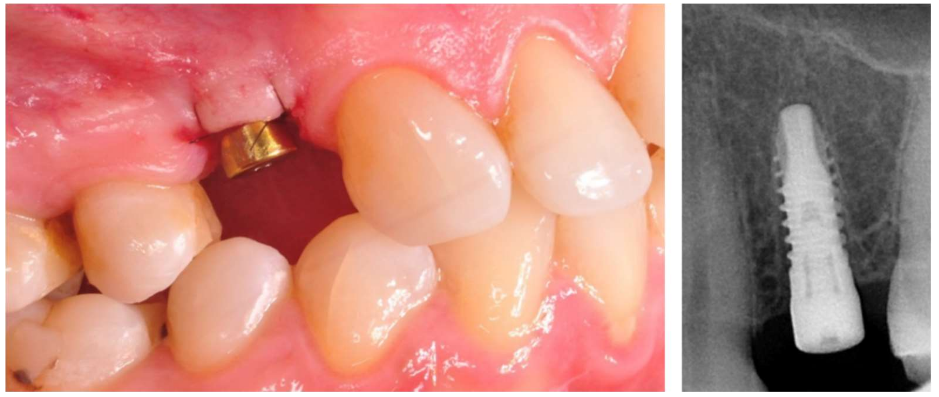
®, Serag Wiessner, Naila, Germany). At the end of the surgery, a RVG image was taken to assess the correctness of the inserted implant (Figure 1). The X-ray tubehead was aimed at right angles (vertically and horizontally) to both the implant and the sensor. A paralleling device was used for this purpose. The surgeries were performed by three members of the team: M.K., J.H. and A.B. Postoperative recommendations included analgesic and anti-inflammatory treatment with Nimesil (Laboratories Menarini SA, Barcelona, Spain) at 200 mg/per day, and rinsing the oral cavity with Eludril Classic (Pierre Fabre S.A, Paris, France) 3 times a day.

2.5. Prosthetic Phase

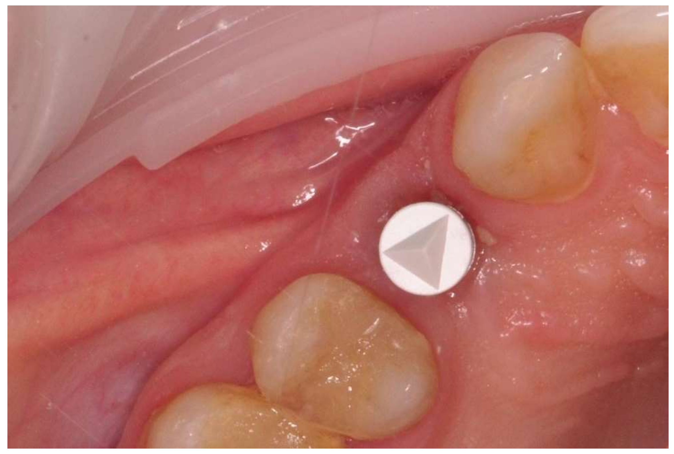
The prosthetic restoration stage started 4 weeks after the implant placement surgery and was prepared in the chairside laboratory by W.G. Patients with no signs of inflammation in the direct vicinity of the implant and with an ISQ (implant’s stability quotient) value of 65 or greater were allowed to participate in the prosthetic protocol. The measurements using the aforementioned device were performed three times in the mesiodistal, buccal and palatal, as well as periapical direction, and the smallest value was considered the cut-off point. Screw-retained implant crowns made of lithium disilicate glass-ceramics, IPS e max CAD LT (Ivoclar Vivadent AG, Schaan, Liechtenstein), were used as the prosthetic restoration materials. After the removal of the healing abutment, the implant bed was cleaned. The scans were taken with an intraoral scanner Sirona Cerec AC Bluecam (DentsplySirona, York, PA, USA) (
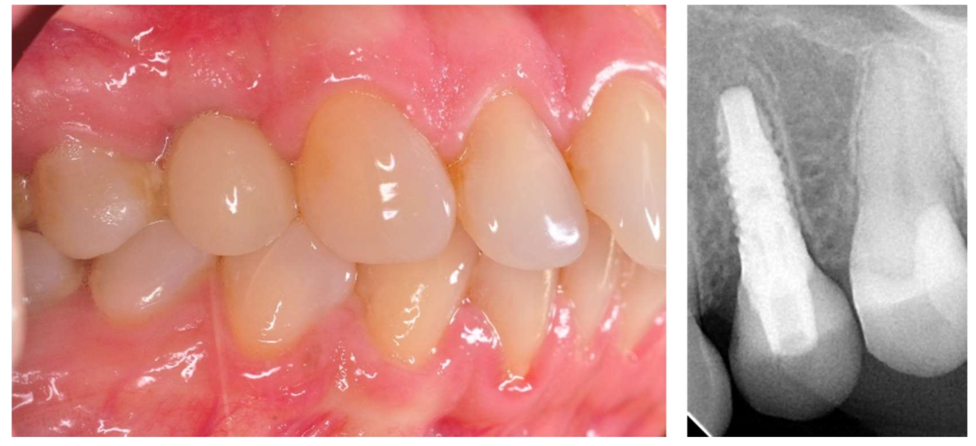
Figure 2). Subsequently, the crown internal surface was etched and then fixed using Multilink Hybrid Abutment cement (Ivoclar Vivadent AG, Schaan, Liechtenstein) on the previously sandblasted titanium base (TiBase) for Sirona Cerec (DentsplySirona, York, PA, USA). The crown was then screwed onto the implant with a force of 25 Ncm. The occluding relations were controlled using articulating paper (Bausch®, Cologne, Germany) with a thickness of 200, 80, and 8 μm. The hole was filled in with Gradia composite (GC Corporation, Tokyo, Japan) and an RVG image was taken (Figure 3). The patients were instructed on proper hygiene around the dental implant.

2.6. Assessment of Implant’s Stability

Values of the implant’s stability quotient (ISQ) were obtained immediately after implant placement (primary stability) and after 4 weeks (secondary stability). For every series of resonance frequency analysis (RFA) measurements, the ISQ values were recorded using an Osstell device in three different directions: vertical, buccal and palatal. A transducer (Smartpegs) was attached to the implant, and ISQs ranging from 1 to 100 were recorded. The Osstells were brought into very close contact with the Smartpegs without touching them, until an audible signal confirmed that the measurement had been taken.

2.7. MBL (Marginal Bone Loss) Assessment Using the Radiological Examination

Before surgery and during the 12-month follow-up, CBCT was performed to assess the marginal bone loss (MBL). The MBL was calculated as follows: first, dimensions were calibrated by the known parameters of the implant diameter and length. Starting from the implant shoulder, distances were measured to the mesial and distal points of the bone to implant contact, parallel to the implant axis. All measurements were taken by P.KR, a member of the research group who was not involved directly in the preparation of the implant.

2.8. Statistical Analyses

To answer the research questions, statistical analyses were performed using the IBM SPSS Statistics 25 software (IBM, New York, USA). The software was used to analyze the basic descriptive statistics together with the Shapiro–Wilk test. To examine the differences between two or more groups, a non-parametric equivalent of variance analysis, the Kruskal–Wallis test, was used. Dunn’s test with the Bonferroni correction was chosen for post hoc comparisons. The relationships between continuous variables were examined by calculating Pearson’s linear correlation coefficient. The value of α = 0.05 was assumed as the significance level.
To check the distribution of continuous variables and to study their compliance with a normal distribution, basic descriptive statistics were used, and the Shapiro–Wilk test of normal distribution was performed. For nominal variables, the frequency and the percentage of individual values in the entire observation pool were calculated. The results were presented separately for the three groups. In the first group, all observations were taken into consideration while the second group included only those observations for which an implant diameter = 3.5 mm, and in the third group the implant diameter = 4.0 mm.

3. Results

3.1. General Data

The average age of all patients (groups one and two combined) was 41.55 (±8.85). Group two consisted of younger patients (40.15 ± 5.38) and group one included generally older patients (42.99 ± 11.30).
Out of 40 implants placed in the surgical phase, each implant achieved a high level of primary stability with an ISQ value of 65 or greater. Subsequently, all patients were admitted to the prosthetic phase. Furthermore, all implants successfully survived the 12-month follow-up period.

3.2. Results of Primary and Secondary Stability

The level of stability dropped within a period of four weeks of observation. The average primary stability (Ostell 0) achieved for a total of 40 implants was 71.59 ISQ (±4.04) and declined to 69.94 ISQ (±3.29) at the prosthetic phase (Ostell 1). The wider diameter of the implant provided a generally higher stability level, as the average results of the primary (72.30 ISQ (±4.56)) and secondary (70.39 ISQ (±3.29)) stability was higher for group two than for group one (70.84 ISQ (±3.39) and 69.50 ISQ (±3.31)), respectively (Table 1 and Figure 4).

3.3. Results of Marginal Bone Loss

The MBL in the 12-month follow-up period was higher in group one (0.26 mm (±0.31)) when compared to group two (0.14 mm (±0.24)) (Table 1 and Figure 5). The averaged MBL for all of the patients (groups one and two) was 0.2 mm (±0.88).

3.4. Results of Statistical Analyses

Positive significant correlations were observed for Ostell 0 and Ostell 1 variables, which means that the higher values of the primary stability were accompanied by higher values of stability measured four weeks after implantation. That correlation was evident in both groups. Furthermore, the statistically significant correlation between the older age of the patient and lower primary and secondary stability values in groups one and two was found. A negative statistical correlation for all implants between Ostell 1 and Ostell 0, as well as between MBL and Ostell 0/Ostell 1 values was found (Table 2, Table 3, Table 4 and Table 5). Additionally, no significant differences between groups with 3.5 and 4.0 mm implants were observed regarding primary (Ostell 0) and secondary (Ostell 1) stability, as well as MBL values (Table 6).

4. Discussion

The values for implant stability can be affected by many factors, though not in the same proportions. Sim and Lang investigated the influence of bone density structure and implant length on stability levels, and reported that the length of the implant influences the implant stability only at the time of the surgery and has lesser impact on further stages of osteointegration. Conversely, bone density seems to influence the stability level in a greater way and on all stages of osteointegration [17]. Subsequently, to standardize the sample population, we selected homogenous bone sites of the frontal aspect of the maxilla as a region of interest.
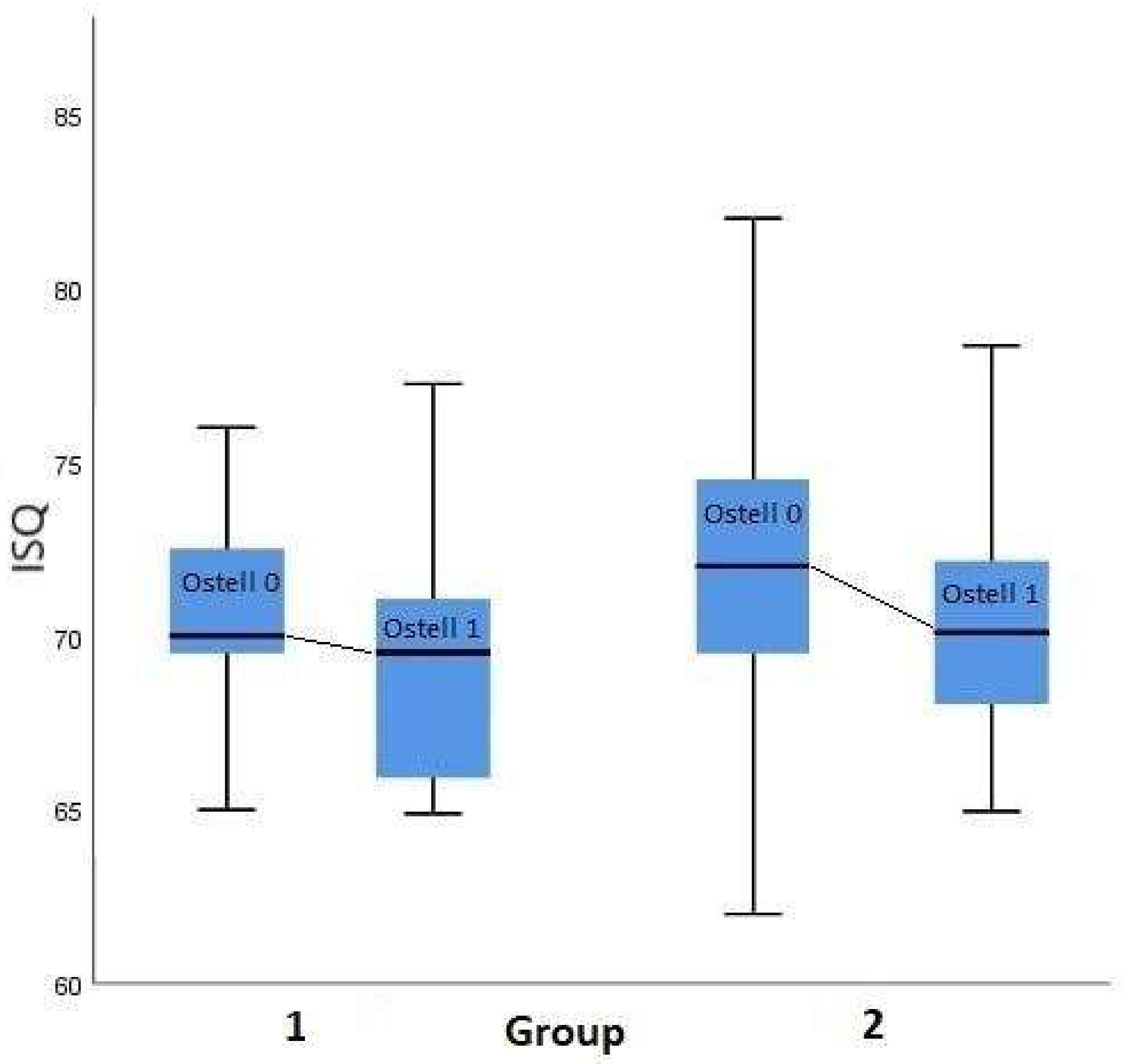
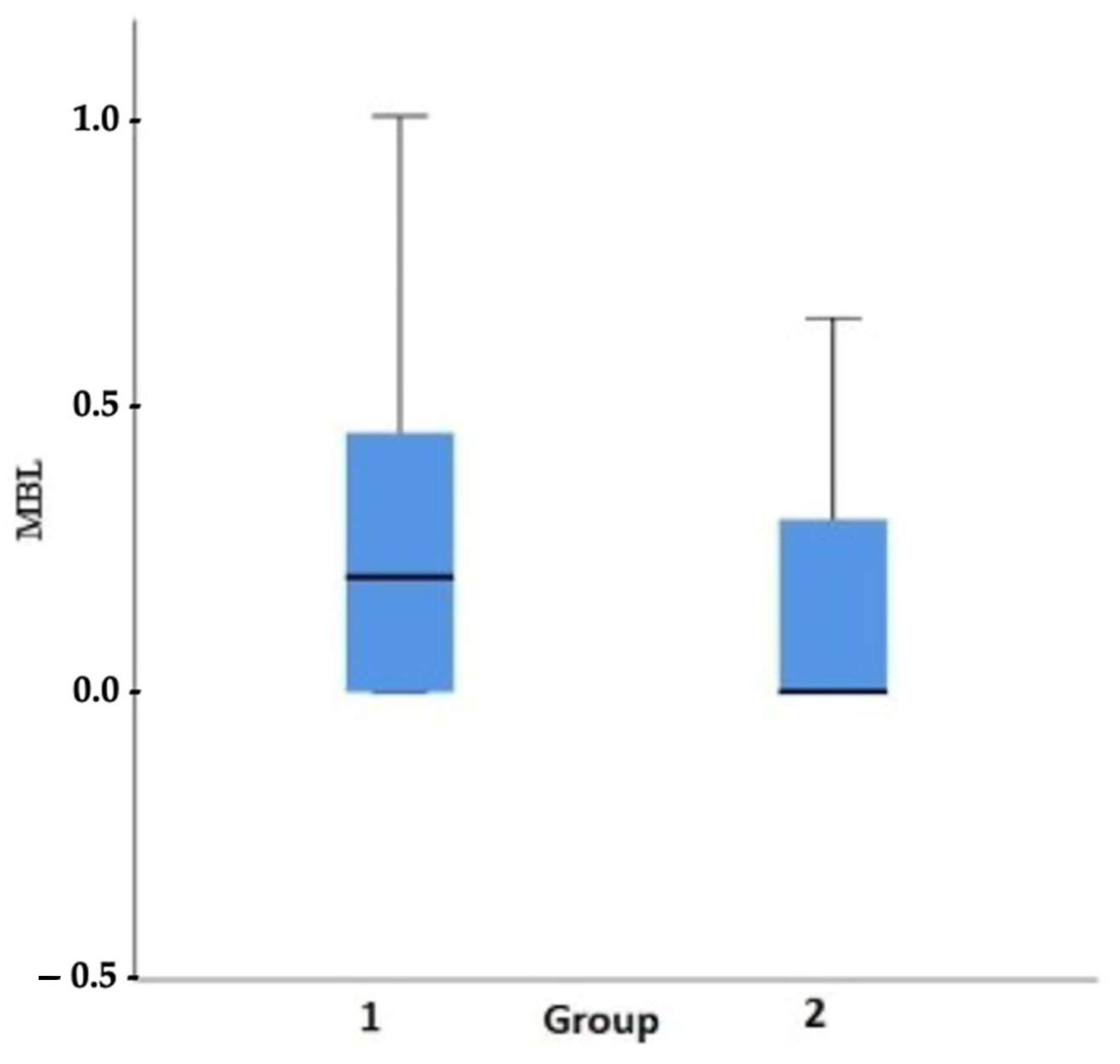
In our study, a decrease in the stability level in the four weeks following the implant insertion was observed (Figure 4). These results are in accordance with other studies and suggest the existence of bone resorption in direct contact with the implant surface. This behavior is attributed to the time dependency of bone remodeling observed at the initial stage. Therefore, the bone shape and remodeling toward the implant surfaces interfere with the bone-implant contact [18,19]. Carmo Filho, in the study on 4.0 and 4.1 mm diameter implants, reported the decline in implant stability at 21 days after surgery for the hydrophilic SLActive implants to 78.8 ± 2.6 ISQ, and to 78.4 ± 3.2 ISQ at 28 days for hydroxyapatite coated implants. However, hydroxyapatite coated implants regain the secondary stability much faster (42 days) when compared to SLActive implants (68 days) [19].
Regarding the primary stability levels, there is a consensus that ISQ values above 70 are optimal for osteointegration to occur, and they enable the consideration of immediate implant loading. In contrast, the ISQ value of 55 for primary stability is the threshold value for the possibility of leaving the implant in place. Below this value, the implant should be replaced with an implant that enables higher primary stability [18,19,20,21]. However, there are studies reporting that modification of the implant’s surface in nano-scale allows for successful early loading even in the case of a lower than optimal primary stability level. Östman et al. compared 242 oxidized surface implants, loaded immediately and delayed. Apart from the relatively low primary stability levels (62.9 ± 4.9 ISQ) of immediately loaded implants, the overall success rate was high (99.2%) in that group. Furthermore, the MBL level for immediately loaded implants during 12 months of observation was 0.78 ± 0.9 mm and seems also acceptable [22].
Subsequent studies addressing this subject demonstrated a correlation between low baseline values of RFA and the potential for implant loss due to lack of osteointegration. Sjöström assessed the primary stability value of 17 implants that were lost within the first year of use. It was found that the average ISQ value in that group was 54.6, whereas the implant group with successful treatments had an average ISQ value of 62.0 [23]. Other studies reported an average primary stability value of 63.3 ISQ in the group of implants that survived the 12-month follow-up period, whereas, in the group with lost implants, the average primary stability value was 56 ISQ [24]. Some studies have found a slightly lower (56 ISQ) threshold value for primary stability, which is necessary for osteointegration to occur [25,26]. That is why it seems that, apart from the established consensus, this issue is still a current topic and further studies are needed for its full development.
Aragoneses et al. included the implant diameter in the assessment of the levels of secondary stability. The ISQ value measured three months after the insertion of implants with a diameter of 3.7 mm was 69.62, while for implants with larger diameters (4.0 mm and 4.3 mm), that value was 72.02 and 69.67, respectively [27]. This finding is in agreement with the following study results (Figure 4 and Figure 5).
In the following study, hydrophilic surface implants were applied. The hydrophilic surface is regarded to osteointegrate faster than other commonly used types of surfaces, including sand blasted and acid etched. Novelino et al. reported that hydrophilic surface implants gain a stability of 70 ISQ in less than five weeks which means that it is 2.24 times faster than implants treated with sand blasting and acid etching [20].
The hydrophilic surface implants modified with hydroxyl ion have been assessed for primary and secondary stability only in two other studies to date. Primary stability in the mentioned studies’ plants amounted to 57.3 (±7.4) ISQ and raised to 71.3 (±8.2) ISQ while loading [28]. The above-mentioned studies mostly focused on measuring the stability of conventionally loaded implants.
An evaluation of the obtained results of MBL shows that they do not deviate from the established norm, according to which MBL should not be greater than 1.5 mm in the first year and then 0.1 mm in each following year. It is important to note that some researchers claim that original remodeling of the alveolar process occurs after implant loading with the aim of restoring biological width [29,30,31,32,33]. However, the condition of the surrounding implant soft tissues seems to play a role in this process. Linkevicius et al. reported significantly lower MBL in a one-year observation when the implant surrounding soft tissue was thicker than 2 mm. The average MBL level in those cases was 0.21 mm and is comparable to our results [29].
The lowest (0.22 ± 0.49 mm) MBL level of early loaded hydrophilic implants with the same, as presented in the following manuscript, observation period (12 months), to date, has been reported by Liaje et al. [30]. This finding of a low MBL was accompanied by high stability values at all time points (above 71 ISQ) and, similar to the presented study findings, (Figure 4) wide implants showed statistically higher stability values than narrow implants. On the contrary, much higher MBL levels than those found in our study for hydrophilic implants were reported by Ryu et al. (0.98 ± 0.61 mm) in 13 months of follow-up, and by Hinkle et al. (0.99 ± 0.29) in 12 months of follow-up [31,32].
In the literature, there are relatively few studies concerning the effects of early loading and changes in stability in connection with MBL. Olsson et al. were among the first who reported the results of studies concerning 68 early-loaded maxillary implants. The value of the average baseline ISQ parameters was 60.1, whereas the implant survival rate was 93.4% and the MBL level was 1.3 ± 0.6 mm at a 12-month follow-up observation [33]. Fischer et al. assessed oxidized-surface implants that were loaded for the period ranging from a few days to 16 days. They achieved an overall success rate of 98.1% and the averaged MBL index was 1.1 mm at a 12-month follow-up observation. In contrast, the ISQ index increased from 63.3 (±6.1) to 66.8 (±5.6) after 12 months [24].
The effect of early loading relative to conventional loading on bone tissue levels (without linking with primary stability levels) was also previously studied in other papers. Degidi et al. assessed immediately loaded implants with a diameter of 3 mm, obtaining the MBL level of 0.85 ± 0.71 mm at a 36-month follow-up observation [34]. In studies concerning early-loaded implants (three weeks), Grandi proved efficacy comparable to immediate loading (with a loss of one implant in both groups) at a 12-month follow-up observation. However, a higher average MBL was found in the group of implants loaded after three weeks (0.390 ± 0.840 mm) when compared to immediately-loaded implants (0.120 ± 0.230 mm), with no statistically significant differences between those groups [35]. Other authors report that there is no effect of implant loading time on marginal bone loss. MBL levels in the mentioned studies ranged from 0 to 1.32 mm in longtime observation [36,37,38,39].
In our previous studies on conventional SLA implants, the average stability level was initially lower (58.67 ± 12.3 ISQ) than in a following study, and raised to 81 ± 5.82 ISQ six months after implant placement and was accompanied by a mean MBL of 0.22 ± 0.46, calculated based on radiographs taken in 36-months of follow-up [40,41].
The present study has some limitations: firstly, the observation period. We decided to evaluate the MBL level in 12 months. In the literature, there exist studies of such methodology; however, the considerable changes in the bone of the alveolar process can occur in the later stages of implant treatment and further studies are needed to confirm all theses of the present study. Secondly, we decided not to measure the stability levels at the final follow-up after 12 months, as the need to remove the implant restoration would emerge under those conditions and could influence implant preservation.

5. Conclusions

With the limitation of our study and 12 months of follow-up, the following conclusions can be made:
(a)
hydrophilic surface implants can be used in a protocol of early functional occlusal loading;
(b)
higher values of primary stability positively influence the values of stability measured four weeks after implantation;
(c)
the age of the patient affects the values of implant stability.

Author Contributions

Conceptualization M.D., M.K. and J.H.; methodology M.D., J.H.; formal analysis, P.K.-R.; clinical investigation A.B., J.H., M.K. and W.G.; data curation M.K.; writing—original draft preparation M.K. and P.K.-R., writing—review and editing M.D. and J.H.; visualization P.K.-R.; supervision, M.D. and J.H. All authors have read and agreed to the published version of the manuscript.

Funding

The study was supported by the Thommen Medical (Grenchen, Switzerland) who donated the implants free of charge (TM-16-001). The study was also partially supported by the University Statutory Grant no. SUB.B040.20.020T.

Institutional Review Board Statement

The study was conducted according to the guidelines of the Declaration of Helsinki and approved by the Local Ethical Committee (229/2019).

Informed Consent Statement

Informed consent was obtained from all subjects involved in the study. Written informed consent has been obtained from the patients to publish this paper.

Data Availability Statement

Not applicable.

Conflicts of Interest

Thommen Medical (Grenchen, Switzerland), the manufacturer of the dental implants used in this study, provided this trial with dental implants. However, the data is the possession of the authors and the sponsor did not interfere with the course of the trial or in the publication of its results whatsoever. The authors declare no other conflicts of interest that could have influenced the outcomes of this manuscript.

Abbreviations

MSCmesenchymal stem cells
APIapproximal plague index
CBCTcone beam computed tomography
RVGradiovisiography
GDPRgeneral data protection regulation
ISQimplant stability quotient
MBLmarginal bone loss
RFAresonance frequency analysis
SLAsand blasted acid-etched

References

  1. Garg, R.; Mishra, N.; Alexander, M.; Gupta, S.K. Implant Survival between Endo-osseous Dental Implants in Immediate Loading, Delayed Loading, and Basal Immediate Loading Dental Implants a 3-Year Follow-up. Ann. Maxillofac. Surg. 2017, 7, 237–244. [Google Scholar] [CrossRef] [PubMed]
  2. Bosshardt, D.D.; Chappuis, V.; Buser, D. Osseointegration of titanium, titanium alloy and zirconia dental implants: Current knowledge and open questions. Periodontology 2000 2017, 73, 22–40. [Google Scholar] [CrossRef] [PubMed]
  3. Di Stefano, D.A.; Arosio, P.; Gastaldi, G.; Gherlone, E. The insertion torque-depth curve integral as a measure of implant primary stability: An in vitro study on polyurethane foam blocks. J. Prosthet. Dent. 2018, 120, 706–714. [Google Scholar] [CrossRef] [PubMed]
  4. Bergkvist, G.; Koh, K.J.; Sahlholm, S.; Klintström, E.; Lindh, C. Bone density at implant sites and its relationship to assessment of bone quality and treatment outcome. Int. J. Oral Maxillofac. Implant. 2010, 25, 321–328. [Google Scholar]
  5. Antonelli, A.; Bennardo, F.; Brancaccio, Y.; Barone, S.; Femiano, F.; Nucci, L.; Minervini, G.; Fortunato, L.; Attanasio, F.; Giudice, A. Can Bone Compaction Improve Primary Implant Stability? An In Vitro Comparative Study with Osseodensification Technique. Appl. Sci. 2020, 10, 8623. [Google Scholar] [CrossRef]
  6. McCullough, J.J.; Klokkevold, P.R. The effect of implant macro-thread design on implant stability in the early post-operative period: A randomized, controlled pilot study. Clin. Oral Implant. Res. 2017, 28, 1218–1226. [Google Scholar] [CrossRef]
  7. Shen, X.; Ma, P.; Hu, Y.; Xu, G.; Zhou, J.; Cai, K. Mesenchymal stem cell growth behavior on micro/nano hierarchical surfaces of titanium substrates. Colloids Surf. B. Biointerfaces 2015, 127, 221–232. [Google Scholar] [CrossRef]
  8. Coelho, P.G.; Jimbo, R.; Tovar, N.; Bonfante, E.A. Osseointegration: Hierarchical designing encompassing the macrometer, micrometer, and nanometer length scales. Dent. Mater. 2015, 31, 37–52. [Google Scholar] [CrossRef]
  9. Matys, J.; Świder, K.; Grzech-Leśniak, K.; Dominiak, M.; Romeo, U. Photobiomodulation by a 635nm Diode Laser on Peri-Implant Bone: Primary and Secondary Stability and Bone Density Analysis-A Randomized Clinical Trial. BioMed Res. Int. 2019, 2019, 2785302. [Google Scholar] [CrossRef] [Green Version]
  10. Chou, W.C.; Wang, R.C.; Huang, C.L.; Lee, T.M. The effect of plasma treatment on the osseointegration of rough titanium implant: A histo-morphometric study in rabbits. J. Dent. Sci. 2018, 13, 267–273. [Google Scholar] [CrossRef]
  11. Lee, B.A.; Kang, C.H.; Vang, M.S.; Jung, Y.S.; Piao, X.H.; Kim, O.S.; Chung, H.J.; Kim, Y.J. Surface characteristics and osteoblastic cell response of alkali-and heat-treated titanium-8tantalum-3niobium alloy. J. Periodontal. Implant. Sci. 2012, 42, 248–255. [Google Scholar] [CrossRef] [PubMed] [Green Version]
  12. Schnitman, P.A.; Wohrle, P.S.; Rubenstein, J.E. Immediate fixed interim prostheses supported by two-stage threaded implants: Methodology and results. J. Oral Implantol. 1990, 16, 96–105. [Google Scholar] [PubMed]
  13. Schnitman, P.A.; Wöhrle, P.S.; Rubenstein, J.E.; DaSilva, J.D.; Wang, N.H. Ten-year results for Brånemark implants immediately loaded with fixed prostheses at implant placement. Int. J. Oral Maxillofac. Implant. 1997, 12, 495–503. [Google Scholar]
  14. Chen, J.; Cai, M.; Yang, J.; Aldhohrah, T.; Wang, Y. Immediate versus early or conventional loading dental implants with fixed prostheses: A systematic review and meta-analysis of randomized controlled clinical trials. J. Prosthet. Dent. 2019, 122, 516–536. [Google Scholar] [CrossRef] [PubMed] [Green Version]
  15. Sim, C.P.; Lang, N.P. Factors influencing resonance frequency analysis assessed by Osstell mentor during implant tissue integration: I. Instrument positioning, bone structure, implant length. Clin. Oral Implant. Res. 2010, 21, 598–604. [Google Scholar] [CrossRef] [PubMed]
  16. Misch, C.E. Density of bone: Effect on treatment planning, surgical approach, and healing. In Contemporary Implant Dentistry; Misch, C.E., Ed.; Mosby: St. Louis, MI, USA, 1993; pp. 469–485. [Google Scholar]
  17. Carmo Filho, L.C.; Marcelo-Machado, R.M.; Castilhos, E.D.; Del Bel Cury, A.A.; Faot, F. Can implant surfaces affect implant stability during osseointegration? A randomized clinical trial. Brazil. Oral Res. 2018, 32, e110. [Google Scholar] [CrossRef] [PubMed]
  18. Chambrone, L.; Shibli, J.A.; Mercúrio, C.E.; Cardoso, B.; Preshaw, P.M. Efficacy of standard (SLA) and modified sandblasted and acid-etched (SLActive) dental implants in promoting immediate and/or early occlusal loading protocols: A systematic review of prospective studies. Clin. Oral Implant. Res. 2015, 26, 359–370. [Google Scholar] [CrossRef]
  19. Norton, M.R.; Åström, M. The Influence of Implant Surface on Maintenance of Marginal Bone Levels for Three Premium Implant Brands: A Systematic Review and Meta-analysis. Int. J. Oral Maxillofac. Implant. 2020, 35, 1099–1111. [Google Scholar] [CrossRef]
  20. Novellino, M.M.; Sesma, N.; Zanardi, P.R.; Laganá, D.C. Resonance frequency analysis of dental implants placed at the posterior maxilla varying the surface treatment only: A randomized clinical trial. Clin. Implant. Dent. Relat. Res. 2017, 19, 770–775. [Google Scholar] [CrossRef] [Green Version]
  21. Shokri, M.; Daraeighadikolaei, A. Measurement of primary and secondary stability of dental implants by resonance frequency analysis method in mandible. Int. J. Dent. 2013, 2013, 506968. [Google Scholar] [CrossRef] [Green Version]
  22. Ostman, P.O.; Hellman, M.; Sennerby, L. Direct implant loading in the edentulous maxilla using a bone density-adapted surgical protocol and primary implant stability criteria for inclusion. Clin. Implant. Dent. Relat. Res. 2005, 1, 60–69. [Google Scholar] [CrossRef]
  23. Sjöström, M.; Sennerby, L.; Nilson, H.; Lundgren, S. Reconstruction of the atrophic edentulous maxilla with free iliac crest grafts and implants: A 3-year report of a prospective clinical study. Clin. Implant. Dent. Relat. Res. 2007, 9, 46–59. [Google Scholar] [CrossRef]
  24. Fischer, K.; Bäckström, M.; Sennerby, L. Immediate and early loading of oxidized tapered implants in the partially edentulous maxilla: A 1-year prospective clinical, radiographic, and resonance frequency analysis study. Clin. Implant. Dent. Relat. Res. 2009, 11, 69–80. [Google Scholar] [CrossRef] [PubMed]
  25. Glauser, R.; Sennerby, L.; Meredith, N.; Rée, A.; Lundgren, A.; Gottlow, J.; Hämmerle, C.H. Resonance frequency analysis of implants subjected to immediate or early functional occlusal loading. Successful vs. failing implants. Clin. Oral. Implant. Res. 2004, 15, 428–434. [Google Scholar] [CrossRef] [PubMed]
  26. Ostman, P.O.; Wennerberg, A.; Albrektsson, T. Immediate occlusal loading of NanoTite PREVAIL implants: A prospective 1-year clinical and radiographic study. Clin. Implant. Dent. Relat. Res. 2010, 12, 39–47. [Google Scholar] [CrossRef]
  27. Aragoneses, J.M.; Suárez, A.; Brugal, V.A.; Gómez, M. Frequency Values and Their Relationship With the Diameter of Dental Implants. Prospective Study of 559 Implants. Implant. Dent. 2019, 28, 279–288. [Google Scholar] [CrossRef] [PubMed]
  28. Makowiecki, A.; Hadzik, J.; Błaszczyszyn, A.; Gedrange, T.; Dominiak, M. An evaluation of superhydrophilic surfaces of dental implants-a systematic review and meta-analysis. BMC Oral Health 2019, 19, 79. [Google Scholar] [CrossRef]
  29. Linkevicius, T.; Puisys, A.; Steigmann, M.; Vindasiute, E.; Linkeviciene, L. Influence of Vertical Soft Tissue Thickness on Crestal Bone Changes Around Implants with Platform Switching: A Comparative Clinical Study. Clin. Implant. Dent. Relat. Res. 2015, 17, 1228–1236. [Google Scholar] [CrossRef] [PubMed]
  30. Liaje, A.; Ozkan, Y.K.; Ozkan, Y.; Vanlioğlu, B. Stability and marginal bone loss with three types of early loaded implants during the first year after loading. Int. J. Oral Maxillofac. Implant. 2012, 27, 162–167. [Google Scholar]
  31. Ryu, H.-S.; Namgung, C.; Heo, Y.-K.; Lee, J.-H.; Lim, Y.-J. Early loading of splinted implants supporting a two-unit fixed partial denture in the posterior maxilla:13-month results from a randomized controlled clinical trial of two different implant systems. Clin. Oral Implant. Res. 2016, 27, 1017–1025. [Google Scholar] [CrossRef]
  32. Hinkle, R.M.; Rimer, S.R.; Morgan, M.H.; Zeman, P. Loading of titanium implants with hydrophilic endosteal surface 3 weeks after insertion: Clinical and radiological outcome of a 12-month prospective clinical trial. J. Oral Maxillofac. Surg. 2014, 72, 1495–1502. [Google Scholar] [CrossRef] [PubMed]
  33. Olsson, M.; Urde, G.; Andersen, J.B.; Sennerby, L. Early loading of maxillary fixed cross-arch dental prostheses supported by six or eight oxidized titanium implants: Results after 1 year of loading, case series. Clin. Implant. Dent. Relat. Res. 2003, 5, 81–87. [Google Scholar] [CrossRef] [PubMed]
  34. Degidi, M.; Nardi, D.; Piattelli, A. Immediate versus one-stage restoration of small-diameter implants for a single missing maxillary lateral incisor: A 3-year randomized clinical trial. J. Periodontol. 2009, 80, 1393–1398. [Google Scholar] [CrossRef]
  35. Grandi, T.; Guazzi, P.; Samarani, R.; Tohme, H.; Khoury, S.; Sbricoli, L.; Grandi, G.; Esposito, M. Immediate, early (3 weeks) and conventional loading (4 months) of single implants: Preliminary data at 1 year after loading from a pragmatic multicenter randomised controlled trial. Eur. J. Oral. Implantol. 2015, 8, 115–116. [Google Scholar]
  36. Grandi, T.; Garuti, G.; Guazzi, P.; Tarabini, L.; Forabosco, A. Survival and success rates of immediately and early loaded implants: 12-month results from a multicentric randomized clinical study. J. Oral Implantol. 2012, 38, 239–249. [Google Scholar] [CrossRef]
  37. Grandi, T.; Guazzi, P.; Samarani, R.; Grandi, G. A 3-year report from a multicentre randomised controlled trial: Immediately versus early loaded implants in partially edentulous patients. Eur. J. Oral Implantol. 2013, 6, 217–224. [Google Scholar]
  38. Kokovic, V.; Jung, R.; Feloutzis, A.; Todorovic, V.S.; Jurisic, M.; Hämmerle, C.H. Immediate vs. early loading of SLA implants in the posterior mandible: 5- year results of randomized controlled clinical trial. Clin. Oral. Implants Res. 2014, 25, 114–119. [Google Scholar] [CrossRef]
  39. Hadzik, J.; Krawiec, M.; Kubasiewicz-Ross, P.; Prylińska-Czyżewska, A.; Gedrange, T.; Dominiak, M. Short Implants and Conventional Implants in The Residual Maxillary Alveolar Ridge: A 36-Month Follow-Up Observation. Med. Sci. Monit. 2018, 24, 5645–5652. [Google Scholar] [CrossRef]
  40. Hadzik, J.; Krawiec, M.; Sławecki, K.; Kunert-Keil, C.; Dominiak, M.; Gedrange, T. The Influence of the Crown-Implant Ratio on the Crestal Bone Level and Implant Secondary Stability: 36-Month Clinical Study. BioMed Res. Int. 2018, 2018, 4246874. [Google Scholar] [CrossRef] [PubMed]
Figure 1. Implant with the healing abutment placed in bicuspid region and left for open healing.
Applsci 11 02958 g001
Figure 2. Implant with scanbody prepared for intraoral scan.
Applsci 11 02958 g002
Figure 3. Implant loaded with the screw-retained crown.
Applsci 11 02958 g003
Figure 4. Results of the primary (Ostell 0) and secondary (Ostell 1) stability in both groups.
Applsci 11 02958 g004
Figure 5. Results of marginal bone loss in both groups.
Applsci 11 02958 g005
Table 1. Descriptive statistics and the normality test to determine distribution for selected variables.
Group VariableMMeSDSk.Kurt.Min.Max.WP
Group 1 and 2 combinedAge41.5540.008.851.302.4926.0068.000.870.001
Ostell 071.5971.004.040.300.7362.0082.000.970.506
Ostell 169.9470.003.290.520.0265.0078.000.960.181
MBL 0.200.000.881.270.810.001.000.75<0.001
1Age42.9538.0011.301.060.7126.0068.000.830.006
Ostell 070.8470.003.390.240.1665.0078.000.960.679
Ostell 169.5069.503.310.690.0865.0077.000.950.477
MBL 0.260.200.311.170.750.001.000.820.003
2Age40.1541.005.38−0.25−0.6631.0050.000.960.673
Ostell 072.3072.004.560.110.8662.0082.000.950.482
Ostell 170.3970.003.290.440.5465.0078.000.950.492
MBL 0.140.000.241.330.070.000.650.62<0.001
M—arithmetic mean, Me—median, SD—standard deviation, Sk.—skewness, Kurt.—kurtosis, Min.—minimum, Max.—maximum, W—Shapiro–Wilk test statistic. The statistically important differences are highlighted in red.
Table 2. Linear correlation coefficient for groups one and two and their significance for the selected continuous variables.
VariableCoefficientAgeOstell 0Ostell 1MBL
AgePearson’s r1−0.280.22−0.06
P<0.0010.0820.2010.735
Ostell 0Pearson’s r−0.2810.58−0.11
P0.082<0.001<0.0010.531
Ostell 1Pearson’s r0.220.581−0.06
P0.2640.4470.5360.623
MBLPearson’s r−0.06−0.11−0.061
P0.7350.5310.736<0.001
Table 3. Linear correlation coefficients for group one and their significance for the selected continuous variables. The statistically important differences are highlighted in red.
VariableCoefficientAgeOstell 0Ostell 1MBL
AgePearson’s r1−0.240.47−0.37
P<0.0010.3250.0470.128
Ostell 0Pearson’s r−0.2410.420.09
P0.325<0.0010.0840.730
Ostell 1Pearson’s r0.470.421−0.05
P0.5840.2220.4300.874
MBLPearson’s r−0.370.09−0.051
P0.1280.7300.837<0.001
Table 4. Linear correlation coefficients for group two and their significance for the selected continuous variables.
VariableCoefficientAgeOstell 0Ostell 1MBL
AgePearson’s r1−0.37−0.110.47
P<0.0010.1050.6630.047
Ostell 0Pearson’s r−0.3710.69−0.20
P0.105<0.0010.0010.422
Ostell 1Pearson’s r−0.110.6910.00
P0.0550.3200.7330.814
MBLPearson’s r0.47−0.200.001
P0.0470.4220.992<0.001
Table 5. Kruskal–Wallis test results for Ostell and MBL variables.
Variabled1 (n = 3)d2 (n = 28) *d3 (n = 5) +Hpη2
Average rankMeIQRAverage rankMeIQRAverage rankMeIQR
Ostell 032.1775.00---21.6372.004.005.7567.003.0013.580.0010.35
Ostell 132.5075.00---18.9870.004.007.4068.003.0011.160.0040.28
MBL17.000.00---19.560.000.4515.200.000.281.660.646−0.01
Note: *—for MBL n = 30, +—for MBL n = 6.
Table 6. The Student’s t-test results for a grouping variable (diameter) and selected dependent variables.
Variable3.5 diameter4.0 diameterTPCohen’s d
MSDMSD
MBL 0.260.310.140.241.300.2030.43
Ostell 070.843.3972.304.56−1.130.266−0.36
Ostell 169.503.3170.393.29−0.810.425−0.27
Age42.9511.3040.155.381.000.3260.32
Publisher’s Note: MDPI stays neutral with regard to jurisdictional claims in published maps and institutional affiliations.

