
Role of Magnetic Anisotropy on the Hyperthermia Efficiency in Spherical Fe3−xCoxO4 (x = 0–1) Nanoparticles
by
Raja Das
1,2,*, 
Ngoc Pham Kim
1,2, 
Supun B. Attanayake
3,
Manh-Huong Phan
3 and
Hariharan Srikanth
3,* 1
Faculty of Materials Science and Engineering, Phenikaa University, Hanoi 12116, Vietnam
2
Phenikaa Research and Technology Institute (PRATI), A&A Green Phoenix Group, 167 Hoang Ngan, Hanoi 13313, Vietnam
3
Department of Physics, University of South Florida, Tampa, FL 33620, USA
*
Authors to whom correspondence should be addressed.
Appl. Sci. 2021, 11(3), 930; https://doi.org/10.3390/app11030930
Submission received: 14 December 2020
/
Revised: 13 January 2021
/
Accepted: 15 January 2021
/
Published: 20 January 2021
(This article belongs to the Special Issue Advances in Translational Nanotechnology)
Abstract
:The use of magnetic nanoparticles in the treatment of cancer using alternating current hyperthermia therapy has shown the potential to replace or supplement conventional cancer treatments, radiotherapy and chemotherapy, which have severe side effects. Though the nearly spherical sub-10 nm iron oxide nanoparticles have their approval from the US Food and Drug Administration, their low heating efficiency and removal from the body after hyperthermia treatment raises serious concerns. The majority of magnetic hyperthermia research is working to create nanomaterials with improved heating efficiency and long blood circulation time. Here, we have demonstrated a simple strategy to enhance the heating efficiency of sub-10 nm Fe3O4 nanoparticles through the replacement of Fe+2 ions with Co+2 ions. Magnetic and hyperthermia experiments on the 7 nm Fe3−xCoxO4 (x = 0–1) nanoparticles showed that the blocking temperature, the coercivity at 10 K, and the specific absorption rate followed a similar trend with a maximum at x = 0.75, which is in corroboration with the theoretical prediction. Our study revealed that the heating efficiency of the Fe3−xCoxO4 (x = 0–1) nanoparticles varies not just with the size and saturation magnetization but also with the magnetocrystalline anisotropy of the particles.

1. Introduction
Functional magnetic nanostructures are of high significance not only for research professionals working in the disciplines of data storage [1], catalysis [2,3], environmental remediation [4], and basic science [5,6,7,8] but also for those working in biomedicine [9,10,11,12,13]. Recently, alternating current (AC) hyperthermia therapy using magnetic nanoparticles, especially spinal ferrites (MFe2O4, M = Mn, Ni, Co, Fe), has become a promising supplementary technique to conventional cancer treatments, radiotherapy and chemotherapy, which have severe side effects [14,15,16,17]. In the treatment involving magnetic hyperthermia, an external AC magnetic field is applied for heating the magnetic nanoparticles in a cancerous area in order to destroy or deactivate the cancerous cells and cause no harm to the healthy ones, resulting in minimum collateral damage. This technique has already shown promising results in cancer therapy; however, the low heating efficiency of the traditionally used spherical iron oxides, particularly magnetite (Fe3O4) and maghemite (γ-Fe2O3) nanoparticles, obstructs their wide application [18,19]. Serious concern has been raised about the number of nanoparticles required for each magnetic hyperthermia treatment because of their low heating efficiency. Recent research has suggested that a significant number of nanoparticles tends to be cumulated in kidneys, liver, spleen, and other parts of the body after hyperthermia treatment [20]. Therefore, there is an increase in demand for highly efficient magnetic nanoparticles so that a minimal dose of nanoparticles can be used to achieve the therapeutic temperature range (40–44 °C) to kill or deactivate the cancerous cells without damaging the healthy ones [21,22,23,24,25,26].
The heating efficiency of the nanoparticles defined in terms of the specific absorption rate (SAR) value is dependent on the saturation magnetization (MS), effective magnetic anisotropy, shape, size and amount of the nanoparticles [27,28,29,30,31]. Smaller nanoparticles, in the sub-10 nm range, have been proven to have a longer circulation time in blood because they can escape the immune system. Moreover, smaller nanoparticles are stable against agglomeration and precipitation, thus avoiding the risk of blood vessel occlusion [32]. However, there is a strong decrease in MS, well below the MS of bulk phase, upon reduction in the size [33]. Both Monte Carlo simulation and experimental results on Fe3O4 nanocubes showed a SAR improvement relative to conventional spherical nanoparticles, due to the enhanced surface anisotropy as a result of the chain-like particle formation [34]. This was further verified by our group, where we have demonstrated a significant enhancement in the SAR value of Fe3O4 nanorods compared to other shapes due to enhanced effective magnetic anisotropy [35,36]. Unfortunately, only nearly spherical iron oxide nanoparticles are currently approved for biomedical applications [37,38,39]. Therefore, a robust strategy is needed to improve the heating efficiency of sub-10 nm iron oxide particles. It has been demonstrated that the heating efficiency of Fe3O4 nanocubes (20 ± 2 nm) can be improved with a small replacement of Fe+2 with Co+2 ions in B-sites, as a result of the increased magnetocrystalline anisotropy in Fe3−xCoxO4 nanoparticles [40].
Even though there is a great potential of Co substituted Fe3−xCoxO4 nanoparticles in improving heating efficiency, the systematical study of sub-10 nm Fe3−xCoxO4 (x = 0–1) nanoparticles [32,41] has attracted much less research effort. This restrains the possibility of using Fe3−xCoxO4 (x = 0–1) nanoparticles for magnetic hyperthermia treatment. To address this challenging issue, we fabricated a 7 nm series of Fe3−xCoxO4 (x = 0–1) nanoparticles, while keeping the size and morphology unchanged. Here, we demonstrate that the magnetocrystalline anisotropy and SAR for these Fe3−xCoxO4 (x = 0–1) nanoparticles follow a similar trend with a maximum at x = 0.75.
2. Materials and Methods
2.1. Synthesis of Fe3−xCoxO4 (x = 0–1) Nanoparticles
Fe3−xCoxO4 (x = 0–1) nanoparticles were fabricated using thermal decomposition of iron acetylacetonate (Fe(acac)3, Sigma-Aldrich 97%, St. Louis, MO, USA) and cobalt acetylacetonate (Co(acac)2, Sigma-Aldrich 97%) [42,43,44,45]. In a typical reaction, the stoichiometric amount of Fe(acac)3 or/and Co(acac)2 (2 mmol) and 1,2 hexadecanediol (Sigma-Aldrich 90%) (10 mmol) were dissolved in 6 mmol of oleic acid (Sigma-Aldrich 90%), 6 mmol of olylamine (Sigma-Aldrich 70%) and 20 mL of benzyl ether (Sigma-Aldrich 98%). For nucleation of the particles, the mixture was stirred in a nitrogen atmosphere while being heated to 200 °C and was kept constant for 2 h, following which it was raised to 280 °C for the growth process. The temperature was kept constant at 280 °C for 1 h, and then the reaction mixture was naturally cooled to ambient temperature (300 K). The resultant Fe3−xCoxO4 (x = 0–1) nanoparticles were washed with ethanol and hexane (3:1) three times. The stoichiometry of the Fe3−xCoxO4 (x = 0–1) nanoparticles was controlled by choosing the appropriate initial ratio of precursors, Fe(acac)3 and Co(acac)2. The experimental conditions for every reaction were kept constant to obtain the same size and morphology of the Fe3−xCoxO4 (x = 0–1) nanoparticles. After washing, the black precipitates were re-dispersed in hexane for storing and further characterization.
2.2. Phase Transfer of Fe3−xCoxO4 (x = 0–1) Nanoparticles
The as-prepared oleic acid and olylamine capped hydrophobic nanoparticles were phase transferred to the aqueous phase using tetramethyl ammonium hydroxide (TMAH) (Sigma-Aldrich ≥ 97%) as a capping agent [32]. The as-prepared nanoparticles were dried by air. For a standard reaction, 100 mg of dried Fe3−xCoxO4 (x = 0–1) nanoparticles was dispersed in a solution containing 500 mg of TMAH in 10 mL of ethanol. For all the phase transfer processes, the ratio of nanoparticles:ethanol:TMAH was kept at 1:10:5. The above mixture was sonicated for 30 min and then washed with water to get rid of extra TMAH. The washed particles were dispersed in water for the hyperthermia measurements. The TEM images of the nanoparticles exhibited no indication of particle size and morphology change due to the phase transfer from the hydrophobic to the aqueous medium [35].
2.3. Characterization of the Nanoparticles
Crystalline phase and morphology were characterized using a Bruker AXS D8 X-ray Diffractometer (XRD) (Bruker, Madison, WI, USA) and a FEI Morgagni 268 Transmission Electron Microscope (TEM) (FEI, Hillsboro, OR, USA) operating at 60 kV, respectively. DC magnetic measurements were done using a vibrating sample magnetometer (VSM) of Physical Property Measurement System (PPMS) from Quantum Design, San Diego, CA, USA. Calorimetric hyperthermia experiments were conducted using a 4.2 kW Ambrell Easyheat Li3542 system (Ambrell, Rochester, NY, USA) with AC fields (0–800 Oe) and frequency (f = 310 kHz).
3. Results
Thermal decomposition of Fe(acac)3 and Co(acac)2 was used to synthesize spherical Fe3−xCoxO4 (x = 0–1) nanoparticles; the specifics are mentioned in the Materials and Methods section. In the thermal decomposition reaction, the decomposition of Fe(acac)3 and Co(acac)2, in the presence of 1,2 hexadecanediol, oleic acid, and olylamine, to cationic metals resulted in the formation of the nanoparticle. In the current case, at 200 °C, the nucleation process occurred, followed by the growth of the nucleus at 280 °C. The segregation of nucleation and the growth process directed the formation of the monodisperse nanoparticles [42,43]. The size and morphology of the particles are dependent on the initial ratio of the precursors, surfactants and solvent.
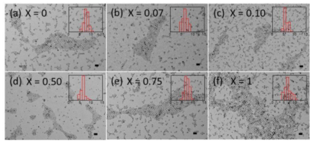
TEM images of Fe3−xCoxO4 (x = 0–1) nanoparticles are displayed in Figure 1. The analysis of the TEM images shows that along with having a nearly monodisperse size distribution, the particles are nearly spherical in shape. It is worth mentioning that the particles are well separated from each other, which indicates the uniform surfactant coating of the nanoparticles’ surface. The mean diameter (D) and a standard deviation (σ) from the fitting of the size histogram to a lognormal distribution are presented in Table 1. The mean diameter of the particles for all compositions is in the range of 6.6 to 7.8 nm. This control over the size of Fe3−xCoxO4 (x = 0–1) nanoparticles allowed us to understand the effect of Co substitution on their magnetic and hyperthermia responses.
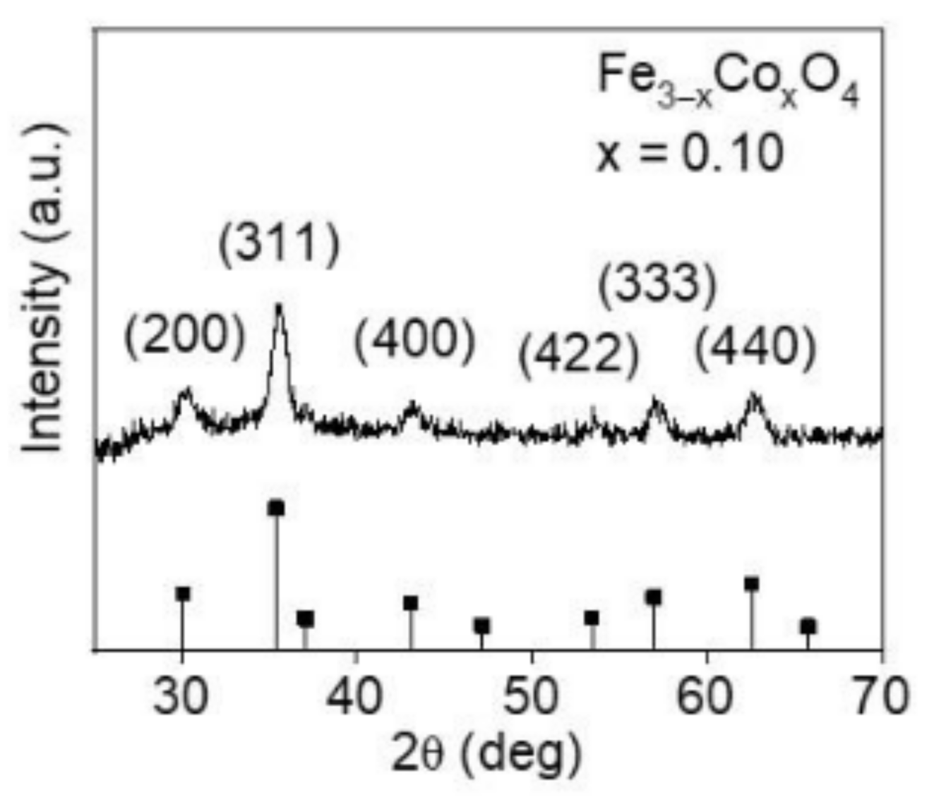
The crystalline phase of the Fe3−xCoxO4 (x = 0–1) nanoparticles was analyzed using XRD. In the XRD pattern of Fe3−xCoxO4 (x = 0.10) displayed in Figure 2, the XRD peaks are indexed to Fe3O4 confirming the formation of the single-phase material. The formation of the single-phase material confirms that the oleic acid and olylamine capping inhibits the oxidation of the nanoparticles.
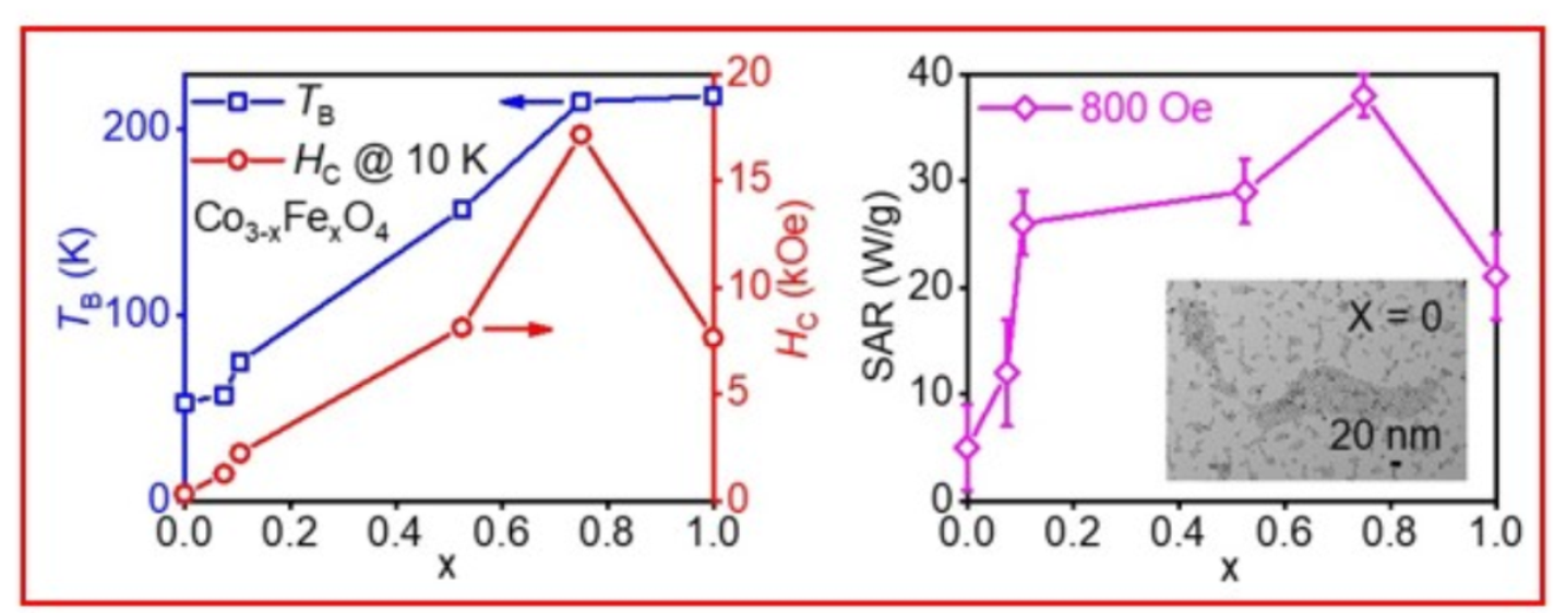
Zero-field-cooled (ZFC) and field-cooled (FC) magnetization vs. temperature in an applied field of 50 Oe for Fe3−xCoxO4 (x = 0–1) nanoparticles is shown in Figure 3a. Monodisperse, non-interacting, and superparamagnetic nanoparticles, which are ferromagnetic in the ground state, are known to show a maximum in the ZFC magnetization curve corresponding to the blocking temperature (TB) when the thermal energy is equivalent to the activation energy. The ZFC magnetization (MZFC(T)) curves for all the samples clearly showed maxima in the measured temperature range. It is evident that the MZFC(T) peak or the TB shifts to a higher temperature with an increase in the Co content. TB vs. Co content is plotted in Figure 3b and Table 2. The sharp increase in TB, with an increase in Co content until x = 0.75, can be attributed to the increase in the magnetocrystalline anisotropy of the nanocrystals [37]. The nearly constant value of TB from x = 0.75 to x = 1 indicates that the magnetocrystalline anisotropy of the nanocrystals does not increase with a further increase in Co content. This is in agreement with previous reports, where a decrease in magnetocrystalline anisotropy in Fe3−xCoxO4 (0.75 ≤ x ≤1) was reported [40,46,47,48,49].
To further verify the variation of magnetocrystalline anisotropy in Fe3−xCoxO4 (x = 0–1) nanoparticles, we measured low temperature (10 K) magnetization as a function magnetic field (M–H) loops (Figure 4a). At 10 K, Fe3−xCoxO4 (x = 0) nanoparticles show coercivity of 330 Oe which increases to 1265 with Co content of x = 0.07 and show a further increase with an increase in the Co content (Table 2). The variation of HC at 10 K as a function of Co content (x = 0–1) is shown in Figure 4b and Table 2. The HC at 10 K increases with an increase in the Co content until x = 0.75 and above, at which point it showed a decreasing trend. Sathya et al. reported that in the series of 20 nm Fe3-xCoxO4 nanocrystals (0.1 ≤ x ≤ 1), with the increase in cobalt content from 0.1 to 0.5, the HC at 5 and 298 K increased; additionally, with a further increase to 1, HC showed a decreasing trend. Yu et al. also observed similar results for 35 nm Fe3−xCoxO4 (x = 0–1) nanoparticles where the maximum in the HC was observed at x = 0.6, which then decreased with a further increase of Co content [50]. These results convey that the size and crystallinity of the nanoparticles determine the maximum of the HC with Co content in Fe3−xCoxO4 nanocrystals (x = 0–1). This is in corroboration with previous experimental and theoretical results [40,46,47,48,49]. It should be noted that although high magnetic anisotropy results in high coercivity, high coercivity does not always mean high magnetic anisotropy as there could be other factors that can account for magnetic anisotropy, such as surface, shape, etc. Therefore, the coercivity trend observed in the present case cannot account for only magnetic anisotropy but, together with other results, it points toward the increase in magnetic anisotropy with the small replacement of Fe+2 with Co+2 ions in B-sites.
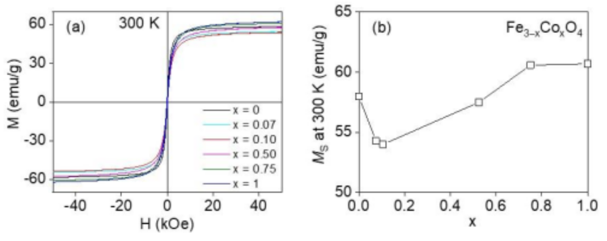
Figure 5a displays the M–H loops at 300 K for Fe3−xCoxO4 (x = 0–1) nanoparticles. For all the nanoparticles, the magnetization saturate is ~9000 Oe, without notable coercivity, HC, and remanence (Figure 5a). The lack of notable HC and remanence at 300 K (above the TB) confirms the superparamagnetic-like nature of the nanoparticles. The saturation magnetization (MS) at 300 K as a function of Co content (x = 0–1) is shown in Figure 5b and Table 2. The values of MS remained almost constant with increasing Co content. These MS values (~60 emu/g) are smaller than those of bulk CoFe2O4 and bulk Fe3O4. The little variation in MS with Co content could be due to the small difference in size and crystallinity of the nanoparticles. The relatively high values of MS for the Fe3−xCoxO4 (x = 0–1) nanoparticles confirm the high crystalline quality. Owing to the enhanced magnetocrystalline anisotropy with the Co addition, the Fe3−xCoxO4 (x = 0–1) nanoparticles are expected to show SAR enhancement. AC hyperthermia experiments via the calorimetric method were conducted on the Fe3−xCoxO4 (x = 0–1) nanoparticles to demonstrate this.
An investigation and comparison of the SAR values of the Fe3−xCoxO4 (x = 0–1) nanoparticles was conducted for the purpose of examining the effect of Co content on the heating efficiency of the Fe3−xCoxO4 (x = 0–1) nanoparticles. The study involved the measurement of temperature vs. time at different strengths of the external AC magnetic field using a nanoparticle concentration of 1 mg/mL in water. The heating rate was directly proportional to the increasing magnetic field, and it was found that an external AC magnetic field HAC ≥ 600 Oe is required to attain the temperature range of 40–44 °C, where tumor cells are deactivated or destroyed. The heating efficiency of the nanoparticles, SAR can be calculated from the initial slope of the T vs. time curves (ΔT/Δt) [51]:
where CP is the heat capacity of the dispersion medium (4.186 J/g K for water) and φ = 1 × 10⁻³ is the mass of Fe3−xCoxO4 (x = 0–1) nanoparticles per unit mass of water.
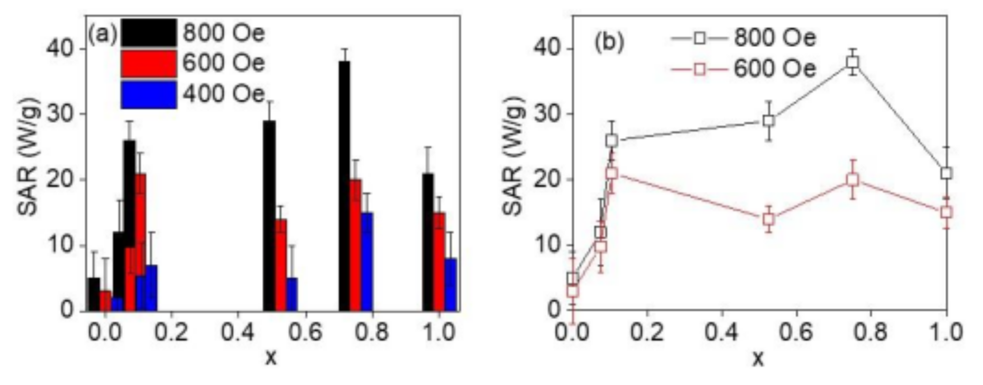
It was established that the SAR values decrease drastically upon reducing the applied external AC magnetic field (Figure 6a). It is generally known that the agglomeration of the nanoparticles, which results in sedimentation, has a negative effect on the SAR value. However, in the present study, the oleic acid and olylamine functionalized hydrophobic nanoparticles were phase transferred to the aqueous phase using TMAH as a capping agent. The TMAH-coated particles remained well dispersed in water during the hyperthermia measurement. Therefore, the effect of particle sedimentation on the hyperthermia response was negligible. The variation of the SAR at external AC applied magnetic fields of 600 and 800 Oe as a function of Co content (Figure 6b). Pure magnetite Fe3−xCoxO4 (x = 0) showed poor heating efficiency even at an external AC applied magnetic field of 800 Oe; however, a reasonable value of the SAR was observed with a small amount of Co content. Similar to the TB and HC at 10 K, the SAR value showed an increase with an increase in the Co content until x = 0.75, above which it showed a decreasing trend (Table 2). This indicates that the SAR value of nanoparticles depends primarily on the magnetic anisotropy of the particles. As discussed above, the magnetocrystalline anisotropy in Fe3−xCoxO4 (x = 0–1) nanoparticles shows a peak at x = 0.75; therefore, the peak in the SAR value at x = 0.75 indicates an important role of the magnetic anisotropy in hyperthermia heating of the present nanoparticles. The relatively smaller values of the SAR for Fe3−xCoxO4 (0.75 ≤ x ≤1) nanoparticles, as compared to 20 nm nanocubes [40], could be attributed to the shape and small size of the nanoparticles. The SAR values are dependent on the size and crystallinity of the nanoparticles and are relatively smaller when the size of the nanoparticles is below 10 nm [52]. Our study demonstrates that the SAR value of the Fe3−xCoxO4 (x = 0–1) nanoparticles varies not just with the size (they are almost the same for all the particles) and Ms but also with magnetocrystalline anisotropy of the particles, on par with the previous reports [35,36]. These results prove that the SAR value of the sub-10 nm Fe3−xCoxO4 nanoparticles can be improved by partial substitution of Fe+2 with Co+2 ions.
4. Conclusions
In summary, we have demonstrated that the heating efficiency (SAR) of the Fe3−xCoxO4 (x = 0–1) nanoparticles improves with an increase in Co content up to x = 0.75, following which it decreases with a further increase of the Co content. Magnetic and hyperthermia measurements on the 7 nm series of spherical Fe3−xCoxO4 nanoparticles have shown that the TB and SAR follow a similar trend with a maximum at x = 0.75. Our study has revealed that the SAR value for the nanoparticles not only depends on the size and saturation magnetization, which were constant for the series, but also primarily on the effective magnetic anisotropy of the particles. Our results are in corroboration with the theoretical findings on Fe3−xCoxO4 (x = 0–1), which showed a maximum of magnetic anisotropy for x to be ~0.6–0.7. The outcome of this study showed that the spherical Fe3−xCoxO4 (x = 0.6–0.7) nanoparticles have the potential of a promising material for magnetic hyperthermia therapy.
Author Contributions
Conceptualization, R.D. and M.-H.P.; methodology, R.D.; formal analysis, R.D.; investigation, R.D., N.P.K. and S.B.A.; resources, R.D., M.-H.P. and H.S.; data curation, R.D., N.P.K. and S.B.A.; writing—original draft preparation, R.D. and M.-H.P.; writing—review and editing, R.D., N.P.K., S.B.A., M.-H.P. and H.S.; supervision, R.D., M.-H.P. and H.S.; funding acquisition, R.D., M.-H.P. and H.S. All authors have read and agreed to the published version of the manuscript.
Funding
This research was funded by Vietnam National Foundation for Science and Technology Development (NAFOSTED), grant number 103.02-2019.314, and Vietnam Ministry of Science and Technology through the national-level project, grant number ĐTĐLCN.17/19. Research at USF was funded by the U.S. Department of Energy, Office of Basic Energy Sciences, Division of Materials Sciences and Engineering, grant number DE-FG02-07ER46438.
Institutional Review Board Statement
Not applicable.
Informed Consent Statement
Not applicable.
Acknowledgments
The work was supported by the Vietnam National Foundation for Science and Technology Development (NAFOSTED) under Grant number 103.02-2019.314. This research was also supported by Vietnam Ministry of Science and Technology through the national-level project ĐTĐLCN.17/19. Research at the University of South Florida was supported by the U.S. Department of Energy, Office of Basic Energy Sciences, Division of Materials Sciences and Engineering under Award No. DE-FG02-07ER46438.
Conflicts of Interest
The authors declare no conflict of interest.
References
- Hyeon, T. Chemical synthesis of magnetic nanoparticles. Chem. Commun. 2003, 8, 927–934. [Google Scholar] [CrossRef]
- Lu, A.-H.; Schmidt, W.; Matoussevitch, N.; Pnnermann, H.B.; Spliethoff, B.; Tesche, B.; Bill, E.; Kiefer, W.; Schuth, F. Nanoengneering of a magnetically separable hydrogenation catalyst. Angew. Chem. Int. Ed. 2004, 43, 4303–4306. [Google Scholar] [CrossRef] [PubMed]
- Tsang, S.C.; Caps, V.; Paraskevas, I.; Chadwick, D.; Thompsett, D. Magnetically separable, carbon-supported nanocatalysts for the manufacture of fine chemicals. Angew. Chem. Int. Ed. 2004, 43, 5645. [Google Scholar] [CrossRef] [PubMed]
- Elliott, D.W.; Zhang, W.-X. Field assessment of nanoscale bimetallic particles for groundwater treatment. Environ. Sci. Technol. 2001, 35, 4922. [Google Scholar] [CrossRef] [PubMed]
- Batlle, X.; Labarta, A. Finite-size effects in fine particles: Magnetic and transport properties. J. Phys. D 2002, 35, R15. [Google Scholar] [CrossRef]
- Sorensen, C.M. Magnetism in Nanoscale Materials in Chemistry. Klabunde, K.J., Ed.; Wiley-Interscience Publication: New York, NY, USA, 2001. [Google Scholar]
- Iwaki, T.; Kakihara, Y. Preparation of high coercivity magnetic FePt nanoparticles by liquid process. J. Appl. Phys. 2003, 94, 6807. [Google Scholar] [CrossRef]
- Lu, A.-H.; Salabas, E.L.; Schuth, F. Magnetic nanoparticles: Synthesis, protection, functionalization, and application. Angew. Chem. Int. Ed. 2007, 46, 1222–1244. [Google Scholar] [CrossRef]
- Noh, S.; Na, W.; Jang, J.; Lee, J.-H.; Lee, E.J.; Moon, S.H.; Lim, Y.; Shin, J.-S.; Cheon, J. Nanoscale magnetism control via surface and exchange anisotropy for optimized ferrimagnetic hysteresis. Nano Lett. 2012, 12, 3716–3721. [Google Scholar] [CrossRef]
- Na, H.B.; Song, I.C.; Hyeon, T. Inorganic nanoparticles for MRI contrast agents. Adv. Mater. 2009, 21, 2133–2148. [Google Scholar] [CrossRef]
- Tirotta, I.; Dichiarante, V.; Pigliacelli, C.; Cavallo, G.; Terraneo, G.; Bombelli, F.B.; Metrangolo, P.; Resnati, G. Magnetic Resonance Imaging (MRI): From Design of Materials to Clinical Applications. Chem. Rev. 2015, 115, 1106–1129. [Google Scholar] [CrossRef]
- Sun, C.; Lee, J.S.H.; Zhang, M. Magnetic nanoparticles in MR imaging and drug delivery. Adv. Drug Deliv. Rev. 2008, 60, 1252–1265. [Google Scholar] [CrossRef] [PubMed] [Green Version]
- Rosi, N.L.; Mirkin, C.A. Nanostructures in biodiagnostics. Chem. Rev. 2005, 105, 1547–1562. [Google Scholar] [CrossRef] [PubMed]
- Hergt, R.; Dutz, S.; Muller, R.; Zeisberger, M. The effect of field parameters, nanoparticle properties and immobilization on the specific heating power in magnetic particle hyperthermia. J. Phys. Condens. Matter 2006, 18, S2935–S2949. [Google Scholar]
- Pankhurst, Q.A.; Connolly, J.; Jones, S.K.; Dobson, J. Applications of magnetic nanoparticles in biomedicine. J. Phys. D Appl. Phys. 2003, 36, R167–R181. [Google Scholar] [CrossRef] [Green Version]
- Pankhurst, Q.A.; Thanh, N.T.K.; Jones, S.K.; Dobson, J. Progress in applications of magnetic nanoparticles in biomedicine. J. Phys. D Appl. Phys. 2009, 42, 224001. [Google Scholar] [CrossRef] [Green Version]
- Colombo, M.; Carregal-Romero, S.; Casula, M.F.; Gutiérrez, L.; Morales, M.P.; Böhm, I.B.; Heverhagen, J.T.; Prosperi, D.; Parak, W.J. Biological applications of magnetic nanoparticles. Chem. Soc. Rev. 2012, 41, 4306–4334. [Google Scholar] [CrossRef]
- Jordan, A. MagForce® Nanotherapy: With tumor specific nanoparticles against cancer. VDI Ber. 2005, 1920, 111. [Google Scholar]
- Hergt, R.; Dutz, S. Magnetic particle hyperthermia–biophysical limitations of a visionary tumour therapy. J. Magn. Magn. Mater. 2007, 311, 187–192. [Google Scholar] [CrossRef]
- Sykes, E.A.; Dai, Q.; Tsoi, K.M.; Hwang, D.M.; Chan, W.C.W. Nanoparticle exposure in animals can be visualized in the skin and analysed via skin biopsy. Nat. Commun. 2013, 5, 3796. [Google Scholar] [CrossRef]
- Roti, J.L. Cellular responses to hyperthermia (40–46 degrees C): Cell killing and molecular events. Int. J. Hyperth. 2008, 24, 3–15. [Google Scholar] [CrossRef]
- Porshokouh, Z.; Salili, S.M.; Masa, J.; Ataie, A.; Das, R.; Phan, M.H.; Srikanth, H. Superparamagnetic iron oxide nanodiscs for hyperthermia therapy: Does size matter? J. Alloys Compd. 2017, 714, 709–714. [Google Scholar]
- Das, R.; Montes, N.R.; Alonso, J.; Amghouz, Z.; Gorria, P.; Blanco, J.A.; Phan, M.H.; Srikanth, H. Boosted hyperthermia therapy by combined AC magnetic and photothermal exposures in Ag/Fe3O4 nanoflowers. ACS Appl. Mater. Interfaces 2016, 8, 25162–25169. [Google Scholar] [CrossRef] [PubMed]
- Brero, F.; Albino, M.; Antoccia, A.; Arosio, P.; Avolio, M.; Berardinelli, F.; Bettega, D.; Calzolari, P.; Ciocca, M.; Corti, M.; et al. Hadron therapy, magnetic nanoparticles and hyperthermia: A promising combined tool for pancreatic cancer treatment. Nanomaterials 2020, 10, 1919. [Google Scholar] [CrossRef] [PubMed]
- Nemec, S.; Kralj, S.; Wilhelm, C.; Hassan, A.A.; Rols, M.P.; Tabi, J.K. Comparison of iron oxide nanoparticles in photothermia and magnetic hyperthermia: Effects of clustering and silica encapsulation on nanoparticles’ heating yield. Appl. Sci. 2020, 10, 7322. [Google Scholar] [CrossRef]
- Salimi, M.; Sarkar, S.; Hashemi, M.; Saber, R. Treatment of breast cancer-bearing BALB/c mice with magnetic hyperthermia using dendrimer functionalized iron-oxide nanoparticles. Nanomaterials 2020, 10, 2310. [Google Scholar] [CrossRef]
- Leboran, I.C.; Baldomir, D.; Boubeta, C.M.; Fesenko, O.C.; del Puerto Morales, M.; Salas, G.; Cabrera, D.; Camarero, J.; Teran, F.J.; Serantes, D. A single picture explains diversity of hyperthermia response of magnetic Nanoparticles. J. Phys. Chem. C 2015, 119, 15698–15706. [Google Scholar] [CrossRef]
- Fortin, J.-P.; Wilhelm, C.; Servais, J.; Ménager, C.; Bacri, J.-C.; Gazeau, F. Size-sorted anionic iron oxide nanomagnets as colloidal mediators for magnetic hyperthermia. J. Am. Chem. Soc. 2007, 129, 2628–2635. [Google Scholar] [CrossRef]
- Nemati, Z.; Alonso, J.; Rodrigo, I.; Das, R.; Garaio, E.; Garcia, J.A.; Orue, I.; Phan, M.H.; Srikanth, H. Improving the heating efficiency of iron oxide nanoparticles by tuning their shape and size. J. Phys. Chem. C 2018, 122, 2367. [Google Scholar] [CrossRef]
- Nemati, Z.; Alonso, J.; Martinez, L.M.; Khurshid, H.; Garaio, E.; Garcia, J.A.; Phan, M.H.; Srikanth, H. Iron oxide nano-octopods with tunable sizes for enhanced hyperthermia. J. Phys. Chem. C 2016, 120, 8370. [Google Scholar] [CrossRef]
- Lavorato, G.; Das, R.; Xing, Y.; Robles, J.; Jochen Litterst, F.; Baggio-Saitovitch, E.; Phan, M.H.; Srikanth, H. Origin and shell-driven optimization of the heating power in core/shell bimagnetic nanoparticles. ACS Appl. Nano Mater. 2020, 3, 1755. [Google Scholar] [CrossRef]
- Fantechi, E.; Innocenti, C.; Albino, M.; Lottini, E.; Sangregorio, C. Influence of cobalt doping on the hyperthermic efficiency of magnetite nanoparticles. J. Magn. Magn. Mater. 2015, 380, 365–371. [Google Scholar] [CrossRef]
- Song, Q.; Zhang, Z.J. Controlled synthesis and magnetic properties of bimagnetic spinel ferrite CoFe2O4 and MnFe2O4 nanocrystals with core-shell architecture. J. Am. Chem. Soc. 2012, 134, 10182–10190. [Google Scholar] [CrossRef] [PubMed]
- Boubeta, C.M.; Simeonidis, K.; Makridis, A.; Angelakeris, M.; Iglesias, O.; Guaardia, P.; Cabot, A.; Yedra, L.; Estradé, S.; Peiró, F.; et al. Learning from nature to improve the heat generation of iron-oxide nanoparticles for magnetic hyperthermia applications. Sci. Rep. 2013, 3, 1652. [Google Scholar] [CrossRef] [PubMed] [Green Version]
- Das, R.; Alonso, J.; Porshokouh, Z.; Kalappattil, V.; Torres, D.; Phan, M.H.; Garaio, E.; Garcia, J.A.; Liamazares, J.L.S.; Srikanth, H. Tunable high aspect ratio iron oxide nanorods for enhanced hyperthermia. J. Phys. Chem. C 2016, 120, 10086–10093. [Google Scholar] [CrossRef]
- Das, R.; Cardarelli, J.A.; Phan, M.H.; Srikanth, H. Magnetically tunable iron oxide nanotubes for multifunctional biomedical applications. J. Alloys Compd. 2019, 789, 323–329. [Google Scholar] [CrossRef]
- Lartigue, L.; Hugounenq, P.; Alloyeau, D.; Clarke, S.P.; Levy, M.; Bacri, J.C.; Bazzi, R.; Brougham, D.F.; Wilhelm, C.; Gazeau, F. Cooperative organization in iron oxide multi-core nanoparticles potentiates their efficiency as heating mediators and MRI contrast agents. ACS Nano 2012, 6, 10935–10949. [Google Scholar] [CrossRef]
- Tabi, J.K.; Corato, R.D.; Lartigue, L.; Marangon, I.; Guardia, P.; Silva, A.K.A.; Luciani, N.; Clement, O.; Flaud, P.; Singh, J.V.; et al. Heat-generating iron oxide nanocubes: Subtle “destructurators” of the tumoral microenvironment. ACS Nano 2014, 8, 4268–4283. [Google Scholar] [CrossRef]
- Anselmo, A.; Mitragotri, S. A review of clinical translation of inorganic nanoparticles. AAPS J. 2015, 17, 1041–1054. [Google Scholar] [CrossRef] [Green Version]
- Sathya, A.; Guardia, P.; Brescia, R.; Silvestri, N.; Pugliese, G.; Nitti, S.; Manna, L.; Pellegrino, T. CoxFe3−xO4 nanocubes for theranostic applications: Effect of cobalt content and particle size. Chem. Mater. 2016, 28, 1769–1780. [Google Scholar] [CrossRef]
- Torres, T.E.; Lima, E., Jr.; Mayoral, A.; Ibarra, A.; Marquina, C.; Ibarra, M.R.; Goya, G.F. Validity of the Neel-Arrhenius model for highly anisotropic CoxFe3−xO4 nanoparticles. J. Appl. Phys. 2005, 118, 183902. [Google Scholar] [CrossRef] [Green Version]
- Sun, S.; Zeng, H. Size-controlled synthesis of magnetite nanoparticles. J. Am. Chem. Soc. 2002, 124, 8204–8205. [Google Scholar] [CrossRef] [PubMed]
- Sun, S.; Zeng, H.; Robinson, D.B.; Raoux, S.; Rice, P.M.; Wang, S.X.; Li, G. Monodisperse MFe2O4 (M = Fe, Co, Mn) nanoparticles. J. Am. Chem. Soc. 2004, 126, 273–279. [Google Scholar] [CrossRef]
- Li, D.; Yun, H.; Diroll, B.T.; Doan-Nguyen, V.V.T.; Kikkawa, J.M.; Murray, C.B. Synthesis and size-selective precipitation of monodisperse nonstoichiometric MxFe3−xO4 (M = Mn, Co) nanocrystals and their DC and AC magnetic properties. Chem. Mater. 2016, 28, 480–489. [Google Scholar] [CrossRef]
- Robles, J.; Das, R.; Glassell, M.; Phan, M.H.; Srikanth, H. Exchange-coupled Fe3O4/CoFe2O4 nanoparticles for advanced magnetic hyperthermia. AIP Adv. 2018, 8, 056719. [Google Scholar] [CrossRef] [Green Version]
- Slonczewski, J.C. Origin of magnetic anisotropy in cobalt-substituted magnetite. Phys. Rev. 1958, 110, 1341. [Google Scholar] [CrossRef]
- Tachiki, M. Origin of magnetocrystalline anisotropy energy in cobalt ferrite. Prog. Theor. Phys. 1960, 23, 1055. [Google Scholar] [CrossRef] [Green Version]
- Nlebedim, I.C.; Snyder, J.E.; Moses, A.J.; Jiles, D.C. Effect of deviation from stoichiometric composition on structural and magnetic properties of cobalt ferrite, CoxFe3−xO4 (x = 0.2 to 1.0). J. Appl. Phys. 2012, 111, 07D704. [Google Scholar] [CrossRef] [Green Version]
- Nlebedim, I.C.; Snyder, J.E.; Moses, A.J.; Jiles, D.C. Anisotropy and magnetostriction in non-stoichiometric cobalt ferrite. IEEE Trans. Magn. 2012, 48, 3084–3087. [Google Scholar] [CrossRef]
- Yu, Y.; Garcia, A.M.; Ning, B.; Sun, S. Cobalt-substituted magnetite nanoparticles and their assembly into ferrimagnetic nanoparticle arrays. Adv. Mater. 2013, 25, 3090–3094. [Google Scholar] [CrossRef]
- Andreu, I.; Natividad, E. Accuracy of available methods for quantifying the heat power generation of nanoparticles for magnetic hyperthermia. Int. J. Hyperth. 2013, 29, 739–751. [Google Scholar] [CrossRef] [Green Version]
- Khandhar, A.P.; Ferguson, R.M.; Simon, J.A.; Krishnan, K.M. Enhancing Cancer Therapeutics Using Size-Optimized Magnetic Fluid Hyperthermia. J. Appl. Phys. 2012, 111, 07B306. [Google Scholar] [CrossRef] [PubMed] [Green Version]
Figure 1.
TEM images of Fe3−xCoxO4 nanoparticles with cobalt content of (a) x = 0, (b) x = 0.07, (c) x = 0.10, (d) x = 0.50, (e) x = 0.75, and (f) x = 1. The inset of each image shows the corresponding diameter histograms. Scale bar is 20 nm in all the images.
Figure 1.
TEM images of Fe3−xCoxO4 nanoparticles with cobalt content of (a) x = 0, (b) x = 0.07, (c) x = 0.10, (d) x = 0.50, (e) x = 0.75, and (f) x = 1. The inset of each image shows the corresponding diameter histograms. Scale bar is 20 nm in all the images.

Figure 2.
XRD pattern of Fe3−xCoxO4 (x = 0.10) nanoparticles. The lower pattern corresponds to the standard cubic Fe3O4.
Figure 2.
XRD pattern of Fe3−xCoxO4 (x = 0.10) nanoparticles. The lower pattern corresponds to the standard cubic Fe3O4.

Figure 3.
(a) Zero-field-cooling (ZFC) and field-cooling (FC) magnetization curve of 7 nm series of Fe3−xCoxO4 (x = 0–1) nanoparticles under 50 Oe magnetic field. (b) The Co content dependence of blocking temperatures of Fe3−xCoxO4 (x = 0–1) nanoparticles.
Figure 3.
(a) Zero-field-cooling (ZFC) and field-cooling (FC) magnetization curve of 7 nm series of Fe3−xCoxO4 (x = 0–1) nanoparticles under 50 Oe magnetic field. (b) The Co content dependence of blocking temperatures of Fe3−xCoxO4 (x = 0–1) nanoparticles.

Figure 4.
(a) M–H curve of 7 nm series of Fe3−xCoxO4 (x = 0–1) nanoparticles at 10 K. (b) Variation of coercivity (HC) at 10 K as a function of Co content.
Figure 4.
(a) M–H curve of 7 nm series of Fe3−xCoxO4 (x = 0–1) nanoparticles at 10 K. (b) Variation of coercivity (HC) at 10 K as a function of Co content.

Figure 5.
(a) M–H curve of 7 nm Fe3−xCoxO4 (x = 0–1) nanoparticles at 300 K. (b) Variation of saturation magnetization (MS) at 300 K as a function of Co content, x.
Figure 5.
(a) M–H curve of 7 nm Fe3−xCoxO4 (x = 0–1) nanoparticles at 300 K. (b) Variation of saturation magnetization (MS) at 300 K as a function of Co content, x.

Figure 6.
(a,b) Specific absorption rate (SAR) of 7 nm series of Fe3−xCoxO4 (x = 0–1) nanoparticles in water, measured at alternating current (AC) fields of 800, 600 and 400 Oe and 310 kHz.
Figure 6.
(a,b) Specific absorption rate (SAR) of 7 nm series of Fe3−xCoxO4 (x = 0–1) nanoparticles in water, measured at alternating current (AC) fields of 800, 600 and 400 Oe and 310 kHz.


Table 1.
Size of nanoparticles corresponding to the Co content in Fe3−xCoxO4 (x = 0–1).
| Fe3−xCoxO4 (x) | Mean Diameter (D) in nm | Standard Deviation (σ) in nm |
|---|---|---|
| 0 | 7.8 | 0.1 |
| 0.07 | 6.7 | 0.1 |
| 0.10 | 6.6 | 0.1 |
| 0.50 | 7.2 | 0.4 |
| 0.75 | 7.1 | 0.2 |
| 1 | 6.5 | 0.1 |
Table 2.
Parameters corresponding to the Co content in Fe3−xCoxO4 (x = 0–1).
| Fe3-xCoxO4 (x) | Blocking Temperature TB (K) | Anisotropy Energy EU = TB × KB (eV) | Saturation Magnetization MS at 300 K (emu/g) | Coercivity HC at 10 K (Oe) | SAR at 800 Oe (W/g) |
|---|---|---|---|---|---|
| 0 | 53 | 0.0045 | 58.0 | 330 | 5 ± 4 |
| 0.07 | 57 | 0.0049 | 54.3 | 1265 | 12 ± 5 |
| 0.10 | 75 | 0.0065 | 54.0 | 2230 | 26 ± 3 |
| 0.50 | 157 | 0.0135 | 57.5 | 8126 | 29 ± 3 |
| 0.75 | 215 | 0.0185 | 60.6 | 17161 | 38 ± 2 |
| 1 | 218 | 0.0187 | 60.7 | 7649 | 21 ± 4 |
Publisher’s Note: MDPI stays neutral with regard to jurisdictional claims in published maps and institutional affiliations. |
© 2021 by the authors. Licensee MDPI, Basel, Switzerland. This article is an open access article distributed under the terms and conditions of the Creative Commons Attribution (CC BY) license (http://creativecommons.org/licenses/by/4.0/).
Share and Cite
MDPI and ACS Style
Das, R.; Kim, N.P.; Attanayake, S.B.; Phan, M.-H.; Srikanth, H. Role of Magnetic Anisotropy on the Hyperthermia Efficiency in Spherical Fe3−xCoxO4 (x = 0–1) Nanoparticles. Appl. Sci. 2021, 11, 930. https://doi.org/10.3390/app11030930
AMA Style
Das R, Kim NP, Attanayake SB, Phan M-H, Srikanth H. Role of Magnetic Anisotropy on the Hyperthermia Efficiency in Spherical Fe3−xCoxO4 (x = 0–1) Nanoparticles. Applied Sciences. 2021; 11(3):930. https://doi.org/10.3390/app11030930
Chicago/Turabian StyleDas, Raja, Ngoc Pham Kim, Supun B. Attanayake, Manh-Huong Phan, and Hariharan Srikanth. 2021. "Role of Magnetic Anisotropy on the Hyperthermia Efficiency in Spherical Fe3−xCoxO4 (x = 0–1) Nanoparticles" Applied Sciences 11, no. 3: 930. https://doi.org/10.3390/app11030930
Note that from the first issue of 2016, this journal uses article numbers instead of page numbers. See further details here.