Article

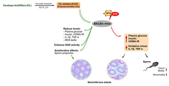
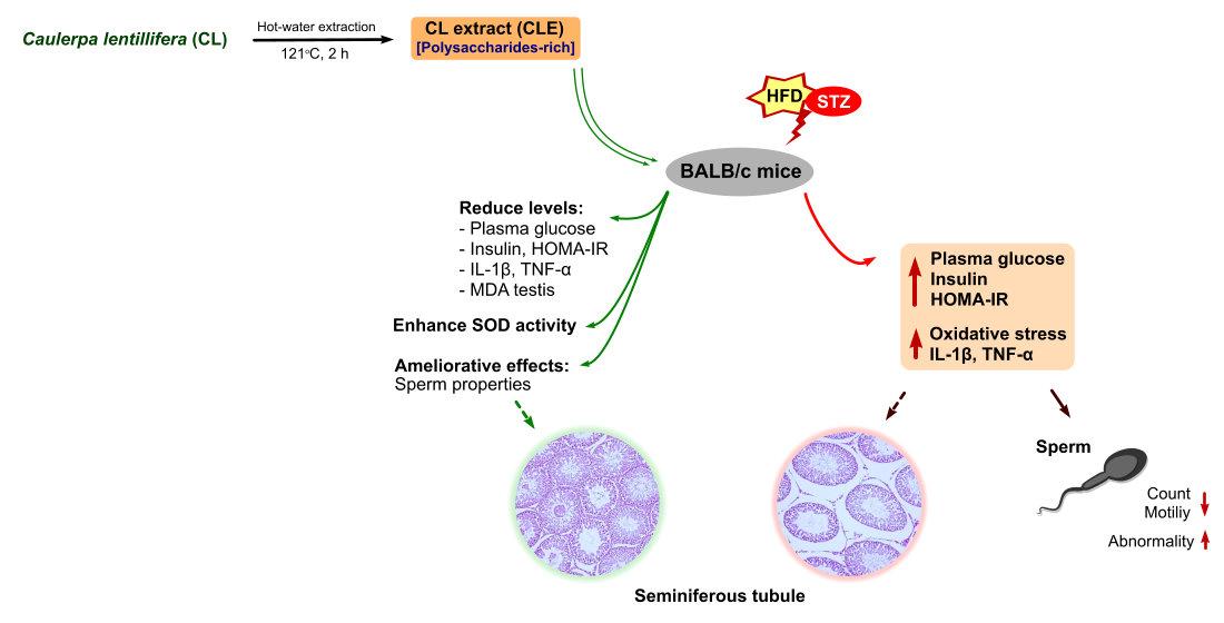
Caulerpa lentillifera Polysaccharides-Rich Extract Reduces Oxidative Stress and Proinflammatory Cytokines Levels Associated with Male Reproductive Functions in Diabetic Mice

1
Department of Food Science, National Taiwan Ocean University, Keelung 20224, Taiwan
2
Fisheries Product Technology, Faculty of Agriculture, Universitas Sriwijaya, Palembang 30862, Indonesia
3
College of Life Science, Fujian Normal University, Fuzhou 350117, China
*
Author to whom correspondence should be addressed.
Appl. Sci. 2020, 10(24), 8768; https://doi.org/10.3390/app10248768
Submission received: 7 November 2020 / Revised: 1 December 2020 / Accepted: 6 December 2020 / Published: 8 December 2020
(This article belongs to the Special Issue Polysaccharides: From Extraction to Applications)

Abstract

:
Diabetes mellitus is a chronic metabolic disease that is positively correlated with reproductive dysfunction. Caulerpa lentillifera is an edible green alga with antioxidant and anti-diabetic properties. This study aims to evaluate the ameliorative effects of a polysaccharides-rich extract from C. lentillifera on the reproductive dysfunctions of diabetic male BALB/c mice induced by a high-fat diet (HFD) supplemented with intraperitoneal injections of streptozotocin (STZ). C. lentillifera was obtained from hot water and converted into a powder form (C. lentillifera extract (CLE)) by freeze drying. Mice were fed an HFD for 4 weeks before supplementing with STZ (30 mg/kg). The diabetic mice were divided into five groups, including a control group, a diabetic (DM) group, a DM with administration of a low-dose CLE treatment (DM+CLE1, 600 mg/kg), a DM with administration of a high-dose of CLE (DM+CLE2, 1000 mg/kg) and a DM with metformin treatment as a positive control (DM+Met, 200 mg/kg) for 6 weeks. The results showed that the CLE administration improved hyperglycemia and insulin resistance. Proinflammatory cytokines such as interleukin-1β and tumor necrosis factor-α were found to decrease in the CLE-treated groups. Additionally, CLE was shown to improve sperm motility and testis morphology. Based on the results, it was confirmed that the polysaccharides-rich extract from C. lentillifera extract was able to prevent diabetes-induced male reproductive dysfunction.

Graphical Abstract

1. Introduction

Diabetes mellitus is a type of metabolic disorder that is considered a serious health problem worldwide. Diabetes is caused either as a result of insufficient insulin secretion (type-1 diabetes) or failure of insulin action (type-2 diabetes). It is characterized by a high glucose level in the blood (hyperglycemia) [1], and proinflammatory cytokines such as interleukin (IL)-1β, IL-6, and IL-2 are increased in the hyperglycemia condition [2]. Dysregulation of tumor necrosis factor-α production is also associated with diabetes [3]. Additionally, hyperglycemia causes an increase in oxidative stress and has a positive association with cell dysfunction in diabetic patients [4,5]. This disease also causes an adverse effect on some organs such as the testis, kidney, liver, and pancreas [6,7]. Reduced levels of some essential hormones, such as luteinizing hormone, follicle-stimulating hormone, and testosterone, as well as low sperm count and motility, have also been seen in diabetic patients [8,9].
Several therapeutic agents have been used to treat diabetes, such as inhibitors of alpha-glucosidase and dipeptidyl peptidase-4, insulin secretagogues, and thiazolidinedione [10]. Unfortunately, some of these agents promote the increased prevalence of cardiovascular disease, gastrointestinal disease, headaches, and nausea [11]. Therefore, studies on novel antidiabetic compounds with less adverse effects is an interesting field and a major challenge for future research in the term of functional foods or natural products.
Marine algae or seaweeds have been widely used for antidiabetic treatments [12]. Seaweeds contain certain bioactive compounds, such as polysaccharides, carotenoids, polyphenols, and phlorotannin, and have been recognized for their beneficial effects on human health [13,14]. Caulerpa lentillifera is a type of green seaweed (also known as sea grapes) widely found in the Indo-Pacific region [15]. Previous studies have reported that C. lentillifera has antioxidant, antibacterial, and anticancer properties [16,17]. Additionally, C. lentillifera also possesses antidiabetic effects by reducing α-glucosidase and dipeptidyl peptidase-IV enzyme activities, while enhancing glucose uptake in adipocytes [18]. Furthermore, the hypoglycemic effects of seaweed extracts such as Sargassum ringgoldianum, Sargassum polycystum, and Ishige okamurae have been reported [19,20,21], and fucoxanthin-rich extract from Laminaria japonica and dietary supplementation of Ulva lactuca and Sargassum angustifolium have exhibited ameliorative effects on reproductive dysfunction [22,23,24]. However, no studies have yet reported on the ameliorative effects of C. lentillifera extract on male reproductive dysfunction under diabetic conditions. We hypothesized that this seaweed extract possesses the ability to reduce reproductive dysfunction under diabetic conditions, and this study aims to evaluate the ameliorative effects of the polysaccharides-rich extract from Caulerpa lentillifera on oxidative stress and proinflammatory cytokines related to reproductive dysfunction using a male diabetic mouse model.

2. Materials and Methods

2.1. Materials

Fresh C. lentillifera seaweed was obtained from Professor Meng-Chou Lee’s Laboratory, Department of Aquaculture, National Taiwan Ocean University (Keelung, Taiwan). Leydig cell line (LC540) was purchased from the Food Industry Research and Development Institute (FIRDI) (Hsinchu, Taiwan). The standard laboratory chow-fed diet (Laboratory Rodent Diet 5001) was purchased from Nutrition International, Inc. (Brentwood, MO, USA). Lard was purchased from MP Biomedicals (Santa Ana, CA, USA). The proinflammatory cytokines, such as tumor necrosis factor (TNF)-α (Cat. No. ARG80206) and interleukin (IL)-1β (Cat. No. ARG80196), were purchased from Arigo Biolaboratories (Hsinchu, Taiwan) and were checked by Enzyme-linked immunosorbent assay (ELISA) kits. The insulin (Cat. No. TCEE50336396T) and testosterone ELISA kits (Cat. No. TCEE30213696T) were purchased from Taiclone Biotechnology Corporation (Taipei, Taiwan). The luteinizing hormone (LH, Cat. No. EM1188) and follicle-stimulating hormone (FSH, Cat. No. EM1035) ELISA kits were purchased from Wuhan Fine Biotech (Wuhan, China), and the kisspeptin (kiss-1, CEC559Mu96) ELISA kit was purchased from Asia Bioscience (Taipei, Taiwan). The glucose (Ref. GL2623), aspartate transferase (AST, Ref. AS1267), alanine aminotransferase (ALT, Ref. AL1268), and enzymatic antioxidants kits, such as superoxide dismutase (SOD, Ref. SD125) and glutathione peroxidase (GPx, Ref. RS505), were purchased from Randox Lab., Ltd. (Crumlin, UK). Streptozotocin (STZ) was purchased from Sigma-Aldrich (St. Louis, MO, USA).

2.2. Caulerpa Lentillifera Extraction

Fresh C. lentillifera seaweed was extracted using the hot-water extraction method, according to the methods of a previous study [25]. Briefly, fresh seaweed was washed three times with water to remove the dirt. The seaweed was cut into a small size using a grinder and extracted using hot water (1:1, w/v) at 121 °C for 2 h. The suspension was centrifuged at 10,000 rpm (4 °C) for 10 min to collect the supernatant. Then, the supernatant was filtered using a paper filter and dried by using a freeze dryer to obtain the C. lentillifera extract (CLE) powder.

2.3. Characterization of Caulerpa Lentillifera Extract

A proximate analysis was performed to determine the content of the CLE. Moisture content was determined in triplicate by drying at 105 °C in an oven to obtain a constant dry weight. The protein content was determined by the Kjeldahl method (Association of Official Analytical Chemists, AOAC method 981.10), and crude fat content was determined by Soxhlet extraction with ethyl ether (AOAC method 991.36). The ash content was determined by complete incineration of the sample in a muffle furnace at 550 °C (AOAC method 930.05). Total sugar was determined by a phenol-sulfuric acid assay according to a previous method [26]. Briefly, the glucose standard was prepared into serial concentrations. The glucose standard and sample extract solutions were added as 1 mL of 80% phenol and 500 µL of 95% sulfuric acid (H2SO4), mixed well and incubated at room temperature for 10 min. After cooling, 200 µL of the solution was pipetted into a 96-well microplate and absorbance was immediately measured at 490 nm using a spectrophotometer.

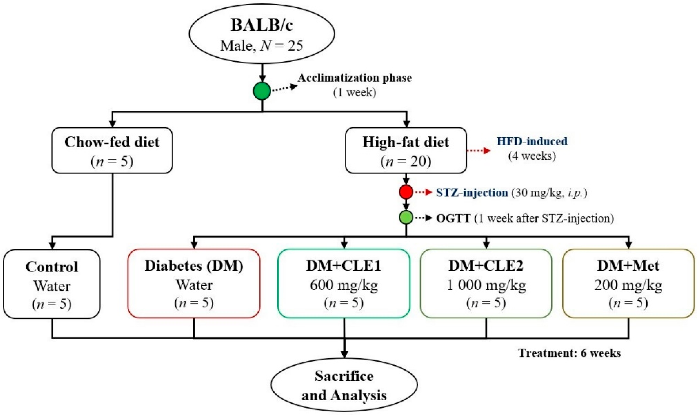
2.4. Animal Experiment

The animal experiment was conducted according to a previous method with some modifications [27]. Six-week-old male BALB/c mice (total of mice, N = 25) were used for the diabetes model. The animals had maintained living conditions (room temperature of 25 °C, 45–50% relative humidity and 12/12-h dark/light cycle) in the Animal Research Center, College of Life Science, National Taiwan Ocean University. All procedures followed the standard of the Institutional Animal Care and Use Committee (IACUC Approval No. 1088011, Date: 23 May 2019) of the National Taiwan Ocean University, Taiwan. Briefly, after a week’s acclimatization, the mice were divided into two main groups: a control group fed a standard commercial diet (five mice) and a diabetes group fed a high-fat diet (HFD, 20 mice) for 4 weeks, as shown in Figure 1. Mice in the diabetes group were intraperitoneally injected with streptozotocin (STZ, 30 mg/kg in citrate buffer, pH 4.5) and divided into four subgroups: a diabetes group (DM, daily oral gavage with water), a DM with a low dose of CLE treatment (CLE1, 600 mg/kg of body weight), a DM with a high dose of CLE (CLE2, 1000 mg/kg), and a DM with metformin as a positive control (metformin, 200 mg/kg) [28]. The mice were treated for 6 weeks before sacrifice, during which time they were placed in an empty chamber and euthanized via CO2 exposure. After sacrifice, whole blood and organs (liver, testis, and hypothalamus) were collected for future analysis.

2.5. Plasma Collection and Homogenized Testis Tissue Preparation

Whole blood was collected on the day of sacrifice using a heparinized syringe to collect it into tubes. The whole blood was then centrifuged at 1000× g for 15 min at 4 °C to separate the plasma. The supernatant (plasma) was pipetted into new collection tubes (1.5 mL conical microtube) and retained for future analysis [29].
The homogenized testis tissue was prepared using a previously published freeze-thaw method [30]. Briefly, the testis tissue (100 mg) was suspended in cold phosphate-buffered saline (PBS, 900 µL). The suspension was homogenized using a micro-tube homogenizer and stored at −20 °C. After 24 h, the freeze-homogenized tissue was thawed at room temperature before use and centrifuged at 5000 rpm for 25 min. The supernatant of the homogenized tissue was collected for future analysis.

2.6. Glucose and Plasma Biochemical Analysis

The plasma glucose concentration was measured by commercial glucose enzymatic kits. Plasma insulin concentration was tested by an insulin ELISA kit. Homeostasis Model Assessment-Insulin Resistance (HOMA-IR) was determined by multiplying the fasting plasma glucose (mmol/L) by the fasting plasma insulin (mU/L), then dividing by 22.5 [31]. The proinflammatory cytokines (i.e., TNF-α and IL-1, kisspeptin (kiss-1)) and the reproductive hormones (i.e., luteinizing hormone, follicle-stimulating hormone, and testosterone) were analyzed by ELISA kits according to their manufacturers’ protocols.

2.7. Enzymatic Antioxidants and Oxidative Stress Assay

The activity of enzymatic antioxidants, such as superoxide dismutase (SOD), catalase (Cat), and glutathione peroxidase (GPx), were measured using commercial kits. The malondialdehyde (MDA) level of the homogenized testis tissue supernatant was used to evaluate lipid peroxidation and was determined according to a previously reported method using MDA reagent (15% (w/v) trichloroacetic acid in 0.25 N HCl and 0.375% (w/v) thiobarbituric acid in 0.25 185 N HCl) [32]. A nitro blue tetrazolium (NBT) test was used to determine the superoxide anion (O2−) level in the sperm [33]. Briefly, the sperm cells were added to 0.2 mL of NBT solution (0.1 mg/mL NBT, 5% Fetal bovine serum, 3% Dimethyl sulfoxide) in the tubes. The mixture was incubated in 10 mL RPMI for 60 min at 37 °C then centrifuged at 500× g for 10 min. The supernatant was removed from the tubes, then the tubes were washed with PBS. Next, 200 μL of DMSO was added to dissolve the purple crystals in the cells, which were placed in an ultrasonic oscillator for 15 min, and 200 μL of the purple solution was pipetted to a 96-well microplate to measure the absorbance at 570 nm using a spectrophotometry. The nitric oxide (NO) assay was conducted based on a previously reported protocol [34]. Briefly, the sperm cells or supernatant of homogenized testis were added to Griess reagent (sulfanilamide dissolved in 5% H3PO4 and N-(1-naphthyl)ethylenediamine dissolved in H2O) at a 1:1 volume ratio. The solution was allowed to react for 15 min at room temperature, then the absorbance was measured at 540 nm using a spectrophotometer.

2.8. Sperm Analysis

The swim-up method was used to collect the sperm from the epididymis, as described previously [35]. Briefly, the epididymis was put into RPMI medium, then the cauda epididymis was cut and shaken at 120 rpm for 10 min. It was centrifuged at 200× g for 5 min, then incubated at 37 °C for 30 min. The liquid in the upper layer was taken and transferred into a new collection tube for future analysis. The sperm count, motility, and sperm abnormality were observed using a hemocytometer under a light microscope according to previous methods [36,37]. Sperm count is the total number of sperm, and sperm motility is the total sperm with the ability to move divided by total sperm count then multiplied by 100. Sperm abnormality is the total sperm with an abnormal form (such as a double head or double tail) divided by the total sperm then multiplied by 100.

2.9. Histopathology Analysis

After sacrifice, one testis was fixed with 4% formaldehyde solution for 2 days. After decalcification, the sections were cut (paraffin-embedded, 5 µm) and stained with hematoxylin and eosin (H&E). The staining process was performed by Li Pie Co. Ltd. (Taichung, Taiwan). The stained section was observed under a light microscope.

2.10. Statistical Analysis

All values were given as mean ± standard deviation (SD). All statistical calculations were done by the SPSS statistics v22.0 (SPSS for Windows Inc., version 22; Chicago, IL, USA) system. One-way ANOVA was used to examine the overall differences between groups, and a Duncan’s multiple range test was used to identify significant differences (p < 0.05) between the groups. All figures produced were drawn using GraphPad Prism 5 (GraphPad Software Inc., San Diego, CA, USA).

3. Results

3.1. Caulerpa Lentillifera Extract Chemical Compositions

The C. lentillifera extract was composed of carbohydrate (69.75 ± 0.86%), protein (10.5 ± 0.77%), lipid (0.83 ± 0.53%), ash (12.5 ± 0.76%), and moisture (6.42 ± 0.53%). The total sugar content of the extract was about 12.49 ± 0.77 µg/mL.

3.2. Effect of Caulerpa Lentillifera on Body and Organs Weight of Diabetic Mice

Table 1 shows that there was no significant difference in the initial body weight of the mice between all groups, and that C. lentillifera extract (CLE) significantly protected the body weight loss after 6 weeks’ treatment. CLE also significantly reduced kidney and abdominal fat weights in diabetic mice, whereas there were no significant effects on spleen and liver weights between the control, DM, and treated groups.

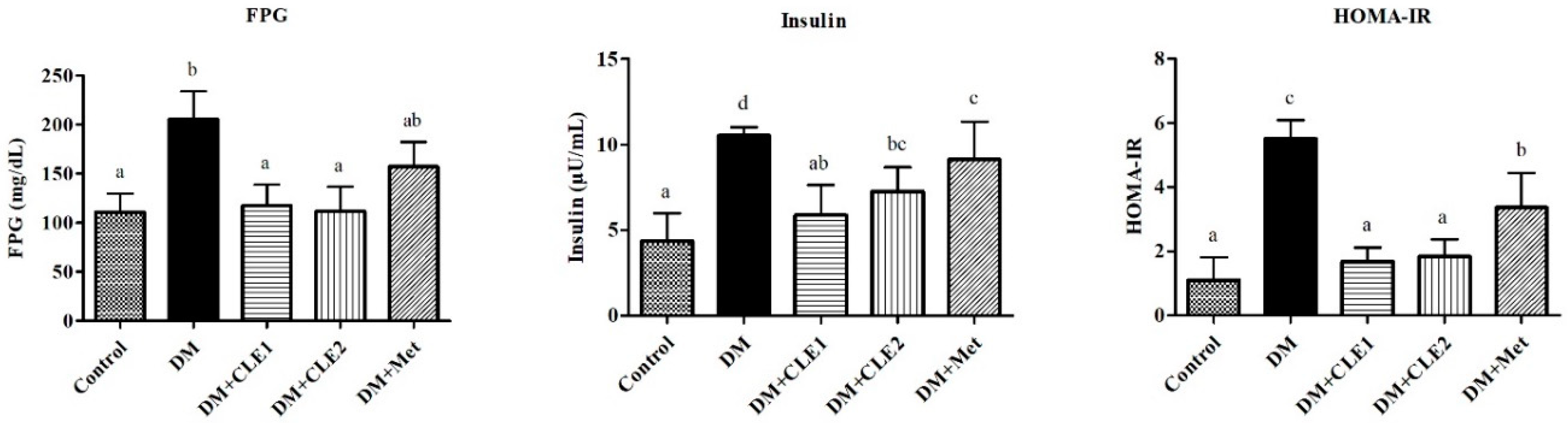
3.3. Caulerpa Lentillifera Extract Improves Plasma Glucose, Insulin, and HOMA-IR Levels in Diabetic Mice

High levels of glucose, insulin, and homeostasis model assessment-insulin resistance (HOMA-IR) were observed in the diabetes (DM) group, as shown in Figure 2. Hypoglycemic effects were observed after treatment with CLE (especially at high doses), which was determined from the decreased level of abovementioned factors. As a positive control, metformin also showed a lowering effect on glucose level. However, there was no significant reduction in glucose level when compared to the DM group. On the other hand, insulin and HOMA-IR levels decreased significantly after metformin treatment.

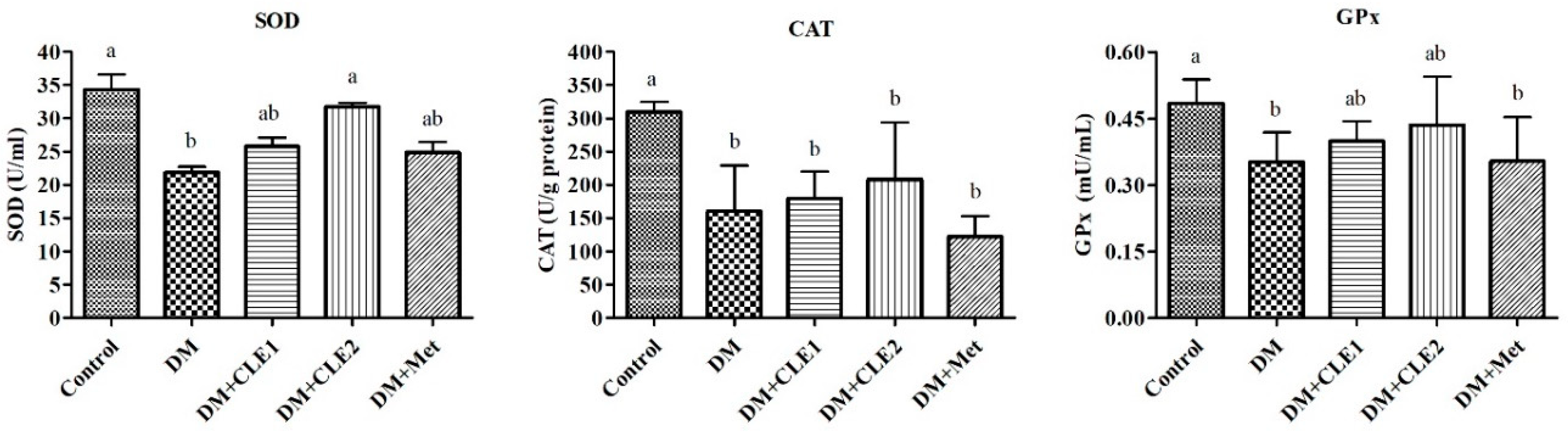
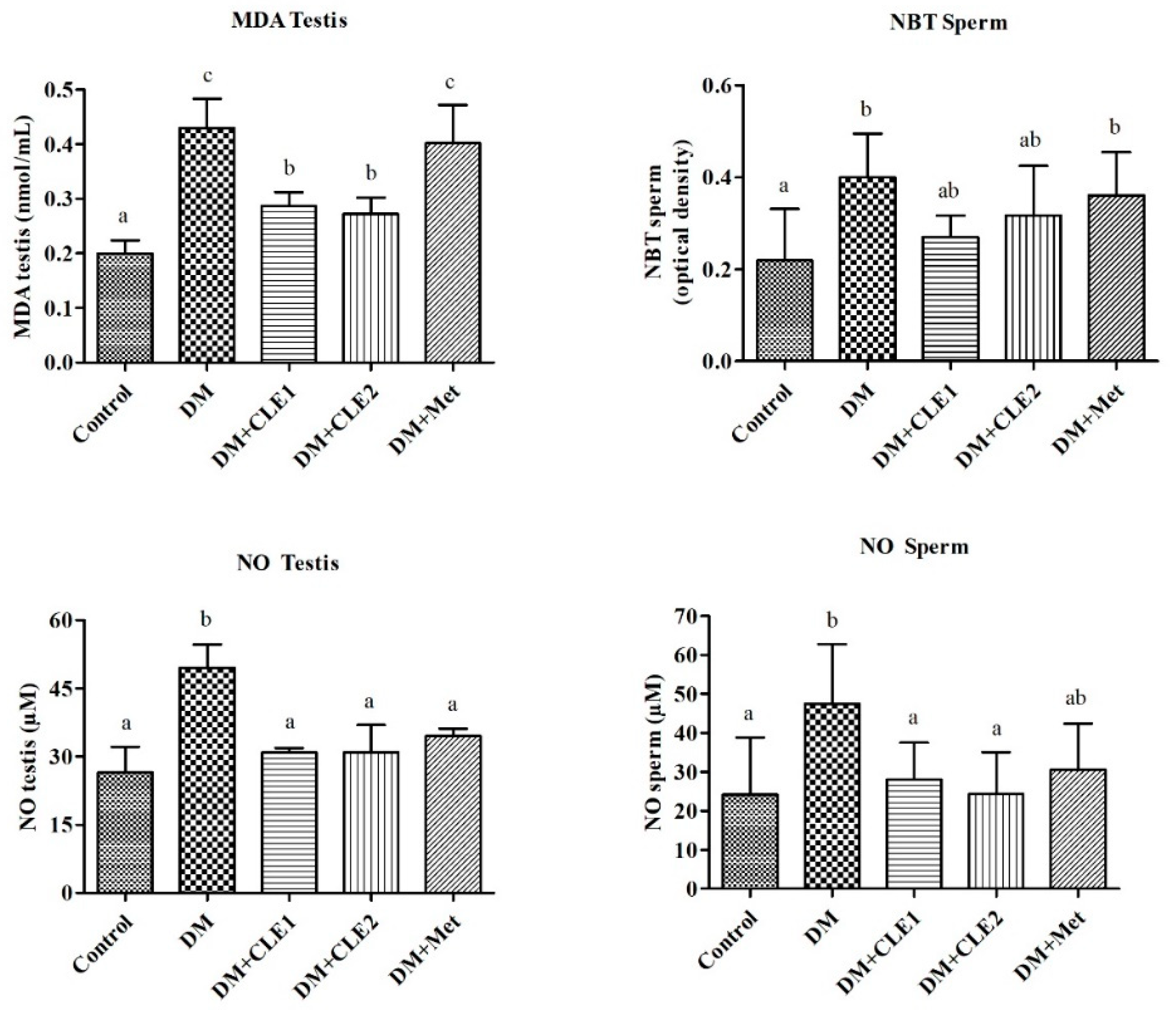
3.4. Caulerpa Lentillifera Extract Ameliorates Oxidative Stress in Diabetic Male Mice

The effect of C. lentillifera extract (CLE) on the activities of plasma enzymatic antioxidants such as superoxide dismutase (SOD), catalase (CAT), and glutathione peroxidase (GPx) are showed in Figure 3. The diabetes (DM) group was characterized by low levels of enzymatic antioxidants, CLE administration significantly enhanced SOD activity, especially in high-dose CLE, and there were no effects of CLE administration on CAT activity. On the other hand, GPx activity increased after CLE treatments. Ultimately, there was no significant difference between CLE-treated and DM groups.
Lipid peroxidation (MDA) in the testis was determined after 6 weeks treatment with C. lentillifera and metformin, as shown in Figure 4. The high value of MDA was shown in the diabetes (DM) group. The CLE treatment group significantly reduced MDA levels compared to the DM group, whereas metformin showed no significant difference from the DM group. The level of nitro blue tetrazolium (NBT) reduction was a quantitative assay for superoxide anion (O2−) production, and nitric oxide (NO) increased in the DM group (Figure 4). The level of NO in the testis and sperm was significantly reduced after 6 weeks treatment with CLE.

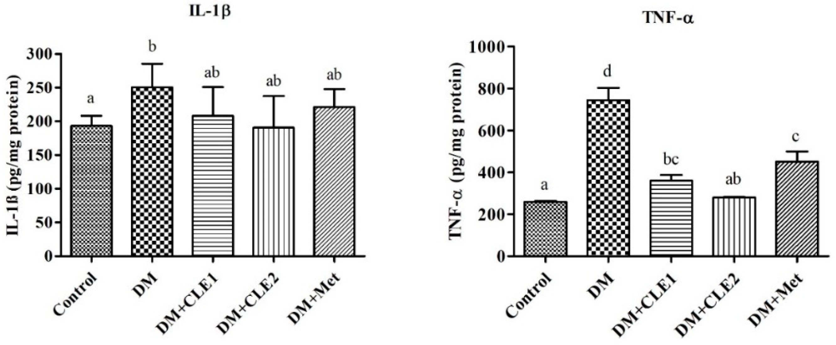
3.5. Caulerpa Lentillifera Extract Reduces Interleukin-1 Beta and Tumor Necrosis Factor-Alpha

The interleukin (IL)-1β and tumor necrosis factor (TNF)-α levels in plasma were increased in the diabetes (DM) group when compared to the control group (Figure 5). The expression of IL-1β in high-dose CLE and metformin treatments were reduced when compared to the DM group. However, there were no significant effects. The TNF-α expression was also significantly reduced in the high-dose CLE group compared to the DM group.

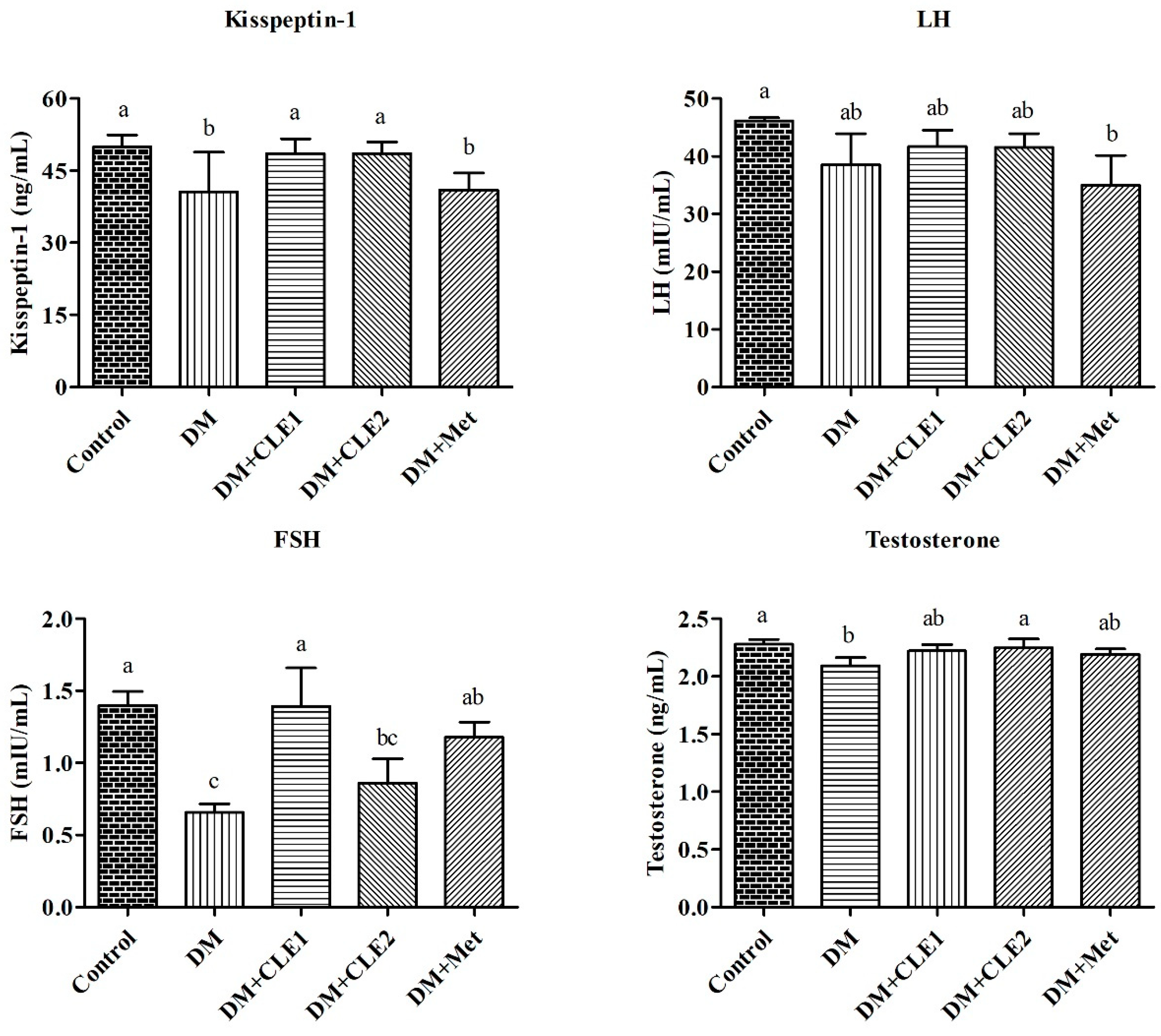
3.6. Caulerpa Lentillifera Extract Administration Improves Levels of Kisspeptin-1 and Reproductive Hormones

The level of kisspeptin-1 was significantly lower in the diabetes (DM) group than the control group. However, this level significantly increased after treatment for 6 weeks with CLE (Figure 6). The levels of follicle-stimulating hormone (FSH) and testosterone in the DM group were also significantly lower than the control group. Oral supplementation with CLE for 6 weeks successfully restored the levels of FSH and testosterone, especially in high doses of CLE. The CLE treatments showed no effects on LH levels when compared to the DM group.

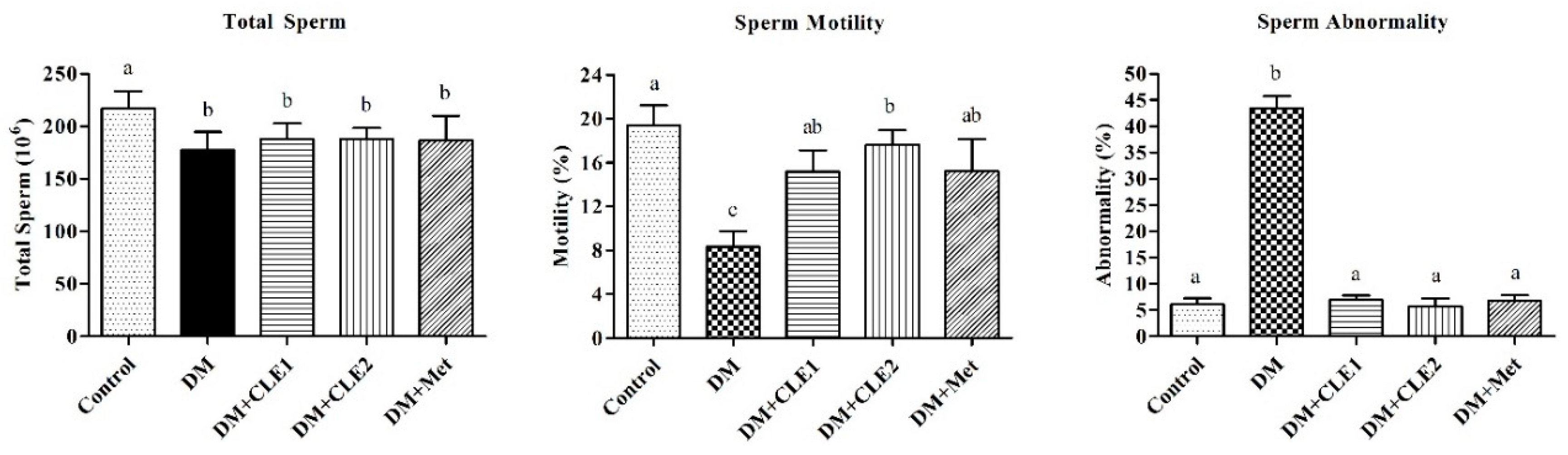
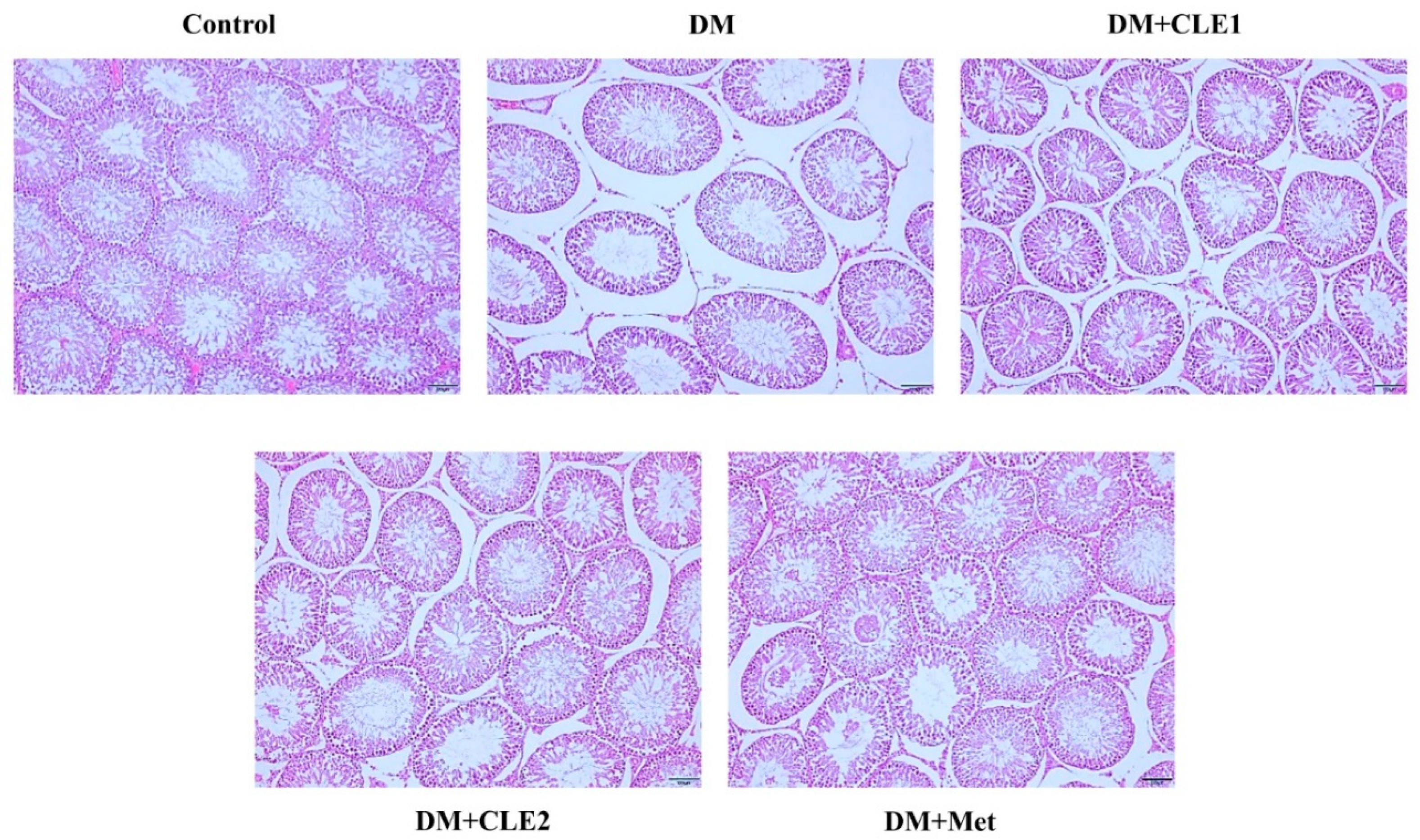
3.7. Effects of Caulerpa Lentillifera on the Sperm and Seminiferous Tubules Morphology

The total sperm and sperm motility of the diabetes (DM) group was significantly lower than the control group, whereas sperm abnormality was significantly higher in the DM group compared to the control and treated groups (Figure 7). Treatment with CLE for 6 weeks not only significantly increased sperm motility, but also significantly reduced sperm abnormality. Hematoxylin and eosin (H&E) staining was used to evaluate seminiferous tubules morphology. The seminiferous tubule structure in the DM group appeared separated when compared to the other groups (Figure 8). Whereas, the high-dose CLE and metformin treatments successfully ameliorated the seminiferous tubule morphology.

4. Discussion

In this study, a diabetic male mouse model induced via a high-fat diet and intraperitoneal injections with streptozotocin was administered C. lentillifera extract for 6 weeks, which successfully ameliorated oxidative stress and some cytokine-related reproductive dysfunction. The C. lentillifera extract contained a high level of carbohydrates. Polysaccharides (a long chain of carbohydrates) are in the major constituent of seaweed, and polysaccharides from marine organisms have been widely used for pharmaceutical, nutraceutical, and cosmetic applications [38]. Seaweed polysaccharides have also been used for the potential treatment of inflammatory bowel disease (IBD), osteoarthritis, and diabetes [12,30,39].
The abdominal fat weight in the diabetes (DM) group was significantly increased compared to the control group. The high fat weight observed was the result of rats fed with a high-fat diet (HFD). However, the CLE treatment significantly reduced abdominal fat weight to a level that was similar to the control group. Previous studies reported increases in the total fat, subcutaneous fat, and visceral fat of mice after being fed an HFD [40,41]. In contrast, total body weight was significantly lowered in the DM group compared to the control and treatment groups. A previous study reported that BALB/c mice do not store excess lipids such as diacylglycerols and triacylglycerols in the liver. This condition is due to differences in lipid oxidation or lipogenesis, which are higher than fatty acid uptake and result in higher body weight [42]. Additionally, early literature also reported that diabetic BALB/c mice showed weight loss 1 week after streptozotocin injection [43].
The present study showed significantly increased levels of fasting plasma glucose and homeostasis measurements of insulin resistance (HOMA-IR) in the diabetes (DM) group compared to the control and treated groups. Type-2 diabetes is characterized by high levels of glucose and insulin resistance, as reported in previous studies [43,44]. Rising insulin concentrations are recognized as insulin resistance (IR) in type-2 diabetes. HOMA-IR has been used previously for IR estimation [45,46]. In this study, the levels of glucose and HOMA-IR were reduced after 6 weeks of oral administration of CLE. According to these data, CLE treatments showed a hypoglycemic effect. Previous studies also showed that seaweed extracts possess hypoglycemic effects, such as the extracts of S. ringgoldianum, S. polycystum, and I. okamurae [19,20,21].
Low levels of enzymatic antioxidants such as superoxide dismutase (SOD), catalase (CAT), and glutathione peroxidase (GPx) were observed in the DM group compared to the control and treated groups. The DM group also showed high levels of lipid peroxidation products and superoxide anions, as analyzed by malondialdehyde (MDA) and nitro-tetrazolium (NBT) assays, respectively. These conditions proved that diabetes is characterized by increasing oxidative stress. A review study reported that oxidative stress can be induced by high glucose levels (hyperglycemia) in diabetic complications [4]. The level of nitric oxide (NO) also increased in the DM group compared to the control and treated-groups. Nitric oxide has been reported as an oxidative stress indicator [47]. The oral administration of CLE for 6 weeks successfully enhanced enzymatic antioxidant levels, especially for SOD and GPx, while reducing lipid peroxidation products and nitric oxide levels.
Increases in pro-inflammatory cytokines such as interleukin (IL)-1β, IL-6, and tumor necrosis factor (TNF)-α have been evaluated in diabetes conditions [48,49]. The present study also recorded increases in pro-inflammatory cytokines such as IL-1β and TNF-α in the DM group. A previous study reported that the levels of these cytokines significantly are increased in a hyperglycemic condition compared to a healthy condition [48], whereas TNF-α is also recognized as an inflammatory mediator to promote IR [50].
Kisspeptin (kiss-1 protein) plays important roles in reproductive function. It is also recognized as an upstream regulator of gonadotropin-releasing hormone (GnRH) in the hypothalamic-pituitary-gonadal (HPG) axis [51]. In the present study, the low levels of kisspeptin-1 and reproductive hormones such as luteinizing hormone (LH), follicle-stimulating hormone (FSH), and testosterone were observed in the DM group. A previous study reported that a low level of the kiss-1 protein was evaluated in a diabetes model. Additionally, levels of LH and FSH were also reduced after STZ injections in the diabetic animal model [52]. The disruption of kiss-1 expression was associated with a low level of gonadotrophin (hypogonadotropic hypogonadism) [53]. The oral administration of CLE for 6 weeks successfully increased kiss-1 protein, FSH, and testosterone levels.
The present study showed low total sperm and sperm motility as well as high sperm abnormality in the DM group compared to the control group. This result proves that disruption of the upstream regulator such as kiss-1 protein and certain male reproductive hormones results in low sperm quality. In men, LH and FSH play an important role in spermatogenesis and maintaining their level of testosterone [54]. A previous study reported that hyperglycemia impairs male reproductive function [55]. In the present study, men in a diabetic condition showed a higher level of sperm DNA fragmentation compared to healthy men, due to the oxidative process, whereas damage to sperm DNA was associated with a reduction in the quality of the sperm [56]. The present study also showed a decrease in seminiferous tubule diameter in the DM group. Previous studies have reported that diabetes can result in a decreased diameter of the seminiferous tubule [57]. In diabetes, testicular dysfunction is caused by seminiferous tubule atrophy, decreased diameter of the tubule, spermatogenesis cell reduction, and cell apoptosis in germ cells [58]. The oral administration of CLE for 6 weeks successfully ameliorated testicular properties, such as increases in sperm motility and reduced sperm abnormality, while protecting seminiferous tubule morphology.
Overall, this study shows that CLE not only possesses an antidiabetic effect, but also improves some proinflammatory cytokines related to male reproductive function. Previous studies have reported that seaweed extracts showed ameliorative effects on reproductive function not only in diabetic models, including dietary supplementation of U. lactuca, fucoxanthin-rich extract from L. japonica, and S. angustifolium [22,23,24,59].

5. Conclusions

C. lentillifera extract (CLE) is rich in polysaccharide. CLE administration was able to reduce pro-inflammatory cytokines, lipid peroxidation, oxidative stress, and insulin resistance. Additionally, CLE showed improved sperm motility and seminiferous tubule morphology. According to these results, C. lentillifera extract successfully prevented diabetes-induced male reproductive dysfunction in mice models. This extract can also be used as a dietary supplement and an alternative treatment for diabetes management.

Author Contributions

Conceptualization, Z.-L.K.; Formal analysis, K.K. and S.S.; Writing—original draft, K.K. and S.S.; Writing—review & editing, S.S., L.H., and Z.-L.K. All authors have read and agreed to the published version of the manuscript.

Funding

This research received no external funding.

Acknowledgments

We are also very grateful that Meng-Chou Lee’s Laboratory Department of Aquaculture National Taiwan Ocean University (Keelung, Taiwan) provided the Caulerpa lentillifera for this study.

Conflicts of Interest

The authors declare no conflict of interest.

References

  1. Wang, P.; Fiaschi-Taesch, N.M.; Vasavada, R.C.; Scott, D.K.; García-Ocaña, A.; Stewart, A.F. Diabetes mellitus—Advances and challenges in human β-cell proliferation. Nat. Rev. Endocrinol. 2015, 11, 201. [Google Scholar] [CrossRef]
  2. Dogan, Y.; Akarsu, S.; Ustundag, B.; Yilmaz, E.; Gurgoze, M.K. Serum il-1β, il-2, and il-6 in insulin-dependent diabetic children. Mediat. Inflamm. 2006, 2006, 1–6. [Google Scholar] [CrossRef] [Green Version]
  3. Swaroop, J.; Naidu, J.N.; Rajarajeswari, D. Association of tnf-α with insulin resistance in type 2 diabetes mellitus. Indian J. Med. Res. 2012, 135, 127–130. [Google Scholar] [CrossRef] [PubMed]
  4. King, G.L.; Loeken, M.R. Hyperglycemia-induced oxidative stress in diabetic complications. Histochem. Cell Biol. 2004, 122, 333–338. [Google Scholar] [CrossRef] [PubMed]
  5. Poitout, V.; Robertson, R.P. Minireview: Secondary β-cell failure in type 2 diabetes—A convergence of glucotoxicity and lipotoxicity. Endocrinology 2002, 143, 339–342. [Google Scholar] [CrossRef] [PubMed]
  6. Abd El-Twab, S.M.; Mohamed, H.M.; Mahmoud, A.M. Taurine and pioglitazone attenuate diabetes-induced testicular damage by abrogation of oxidative stress and up-regulation of the pituitary–gonadal axis. Can. J. Physiol. Pharmacol. 2016, 94, 651–661. [Google Scholar] [CrossRef]
  7. Aksu, I.; Baykara, B.; Kiray, M.; Gurpinar, T.; Sisman, A.R.; Ekerbicer, N.; Tas, A.; Gokdemir-Yazar, O.; Uysal, N. Serum igf-1 levels correlate negatively to liver damage in diabetic rats. Biotech. Histochem. 2013, 88, 194–201. [Google Scholar] [CrossRef]
  8. Agbaje, I.M.; Rogers, D.A.; McVicar, C.M.; McClure, N.; Atkinson, A.B.; Mallidis, C.; Lewis, S.E.M. Insulin dependant diabetes mellitus: Implications for male reproductive function. Hum. Reprod. 2007, 22, 1871–1877. [Google Scholar] [CrossRef] [Green Version]
  9. Dandona, P.; Dhindsa, S.; Chandel, A.; Chaudhuri, A. Hypogonadotropic hypogonadism in men with type 2 diabetes. Postgrad. Med. 2015, 121, 45–51. [Google Scholar] [CrossRef]
  10. Marín-Peñalver, J.J.; Martín-Timón, I.; Sevillano-Collantes, C.; del Cañizo-Gómez, F.J. Update on the treatment of type 2 diabetes mellitus. World J. Diabetes 2016, 7, 354–395. [Google Scholar] [CrossRef]
  11. Chaudhury, A.; Duvoor, C.; Reddy Dendi, V.S.; Kraleti, S.; Chada, A.; Ravilla, R.; Marco, A.; Shekhawat, N.S.; Montales, M.T.; Kuriakose, K.; et al. Clinical review of antidiabetic drugs: Implications for type 2 diabetes mellitus management. Front. Endocrinol. 2017, 8, 6. [Google Scholar] [CrossRef] [PubMed] [Green Version]
  12. Sharifuddin, Y.; Chin, Y.-X.; Lim, P.-E.; Phang, S.-M. Potential bioactive compounds from seaweed for diabetes management. Mar. Drugs 2015, 13, 5447–5491. [Google Scholar] [CrossRef] [PubMed] [Green Version]
  13. Kadam, S.U.; Prabhasankar, P. Marine foods as functional ingredients in bakery and pasta products. Food Res. Int. 2010, 43, 1975–1980. [Google Scholar] [CrossRef]
  14. Lee, S.-H.; Jeon, Y.-J. Anti-diabetic effects of brown algae derived phlorotannins, marine polyphenols through diverse mechanisms. Fitoterapia 2013, 86, 129–136. [Google Scholar] [CrossRef]
  15. de Gaillande, C.; Payri, C.; Remoissenet, G.; Zubia, M. Caulerpa consumption, nutritional value and farming in the indo-pacific region. J. Appl. Phycol. 2016, 29, 2249–2266. [Google Scholar] [CrossRef]
  16. Yap, W.-F.; Tay, V.; Tan, S.-H.; Yow, Y.-Y.; Chew, J. Decoding antioxidant and antibacterial potentials of malaysian green seaweeds: Caulerpa racemosa and Caulerpa lentillifera. Antibiotics 2019, 8, 152. [Google Scholar] [CrossRef] [Green Version]
  17. Fedorov, S.; Ermakova, S.; Zvyagintseva, T.; Stonik, V. Anticancer and cancer preventive properties of marine polysaccharides: Some results and prospects. Mar. Drugs 2013, 11, 4876–4901. [Google Scholar] [CrossRef] [Green Version]
  18. Sharma, B.R.; Rhyu, D.Y. Anti-diabetic effects of Caulerpa lentillifera: Stimulation of insulin secretion in pancreatic β-cells and enhancement of glucose uptake in adipocytes. Asian Pac. J. Trop. Biomed. 2014, 4, 575–580. [Google Scholar] [CrossRef] [Green Version]
  19. Lee, C.-W.; Han, J.-S. Hypoglycemic effect of Sargassum ringgoldianum extract in stz-induced diabetic mice. Prev. Nutr. Food Sci. 2012, 17, 8–13. [Google Scholar] [CrossRef] [Green Version]
  20. Motshakeri, M.; Ebrahimi, M.; Goh, Y.M.; Matanjun, P.; Mohamed, S. Sargassum polycystumreduces hyperglycaemia, dyslipidaemia and oxidative stress via increasing insulin sensitivity in a rat model of type 2 diabetes. J. Sci. Food Agric. 2013, 93, 1772–1778. [Google Scholar] [CrossRef]
  21. Min, K.-H.; Kim, H.-J.; Jeon, Y.-J.; Han, J.-S. Ishige okamurae ameliorates hyperglycemia and insulin resistance in c57bl/ksj-db/db mice. Diabetes Res. Clin. Pract. 2011, 93, 70–76. [Google Scholar] [CrossRef] [PubMed]
  22. Kong, Z.-L.; Sudirman, S.; Hsu, Y.-C.; Su, C.-Y.; Kuo, H.-P. Fucoxanthin-rich brown algae extract improves male reproductive function on streptozotocin-nicotinamide-induced diabetic rat model. Int. J. Mol. Sci. 2019, 20, 4485. [Google Scholar] [CrossRef] [PubMed] [Green Version]
  23. Okab, A.B.; Samara, E.M.; Abdoun, K.A.; Rafay, J.; Ondruska, L.; Parkanyi, V.; Pivko, J.; Ayoub, M.A.; Al-Haidary, A.A.; Aljumaah, R.S.; et al. Effects of dietary seaweed (ulva lactuca) supplementation on the reproductive performance of buck and doe rabbits. J. Appl. Anim. Res. 2013, 41, 347–355. [Google Scholar] [CrossRef]
  24. Sobhani, A.; Eftekhaari, T.E.; Shahrzad, M.E.; Natami, M.; Fallahi, S. Antioxidant effects of brown algae sargassum on sperm parameters. Medicine 2015, 94, e1938. [Google Scholar] [CrossRef]
  25. Fujiki, K.; Matsuyama, H.; Yano, T. Effect of hot-water extracts from marine algae on resistance of carp and yellowtail against bacterial infections. Sci. Bull. Fac. Agric.-Kyushu Univ. (Jpn.) 1993, 47, 137–141. [Google Scholar]
  26. Nielsen, S.S. Total carbohydrate by phenol-sulfuric acid method. In Food Analysis Laboratory Manual; Springer: Cham, Switzerland, 2017; pp. 137–141. [Google Scholar]
  27. Sharma, B.; Kim, H.; Rhyu, D. Caulerpa lentillifera extract ameliorates insulin resistance and regulates glucose metabolism in c57bl/ksj-db/db mice via pi3k/akt signaling pathway in myocytes. J. Transl. Med. 2015, 13, 62. [Google Scholar] [CrossRef] [Green Version]
  28. Oliveira, W.H.; Nunes, A.K.; França, M.E.R.; Santos, L.A.; Lós, D.B.; Rocha, S.W.; Barbosa, K.P.; Rodrigues, G.B.; Peixoto, C.A. Effects of metformin on inflammation and short-term memory in streptozotocin-induced diabetic mice. Brain Res. 2016, 1644, 149–160. [Google Scholar] [CrossRef]
  29. Sudirman, S.; Chang, H.-W.; Chen, C.-K.; Kong, Z.-L. A dietary polysaccharide from Eucheuma cottonii downregulates proinflammatory cytokines and ameliorates osteoarthritis-associated cartilage degradation in obese rats. Food Funct. 2019, 10, 5697–5706. [Google Scholar] [CrossRef]
  30. Sudirman, S.; Hsu, Y.H.; He, J.L.; Kong, Z.L. Dietary polysaccharide-rich extract from Eucheuma cottonii modulates the inflammatory response and suppresses colonic injury on dextran sulfate sodium-induced colitis in mice. PLoS ONE 2018, 13, e0205252. [Google Scholar] [CrossRef] [Green Version]
  31. Sudirman, S.; Hsu, Y.-H.; Johnson, A.; Tsou, D.; Kong, Z.-L. Amelioration effects of nanoencapsulated triterpenoids from petri dish-cultured Antrodia cinnamomea on reproductive function of diabetic male rats. Int. J. Nanomed. 2018, 13, 5059–5073. [Google Scholar] [CrossRef] [Green Version]
  32. Placer, Z.A.; Cushman, L.L.; Johnson, B.C. Estimation of product of lipid peroxidation (malonyl dialdehyde) in biochemical systems. Anal. Biochem. 1966, 16, 359–364. [Google Scholar] [CrossRef]
  33. Tunc, O.; Thompson, J.; Tremellen, K. Development of the nbt assay as a marker of sperm oxidative stress. Int. J. Androl. 2008, 33, 13–21. [Google Scholar] [CrossRef]
  34. Balercia, G.; Moretti, S.; Vignini, A.; Magagnini, M.; Mantero, F.; Boscaro, M.; Ricciardo-Lamonica, G.; Mazzanti, L. Role of nitric oxide concentrations on human sperm motility. J. Androl. 2004, 25, 245–249. [Google Scholar] [CrossRef] [PubMed]
  35. Yokoi, K.; Uthus, E.O.; Nielsen, F.H. Nickel deficiency diminishes sperm quantity and movement in rats. Biol. Trace Elem. Res. 2003, 93, 141–154. [Google Scholar] [CrossRef]
  36. Sönmez, M.; Türk, G.; Yüce, A. The effect of ascorbic acid supplementation on sperm quality, lipid peroxidation and testosterone levels of male wistar rats. Theriogenology 2005, 63, 2063–2072. [Google Scholar] [CrossRef] [PubMed] [Green Version]
  37. Ateşşahin, A.; Türk, G.; Yilmaz, S.; Sönmez, M.; Sakin, F.; Çeribasi, A.O. Modulatory effects of lycopene and ellagic acid on reproductive dysfunction induced by polychlorinated biphenyl (aroclor 1254) in male rats. Basic Clin. Pharmacol. Toxicol. 2010, 106, 479–489. [Google Scholar] [CrossRef] [Green Version]
  38. Ruocco, N.; Costantini, S.; Guariniello, S.; Costantini, M. Polysaccharides from the marine environment with pharmacological, cosmeceutical and nutraceutical potential. Molecules 2016, 21, 551. [Google Scholar] [CrossRef]
  39. Sudirman, S.; Ong, A.D.; Chang, H.W.; Kong, Z.L. Effect of fucoidan on anterior cruciate ligament transection and medial meniscectomy induced osteoarthritis in high-fat diet-induced obese rats. Nutrients 2018, 10, 686. [Google Scholar] [CrossRef] [Green Version]
  40. Ludgero-Correia, A.; Aguila, M.B.; Mandarim-de-Lacerda, C.A.; Faria, T.S. Effects of high-fat diet on plasma lipids, adiposity, and inflammatory markers in ovariectomized c57bl/6 mice. Nutrition 2012, 28, 316–323. [Google Scholar] [CrossRef] [Green Version]
  41. Cho, Y.-R.; Lee, J.A.; Kim, Y.Y.; Kang, J.-S.; Lee, J.-H.; Ahn, E.-K. Anti-obesity effects of Clausena excavata in high-fat diet-induced obese mice. Biomed. Pharmacother. 2018, 99, 253–260. [Google Scholar] [CrossRef]
  42. Montgomery, M.K.; Hallahan, N.L.; Brown, S.H.; Liu, M.; Mitchell, T.W.; Cooney, G.J.; Turner, N. Mouse strain-dependent variation in obesity and glucose homeostasis in response to high-fat feeding. Diabetologia 2013, 56, 1129–1139. [Google Scholar] [CrossRef] [PubMed]
  43. Omaña-Molina, M.; Sanchez-Rocha, R.; Hernandez-Martinez, D.; Romero Grijalva, M.; Salinas-Lara, C.; Rodriguez-Sosa, M.; Juarez-Avelar, I.; Salazar-Villatoro, L.; Gonzalez-Robles, A.; Mendez-Cruz, A.R.; et al. Type 2 diabetes mellitus balb/c mice are more susceptible to granulomatous amoebic encephalitis: Immunohistochemical study. Exp. Parasitol. 2017, 183, 150–159. [Google Scholar] [CrossRef] [PubMed]
  44. Shafiee, F.; Khoshvishkaie, E.; Davoodi, A.; Dashti Kalantar, A.; Bakhshi Jouybari, H.; Ataee, R. The determination of blood glucose lowering and metabolic effects of Mespilus germanica L. Hydroacetonic extract on streptozocin-induced diabetic balb/c mice. Medicines 2018, 5, 1. [Google Scholar] [CrossRef] [PubMed] [Green Version]
  45. Gayoso-Diz, P.; Otero-González, A.; Rodriguez-Alvarez, M.X.; Gude, F.; García, F.; De Francisco, A.; Quintela, A.G. Insulin resistance (homa-ir) cut-off values and the metabolic syndrome in a general adult population: Effect of gender and age: Epirce cross-sectional study. BMC Endocr. Disord. 2013, 13, 47. [Google Scholar] [CrossRef] [Green Version]
  46. Qu, H.-Q.; Li, Q.; Rentfro, A.R.; Fisher-Hoch, S.P.; McCormick, J.B. The definition of insulin resistance using homa-ir for americans of mexican descent using machine learning. PLoS ONE 2011, 6, e21041. [Google Scholar] [CrossRef] [Green Version]
  47. Pierini, D.; Bryan, N.S. Nitric oxide availability as a marker of oxidative stress. In Advanced Protocols in Oxidative Stress III; Humana Press: New York, NY, USA, 2015; pp. 63–71. [Google Scholar]
  48. Fatima, N.; Faisal, S.M.; Zubair, S.; Ajmal, M.; Siddiqui, S.S.; Moin, S.; Owais, M. Role of pro-inflammatory cytokines and biochemical markers in the pathogenesis of type 1 diabetes: Correlation with age and glycemic condition in diabetic human subjects. PLoS ONE 2016, 11, e0161548. [Google Scholar] [CrossRef] [Green Version]
  49. Gouda, W.; Mageed, L.; Abd El Dayem, S.M.; Ashour, E.; Afify, M. Evaluation of pro-inflammatory and anti-inflammatory cytokines in type 1 diabetes mellitus. Bull. Natl. Res. Cent. 2018, 42, 14. [Google Scholar] [CrossRef]
  50. Gomes, B.F.; Accardo, C.d.M. Immunoinflammatory mediators in the pathogenesis of diabetes mellitus. Einstein (São Paulo) 2019, 17, eRB4596. [Google Scholar] [CrossRef] [Green Version]
  51. Colledge, W.H. Gpr54 and kisspeptins. In Orphan g Protein-Coupled Receptors and Novel Neuropeptides; Springer: Berlin/Heidelberg, Germany, 2008; pp. 117–143. [Google Scholar]
  52. Castellano, J.M.; Navarro, V.M.; Fernández-Fernández, R.; Roa, J.; Vigo, E.; Pineda, R.; Dieguez, C.; Aguilar, E.; Pinilla, L.; Tena-Sempere, M. Expression of hypothalamic kiss-1 system and rescue of defective gonadotropic responses by kisspeptin in streptozotocin-induced diabetic male rats. Diabetes 2006, 55, 2602–2610. [Google Scholar] [CrossRef] [Green Version]
  53. de Roux, N.; Genin, E.; Carel, J.C.; Matsuda, F.; Chaussain, J.L.; Milgrom, E. Hypogonadotropic hypogonadism due to loss of function of the kiss1-derived peptide receptor gpr54. Proc. Natl. Acad. Sci. USA 2003, 100, 10972–10976. [Google Scholar] [CrossRef] [Green Version]
  54. Conn, P.D.P.M.; Crowley, W.F. Gonadotropin-releasing hormone and its analogs. Annu. Rev. Med. 1994, 45, 391–405. [Google Scholar] [CrossRef] [PubMed]
  55. Maresch, C.C.; Stute, D.C.; Alves, M.G.; Oliveira, P.F.; de Kretser, D.M.; Linn, T. Diabetes-induced hyperglycemia impairs male reproductive function: A systematic review. Hum. Reprod. Update 2018, 24, 86–105. [Google Scholar] [CrossRef] [PubMed] [Green Version]
  56. Ding, G.-L.; Liu, Y.; Liu, M.-E.; Pan, J.-X.; Guo, M.-X.; Sheng, J.-Z.; Huang, H.F. The effects of diabetes on male fertility and epigenetic regulation during spermatogenesis. Asian J. Androl. 2015, 17, 948–953. [Google Scholar] [PubMed]
  57. Kotian, S.; Kumar, A.; Mallik, S.; Bhat, N.; Souza, A.; Pandey, A. Effect of diabetes on the male reproductive system—A histomorphological study. J. Morphol. Sci. 2019, 36, 017–023. [Google Scholar] [CrossRef] [Green Version]
  58. Gumustekin, M. Hgf/c-met pathway has a role in testicular damage in diabetes induced by streptozotocin. Acta Endocrinol. (Buchar.) 2017, 13, 17–22. [Google Scholar] [CrossRef]
  59. Wang, P.-T.; Sudirman, S.; Hsieh, M.-C.; Hu, J.-Y.; Kong, Z.-L. Oral supplementation of fucoxanthin-rich brown algae extract ameliorates cisplatin-induced testicular damage in hamsters. Biomed. Pharmacother. 2020, 125, 109992. [Google Scholar] [CrossRef]
Figure 1. Flowchart showing the effects of Caulerpa lentillifera extract on diabetic male mice fed a high-fat diet with streptozotocin. CLE, Caulerpa lentillifera extract; HFD, high-fat diet; Met, metformin; OGTT, oral glucose tolerant test; STZ, streptozotocin.
Applsci 10 08768 g001
Figure 2. The value of fasting plasma glucose (FPG), insulin, and HOMA-IR in diabetic mice after treatment for 6 weeks. Data are shown as mean ± SD (n = 5). The values with different letters (a–d) represent significant difference (p < 0.05) as analyzed by a Duncan’s multiple range test. DM, diabetes; DM+CLE1, diabetes and 600 mg/kg of C. lentillifera; DM+CLE2, diabetes and 1000 mg/kg of C. lentillifera; DM+Met, diabetes and 200 mg/kg of metformin; HOMA-IR, homeostatic model assessment of insulin resistance.
Applsci 10 08768 g002
Figure 3. The SOD, CAT, and GPx activities in the plasma of diabetic mice after treatment for 6 weeks. Data are shown as mean ± SD (n = 5). The values with different letters (a–b) represent significant difference (p < 0.05) as analyzed by a Duncan’s multiple range test. SOD, superoxide dismutase; CAT, catalase; GPx, glutathione peroxidase; DM, diabetes; DM+CLE1, diabetes and 600 mg/kg of C. lentillifera; DM+CLE2, diabetes and 1000 mg/kg of C. lentillifera; DM+Met, diabetes and 200 mg/kg of metformin.
Applsci 10 08768 g003
Figure 4. The levels of MDA, NBT, and NO in the diabetic mice after treatment for 6 weeks. Data are shown as mean ± SD (n = 5). The values with different letters (a–c) represent significant difference (p < 0.05) as analyzed by a Duncan’s multiple range test. DM, diabetes; DM+CLE1, diabetes and 600 mg/kg of C. lentillifera; DM+CLE2, diabetes and 1000 mg/kg of C. lentillifera; DM+Met; diabetes and 200 mg/kg of metformin; MDA, malondialdehyde; NBT, nitro blue tetrazolium; NO, nitric oxide.
Applsci 10 08768 g004
Figure 5. The levels of IL-1β and TNF-α in diabetic mice after treatment for 6 weeks. Data are shown as mean ± SD (n = 5). The values with different letters (a–d) represent significant difference (p < 0.05) as analyzed by a Duncan’s multiple range test. IL-1β, interleukin-1β; TNF-α, tumor necrosis factor-α; DM, diabetes; DM+CLE1, diabetes and 600 mg/kg of C. lentillifera; DM+CLE2, diabetes and 1000 mg/kg of C. lentillifera; DM+Met, diabetes and 200 mg/kg of metformin; IL-1β, interleukin-1 beta; TNF-α, tumor necrosis factor-alpha.
Applsci 10 08768 g005
Figure 6. The levels of kisspeptin-1, LH, FSH, and testosterone in the plasma of diabetic mice after treatment for 6 weeks. Data are shown as mean ± SD (n = 5). The values with different letters (a–c) represented significant difference (p < 0.05) as analyzed by a Duncan’s multiple range test. DM, diabetes; DM+CLE1, diabetes and 600 mg/kg of C. lentillifera; DM+CLE2, diabetes and 1000 mg/kg of C. lentillifera; DM+Met, diabetes and 200 mg/kg of metformin; LH, luteinizing hormone; FSH, follicle-stimulating hormone.
Applsci 10 08768 g006
Figure 7. The sperm parameters were analyzed by determining the total sperm count, motility, and abnormality of the diabetic mice after treatment for 6 weeks. Data are shown as mean ± SD (n = 5). The values with different letters (a–b) represent significant difference (p < 0.05) as analyzed by a Duncan’s multiple range test. DM, diabetes; DM+CLE1, diabetes and 600 mg/kg of C. lentillifera; DM+CLE2, diabetes and 1000 mg/kg of C. lentillifera; DM+Met, diabetes and 200 mg/kg of metformin.
Applsci 10 08768 g007
Figure 8. The representatives of the seminiferous tubule sections of each group were stained with hematoxylin and eosin (H&E) in diabetic mice after treatment for 6 weeks (n = 1). Magnification, 200 ×. DM, diabetes; DM+CLE1, diabetes and 600 mg/kg of C. lentillifera; DM+CLE2, diabetes and 1000 mg/kg of C. lentillifera; DM+Met, diabetes and 200 mg/kg of metformin.
Applsci 10 08768 g008
Table 1. Body weight and organ weight (% of body weight) of diabetic mice after treatment for 6 weeks.
Parameters (g)ControlDMDM+CLE1DM+CLE2DM+Met
Initial BW
Final BW
22.25 ± 0.13 a21.75 ± 0.34 a20.97 ± 0.83 a21.80 ± 0.74 a21.44 ± 0.42 a
28.74 ± 0.63 a23.87 ± 0.63 c26.66 ± 0.67 b27.10 ± 1.05 b26.64 ± 0.36 b
Spleen0.55 ± 0.13 a0.43 ± 0.11 a0.63 ± 0.13 a0.58 ± 0.13 a0.53 ± 0.14 a
Kidney1.42 ± 0.09 a1.66 ± 0.09 b1.51 ± 0.07 a1.48 ± 0.14 a1.46 ± 0.09 a
Liver4.34 ± 0.43 a4.03 ± 0.26 a4.08 ± 0.37 a4.03 ± 0.53 a4.02 ± 0.33 a
Abdominal Fat1.66 ± 0.51 a2.07 ± 0.52 b1.92 ± 0.12 b1.75 ± 0.45a2.17 ± 0.36 b
Data are shown as mean ± SD (n = 5). The values with different letters (a–c) in same row represent significant difference (p < 0.05) as analyzed by a Duncan’s multiple range test. BW, body weight; DM, diabetes; DM+CLE1, diabetes and 600 mg/kg of C. lentillifera; DM+CLE2, diabetes and 1000 mg/kg of C. lentillifera; DM+Met, diabetes and 200 mg/kg of metformin.
Publisher’s Note: MDPI stays neutral with regard to jurisdictional claims in published maps and institutional affiliations.

