Article

Curcumin Nanocrystals: Production, Physicochemical Assessment, and In Vitro Evaluation of the Antimicrobial Effects against Bacterial Loading of the Implant Fixture

1
Department of Prosthodontics, Faculty of Dentistry, Tabriz University of Medical Sciences, Tabriz 51368, Iran
2
Dental and Periodontal Research Center, Tabriz University of Medical Sciences, Tabriz 51368, Iran
3
Department of Oral and Maxillofacial Surgery, Faculty of Dentistry, Tabriz University of Medical Sciences, Tabriz 51368, Iran
4
Infectious and Tropical Diseases Research Center, Tabriz University of Medical Sciences, Tabriz 51368, Iran
5
Pharmacology and Toxicology Department, Maragheh University of Medical Sciences, Maragheh 55181, Iran
6
Center of Experimental Orthopaedics, Saarland University Medical Center, 66421 Homburg, Saar, Germany
*
Authors to whom correspondence should be addressed.
Appl. Sci. 2020, 10(23), 8356; https://doi.org/10.3390/app10238356
Submission received: 29 October 2020 / Revised: 15 November 2020 / Accepted: 19 November 2020 / Published: 25 November 2020
(This article belongs to the Section Nanotechnology and Applied Nanosciences)

Abstract

:

Featured Application

Medicinal plants derivatives in the form of nanocrystals can be utilized inside the implant fixture as antimicrobial agents in order to more stabilization and success of the implant.

Abstract

Background: This study aimed to prepare and study physicochemical properties as well as the antibacterial action of curcumin nanocrystals inside the implant fixture against Escherichia coli (E. coli), Staphylococcus aureus (S. aureus), and Enterococcus faecalis (E. faecalis). Methods: Curcumin nanocrystals were prepared via precipitation combined with the spray drying method. The produced curcumin nanocrystals were characterized by dynamic light scattering (DLS), scanning electron microscopy (SEM), powder X-ray diffraction (PXRD), and Fourier transform infrared spectroscopy (FTIR). Moreover, the in vitro antimicrobial effect of curcumin nanocrystals inside the implant fixture was assessed against E. coli, S. aureus, and E. faecalis. All implant-abutment assemblies were immersed in bacterial suspensions and were incubated at 24, 48, and 72 h. The contents of each implant were cultured to count the colony of bacteria at 37 °C for 24 h. Results: The prepared curcumin nanocrystals with a mean particle size of 95 nm and spherical morphology exhibited a removal rate of 99.99% for all bacteria. In addition, the colony-forming unit (CFU) of bacteria in exposure to nanocrystals significantly was reduced (p < 0.010) by increasing the time. Conclusions: Curcumin nanocrystals can be used inside the implant fixture as an antimicrobial agent in order to more stabilization of the implant.

1. Introduction

One of the main reasons for the failure of the implant treatment is peri-implantitis, which defines as a destructive inflammatory process around the osseointegrated implant because of the bacterial colonization. An important problem in structures of two-piece implants is microbial leakage in the abutment implant interface, which is directly related to peri-implantitis and inflammatory response [1]. A wide group of bacteria can penetrate and contaminate micro-gaps [2]. These bacteria, which have been detected in numerous studies as pathogens causing periodontal diseases and peri-implant infections include Prevotella intermedia, Fusobacterium nucleatum, Bacteroides forsythus, Porphyromonas gingivalis, Peptostreptococcus micros, Actinobacillus actinomycetemcomitans, Escherichia coli (E. coli), Staphylococcus aureus (S. aureus), and Enterococcus faecalis (E. faecalis) [2,3,4,5,6].
Curcumin (1,7-bis (4-hydroxy-3-mrthoxyphenyl) -1,6-heptadiene-3,5-dione) is extracted from rhizome of the turmeric [7,8]. Curcumin has long been utilized as a safe coloring agent in Asia and is applied in traditional medicine for several therapeutic purposes [9]. Numerous studies have shown antibacterial, antifungal, antiviral, and antimalarial activities of the curcumin. Because of the extensive antimicrobial effects of this phytochemical and its high safe dose (12 g daily) in human clinical trials, it is utilized as a basis for the fabrication of antimicrobial drugs [10,11]. For example, yarns prepared by combining curcumin displayed antibacterial actions against E. coli by 30% and S. aureus by 45% in up to 30 times of washing [12]. The combination of antibacterial agents with curcumin has also been employed to prepare gels and facial emulsions to improve wound healing and skin-care [13]. Moreover, curcumin inhibits the growth and proliferation of E. coli by preventing the creation of the FTsZ factor and prevents SOS responses arising from levofloxacin in E. coli at a concentration of 8 µg/mL [14]. Additionally, curcumin can inhibit the growth of methicillin-resistant S. aureus (MRSA) at concentrations of 125–250 µg/mL. Furthermore, curcumin can synergically increase the antimicrobial effects of some antibiotics such as ciprofloxacin, ampicillin, and oxacillin [15]. Previous studies also showed that the local uses of curcumin gel decreased gingival inflammation and improved the severity of disease [16,17,18,19,20]. Moreover, there is evidence that supports curcumin effectively prevents the activation of inflammatory mediators and has therapeutic effects on periodontal diseases [16,17,19]. Furthermore, Cirano et al. investigated the effect of curcumin on bone healing around implants and in relation to challenging critical-sized defects. They showed that curcumin improved bone volume and increased bone-implant contact in rat animal model [21]. It was reported the promising effects of curcumin in the control of alveolar bone loss during experimental periodontitis [22,23]. Other studies have confirmed the protective properties of curcumin against bone deterioration in glucocorticoid-induced secondary osteoporosis and its potential effect on the management of postmenopausal osteoporosis [24,25].
Despite all the excellent therapeutic activities of curcumin, the bioavailability of this beneficial agent is low because of its low water solubility. Curcumin is poorly absorbed orally and can be metabolized rapidly and removed from the circulatory system. The main way to increase bioavailability of curcumin is to use the nanoparticulated form of curcumin [26,27,28,29,30]. The topical administration of curcumin-loaded nanoparticles efficiently prevented bone resorption and inflammation related with experimental periodontal disease [31].
The present study aimed at preparing and investigating physicochemical properties as well as assessing the antibacterial action of nanocurcumin inside the implant fixture against E. coli, S. aureus, and E. faecalis at 24, 48, and 72 h.

2. Materials and Methods

2.1. Preparation of Curcumin Nanocrystals

Curcumin nanocrystals were prepared by combining the evaporative precipitation of nanosuspension and spray drying techniques. Briefly, curcumin (Sigma-Aldrich, Oakville, ON, Canada) was dissolved in ethanol (Merck, Germany) and hexane (Merck) was quickly added to it to achieve a solution at 8 mg/mL. The ethanol/hexane ratio was 1/30 by volume. Curcumin nanocrystals were obtained by solvent evaporation using a rotary evaporator. An YC-015 experimental spray drier (Shanghai, China) was used for spray-drying of curcumin nanocrystals with an operating condition as follows: inlet temperature of 150 °C, the outlet temperature of 80 °C, and liquid feed rate of 1.5 mL/min. A magnetic stirrer was used to keep the suspension homogenized. Dried curcumin nanocrystals were collected in small bottles and maintained in separate desiccators for further study [32,33].

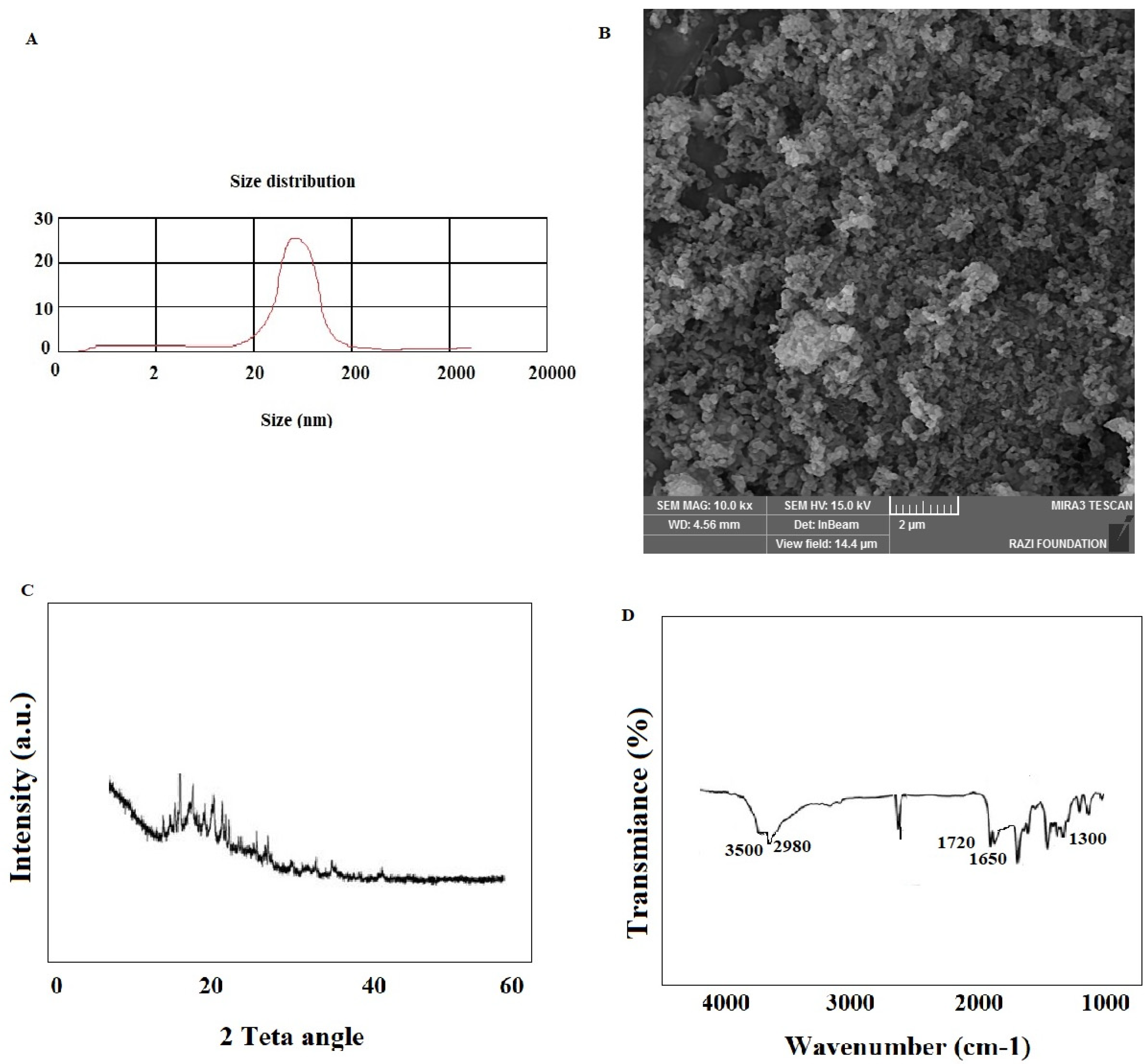
2.2. Particle Size Characterization

The mean size of curcumin nanocrystals was measured by dynamic light scattering (DLS) technique (Malvern, UK) at 25 °C using an argon laser beam at 633 nm and 90° scattering angle. 0.1 g of curcumin nanocrystals were weighted and dispersed in 50 mL deionized water by sonication (Power 500 W, amplitude 20%, reaction time: 20 min) at 25 °C.

2.3. Scanning Electron Microscopy (SEM)

Surface morphology of the prepared curcumin nanocrystals examined by SEM (SEM, TESCAN, Warrendale, PA, USA). Samples were fixed on stubs via a double-sided tape and coated with gold under a high-vacuum atmosphere and then observed at an acceleration voltage of 10 kV.

2.4. Fourier Transform Infrared Spectroscopy (FTIR) and Powder X-ray Diffraction (PXRD)

FTIR spectra were assessed by means of an FTIR spectrometer (Shimadzu 8400S, Japan). The powder of curcumin nanocrystals was mixed with potassium bromide of IR grade and compressed via an IR pellet manufacturing machine.
PXRD of the prepared curcumin nanocrystals was imaged by an X-ray diffractometer (Philips TW 1710 diffractometer with Cu-Kα incident radiation regulated at 40 kV and 30 mA). The curcumin nanocrystals powder was placed in the sample slot and pressed smoothly with frosted glass. Then, the sample was put into an instrument with a scan speed at 0.04°/min, and the pattern was recorded over 2θ, with the angle ranged from 10° to 60° [34].

2.5. Bacteria Preparation

Bacteria were prepared at the Faculty of Medicine, Tabriz University of Medical Sciences, Tabriz, Iran (ethical code of IR.TBZMED.VCR.REC.1398.230). The bacteria suspensions were prepared by cultivating E. coli (ATCC 25922), S. aureus (ATCC 6538), and E. faecalis (ATCC 29212) in brain heart infusion (BHI) broth and incubating it for 24 h at 37 °C. Subsequently, the bacterial suspension was prepared to reach a density to 0.5 McFarland standards.

2.6. Implant Experiment Groups

Twenty-seven implants (DIO CO., Busan, Korea) were mounted and classified into three groups including curcumin nanocrystals, distilled water (negative control), and chlorhexidine (Sigma-Aldrich Canada Co.; positive control, 2% w/v). The torque tester was utilized to close each abutment in fixed torque of 30 Ncm. For the preparation of nanocurcumin solution, 600 mg of the prepared curcumin nanocrystal’s powder was weighted and dissolved in 10 mL of distilled water to prepare a solution with a concentration of 60 mg/mL. Then, it was stirred for 30 min in a little dark bottle at room temperature.

2.7. Microbial Sampling and Detection

The implants were removed from their packaging under sterile conditions. Next, the implants were held with sterile pliers to allow a firm torque action and kept in a vertical position. In the curcumin nanocrystals group, curcumin nanocrystals (60 mg/mL) with a volume of 10 µL were used to the internal cavity of the implant. As a negative control, 10 µL of distilled water was injected into the implant fixation. As a positive control, 10 µL of chlorhexidine was used. All implant abutment assemblies were submerged in sterile tubes containing 5 mL of bacteria suspension and were incubated at 37 °C for 24, 48, and 72 h. After incubation, abutments were separated from implants by the hexdriver. Then, 0.01 mL of implant contents was removed by a micro-syringe and then inoculated on the nutrient agar (Quelab, Montréal, QC, Canada). In addition, the diluted suspension (1:10, 1:102, and 1:103, 104 and 1:105) were prepared using the 0.01 mL of implants content and inoculated on the surface of nutrient agar plates. Plates were incubated for 24 at 35° and CFU was calculated by following formula:
CFU/mL = Number of colony × Dilution of samples × 10−2 (because the dilutions were prepared by 0.01 mL of implants content)/volume of culture plate.
Plates were selected for counting on which the bacteria colonies grow separately and did not overlap [35]. All steps were repeated three times.

2.8. Statistical Analysis

The results were reported as mean ± standard deviation and frequency (percentage). SPSS 20 (IBM Company, New York, NY, USA) was used to analyze the data with p value of <0.05 being considered significant. Kolmogorov–Smirnov test was used to test the data normality. A repeated measure ANOVA was used for the comparison among the groups.

3. Results and Discussion

3.1. Characterization of Curcumin Nanocrystals

The prepared curcumin nanocrystals showed a mean particle size of 95 nm (Figure 1A). In addition, the SEM results presented that the curcumin nanocrystals showed spherical, uniform small particle sizes (Figure 1B). It was found that some fine crystals adhered with each other during spraying by electrostatic and van der Waals forces, and the particle size became larger. During spray drying, the nanocrystals were sprayed in the form of many small droplets and each droplet contained many fine particles. These particles aggregated together after the water evaporated instantly, and the aggregates were hard to break into individual particles [33].
The prepared curcumin nanocrystals exhibited the lower PXRD peak intensities compared with the bulk curcumin (in references), suggesting the lower crystallinity and the smaller sizes [33], which was consistent with our SEM results (Figure 1C).
The chemical composition of curcumin nanocrystals was assessed by FTIR spectra (Figure 1D) and there were no obvious differences between the absorption bands of nanocurcumin and the bulk curcumin (in references) in the whole area of absorption bands. FTIR analysis displayed absorption at 1720 cm−1 for C=O stretching of the ester group, at 1650 cm−1 for C=O of the ketone group, at 1300 cm−1 for the ether group, and at 2980 cm−1 for aromatic stretching. A sharp band at about 3500 cm−1 and a broad peak at 3200–3500 cm−1 in the spectrum have been attributed to the –OH group stretching vibration [36]. It could be determined that spray drying did not alternate the chemical compositions of curcumin [33].

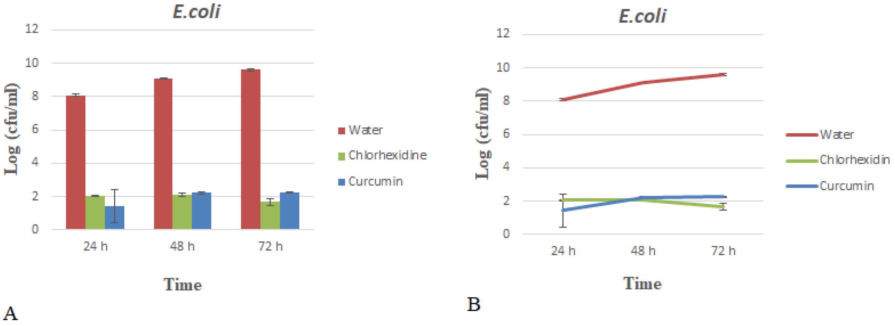
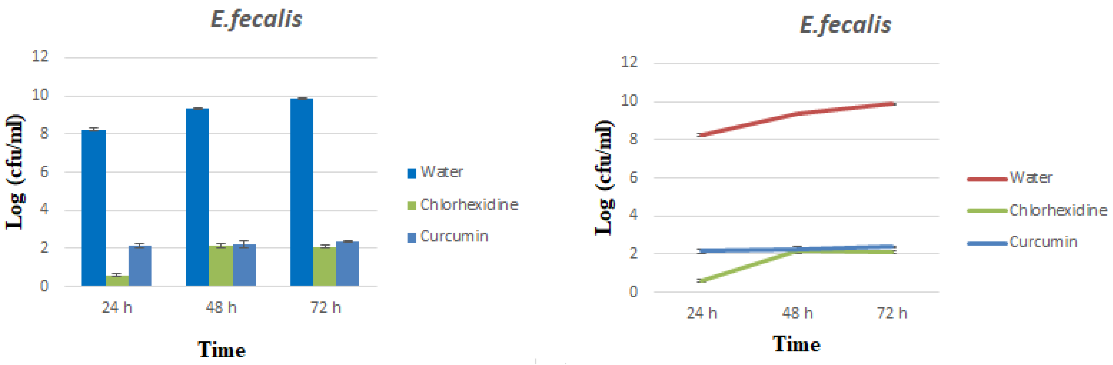
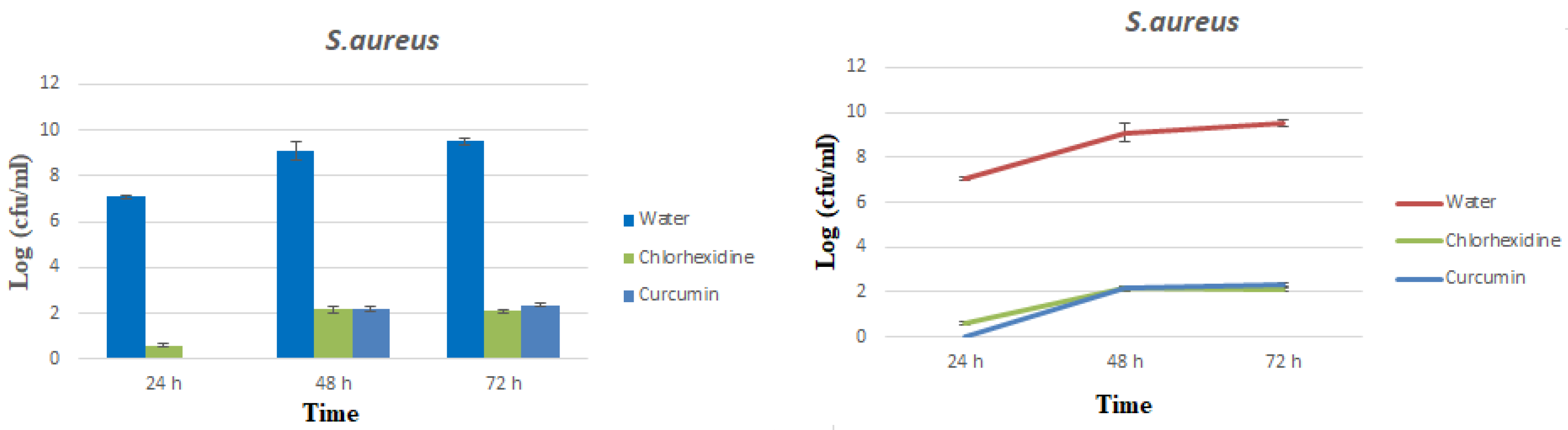
3.2. Microbial Analyses

Infections after implant placement are the main reasons for the failure of the implant treatment. Failed therapies are associated with a type of microbial flora that is directly related to periodontitis [17,18]. Various studies have reported the prevalence of infection by various bacteria in implants, involved in the occurrence of periodontitis or dental abscesses, including Staphylococci, Coliform bacteria, and Candida species [37,38,39].
The pharmacological effects of curcumin, including anti-inflammatory, antioxidant, anti-cancer, anti-diabetic, and antibacterial activities against S. aureus, E. coli, Bacillus cereus, and Helicobacter pylori have been reported [40]. Furthermore, curcumin has been identified with very low solubility, chemical instability, and poor bioavailability. Therefore, the use of nanotechnology has been considered to improve the bioavailability of curcumin and its high stability in the bloodstream and reduce its toxic effects. Moreover, the synthesis of curcumin as a nano-crystalline has different characteristics compared with its mass state due to its small size [35].
Results of the present study indicated that the CFU amounts for curcumin nanocrystals significantly were decreased (p < 0.010) compared with the CFU amounts for water (negative control). In addition, by increasing the time from 24 to 48 and 72 h, the CFU amounts for curcumin nanocrystals significantly decreased (p < 0.010).
The results also displayed that the removal rates (R%) of bacteria by curcumin nanocrystals were higher than 99.99% in all three types of bacteria. Table 1 presents a comparison of removal rates for all three types of bacteria. A comparison of the mean growth of bacteria in all three solutions separately for bacteria at different times is shown in Table 1 and Figure 2, Figure 3 and Figure 4.
Such results may be due to the different functions of nanoparticles in bacteria entry and removal. Depending on the particle size as well as the type of bacteria, the nanoparticles exert their antibacterial effects on bacteria by various mechanisms. The mechanisms are different from what has been reported for the mechanism of action of common antibiotics. To this end, the use of nanoparticles with antimicrobial properties is a way to overcome the microbial resistance of antibiotics. The nanoparticles used in the present study had an average particle size of 95 nm. According to scientific texts, physicochemical properties (size, shape, and surface properties) and doses of nanoparticles are effective in their antimicrobial effects [41]. Studies indicate that nanoparticles in the range below 100 nm generally have the ability to disrupt cell membrane functions by binding to the surface of cell membranes with a high affinity compared with larger nanoparticles. Such an effect is more prevalent in smaller nanoparticles due to its larger surface area [42,43]. The interaction of membrane–nanoparticles causes local pores in the membrane and largely damages the bacteria due to the entry of nanoparticles into the bacteria and the interaction of intracellular proteins (especially protein-rich in sulfur) and DNA. Another possible mechanism for antibacterial effects of antimicrobial nanoparticles is their binding to the bacterial membrane, and their gradual entry into the cytoplasm and disrupting the bacterial functions. Some drug nano-carriers are also able to mix with the bacterial cell wall and inject their antimicrobial substance into the bacterium [43,44].
In a study by Neelakantan et al. who studied the effects of curcumin against E. faecalis, the authors found that in two-day and ten-week biofilms, curcumin showed full inhibition that was significantly lower in terms of CFU re-obtained from chlorhexidine and saline [45]. Jahromi et al. found that curcumin loaded in nanoparticles significantly inhibited S. aureus in the infectious skin of rats. The results indicated that the use of nanoparticles increased the bactericidal effect of curcumin [46]. In fact, the gradual release of curcumin from nanoparticles occurred at the site of infection and properly inhibited the bacterial infection [47]. Results of a study by Bhawana et al. indicated that nanoparticles loaded with curcumin had more antimicrobial effects on a variety of fungi [48]. Singh et al. reported similar results and showed the antimicrobial and antibacterial properties of curcumin after its use in infectious therapies [49]. In another study by Rai et al. who investigated the antibacterial effects of curcumin, findings indicated that curcumin inhibited the growth of S. aureus [49]. The disinfectant, anti-inflammatory and antioxidant properties of curcumin have been taken into consideration in some studies [50]. Results of a study by Desai et al. who examined the reduction of bacterial contamination of internal and external dental implants using silver nanoparticles indicated that no bacterial growth was observed in groups containing silver nanoparticles. The absence of bacterial growth in the experimental groups suggested that silver particles were highly effective in inhibiting the bacterial growth inside the implants after 24 h in contact with bacterial suspensions (E. coli, S. aureus, and Salmonella) [35].
Curcumin can reduce the activity of NF-KB and prevent the leakage of inflammatory macromolecules, thereby reducing the inflammation caused by this bacterium [51,52]. Furthermore, curcumin in a concentration of 125–250 mg/mL can prevent the growth of 10 different strains of MRSA. Furthermore, mixing curcumin with any of the antibiotics, including ampicillin, oxacillin, and ciprofloxacin, can decrease the minimum inhibitory concentrations (MICs) of antibiotics due to a synergic effect [15]. Curcumin prevents the growth and proliferation of E. coli by inhibiting the FTsZ factor formation and can also inhibit the SOS responses due to levofloxacin in E. coli at a concentration of 8 µg/mL [14]. In addition, mixing curcumin with antibiotics such as ampicillin, oxacillin, and ciprofloxacin can decrease the MICs of bacteria [15].
Given the pharmacological resistance of bacteria to antibiotics, the production and optimization of nano-systems have been considered for drug delivery to target tissues. Drug-carrying nano-systems have useful features that did not exist before converting to nano-drug [53,54]. Nano-curcumin has increased its antimicrobial power against microorganisms such as E. coli, S. aureus, and Bacillus subtilis, Aspergillus, and Saccharomyces cerevisiae. Curcumin has been also used as an antibiotic against Yersinia enterocolitica and B. cereus [48,55].
Then introducing curcumin into implant fixture can present some potential advantages. At first, the chemical antimicrobial agents can be gradually replaced with antibacterial plant derivative such as curcumin in the future. This replacement will not only overcome the microbial resistance but will be also a solution to reduce the use of chemicals (like chlorhexidine, as gold standard antimicrobial material in dentistry) and their side effects and toxicity. It can not only increase the success of implant therapy (due to a decrease in the failure of the implant), but also its beneficial effects for gingival and its positive effect on bone–implant contact can lead to a further stabilization and success of the implant.

4. Conclusions

The use of dental implants has significantly increased in modern dentistry and has become a predictable choice in the replacement of missing teeth. Despite the fact that implant treatments are generally successful treatments, failure of implant treatments can be due to biological, mechanical, iatrogenic, or functional factors that lead to bacterial infections of the peri-implant tissue and implant overloading. Identification of these factors and their importance definitely affect the success of implant treatment. In many cases, the causes of these failures remain unknown. Therefore, researchers are always looking for causes of failure or complications of implant treatments. The present study investigated the antimicrobial effects of curcumin nanoparticles on bacteria. According to the results, nano-curcumin solution was able to completely remove all three types of bacteria. Results of the present study indicated that the CFU amounts for curcumin nanocrystals solution significantly were decreased (p < 0.010) compared with the CFU amounts for water (as negative control). In addition, by increasing the time from 24 to 72 h, the CFU amounts for curcumin nanocrystals solution significantly were decreased (p < 0.010). The results also showed that the removal rates (R%) of bacteria by nano-curcumin solution were higher than 99.99% in all bacteria. Then, due to the antimicrobial effects of nanocurcumin, its beneficial effects for gingival and its positive effect on bone–implant contact, nanocurcumin can be used inside the implant fixture to use antimicrobial effects and further stabilization and success of the implant. In addition, since the growth of the bacteria on the implant is in the form of biofilm; therefore, the assessment of the antibiofilm effects of the prepared curcumin nanocrystals will be suggested as a topic for future studies.

Author Contributions

R.N., M.A.G., M.Y.M., S.S., A.E., and B.K. made a significant contribution to the research steps or the drafting of the manuscript. S.M.D. and M.C. contributed to the drafting and scientific revision of the manuscript. S.M.D. and M.C. are the corresponding authors of the manuscript. All authors have read and agreed to the published version of the manuscript.

Funding

The Vice Chancellor for Research at Tabriz University of Medical Sciences provided financial support for this research.

Acknowledgments

This article was written based on a dataset from a thesis registered at the Faculty of Dentistry, Tabriz University of Medical Sciences (number 63041). The Vice Chancellor for Research at Tabriz University of Medical Sciences provided financial support for this research that is greatly acknowledged.

Conflicts of Interest

The authors declare that they have no conflict of interest.

References

  1. Lauritano, D.; Moreo, G.; Lucchese, A.; Viganoni, C.; Limongelli, L.; Carinci, F. The Impact of Implant–Abutment Connection on Clinical Outcomes and Microbial Colonization: A Narrative Review. Materials 2020, 13, 1131. [Google Scholar] [CrossRef] [Green Version]
  2. Passos, S.P.; May, L.G.; Faria, R.; Özcan, M.; Bottino, M.A. Implant-abutment gap versus microbial colonization: Clinical significance based on a literature review. J. Biomed. Mater. Res. Part B Appl. Biomater. 2013, 101, 1321–1328. [Google Scholar]
  3. Van Winkelhoff, A.J.; Goené, R.J.; Benschop, C.; Folmer, T. Early colonization of dental implants by putative periodontal pathogens in partially edentulous patients. Clin. Oral Implant. Res. 2000, 11, 511–520. [Google Scholar]
  4. Sharifi, S.; Vahed, S.Z.; Ahmadian, E.; Dizaj, S.M.; Eftekhari, A.; Khalilov, R.; Ahmadi, M.; Hamidi-Asl, E.; Labib, M. Detection of pathogenic bacteria via nanomaterials-modified aptasensors. Biosens. Bioelectron. 2020, 150, 111933. [Google Scholar] [PubMed]
  5. Haghshenas, B.; Nami, Y.; Haghshenas, M.; Barzegari, A.; Sharifi, S.; Radiah, D.; Rosli, R.; Abdullah, N. Effect of addition of inulin and fenugreek on the survival of microencapsulated Enterococcus durans 39C in alginate-psyllium polymeric blends in simulated digestive system and yogurt. Asian J. Pharm. Sci. 2015, 10, 350–361. [Google Scholar]
  6. Barzegari, A.; Kheyrolahzadeh, K.; Khatibi, S.M.H.; Sharifi, S.; Memar, M.Y.; Vahed, S.Z. The Battle of Probiotics and Their Derivatives Against Biofilms. Infect. Drug Resist. 2020, 13, 659–672. [Google Scholar]
  7. Ammon, H.P.T.; Anazodo, M.I.; Safayhi, H.; Dhawan, B.N.; Srimal, R.C. Curcumin: A Potent Inhibitor of Leukotriene B4Formation in Rat Peritoneal Polymorphonuclear Neutrophils (PMNL). Planta Med. 1992, 58, 226. [Google Scholar]
  8. Sharifi, S.; Vahed, S.Z.; Ahmadian, E.; Dizaj, S.M.; Abedi, A.; Khatibi, S.M.H.; Samiei, M. Stem Cell Therapy: Curcumin Does the Trick. Phytother. Res. 2019, 33, 2927–2937. [Google Scholar]
  9. Araújo, C.; Leon, L. Biological activities of Curcuma longa L. Memórias do Instituto Oswaldo Cruz 2001, 96, 723–728. [Google Scholar]
  10. Artico, M.; Di Santo, R.; Costi, R.; Novellino, E.; Greco, G.; Massa, S.; Tramontano, E.; Marongiu, M.E.; De Montis, A.; La Colla, P. Geometrically and Conformationally Restrained Cinnamoyl Compounds as Inhibitors of HIV-1 Integrase: Synthesis, Biological Evaluation, and Molecular Modeling. J. Med. Chem. 1998, 41, 3948–3960. [Google Scholar]
  11. Anand, P.; Kunnumakkara, A.B.; Newman, R.A.; Aggarwal, B.B. Bioavailability of Curcumin: Problems and Promises. Mol. Pharm. 2007, 4, 807–818. [Google Scholar] [PubMed]
  12. Han, S.; Yang, Y. Antimicrobial activity of wool fabric treated with curcumin. Dyes Pigments 2005, 64, 157–161. [Google Scholar]
  13. Varaprasad, K.; Vimala, K.; Ravindra, S.; Reddy, N.N.; Reddy, G.V.S.; Raju, K.M. Fabrication of silver nanocomposite films impregnated with curcumin for superior antibacterial applications. J. Mater. Sci. Mater. Med. 2011, 22, 1863–1872. [Google Scholar] [PubMed]
  14. Bellio, P.; Brisdelli, F.; Perilli, M.; Sabatini, A.; Bottoni, C.; Segatore, B.; Setacci, D.; Amicosante, G.; Celenza, G. Curcumin inhibits the SOS response induced by levofloxacin in Escherichia coli. Phytomedicine 2014, 21, 430–434. [Google Scholar] [PubMed]
  15. Mun, S.-H.; Joung, D.-K.; Kim, Y.S.; Kang, O.-H.; Kim, S.-B.; Seo, Y.-S.; Kim, Y.-C.; Lee, D.-S.; Shin, D.-W.; Kweon, K.-T.; et al. Synergistic antibacterial effect of curcumin against methicillin-resistant Staphylococcus aureus. Phytomedicine 2013, 20, 714–718. [Google Scholar] [PubMed]
  16. Farjana, H.N.; Chandrasekaran, S.; Gita, B. Effect of Oral Curcuma Gel in Gingivitis Management—A Pilot Study. J. Clin. Diagn. Res. 2014, 8, ZC08–ZC10. [Google Scholar]
  17. Guimaraes-Stabili, M.R.; De Aquino, S.G.; Curylofo, F.D.A.; Tasso, C.O.; Rocha, F.R.G.; De Medeiros, M.C.; De Pizzol, J.P.; Cerri, P.S.; Romito, G.A.; Rossa, C. Systemic administration of curcumin or piperine enhances the periodontal repair: A preliminary study in rats. Clin. Oral Investig. 2019, 23, 3297–3306. [Google Scholar]
  18. Malekzadeh, M.; Kia, S.J.; Mashaei, L.; Moosavi, M.-S. Oral n ano-curcumin on gingival inflammation in patients with gingivitis and mild periodontitis. Clin. Exp. Dent. Res. 2020. [Google Scholar]
  19. Ghavimi, M.A.; Shahabadi, A.B.; Jarolmasjed, S.; Memar, M.Y.; Dizaj, S.M.; Sharifi, S. Nanofibrous asymmetric collagen/curcumin membrane containing aspirin-loaded PLGA nanoparticles for guided bone regeneration. Sci. Rep. 2020, 10, 1–15. [Google Scholar]
  20. Ahmadian, E.; Maleki, S.; Sharifi, S.; Eftekhari, A.; Samiei, M. Hyaluronic acid hydrogel nanoscaffolds: Production and assessment of the physicochemical properties. Eurasian Chem. Commun. 2020, 2, 51–58. [Google Scholar]
  21. Cirano, F.R.; Pimentel, S.; Casati, M.; Correa, M.; Pino, D.; Messora, M.; Silva, P.H.F.; Ribeiro, F.V. Effect of curcumin on bone tissue in the diabetic rat: Repair of peri-implant and critical-sized defects. Int. J. Oral Maxillofac. Surg. 2018, 47, 1495–1503. [Google Scholar] [PubMed]
  22. Shang, W.; Zhao, L.-J.; Dong, X.-L.; Zhao, Z.-M.; Li, J.; Zhang, B.-B.; Cai, H. Curcumin inhibits osteoclastogenic potential in PBMCs from rheumatoid arthritis patients via the suppression of MAPK/RANK/c-Fos/NFATc1 signaling pathways. Mol. Med. Rep. 2016, 14, 3620–3626. [Google Scholar] [PubMed] [Green Version]
  23. Theodoro, L.H.; Ferro-Alves, M.L.; Longo, M.; Nuernberg, M.A.A.; Ferreira, R.P.; Andreati, A.; Ervolino, E.; Duque, C.; Garcia, V.G. Curcumin photodynamic effect in the treatment of the induced periodontitis in rats. Lasers Med. Sci. 2017, 32, 1783–1791. [Google Scholar]
  24. Hussan, F.; Ibraheem, N.G.; Kamarudin, T.A.; Shuid, A.N.; Soelaiman, I.N.; Othman, F. Curcumin Protects against Ovariectomy-Induced Bone Changes in Rat Model. Evid. Based Complement. Altern. Med. 2012, 2012, 1–7. [Google Scholar]
  25. Chen, Z.; Xue, J.; Shen, T.; Mu, S.; Fu, Q. Curcumin alleviates glucocorticoid-induced osteoporosis through the regulation of the Wnt signaling pathway. Int. J. Mol. Med. 2016, 37, 329–338. [Google Scholar]
  26. Singh, A.K.; Jiang, Y.; Gupta, S.; Younus, M.; Ramzan, M. Anti-Inflammatory Potency of Nano-Formulated Puerarin and Curcumin in Rats Subjected to the Lipopolysaccharide-Induced Inflammation. J. Med. Food 2013, 16, 899–911. [Google Scholar]
  27. Salatin, S. Nanoparticles as potential tools for improved antioxidant enzyme delivery. J. Adv. Chem. Pharm. Mater. (JACPM) 2018, 1, 65–66. [Google Scholar]
  28. Sharifi, S.; Fathi, N.; Memar, M.Y.; Khatibi, S.M.H.; Khalilov, R.; Negahdari, R.; Vahed, S.Z.; Dizaj, S.M. Anti-microbial activity of curcumin nanoformulations: New trends and future perspectives. Phytother. Res. 2020, 34, 1926–1946. [Google Scholar]
  29. Ghavimi, M.A.; Negahdari, R.; Shahabadi, A.B.; Sharifi, S.; Kazeminejad, E.; Shahi, S.; Dizaj, S.M. Preparation and study of starch/ collagen/ polycaprolactone nanofiber scaffolds for bone tissue engineering using electrospinning technique. Eurasian Chem. Commun. 2020, 2, 122–127. [Google Scholar]
  30. Sharifi, S.; Samani, A.A.; Ahmadian, E.; Eftekhari, A.; Derakhshankhah, H.; Jafari, S.; Mokhtarpour, M.; Vahed, S.Z.; Salatin, S.; Dizaj, S.M. Oral delivery of proteins and peptides by mucoadhesive nanoparticles. Biointerface Res. Appl. Chem. 2019, 9, 3849–3852. [Google Scholar]
  31. Zambrano, L.M.G.; Brandao, D.A.; Rocha, F.R.G.; Marsiglio, R.P.; Longo, I.B.; Primo, F.L.; Tedesco, A.C.; Guimaraes-Stabili, M.R.; Junior, C.R. Local administration of curcumin-loaded nanoparticles effectively inhibits inflammation and bone resorption associated with experimental periodontal disease. Sci. Rep. 2018, 8, 1–11. [Google Scholar]
  32. Ghaffari, F.; Moghaddam, A.H.; Zare, M. Neuroprotective Effect of Quercetin Nanocrystal in a 6-Hydroxydopamine Model of Parkinson Disease: Biochemical and Behavioral Evidence. Basic Clin. Neurosci. 2018, 9, 317. [Google Scholar] [PubMed]
  33. Hu, L.; Kong, D.; Hu, Q.; Gao, N.; Pang, S. Evaluation of High-Performance Curcumin Nanocrystals for Pulmonary Drug Delivery Both In Vitro and In Vivo. Nanoscale Res. Lett. 2015, 10, 381. [Google Scholar] [PubMed] [Green Version]
  34. Noroozi Pesyan, N.; Gholsanamloo, V.; Moradi Par, M.; Rashidnejad, H.; Gharib, A.; Nejati, K. Synthesis, characterization and spectroscopic properties of new azo dyes derived from aniline derivatives based on acetylacetone and azo-metal (II) complexes and singular value decomposition (SVD) investigation. Iran. Chem. Commun. 2019, 7, 1–19. [Google Scholar]
  35. Desai, C.T.; Desai, S.J.; Marjadi, D.S.; Shah, G.S. Diminution of Internal Bacterial Contamination of External Dental Implants Using Silver Nanoparticles. JANUARY 2018, 4, 115–119. [Google Scholar]
  36. El-Rahman, S.; Al-Jameel, S. Protection of curcumin and curcumin nanoparticles against cisplatin induced nephrotoxicity in male rats. Sch. Acad. J. Biosci. 2014, 2, 214–223. [Google Scholar]
  37. Alcoforado, G.A.; Rams, T.E.; Feik, D.; Slots, J. Microbial aspects of failing osseointegrated dental implants in humans. J. Parodontol. 1991, 10, 11–18. [Google Scholar]
  38. Leonhardt, Å.; Renvert, S.; Dahlén, G. Microbial findings at failing implants. Clin. Oral Implant. Res. 1999, 10, 339–345. [Google Scholar]
  39. Salvi, G.E.; Fürst, M.M.; Lang, N.P.; Persson, G.R. One-year bacterial colonization patterns of Staphylococcus aureus and other bacteria at implants and adjacent teeth. Clin. Oral Implant. Res. 2008, 19, 242–248. [Google Scholar]
  40. Menon, V.P.; Sudheer, A.R. Antioxidant and Anti-Inflammatory Properties of Curcumin. In The Molecular Targets and Therapeutic Uses of Curcumin in Health and Disease; Springer: New York, NY, USA, 2007; pp. 105–125. [Google Scholar]
  41. Marambio-Jones, C.; Hoek, E.M.V. A review of the antibacterial effects of silver nanomaterials and potential implications for human health and the environment. J. Nanopart. Res. 2010, 12, 1531–1551. [Google Scholar]
  42. Dizaj, S.M.; Lotfipour, F.; Barzegar-Jalali, M.; Zarrintan, M.H.; Adibkia, K. Antimicrobial activity of the metals and metal oxide nanoparticles. Mater. Sci. Eng. C 2014, 44, 278–284. [Google Scholar]
  43. Salatin, S.; Maleki Dizaj, S.; Yari Khosroushahi, A. Effect of the surface modification, size, and shape on cellular uptake of nanoparticles. Cell Biol. Int. 2015, 39, 881–890. [Google Scholar]
  44. Wang, L.; Hu, C.; Shao, L. The antimicrobial activity of nanoparticles: Present situation and prospects for the future. Int. J. Nanomed. 2017, 12, 1227. [Google Scholar]
  45. Neelakantan, P.; Subbarao, C.; Sharma, S.; Subbarao, C.V.; Garcia-Godoy, F.; Gutmann, J.L. Effectiveness of curcumin against Enterococcus faecalis biofilm. Acta Odontol. Scand. 2013, 71, 1453–1457. [Google Scholar] [PubMed]
  46. Mofazzal Jahromi, M.A.; Rajayi, H.; Al-Musawi, S.; Pirestani, M.; Fasihi Ramandi, M.; Ahmadi, K.; Sharifzadeh Peivasti, V.; Mohammad Hassan, Z.; Kamali, M.; Mirnejad, R. Evaluation of antibacterial effect of curcumin loaded chitosan nanoparticles. J. Fasa Univ. Med. Sci. 2015, 5, 134–141. [Google Scholar]
  47. Anitha, A.; Maya, S.; Deepa, N.; Chennazhi, K.; Nair, S.; Tamura, H.; Jayakumar, R. Efficient water soluble O-carboxymethyl chitosan nanocarrier for the delivery of curcumin to cancer cells. Carbohydr. Polym. 2011, 83, 452–461. [Google Scholar]
  48. Basniwal, R.K.; Buttar, H.S.; Jain, V.K.; Jain, N. Curcumin Nanoparticles: Preparation, Characterization, and Antimicrobial Study. J. Agric. Food Chem. 2011, 59, 2056–2061. [Google Scholar]
  49. Singh, R.K.; Rai, D.; Yadav, D.; Bhargava, A.; Balzarini, J.; De Clercq, E. Synthesis, antibacterial and antiviral properties of curcumin bioconjugates bearing dipeptide, fatty acids and folic acid. Eur. J. Med. Chem. 2010, 45, 1078–1086. [Google Scholar]
  50. Negi, P.; Jayaprakasha, G.K.; Rao, L.J.M.; Sakariah, K.K. Antibacterial Activity of Turmeric Oil: A Byproduct from Curcumin Manufacture. J. Agric. Food Chem. 1999, 47, 4297–4300. [Google Scholar]
  51. Foryst-Ludwig, A.; Neumann, M.; Schneider-Brachert, W.; Naumann, M. Curcumin blocks NF-κB and the motogenic response in Helicobacter pylori-infected epithelial cells. Biochem. Biophys. Res. Commun. 2004, 316, 1065–1072. [Google Scholar]
  52. Sintara, K.; Thong-Ngam, D.; Patumraj, S.; Klaikeaw, N.; Chatsuwan, T. Curcumin suppresses gastric NF-κB activation and macromolecular leakage inHelicobacter pylori-infected rats. World J. Gastroenterol. 2010, 16, 4039. [Google Scholar] [PubMed]
  53. Elsagh, A. Quantum study of solvent effect with POPC phospholipid bilayers in a cell membrane and its impact on active and targeted drug delivery. Eurasian Chem. Commun. 2020, 2, 440–455. [Google Scholar]
  54. Afshar, M.; Khojasteh, R.R.; Ahmadi, R. Adsorption of lomustin anticancer drug on the surface of carbon nanotube: A theoretical study. Eurasian Chem. Commun. 2020, 2, 595–603. [Google Scholar]
  55. Wang, Y.-F.; Lu, Z.; Wu, H.; Lv, F. Study on the antibiotic activity of microcapsule curcumin against foodborne pathogens. Int. J. Food Microbiol. 2009, 136, 71–74. [Google Scholar]
Figure 1. Physicochemical characterization of the prepared nanocurcumin. (A) Particle size distribution, (B) SEM image, (C) powder X-ray diffraction (PXRD) pattern, and (D) Fourier transform infrared spectroscopy (FTIR) peak.
Applsci 10 08356 g001
Figure 2. Comparison of the mean growth rates of E. coli in different groups; (A) column graph, (B) liner graph (nanocurcumin (60 mg/mL), chlorhexidine (2%)).
Applsci 10 08356 g002
Figure 3. Comparison of the mean growth rates of E. faecalis in different groups; column graph and liner graph (nanocurcumin (60 mg/mL), chlorhexidine (2%)).
Applsci 10 08356 g003
Figure 4. Comparison of the mean growth rates of S. aureus in different groups; column graph and liner graph (nanocurcumin (60 mg/mL), chlorhexidine (2%)).
Applsci 10 08356 g004
Table 1. Comparison of mean growth rates of bacteria in different groups and times.
BacteriaTime (h)Water Log (cfu/mL)Chlorhexidine Log (cfu/mL)R * (%)Curcumin Log (cfu/mL)R ** (%)
E. coli248.082.0499.991.4199.99
489.102.0999.992.2299.99
729.611.671002.2499.99
E. faecalis248.220.591002.1699.99
489.342.1599.992.2599.99
729.872.091002.3799.99
S. aureus247.080.5699.990100
489.091.101002.1699.99
729.512.121002.3599.99
R * = (CFU of water–CFU of chlorhexidine)/CFU of water × 100. R ** = (CFU of water–CFU of curcumin)/CFU of water × 100.
Publisher’s Note: MDPI stays neutral with regard to jurisdictional claims in published maps and institutional affiliations.

