Article

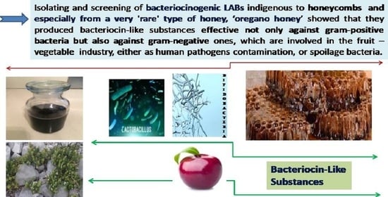
Effectiveness of Bacteriocin-Producing Lactic Acid Bacteria and Bifidobacterium Isolated from Honeycombs against Spoilage Microorganisms and Pathogens Isolated from Fruits and Vegetables

1
School of Agriculture, University of Ioannina, 47100 Arta, Greece
2
Department of Agricultural Development, Laboratory of Microbiology, Biotechnology and Hygiene, Democritus University of Thrace, 67100 Xanthi, Orestiada, Greece
3
Department of Food Science and Technology, University of the Peloponnese, Antikalamos, 24100 Kalamata, Greece
4
Medical School, Laboratory of Hygiene and Environmental Protection, Democritus University of Thrace, 67100 Alexandroupolis, Greece
*
Author to whom correspondence should be addressed.
Appl. Sci. 2020, 10(20), 7309; https://doi.org/10.3390/app10207309
Submission received: 29 August 2020 / Revised: 28 September 2020 / Accepted: 15 October 2020 / Published: 19 October 2020

Abstract

:
Screening natural products for bacteriocin-producing bacteria may be the equilibrium point between the consumer demand for mild processing and the industry’s need for hazard control. Raw unprocessed honeycombs filled with oregano honey from the alpine mountainous territory of Epirus, Greece were screened for bacteriocinogenic lactic acid bacteria and Bifidobacterium spp., with inhibitory action towards some pathogens and spoilage microorganisms isolated from fresh fruits and vegetables (number and type of strains: three E. coli, two L. monocytogenes, two Salmonella spp., two B.cereus, two Erwinia spp., one Xanthomonas spp., L. innocua (ATCC 33090TM) and E. coli 0157:H7 (ATCC 69373)). Among the 101 collected isolates (73 Lactobacillus, 8 Lactococcus, 8 Leuconostoc and 12 Bifidobacterium species) from the oregano honeycombs (an original finding since there are no other reports on the microbial biodiversity of the flora of the oregano honey), 49 strains of lactic acid bacteria (LAB) and Bifidobacterium spp. were selected and tested for their bacteriocin-producing capacity (34 Lactobacillus, 6 Lactococcus, 5 Leuconostoc and 4 Bifidobacterium). The antibacterial activity exerted by the tested LAB and Bifidobacterium strains was not of the same potency. Our results suggest that the main molecules involved in the antimicrobial activity are probably bacteriocin-like substances (a conclusion based on reduced antibacterial activity after the proteolytic treatment of the cell-free supernatant of the cultures) and this antimicrobial activity is specific for the producing strains as well as for the target strains. The spoilage bacteria as well as the reference microorganisms showed increased resistance to the bacteriocin-like substances in comparison to the wild-type pathogens.

Graphical Abstract

1. Introduction

According to EUROSTAT (ec.europa.eu) surveys, half of the EU population eats at least one portion of fruits and vegetables on a daily basis and this trend is increasing (the southern states are in the top three EU Member States in daily intake of fruit: Italy (85%), Portugal (81%) and Spain (77%)) [1]. Unsurprisingly, the WHO/FAO Joint Expert Consultation Report on Diet, Nutrition and the Prevention of Chronic Diseases states that at least 400 g of fruits and vegetables per day are needed to prevent heart disease, certain types of cancer, diabetes and obesity [2]. Given these statistics, important food quality and food safety issues emerge. Fruits and vegetables are rich in humidity and carbohydrate content and hence characterized as ideal habitats for bacterial and fungal growth [3]. Among others, factors such as the presence of various parasites and the quality and origin of manure, harvest and postharvest treatment and skin lesions are of utmost importance for their contamination with pathogens and spoilage microorganisms. Various studies have shown the presence of Salmonella spp., E. coli O157:H7, Listeria monocytogenes, Bacillus cereus, Campylobacter spp., Yersinia enterocolitica, Clostridium botulinum and other pathogens, in addition to some viruses and parasites in fruits and vegetables [4,5]. Spoilage bacteria such as Erwinia carotovora and Xanthomonas campestris cause various types of lesions regarding texture and color thus reducing their quality and commercial value [6].
There are modern trends in food processing concerning food safety and prolonging shelf life. Consumers prefer minimally processed foods that are free of chemical preservatives. Given these demands, and the increasing resistance of pathogens and spoilage bacteria to antibiotics and other chemicals, the food industry is seeking alternative means of food preservation [7,8].Consequently, there is an increasing interest in so-called “green technologies,” including novel approaches to the minimal processing of food as well as the use of microbial metabolites such as bacteriocins on an industrial scale for “biopreservation” [9].
In order to control, or even prevent, the growth of such microorganisms, the application of bacteriocins-producing lactic acid bacteria (LAB) has been proposed [10]. LAB can be isolated from many raw wild fruits, vegetables and flowers [11,12]. Moreover, lactic and acetic acid bacteria can promote the spoilage of fruits, vegetables, fruit juices and beverages as residents at the outer layer of the skin of fruits and vegetables [6]. These bacteria are known for the fermentation of carbohydrates and the production of various organic acids such as lactic acid, which significantly lower the pH of fermented foods [12]. They also produce other compounds with antimicrobial action such as hydrogen peroxide, acetaldehyde and bacteriocins [13,14,15].
Bacteriocins are peptides with natural antibacterial activity, and there is a reason for their comparison to antibiotics. Many researchers propose the term “biological preservatives of foods” and stress the fact that bacteriocins are not used for clinical therapeutic purposes as antibiotics are [16]. They are synthesized in the ribosomes of the bacterial cell and secreted extracellularly. Bacteriocins are most effective against Gram-positive bacteria. The spectrum of their activity varies from narrow (against one species) to broad (against several species) [17,18]. The main advantage of bacteriocins is that their presence does not change the sensorial characteristics of foods. Their usage enables the reduction of the intensity of other means of preservation such as heat. These two characteristics make bacteriocins’ application compatible with modern consumer demands for the minimal and more natural treatment of foods [19]. Bacteriocins can be preferably added to foods as compounds rather than cultures of bacteriocinogenic LAB because, in the latter case, LAB can ferment the carbohydrates of foods [20].
Nisin, pediocin, enterocin AS-48, bovicin, enterocin 416K1 and bificin C6165 are some bacteriocins already tested against spoilage bacteria and pathogens but only the first two were granted approval as food additives. However, they are mostly used in other foods and their usage in the fruit and vegetable industry is still limited [19].
LAB have been established as “generally regarded as safe” (GRAS), a fact that makes them attractive candidates for industrial utilization [21,22]. There is ongoing research for new strains with potentially superior properties, such as being probiotic, and the production of active bacteriocins. Sources of such LAB are various natural products, which could be an ideal ecological niche for these microorganisms. Oregano honey is a very special and extremely rare natural product. It is produced by honeybees grazing on wild oregano plants in the alpine mountainous territory of Epirus, Greece. It is difficult to find it since there are very few producers. Unlike other types of honey, oregano honey has a bitter taste. It is consumed by rural populations of the area and data from local people suggest they believe oregano honey possesses therapeutic properties against various infections and gastrointestinal disorders.
The aim of this study is to screen the diverse autochthonous microbiota isolated from the honeycombs of oregano-grazing bees for LAB and Bifidobacterium strains and determine the antibacterial activity of these bacteriocin-producing isolates against some pathogens and spoilage microorganisms isolated from fresh fruits and vegetables.

2. Materials and Methods

2.1. Sample Collection

2.1.1. Fruits and Vegetables

All samples were purchased from open fruit markets in different areas of Epirus, Greece. In total, 20 each of pears, apples, peaches, tomatoes, cucumbers and red peppers, 40 white cabbages and 60 carrots were collected in sterile plastic bags and brought within an hour to the laboratory for analysis. The samples were of reasonably good quality and chosen for their slight lesions or minor skin ruptures (Figure 1). Fifty commercially sealed bags of minimally processed fresh-cut products ready-to-use (RTU) were sampled from various supermarkets and other retail stores.

2.1.2. Honeycomb Filled with Oregano Honey

A total of 30 raw unprocessed honeycombs filled with oregano honey, were received from local producers. A portion of 1000 g samples (from each local producer) were weighed and placed in a dark, sterile glass container under sterile conditions and homogenized. The samples did not contain any additives or diluents and had not been previously heated. They were evaluated for their microbiological quality by inoculation into blood agar (Columbia agar base with 5% sheep blood, Becton Dickinson), egg yolk agar, mannitol-egg yolk-polymyxin (MYP) agar and incubated aerobically at 30 °C and 37 °C for 48 h. Samples that showed growth of bacteria or growth of more than 4–5 colonies of yeasts were excluded from the study; only 4 samples were excluded from our study. The samples were stored at 5 °C in the dark in the Laboratory of School of Agricultural Science (University of Ioannina) to prevent photodegradation until being used.

2.2. Isolation of Spoilage and Pathogens Organisms

The bacterial flora of fruits or vegetables was collected by the following method. The entire product was placed in a sterile sample bag and a sufficient volume of enrichment broth (sterile 0.1% buffered peptone water (BPW, Oxoid)) was added in order to totally submerge the fruit or vegetable. The samples were then agitated and rubbed separately in the nylon sterile bag for 10 min to suspend surface microbes [23]. At the same time, the areas and tissues with lesions were removed using a sterile knife and placed into the same sterile bag as above. Then, various dilutions under aseptic conditions were made and different selective media and incubation conditions were used to isolate specific bacterial species, as described in the following sections. After incubation, the numbers of CFU were counted and different types of colonies were isolated. The distinct colonies were screened and selected on the basis of morphology and cultural characteristics and identified on the basis of standard tests.

2.2.1. Detection of E.coli

To enumerate the E. coli, a technique employing two media(eosin methylene blue (EMB) agar (Merck) at 37 °C for 24 h and chromogenic medium tryptone bile agar containing 5-bromo-4-chloro-3-indolyl-B D-glucuronic acid (BCIG) (tryptone bile × glucuronide agar) (Merck)) was used. The second medium relies on the use of bile salts and an elevated incubation at 44 °C to suppress competitor organisms. The chromogenic TBX was used to indicate the presence of β-glucuronidase activity, which is common in 95% of E. coli strains [24].

2.2.2. Detection of Salmonella Spp.

A method based on primary enrichment in Erlenmeyer flasks containing 225 mL of lactose broth was used (the procedure consisted of very thorough stirring at first then leaving the flasks at room temperature for 60 min. pH was measured and adjusted to 6.8 ± 0.2 with 1N NaOH or 1N HCl followed by incubation at 35 ± 2 °C for 24 ± 2 h). Subsequently, a secondary selective enrichment took place with the use of two media (Rappaport–Vassiliadis (RV) medium and tetrathionate (TT) broth) and incubation at 42 ± 0.2 °C for 24 ± 2 h (circulating, thermostatically controlled water bath), incubated at 35 ± 2.0 °C for 24 ± 2 h, respectively. Finally, 10 µL from each incubated TT broth and RV medium were streaked on bismuth sulfite (BS) agar, xylose lysine deoxycholate (XLD) agar and Hektoen enteric (HE) agar. Then, using a range of biochemical tests, suspect Salmonella spp. isolates were identified using the VITEK system [23,25,26,27].

2.2.3. Detection of Shigella spp.

The enrichment Shigella broth containing 3.0 μg/mL of novobiocin was used and the plates were incubated anaerobically (using the Anaerocult® A system (Merck) in an anaerobic jar) at 44 °C for 20 h. Next, surface spreading took place (spread plate method) on MacConkey agar followed by anaerobic incubation at 42 °C for 24 h. All the isolated colonies were characterized biochemically [26].

2.2.4. Detection of L. monocytogenes

Detection began with the enrichment of Listeria spp. using the Fraser broth (Oxoid), which contains selective agents including nalidixic acid, acriflavine and lithium chloride, to suppress most of the competitors. Afterwards, the broth was streaked onto Listeria Palcam agar (Merck) and chromogenic medium (agar Listeria according to Ottaviani and Agosti (ALOA)) incubated at 37 °C for 24 h [28]. Characteristic colonies were Gram-stained and tested for motility, oxidase and catalase activity, followed by identification with the API Listeria system (bioMérieux®, MarcyI’Etoile, France) [24].

2.2.5. Detection of P. fluorescens

For P. fluorescens, the plate count agar (PCA) and cetrimide agar (bioMérieux®) were used. The incubation conditions were 35 °C and 48 h and 30 °C and 48 h, respectively. The isolates were stored at −20 °C for further tests, such as Gram-staining, the catalase, oxidase and motility tests, starch hydrolysis, fluorescent pigment and gelatin liquefaction [29].

2.2.6. Detection of B. cereus

For the qualitative detection of B. cereus, a procedure for strengthening the strains of this species took place with the use of trypticase-soy-polymyxin (TSB) broth (Oxoid) at 30 °C for 48 h. Then, cultures were streaked on the mannitol-egg yolk-polymyxin (MYP) agar plate (Oxoid) and chromogenic plate (BACARA™, bioMérieux) and incubated at 30 °C for 24 h. Colonies with a pink sparkle in blue or blue-green precipitation on the chromogenic plate were picked for further testing to identify the presence of B. cereus in the examined samples of fruits and vegetables (parasporal crystal observation root growth observation was conducted, along with hemolysis, catalase, motility, nitrate reduction, casein decomposition, lysozyme tolerance, glucose utilization and acetyl methyl alcohol tests) [30].

2.2.7. Detection of Other Spoilage Bacteria, Yeasts and Molds

For the estimation of other spoilage bacteria, yeasts and molds, the nutrient agar, oxytetracycline glucose yeast extract (OGYE) (Sigma-Aldrich, St. Louis, MO, USA), dichloran Rose Bengal chloramphenicol (DRBC) agar (Fluka, Germany) and a sterile fresh medium of potato dextrose agar (PDA) (Merck) were used and incubated at 28 °C for at least 5 days or until fungal proliferation occurred on the medium surface [31,32,33].
In the present study, crystal violet pectate (CVP) medium [34] was used. Surface spreading took place from a dilution series prepared in saline supplemented with 0.05% w/v ascorbic acid, followed by plating on the CVP medium [35].

2.2.8. Detection of C. perfringens

For the detection of C. perfringens (vegetative and spore forms), a pre-enrichment technique was performed in PBS for 2 h, followed by decimal dilutions in lactose sulfite broth (LS), which was the growth medium used. The composition of the broth as per the Council of Europe [36] is as follows: 5 g tryptic digest of casein, 2.5 g yeast extract, 2.5 g sodium chloride, 2.5 g lactose, 0.3 g L-cysteine hydrochloride and 1 L distilled water. The broth (9 mL) was distributed into screw cap tubes, which contained inverted Durham tubes, and sterilized by autoclaving at 115 °C for 20 min. After cooling to 45–50 °C, a filter-sterilized solution of 1.2% sodium metabisulfite (0.5 mL) and 1.0% ferric ammonium citrate (0.5 mL) was added to each tube. Reduced conditions in the medium were accomplished via boiling and due to the presence of cysteine. To examine the samples for vegetative cells, filter membranes were placed into tubes and incubation was performed aerobically in a water bath at 46 °C for 24 h. Additionally, a second aliquot (100 mL) of each sample was heated for 15 min at 80 °C for spore germination (and vegetative cell destruction), cooled under running water and filtered. The filter was then placed into the LS broth as described above. The interpretation of results (positive samples) was based on the resulting turbidity from lactose fermentation, the presence of iron sulfide (black precipitate) and the presence of gas (H2S) visible in the inverted Durham tube within 24 h at 46 °C [37,38].

2.3. Isolation of LAB and Bifidobacterium

In order to investigate the LAB in the stored samples of honeycombs filled with oregano honey, 100 g of each honeycomb was weighed, placed in a sterile Stomacher blender bag under completely sterile conditions and homogenized for 2 min using a Stomacher 400 Circulator. Then, 900 mL of peptone saline diluent were added to give a total volume of 1 L (0.9% w/v NaCl, 0.1% w/v Tween 80 and 0.1% w/v peptone). The above homogenization technique was repeated for 2 min [39] and 10-fold serial dilutions were prepared in normal saline diluents. Aliquots of 0.1 mL of each 10-fold serial dilution were spread on modified MRS agar (Oxoid) adjusted to an agar content of 1.5% w/v (Agar, Merck) and supplemented with 0.3% w/v CaCO3 (Merck) [40]. The plates for the recovery of facultative and strict anaerobe LAB and Bifidobacterium were incubated for 3–4 days at 37 °C under anaerobic conditions using anaerobic jars with Anaerocult® A gas packs (Merck) [41]. Following incubation, colonies from the modified MRS agar of a different morphology with a surrounding clear zone in the agar, indicating acid production, were transferred to the MRS agar without CaCO3 to grow pure cultures under conditions as described above. In a general approach, cultures were identified as LAB species or Bifidobacterium by evaluating their morphological, cultural, physiological and biochemical characteristics by the procedures described in Bergey’s Manual of Systematic Bacteriology. At least 10 selected isolates from each plate were initially identified based on the following: microscopic examination of Gram-stained cells, motility, catalase and oxidase reactions, gas production from glucose and growth in anaerobic conditions. Finally, phenotypic observations and biochemical tests were performed using the VITEK2 Compact System [42,43]. Τhe presumptive LAB isolates were further characterized according to their registered protein and peptide information analysis results with the help of the Microflex LT MALDI-TOF MS (matrix-assisted laser desorption ionization time-of-flight mass spectrometry) (Bruker Daltonics, Bremen, Germany), which is a new technology for identifying LAB cultures. Mass spectra were processed using BioTyper software (version 3.0; Bruker Daltonics) running with the BioTyper database (version DB-5989). Matching between experimental MALDI-TOF MS profiles obtained from bacteria isolates and the reference MALDI-TOF MS profiles was expressed by BioTyper according to a Log(Score). Analyses were conducted in accordance with the manufacturer’s instructions and performed twice for each isolate. Results (BioTyper score value, created by an automatic mass spectra comparison with the Bruker library) with a score value >2.3 were interpreted as identified at the species level with a high probability. A score value between 1.7 and 2.0 implies probability only at the genus level [44,45].

2.4. Determination of the Antibacterial Activity of LAB and Bifidobacterium against the Isolated Pathogens and Spoilage Bacteria

Preparation of the Cell-Free Supernatant (CFS)

In short, MRS broth (20 mL) was inoculated separately with the LAB isolates previously characterized and incubated anaerobically at 37 °C for 72 h. After incubation, a cell-free supernatant was obtained by centrifuging the bacterial culture at 10,000× g for 10 min at 4 °C, followed by filtration of the supernatant through a 0.22 μm syringe filter with a low protein binding capacity membrane (cellulose acetate, Rotilabo; syringe filter, Carl Roth).
For Bifidobacterium isolates, the procedure was carried out as follows. The strains were inoculated into the MRS broth at 1% (v/v). After 16 h of incubation at 37 °C, each culture supernatant was collected by centrifugation at 22,100× g for 30 min. The supernatant was sterilized by filtration through 0.45 and 0.22 µm poresize filters sequentially and precipitated with 70% ammonium sulfate. The precipitate was stored at 4 °C for 18 h. Subsequently, the precipitate was collected after centrifugation at 10,000× g for 30 min and dissolved in a minimum amount of deionized water.
The agar diffusion bioassay was used to screen for bacteriocin-producing LAB or Bifidobacterium strains among the 49 isolates from honeycomb filled with oregano honey, against selected (the most typical) bacterial pathogens and spoilage, isolated from fresh fruits and vegetables purchased in open markets and retail stores. These comprised: 3 E. coli strains, 2 Salmonella spp. strains, 2 L. monocytogenes strains, 2 B. cereus strains, 2 Erwinia spp. strains and 1 Xanthomonas spp. strain. Targeted indicator organisms were used as reference: L. innocua (ATCC 33090TM), E. coli 0157:H7 (ATCC 69373) [17,46].
One milliliter of each pathogen or spoilage fruits or vegetables organism (approximately 7 × 10⁵ cfu/L) was inoculated into 15 mL of semisolid BHI agar (BHI broth powder), supplemented with 0.7% agar (Merck) maintained at 50 °C and then poured into a Petri dish. After solidification, three wells of 5 mm diameter and 2 mm depth were made in the agar. (a) In the first well, 50 μL of the untreated CFS aliquot from each LAB or Bifidobacterium isolate were added. (b) The CFS was treated further and was adjusted to pH 6.0 with 1 mol/L NaOH in order to rule out possible observed antibacterial activity caused by acid formation. Then, 50 μL of the pH-adjusted CFS were filtered and added to the second well. (c) The neutralized CFS described above was then treated with a final concentration of 1 mg mL−1 of catalase (Merck) at 20–25 °C for 30 min to eliminate the possible inhibitory action of H2O2. It was filtered and immediately was placed in the third well. In summary, if inhibition zones were present in the third well, the LAB or Bifidobacterium isolates were considered to be able to produce bacteriocin-like inhibitory substances (BLIS). The BHI plates were incubated aerobically at 37 °C for 24 h or anaerobically at 37 °C for 24 h (in order to obtain the ideal growth requirements for each strain). The inhibition zone was measured using an electronic caliper with a digital display [15,41]. Each screening test for the bacteriocin was performed in triplicate and the results are reported as mean values.
The isolated crude CFS was evaluated to discover whether inhibition was due to BLIS. For that purpose, the selected enzymes were tested on the cell-free supernatant. The CFS displaying antimicrobial potential after acid neutralization and H2O2 elimination was treated with proteolytic enzymes, among others, including trypsin, pepsin, lipase and α-amylase. The proteolytic enzymes were dissolved in 40 mM Tris-HCl (pH, 8.2), 0.002 M HCl (pH, 7) and 0.05 M sodium phosphate (pH, 7.0), respectively, to a concentration of 0.1 mg mL−1, while lipase and α-amylase were dissolved in 0.1 M potassium phosphate (pH, 6.0) and 0.1 M potassium phosphate (pH, 7.0), respectively, to a final concentration of 0.1 mg/mL. Equal aliquots of both filter-sterilized CFS of each test strain and enzyme solution were mixed. Each enzyme was incubated at 37 °C for 2 h and heated to 100 °C for 5 min to inactivate the enzymes. These sample mixtures and controls (without enzyme treatment) were inoculated with the indicator strains L. innocua (ATCC 33090TM), E. coli 0157:H7 (ATCC 69373). The treated inoculated CFS aliquots were then incubated in a solution at 37 °C for 2 h, after which the retention of bacteriocin-like substances (BLS) in the treated samples was determined by the agar diffusion method as described above. If the antibacterial activity was negative after the enzymatic treatment, the isolate was regarded as positive for producing a bacteriocin [47,48].

2.5. Statistical Analysis

Inhibition zones from the susceptibility experiments were estimated in millimeters and categorized into four classes according to their diameter, i.e., Class I: 0–10 mm, Class II: 10.1–12 mm, Class III: 12.1–14 mm and Class IV: 14.1–18 mm. A Pearson’s chi-squared test was used to screen for independence between categories and treatments of CFS (A–C, as described above) and classes of inhibition zones (I–IV) [49]. Analysis was performed using SPSS v. 19 (IBM Corp, Armonk, NY, USA) at a significance (alpha (α)) level of 0.05.

3. Results

Table 1 shows the isolated LAB and Bifidobacterium strains from the oregano honeycombs. In total, 101 strains were isolated belonging to 16 species, a novel finding regarding the biodiversity of the natural microflora of the oregano honeycombs. The majority of these strains were Lactobacillus (n = 73, 72.27%), followed by Bifidobacterium (n = 12, 11.88%), Lactococcus (n = 8, 7.92%) and Leuconostoc (n = 8, 7.92%).
For the purposes of the present study, 49 strains of LAB and Bifidobacterium were selected and tested for their bacteriocin-producing capacity. The selected strains had a higher BioTyper score value (not including L. insectis) than the others and thus were considered the most typical of their species. Of these strains, 34 belonged to the Lactobacillus genus, 6 to the Lactococcus genus, 5 to the Leuconostoc genus and 4 to the Bifidobacterium genus. The 34 Lactobacillus strains were classified into 12 genera as follows: three L. insectis strains, two L. kunkeei strains, three L. casei strains, three L. fermentum strains, two L. curvatus strains, three L. paracasei subsp. paracasei strains, two L. plantarum strains, three L. reuteri strains, three L. sakei strains, two L. salivarius strains, three L. pentosus strains and five L. kefiri strains. The six Lactococcus strains were classified into two species: three L. lactis strains and three L. lactis subsp. cremoris strains.
Table 2 shows the pathogen and spoilage bacteria isolated from the fruit and vegetable samples (n = 311) purchased from local open markets and retail stores. It is obvious that apart from the spore formation of C. perfringens (n = 117, 37.62%), Salmonella spp. is the most frequently isolated microorganism (n = 27, 8.68%), its isolates originating from the RTU salad (12 strains out of 27, 44.44%), tomatoes (6 out of 27 strains), white cabbage (5 out of 27 strains) and apples (4 out of 27 strains). L. monocytogenes (n = 3), Listeria spp. (n = 10) and E. coli (n = 20) were also isolated.
In general, inhibition zones from 0 to 18 mm were observed. All pathogens and spoilage bacteria (n = 14) showed susceptibility to the bacteriocins produced by the LAB and Bifidobacterium strains. However, the degree of this susceptibility varied significantly depending on the bacteriocin-like substances (BLS)-producing strain as well as the pathogen target cell (Table 3). In order to assess these discrepancies, the inhibition zones were classified into four classes: Class I, 0–10 mm, characterized a weak inhibitory result; Class II, 10.1–12 mm, characterized by a moderate inhibitory result; Class III, 12.1–14 mm, characterized by a moderate to strong inhibitory result; and Class IV, 14.1–18 mm, characterized by a strong inhibitory result. All LAB and Bifidobacterium strains were classified with respect to the inhibition zone they produced under each of the three treatments and for each of the 14 pathogens and spoilage bacteria (Table 3). The difference in the distribution (frequencies) of the inhibitory result of the LAB and Bifidobacterium strains was statistically assessed (chi-squared test, significance level of p < 0.05).
The CFS was left untreated or neutralized (pH = 6.0). In the case of neutralization, it was treated with catalase to eliminate hydrogen peroxide. For the sake of convenience, we named the three types of CFS as follows: (a) untreated CFS, (b) neutralized CFS and (c) neutralized CFS treated with a 1 mg mL−1concentration of catalase. In order to assess the effect of the different treatments (Treatments A–C) to the distribution of the inhibitory potency of the LAB and Bifidobacterium strains, the comparison concerned the different categories/treatments (A–C) for every pathogen (same pathogen but different treatment). For example, in the case of E. coli strain No.1 (Table 3), there is no significant difference in the distribution of the inhibitory potency of the LAB and Bifidobacterium strain for the three treatments. In other words, the different treatments did not significantly affect the number of LAB and Bifidobacterium strains in the different potency classes (I–IV). The same conclusion was derived for the other two E. coli strains, the E. coli O157:H7 strain, the two L. monocytogenes strains, the two Salmonella spp. strains and the two B. cereus strains (Table 3). In the cases of the above-mentioned pathogens, the different categories/treatments (A–C) did not affect the distribution of the inhibitory potency of the LAB and Bifidobacterium. However, the picture is totally different in the cases of the plant pathogens (two E. carotovora strains and one Xanthomonas spp. strain) as well as the L. innocuastrain (ATCC 33090TM). The inhibitory potency of the LAB and Bifidobacterium strains was reduced from Category A to Category/Treatment B and from Category/Treatment B to Category/Treatment C when the target cell was L. innocua2 = 13.7806, p = 0,008029, df = 6).In all plant pathogens, Categories/Treatments B and C significantly reduced the inhibitory potency of the LAB and Bifidobacterium strains (χ2 = 18.2566, p = 0.005622, df = 6 for E. carotovora strain No. 1, χ2 = 16.6212, p = 0.01078, df = 6 for E. carotovora strain No. 2 and χ2 = 39.404, p < 0.0001, df = 6 for Xanthomonas spp.).
In order to compare the difference in susceptibility of the different pathogens and spoilage bacteria (target cells of the BLS) to the BLS of the LAB and Bifidobacterium strains, a statistical comparison was made between pathogens of the same species and for the same category/treatment (Category A, or the same treatment as either B or C, but different target cell pathogens of the same species). For example, the three E. coli strains showed no significant difference in the distribution of inhibitory potency to the various classes of BLS-producing LAB and Bifidobacterium strains under the same category/treatment (A (untreated), B or C). However, when the E. coli O157:H7 strain was introduced in the comparison scheme, the perspective changed dramatically. It appears that the latter strain is far more resistant (less susceptible) to the BLS than the other three E. coli strains, no matter the treatment (χ2 = 29.0012, p < 0.0001, df = 9 for Treatment A, χ2 = 33.7242, p < 0.0001, df = 9 for Treatment B and χ2 = 30.4993, p < 0.0001, df = 9 for Treatment C). The different treatments did not significantly affect the two Salmonella strains with respect to the distribution of the inhibitory potency to the various classes of BLS-producing LAB and Bifidobacteria strains. The same conclusion is also valid for the two strains of L. monocytogenes. However, when the two L. monocytogenes strains were compared to the L. innocua (ATOC 33090TM) strain belonging to the same genus, the differences became significant, showing that the L. innocua (ATOC 33090TM) strain is more resistant (less susceptible) to the BLS for Treatments B and C but not Treatment A (χ2 = 10.0342, p = 0.039856, df = 4 for Treatment B and χ2 = 14.8155, p = 0.0051, df = 4 for Treatment C). B. cereus strains showed significant differences under all treatments with strain No. 1 being more vulnerable than strain No. 2 (χ2 = 4.3457, p = 0.037102, df = 1 for Treatment A, χ2 = 5.0178, p = 0.025069, df = 1 for Treatment B and χ2 = 7.1273, p = 0.007592, df = 1 for Treatment C). Finally, the two E. carotovora strains showed no significant differences for Treatments A and B but showed significant differences for Treatment C (χ2 = 9.7695, p = 0.020631, df = 3), meaning that when the CFS of LAB and Bifidobacterium strains was treated under Treatment C, E. carotovora strain No. 1 was more vulnerable than strain No. 2.
The CFS from the isolated LAB and Bifidobacterium strains from oregano honey were treated with different enzymes to verify the proteinaceous nature of the inhibitory substances (Table 4). For that purpose, proteolytic enzymes, among others such as trypsin, pepsin, lipase and α-amylase, were used. The data show that almost all LAB and Bifidobacterium strains were susceptible and no inhibition zones were observed after the enzymatic treatment of the CFS, a finding suggesting that the causative agent of the inhibition zone observed prior to enzymatic treatment was due to substances of protein origin like BLS.

4. Discussion

Fruits and vegetables are staples in human diets worldwide. In developed countries, there is a constantly increasing demand for these products, not to mention that social nutritional movements such as vegetarianism and veganism are gaining followers on a daily basis. From this perspective, the safety of fruits and vegetables is a public health issue of top priority.
In the present study, we purchased fruits (apples, pears and peaches) and vegetables (tomatoes, red peppers, white cabbage, carrots and RTU salad) from local open markets and retail stores. Table 2 shows the pathogens and spoilage bacteria isolated from these products, including E. coli, L. monocytogenes and Salmonella spp. These findings underline the danger that fruits and vegetables can carry a load of pathogens, which, under certain conditions, may initiate an outbreak. Twelve of these strains were selected and used as target cell indicators of the production of bacteriocins from LAB and Bifidobacterium strains.
Contamination with such pathogens can easily occur during preharvest (soil, manure, irrigation water), harvest (handling) and postharvest (transportation, storage, etc.) treatments. Because these products, and particularly the fruits, are eaten raw, they have been described as causative agents of outbreaks [5,50,51,52,53]. Although practices such as washing and sanitation reduce the microbial load to some extent, they do not eliminate the dangerous pathogens, e.g., E. coli O157:H7, Salmonella spp. and L. monocytogenes [54,55].
Furthermore, consumers expect and demand “natural and mild” means to preserve food safety from the industry—means that do not alter the sensorial characteristics of food and, of course, pose any health hazards to the consumer [52,56]. Bacteriocins, or BLS in general, seem to be an ideal solution to satisfy these demands since they are safe for consumers, natural in their origin and, by exerting antimicrobial activity, can extend the shelf life of fruits and vegetables [57,58]. Nature and natural products are excellent sources of BLS-producing bacteria. Our results revealed 101 strains of 16 species of LAB and Bifidobacteria (Table 1) isolated from oregano honeycombs, indicating an impressive biodiversity with the potential to harness BLS-producing microorganisms. This is a novel finding since there are no other reports in the literature concerning the microbiology of oregano honey. The wild types of Lactobacilli are stable and adapted to a variety of environments. They survive by (a) the production of organic acids, mainly lactic acid, as byproducts of the fermentation of carbohydrates, (b) the production of substances with antibacterial activity such as hydrogen peroxide and (c) the production of BLS.
In our study, and in order to analyze the antimicrobial effect of the LAB and Bifidobacterium strains isolated from oregano honeycombs to pathogens and spoilage bacteria isolated from fruits and vegetables, the cell-free supernatant (CFS) fluid of the cultures underwent three different categories and treatments: it was left untreated (Category A), modified to pH 6 by the addition of NaOH to eliminate organic acids (Treatment B) and, finally, in addition to NaOH, catalase was added to eliminate hydrogen peroxide (Treatment C). Our results (Table 3) show that in 10 out of 14 pathogens and spoilage bacteria, the distribution of the antibacterial potency of LAB and Bifidobacterium (expressed in millimeters of inhibition zones) was not affected by the different treatments of the CFS. This finding strongly suggests that, firstly, this antimicrobial activity was due to the BLS, and secondly, BLS’ activity overshadows the antimicrobial effect of organic acids and hydrogen peroxide.
For example, in the case of E. coli strain No. 1, the three different treatments of CFS did not significantly change the distribution of LAB and Bifidobacterium to the different classes of inhibition zones (Table 3), which means that the potency of their antimicrobial action was not significantly modified when the organic acids and hydrogen peroxide were eliminated from the CFS. It follows that it was the action of BLS that exerted the inhibition zones observed and the contribution of organic acids and hydrogen peroxide to this effect was minimal, if not negligible. If the contribution of organic acids or activity of hydrogen peroxide was important, the distribution of the LAB and Bifidobacterium would have been significantly different. The exact same conclusions are also valid in the cases of the other two strains of E. coli, the two strains of L. monocytogenes, the two strains of Salmonella spp., the two strains of B. cereus and the reference strain E. coli O157:H7.
In the case of L. innocua (ATOC 33090TM), the picture is completely different (Table 3). Under Category A, 10 strains showed inhibition zones between 12.1 and 14.0 mm (Class III) and four strains showed inhibition zones between 14.1 and 18 mm (Class IV). When the organic acids were eliminated by the addition of NaOH (Treatment B), five strains showed inhibition zones between 12.1 and 14.0 mm (Class III) and one strain showed inhibition zones between 14.1 and 18 mm (Class IV). Finally, when hydrogen peroxide was eliminated by the addition of catalase (Treatment C), four strains showed inhibition zones between 12.1 and 14.0 mm (Class III) and no strains showed inhibition zones between 14.1 and 18 mm (Class IV). The entire Class IV antibacterial action was due to organic acids (in three out of four strains) and to a lesser extent to hydrogen peroxide (one strain out of four). In Class III, the antibacterial activity of 5 out of 10 strains was due to organic acid production, while the antibacterial activity of one strain was due to hydrogen peroxide. Similar findings are also valid in the case of the two E. carotovora strains and the Xanthomonas strain. A possible explanation is that these bacteria were, to some extent, resistant to the activity of the BLS produced by LAB and Bifidocaterium, thus leaving space for the organic acids and hydrogen peroxide to act.
Another indirect way to check the susceptibility of each pathogen and spoilage bacteria is to compare the bacterial target cells of each species under the same treatment. The three E.coli strains, isolated from fruits and vegetables, showed exactly the same sensitivity to the bacteriocins produced by the LAB and Bifidobacteriums trains under Category A (Table 3). A similar observation was valid for Treatments B and C. However, when compared with the reference E. coli O157:H7 strain, significant differences arose for all treatments, showing that the latter strain is more resistant to the BLS than the others. The same resistance to the BLS showed the reference L. innocua strain (a surrogate microorganism for L. monocytogenes) while the two strains of L. monocytogenes, when compared with each other under each treatment, showed no differences. It seems that the wild types are more sensitive to the action of BLIS than the standard registered strains. The two Salmonella spp. strains also showed no differences when compared under the same treatment. However, the two B. cereus strains showed significant differences under all treatments, implying that strain No. 1 was more susceptible than strain No. 2. Finally, the E. carotovora strains showed no significant differences under Category A and Treatment B, but showed significant differences under Treatment C. Since in Treatment C the organic acids and the hydrogen peroxide are eliminated, this finding suggests that E. carotovora strain No. 1 was more susceptible to the action of BLS than strain No. 2. These findings strongly point out that susceptibility to the BLS is not only species-specific but also, even within the same species, for some species, it is cell-specific. Of course, the possibility of the low concentration of BLIS in the CFS should be considered, as some LAB or Bifidobacterium strains could be poor producers of otherwise potent BLS. In such cases, the inhibitory effect of BLS to the target cells would be dose-responsive.
However, other studies in which BLS-producing LAB isolates were tested for their antimicrobial effects against Gram-negative and Gram-positive bacteria, as well as common spoilage fungi in fruits and vegetables, highlight that untreated CFS inhibited all tested bacteria and fungi except for E. coli, when, at the same time, after pH neutralization and H2O2 elimination, the CFS inhibited only L. innocua [17,59]. In a second statement, emphasizing the fact that the strong antimicrobial effects associated with L. lactis and E. faecium appeared to be a direct result of the organic acids and the H2O2 present in the CFS rather than the BLS [16], Hajikhani et al. [60] reported that antimicrobial activity compounds were produced by Enterococci against Pseudomonas aeruginosa and Proteus vulgaris, but left E. coli and Yersinia enterocolitica untouched.
Enzymatic treatment of the CFS strongly suggests that the inhibitory factor causing the inhibitory zones observed in our study is of protein origin (Table 4). Similar data were reported by Ghanbari et al. [61], who observed the inactivation of antimicrobial activity by the action of proteolytic enzymes and concluded that the particular finding was an indication of the proteinaceous nature of BLS. Additionally, the elimination of the inhibition zone promoted by α-amylase indicates that the BLS produced by LAB may be glycoproteins (carbohydrate moiety), which require both the glyco and the protein portion of the molecule in order to be active [62].

5. Conclusions

The isolation of LAB and Bifidobacterium strains originating from oregano honey, a very rare product, revealed the impressive biodiversity of these microorganisms. This finding is novel, and this study is the first report on the subject in the literature. Further screening of these microorganisms showed that they produced bacteriocin-like substances, which are effective not only against Gram-positive bacteria but also Gram-negative bacteria. These microorganisms are involved in the fruit or vegetable industry, either as human pathogen contamination or spoilage bacteria. This is an important finding since it is commonly accepted by the scientific community that these peptide-like substances are mainly active against Gram-positive bacteria.

Author Contributions

C.V., G.R., A.T. (Athina Tzora) and E.B.; methodology, G.R. and A.A.; software, C.V., A.T. (Athina Tzora), I.S., A.T. (Anastasios Tsinas). and E.B.; validation, A.A., A.T. (Anastasios Tsinas), G.R. and T.V.; formal analysis, C.V. and A.A.; investigation, C.V., A.T. (Athina Tzora), I.S. and E.B.; resources, C.V., A.A., G.R. and T.V.; data curation, C.V. and G.R.; writing—original draft preparation, C.V., A.T. (Athina Tzora), T.V. and E.B.; writing—review and editing, C.V., T.V. and E.B.; visualization, C.V., A.T. (Athina Tzora) and, E.B.; supervision, C.V., A.T. and E.B.; project administration. All authors have read and agreed to the published version of the manuscript.

Funding

This research received no external funding.

Conflicts of Interest

The authors declare no conflict of interest.

References

  1. Eurostat. Frequency of Fruit and Vegetables Consumption by Sex, Age and Degree of Urbanization. Available online: https://appsso.eurostat.ec.europa.eu/nui/show.do?dataset=hlth_ehis_fv1u&lang=en (accessed on 10 September 2020).
  2. Nishida, C.; Uauy, R.; Kumanyika, S.; Shetty, P. The joint WHO/FAO expert consultation on diet, nutrition and the prevention of chronic diseases: Process, product and policy implications. Public Health Nutr. 2004, 7, 245–250. [Google Scholar] [CrossRef] [PubMed] [Green Version]
  3. Garg, M.; Singh, V.K. Carbohydrate metabolism during fruit spoilage. Biotechnol. Hortic. 2016, 178–197. [Google Scholar]
  4. Luna-Guevara, J.J.; Arenas-Hernandez, M.M.P.; De La Peña, C.M.; Silva, J.L.; Ramos-Cassellis, M.E. The role of pathogenic E. coli in fresh vegetables: Behavior, contamination factors, and preventive measures. Int. J. Microbiol. 2019, 2019, 1–10. [Google Scholar] [CrossRef] [PubMed] [Green Version]
  5. Beuchat, L.R. Ecological factors influencing survival and growth of human pathogens on raw fruits and vegetables. Microbes Infect. 2002, 4, 413–423. [Google Scholar] [CrossRef]
  6. Spoilage of vegetables and fruits. Food Microbiol. Princ. Pract. 2016, 337–363. [CrossRef]
  7. European Data Journalism Network. The Antibiotic Resistance Crisis Deepens. Available online: https://www.europeandatajournalism.eu/eng/News/Data-news/The-antibiotic-resistance-crisis-deepens (accessed on 15 September 2020).
  8. Fair, R.J.; Tor, Y. Antibiotics and bacterial resistance in the 21st century. Perspect. Med. Chem. 2014, 6, PMC.S14459-64. [Google Scholar] [CrossRef] [PubMed] [Green Version]
  9. Morales, J.L.; Gutiérrez-Méndez, N.; Rivera-Chavira, B.E.; Pérez-Vega, S.B.; Nevárez-Moorillón, G.V. Biocontrol processes in fruits and fresh produce, the use of lactic acid bacteria as a sustainable option. Front. Sustain. Food Syst. 2018, 2, 1–13. [Google Scholar] [CrossRef] [Green Version]
  10. Agriopoulou, S.; Stamatelopoulou, E.; Sachadyn-Król, M.; Varzakas, T. Lactic acid bacteria as antibacterial agents to extend the shelf life of fresh and minimally processed fruits and vegetables: Quality and safety aspects. Microorganism 2020, 8, 952. [Google Scholar] [CrossRef]
  11. Fessard, A.; Remize, F. Genetic and technological characterization of lactic acid bacteria isolated from tropically grown fruits and vegetables. Int. J. Food Microbiol. 2019, 301, 61–72. [Google Scholar] [CrossRef]
  12. Rodríguez, L.G.R.; Mohamed, F.; Bleckwedel, J.; Medina, R.; De Vuyst, L.; Hebert, E.M.; Mozzi, F. Diversity and functional properties of lactic acid bacteria isolated from wild fruits and flowers present in northern argentina. Front. Microbiol. 2019, 10, 1091. [Google Scholar] [CrossRef]
  13. Thokchom, S.; Joshi, S.R. Probiotic and bacteriocin efficacy of lactic acid bacteria from traditionally fermented foods: A review. Assam Univ. J. Sci. Technol. 2012, 10, 142–155. [Google Scholar]
  14. Buckenhüskes, H. Selection criteria for lactic acid bacteria to be used as starter cultures for various food commodities. FEMS Microbiol. Rev. 1993, 12, 253–271. [Google Scholar] [CrossRef]
  15. Mokoena, M.P.; Mutanda, T.; Olaniran, A.O. Perspectives on the probiotic potential of lactic acid bacteria from African traditional fermented foods and beverages. Food Nutr. Res. 2016, 60, 1–12. [Google Scholar] [CrossRef] [Green Version]
  16. Cleveland, J.; Montville, T.J.; Nes, I.F.; Chikindas, M.L. Bacteriocins: Safe, natural antimicrobials for food preservation. Int. J. Food Microbiol. 2001, 71, 1–20. [Google Scholar] [CrossRef]
  17. Yang, E.; Fan, L.; Jiang, Y.; Doucette, C.; Fillmore, S. Antimicrobial activity of bacteriocin-producing lactic acid bacteria isolated from cheeses and yogurts. AMB Express 2012, 2, 48. [Google Scholar] [CrossRef] [Green Version]
  18. Cotter, P.D.; Hill, C.; Ross, R.P. Bacteriocins: Developing innate immunity for food. Nat. Rev. Genet. 2005, 3, 777–788. [Google Scholar] [CrossRef] [PubMed]
  19. Barbosa, A.A.T.; Mantovani, H.C.; Jain, S. Bacteriocins from lactic acid bacteria and their potential in the preservation of fruit products. Crit. Rev. Biotechnol. 2017, 37, 852–864. [Google Scholar] [CrossRef]
  20. Silva, C.C.G.; Silva, S.P.M.; Ribeiro, S.C. Application of bacteriocins and protective cultures in dairy food preservation. Front. Microbiol. 2018, 9, 594. [Google Scholar] [CrossRef]
  21. Bouchard, D.S.; Seridan, B.; Saraoui, T.; Rault, L.; Germon, P.; Gonzalez-Moreno, C.; Nader-Macias, F.M.E.; Baud, D.; François, P.; Chuat, V.; et al. Lactic Acid bacteria isolated from bovine mammary microbiota: Potential allies against bovine mastitis. PLoS ONE 2015, 10, e0144831. [Google Scholar] [CrossRef]
  22. Dhundale, V.R.; Hemke, V.; Desai, D.; Dhundale, P. Evaluation and exploration of lactic acid bacteria for preservation and extending the shelf life of fruit. Int. J. Fruit Sci. 2018, 18, 1–14. [Google Scholar] [CrossRef]
  23. Seow, J.; Agoston, R.; Phua, L.; Yuk, H.-G. Microbiological quality of fresh vegetables and fruits sold in Singapore. Food Control 2012, 25, 39–44. [Google Scholar] [CrossRef]
  24. Ghaffar, N.A.; Hazman, Y.M.; Nabil, O.N.A. Rapid detection of pathogenic bacteria in vegetables and fruits in Egyptian Farms. J. Am. Sci. 2014, 10, 242–252. [Google Scholar]
  25. FDA. Bacteriological Analytical Manual (BAM) Chapter 5: Salmonella. Available online: https://www.fda.gov/food/laboratory-methods-food/bam-chapter-5-salmonella (accessed on 10 September 2020).
  26. Lennox, J.A.; Matthew, E.; Uwamere, E.; Chinyere, O.; Okpako, E.C. Incidence of Salmonella and Shigella species on some selected fruits and vegetables obtained from open area markets in Calabar Metropolis. Int. J. Curr. Microbiol. App. Sci. 2015, 4, 262–268. [Google Scholar]
  27. Sospedra, I.; Rubert, J.; Soriano, J.; Mañes, J. Survey of microbial quality of plant-based foods served in restaurants. Food Control 2013, 30, 418–422. [Google Scholar] [CrossRef]
  28. Ottaviani, F.; Ottaviani, M.; Agosti, M. Differential Agar Medium for Listeria monocytogenes. Quimper Froid. Symp. Proc. 1997, 6, 16–18. [Google Scholar]
  29. Julien, C.-K.; Edith, A.A.; Thomas, A.D.; Mireille, D.; Coulibaly-Kalpy, J.; Agbo, E.A.; Dadie, T.A.; Dosso, M. Microbiological quality of raw vegetables and ready to eat products sold in Abidjan (CtedIvoire) markets. Afr. J. Microbiol. Res. 2017, 11, 204–210. [Google Scholar] [CrossRef] [Green Version]
  30. Yu, P.; Yu, S.; Wang, J.; Guo, H.; Zhang, Y.; Liao, X.; Zhang, J.; Wu, S.; Gu, Q.; Xue, L.; et al. Bacillus cereus isolated from vegetables in China: Incidence, genetic diversity, virulence genes, and antimicrobial resistance. Front. Microbiol. 2019, 10, 948. [Google Scholar] [CrossRef]
  31. Roach, R.; Mann, R.; Gambley, C.G.; Shivas, R.G.; Rodoni, B. Identification of Xanthomonas species associated with bacterial leaf spot of tomato, capsicum and chilli crops in eastern Australia. Eur. J. Plant Pathol. 2017, 150, 595–608. [Google Scholar] [CrossRef] [Green Version]
  32. Matei, G.M.; Matei, S.; Matei, A.; Cornea, C.P.; Draghici, E.M.; Jerca, I.O. Bioprotection of fresh food products against blue mold using lactic acid bacteria with antifungal properties. Rom. Biotechnol. Lett. 2016, 21, 11201–11208. [Google Scholar]
  33. Al-Hindi, R.R.; Alnajada, R.A.; Saleh, A.H.M. Isolation and identification of some fruit spoilage fungi: Screening of plant cell wall degrading enzymes. Afr. J. Microbiol. Res. 2011, 5, 443–448. [Google Scholar]
  34. Cuppels, D. Evaluation of selective media for isolation of soft-rot bacteria from soil and plant tissue. Phytopathology 1974, 64, 468. [Google Scholar] [CrossRef]
  35. Hadas, R.; Kritzman, G.; Gefen, T.; Manulis, S. Detection, quantification and characterization of Erwinia carotovora ssp. carotovora contaminating pepper seeds. Plant Pathol. 2001, 50, 117–123. [Google Scholar] [CrossRef]
  36. Council of Europe. European pharmacopoeia suppl.4.2, chap.2.6.13. In Test for Specified Micro-Organisms, 4th ed.; Council of Europe: Strasbourg, France, 2002. [Google Scholar]
  37. Savvaidis, T.K.I. Bacterial indicators and metal ions in high mountain lake waters. Microb. Ecol. Health Dis. 2001, 13, 147–152. [Google Scholar] [CrossRef]
  38. Bezirtzoglou, E.; Romond, C. Occurrence of Lactobacillus sp. in newborns delivered by caesarean section. Rev. Med. Microbiol. 1997, 8, 101–103. [Google Scholar] [CrossRef]
  39. Tajabadi, N. Comparison of lactic acid bacteria and bifidobacteria from honey stomachs and honeycombs of Giant honeybee (Apis dorsata) in Kedah and Terengganu. Master’s Thesis, Universiti Putra Malaysia, Selangor, Malaysia, 2010. [Google Scholar]
  40. Hwanhlem, N.; Buradaleng, S.; Wattanachant, S.; Benjakul, S.; Tani, A.; Maneerat, S. Isolation and screening of lactic acid bacteria from Thai traditional fermented fish (Plasom) and production of Plasom from selected strains. Food Control 2011, 22, 401–407. [Google Scholar] [CrossRef]
  41. Tajabadi, N.; Mardan, M.; Manap, M.Y.A.; Shuhaimi, M.; Meimandipour, A.; Nateghi, L. Detection and identification of Lactobacillus bacteria found in the honey stomach of the giant honeybee Apis dorsata. Apidologie 2011, 42, 642–649. [Google Scholar] [CrossRef]
  42. Feizabadi, F.; Sharifan, A.; Tajabadi, N. Isolation and identification of lactic acid bacteria from stored Apis mellifera honey. J. Apic. Res. 2020, 1–6. [Google Scholar] [CrossRef]
  43. Funke, G.; Monnet, D.; Debernardis, C.; Von Graevenitz, A.; Freney, J. Evaluation of the VITEK 2 system for rapid identification of medically relevant gram-negative rods. J. Clin. Microbiol. 1998, 36, 1948–1952. [Google Scholar] [CrossRef] [PubMed] [Green Version]
  44. Karaduman, A.; Ozaslan, M.O.; Kilic, I.H.; Bayil-Oguzkan, S.; Kurt, B.S.; Erdogan, N. Identification by using MALDI-TOF mass spectrometry of lactic acid bacteria isolated from non-commercial yogurts in southern Anatolia, Turkey. Int. Microbiol. 2017, 20, 25–30. [Google Scholar]
  45. Bungenstock, L.; Abdulmawjood, A.; Reich, F. Evaluation of antibacterial properties of lactic acid bacteria from traditionally and industrially produced fermented sausages from Germany. PLoS ONE 2020, 15, e0230345. [Google Scholar] [CrossRef]
  46. Herreros, M.; Sandoval, H.; González, L.; Castro, J.M.; Fresno, J.M.; Tornadijo, M. Antimicrobial activity and antibiotic resistance of lactic acid bacteria isolated from Armada cheese (a Spanish goats’ milk cheese). Food Microbiol. 2005, 22, 455–459. [Google Scholar] [CrossRef]
  47. Djadouni, F.; Mebrouk, K.; Miloud, H. Control of E. coli and spoilage microorganisms in tomato sauce and paste using a synergistic antimicrobial formula. J. Chem. Pharmac. Res. 2015, 7, 1352–1360. [Google Scholar]
  48. Smaoui, S.; Elleuch, L.; Bejar, W.; Karray-Rebai, I.; Ayadi, I.; Jaouadi, B.; Mathieu, F.; Chouayekh, H.; Bejar, S.; Mellouli, L. Inhibition of fungi and gram-negative bacteria by bacteriocin BacTN635 produced by Lactobacillus plantarum sp. TN635. Appl. Biochem. Biotechnol. 2009, 162, 1132–1146. [Google Scholar] [CrossRef] [PubMed] [Green Version]
  49. Dawson, B.; Trapp, R.G. Basic & Clinical Biostatistics, 4th ed.; McGraw-Hill Education LLC: New York, NY, USA, 2004. [Google Scholar]
  50. Bintsis, T. Foodborne pathogens. AIMS Microbiol. 2017, 29, 529–563. [Google Scholar] [CrossRef]
  51. Artés, F.; Allende, A. Minimal fresh processing of vegetables, fruits and juices. In Emerging Technologies for Food Processing; Elsevier BV: Amsterdam, The Netherlands, 2005; pp. 677–716. [Google Scholar]
  52. Allende, A.; Tomas-Barberan, F.; Gil, M.I. Minimal processing for healthy traditional foods. Trends Food Sci. Technol. 2006, 17, 513–519. [Google Scholar] [CrossRef]
  53. Abadias, M.; Usall, J.; Anguera, M.; Solsona, C.; Viñas, I. Microbiological quality of fresh, minimally-processed fruit and vegetables, and sprouts from retail establishments. Int. J. Food Microbiol. 2008, 123, 121–129. [Google Scholar] [CrossRef]
  54. Powell, D.; Luedtke, A. Fact Sheet: A Timeline of Fresh Juice Outbreaks. 2000. Available online: http://www.foodsafety.ksu.edu/en/article-details.php?a=2&c=6&sc=37&id=427 (accessed on 16 September 2020).
  55. CDC. Multistate Outbreak of Human Salmonella Newport Infections Linked to Raw Alfalfa Sprouts (Final Update). Available online: https://www.cdc.gov/salmonella/2010/newport-alfalfa-sprout-6-29-10.html (accessed on 17 September 2020).
  56. Xiao, Y.; Su, C.; Ouyang, Y.; Zhang, B. Trends of vegetables and fruits consumption among Chinese adults aged 18 to 44 years old from 1991 to 2011. Zhonghualiuxingbingxue Za Zhi 2015, 36, 232–236. [Google Scholar]
  57. Barbosa, A.A.T.; De Araújo, H.G.S.; Matos, P.N.; Carnelossi, M.A.G.; De Castro, A.A. Effects of nisin-incorporated films on the microbiological and physicochemical quality of minimally processed mangoes. Int. J. Food Microbiol. 2013, 164, 135–140. [Google Scholar] [CrossRef]
  58. Junior, A.A.D.O.; Couto, H.G.S.D.A.; Barbosa, A.A.T.; Carnelossi, M.A.G.; De Moura, T.R. Stability, antimicrobial activity, and effect of nisin on the physico-chemical properties of fruit juices. Int. J. Food Microbiol. 2015, 211, 38–43. [Google Scholar] [CrossRef]
  59. Sharpe, V.D. Bioproservation of Fresh-Cut Salads Using Bacteriocinogenic Lactic Acid Bacteria Isolate from Commercial Produce. Master’s Thesis, Dalhousie University, Halifax, NS, Canada, 2009. [Google Scholar]
  60. Hajikhani, R.; Beyatli, Y.; Aslim, B. Antimicrobial activity of enterococci strains isolated from white cheese. Int. J. Dairy Technol. 2007, 60, 105–108. [Google Scholar] [CrossRef]
  61. Ghanbari, M.; Jami, M.; Kneifel, W.; Domig, K.J. Antimicrobial activity and partial characterization of bacteriocins produced by lactobacilli isolated from Sturgeon fish. Food Control 2013, 32, 379–385. [Google Scholar] [CrossRef]
  62. Heredia-Castro, P.Y.; Méndez-Romero, J.I.; Hernández-Mendoza, A.; Acedo-Félix, E.; González-Córdova, A.F.; Vallejo-Cordoba, B. Antimicrobial activity and partial characterization of bacteriocin-like inhibitory substances produced by Lactobacillus spp. isolated from artisanal Mexican cheese. J. Dairy Sci. 2015, 98, 8285–8293. [Google Scholar] [CrossRef] [Green Version]
Figure 1. Photos of the samples. Their quality was reasonably good and they were chosen for their slight lesions and minor skin ruptures in order to isolate pathogens and spoilage bacteria.
Applsci 10 07309 g001
Table 1. Isolated species of lactic acid bacteria (LAB) and Bifidobacterium spp. From the oregano honey honeycomb.
Speciesn
Lactobacillus insectis10
Lactobacillus kunkeei10
Lactobacillus casei7
Lactobacillus fermentum6
Lactobacillus curvatus4
Lactobacillus paracasei subsp. paracasei5
Lactobacillus plantarum7
Lactobacillus reuteri4
Lactobacillus sakei7
Lactobacillus salivarius3
Lactobacillus pentosus5
Lactobacillus kefiri5
Lactococcus lactis4
Lactococcus lactis subsp. cremoris4
Leuconostoc spp.8
Bifidobacterium spp.12
Total101
Table 2. Number of isolated microorganisms from the surface of various fruits or vegetables and the entire vegetable content in ready-to-eat (RTU) commercially available salads.
Isolated
Species
Apples
(n = 20) *
Pears
(n = 20)
Peaches
(n = 20)
Tomatoes
(n = 20)
Cucumbers
(n = 20)
Red Pepper
(n = 20)
White Cabbage
(n = 40)
Carrots
(n = 40)
RTU
(n = 50)
E. coli1--6343-3
Salmonella spp.4--6--5-12
Shigella spp.---1---3-
Listeria spp.---2---62
L. monocytogenes------2-1
B. cereus--12-4232
C. perfringens
(vegetative forms)
-15-2-4-9
C. perfringens
(spore forms)
1112131276202511
Clostridium spp.23-3--343
Erwinia spp.-163---4-
Xanthomonas spp.43223-43-
P. fluorescens2152233-2
Aspergillus spp.4-3-4-17-
Penicillium spp.34--12-3-
* Number of samples examined; RTU: packages of ready-to-eat commercially available salads.
Table 3. Distribution of bacteriocin-producing LAB and Bifidobacterium, with respect to their class of inhibition zone against various pathogens.
E. coli (1)E. coli (2)E. coli (3)E. coli O157:H7
(ATCC 69373)
Class of Inhibition
Zone
ABCABCABCABC
I (0–10 mm)101010111111101211252634
II (10.1–12 mm)4131571112381310169
III (12.1–14 mm)1711131719161813141156
IV (14.1–18 mm)18151114810181611320
L. monocytogenes (1)L. monocytogenes (2)L. innocua (ATCC 33090TM)
I (0–10 mm)282829303030312834
II (10.1–12 mm)56626241511
III (12.1–14 mm)34766121054
IV (14.1–18 mm)131171175410
Salmonella spp. (1)Salmonella spp. (2)
I (0–10 mm)910108910
II (10.1–12 mm)21117457
III (12.1–14 mm)10111181613
IV (14.1–18 mm)181721201919
B. cereus (1)B. cereus (2)
I (0–10 mm)394040464748
II (10.1–12 mm)876321
III (12.1–14 mm)123000
IV (14.1–18 mm)100000
Erwinia spp. (1)Erwinia spp. (2)Xanthomonas spp.
I (0–10 mm)713183455713
II (10.1–12 mm)1219178202242123
III (12.1–14 mm)171312241919261812
IV (14.1–18 mm)134214631431
A: cell-free supernatant (CFS) of LAB isolates without any treatment; B: cell-free supernatant (CFS) with pH neutralized to 6.0; C: cell-free supernatant (CFS) with pH neutralized to 6.0 and H2O2 eliminated; I, II, III, IV: classes of inhibition zones (I: 0–10 mm, II: 10.1–12 mm, III: 12.1–14 mm and IV: 14.1–18 mm).
Table 4. The effect of pepsin, trypsin, α-amylase and lipase treatment on the inhibitory activity of bacteriocin-like substances (BLS)produced by the LAB and Bifidobacterium isolates against indicators (L. innocua (ATCC 33090TM) and E. coli 0157:H7 (ATCC 69373) strains).
Isolated LAB and BifidobacteriaControl *Listeria innocua (ATCC 33090TM)ControlE. coli 0157:H7 (ATCC 69373)
PepsinTrypsinα-AmylaseLipasePepsinTrypsinα-AmylaseLipase
L. insectis 1n.d.n.d.n.d.n.d.n.d.n.d.n.d.n.d.n.d.n.d.
L. insectis 2+ **− ***n.d.n.d.n.d.n.d.n.d.n.d.n.d.n.d.
L. insectis 3+
L. kunkeei 1+n.d.n.d.n.d.n.d.n.d.
L. kunkeei 2n.d.n.d.n.d.n.d.n.d.n.d.n.d.n.d.n.d.n.d.
L. casei 1+ n.d.n.d.
L. casei 2+
L. casei 3+n.d.n.d.n.d.n.d.
L. fermentum 1+
L. fermentum 2+
L. fermentum 3+n.d.n.d.n.d.n.d.n.d.
L. curvatus 1n.d.n.d.n.d.n.d.n.d.n.d.n.d.n.d.n.d.n.d.
L. curvatus 2+n.d.n.d.n.d.n.d.n.d.
L. paracasei subsp. Paracasei 1+
L. paracasei subsp. Paracasei 2+
L. paracasei subsp. Paracasei 3++
L. plantarum 1+
L. plantarum 2+
L. reuteri 1+
L. reuteri 2+
L. reuteri 3+
L. sakei 1+
L. sakei 2+
L. sakei 3+
L. salivarius 1+
L. salivarius 2+
L. pentosus 1+
L. pentosus 2+
L. pentosus 3+
L. kefiri 1+n.d.n.d.n.d.n.d.
L. kefiri 2+n.d.n.d.n.d.n.d.
L. kefiri 3+n.d.n.d.n.d.n.d.
L. kefiri 4+
L. kefiri 5+
Lact. lactis 1n.d.n.d.n.d.n.d.n.d.n.d.n.d.n.d.n.d.n.d.
Lact. lactis 2+n.d.n.d.n.d.n.d.n.d.
Lact. lactis 3+n.d.n.d.n.d.n.d.n.d.
Lact. lactis subsp. cremoris 1++
Lact. lactis subsp. cremoris 2++
Lact. lactis subsp. cremoris 3+
Leuconostoc spp. (1)+n.d.n.d.n.d.n.d.
Leuconostoc spp. (2)+
Leuconostoc spp. (3)++
Leuconostoc spp. (4)+
Leuconostoc spp. (5)+n.d.n.d.n.d.n.d.
Bifidobacterium spp. (1)+n.d.n.d.n.d.n.d.
Bifidobacterium spp. (2)+n.d.n.d.n.d.n.d.n.d.
Bifidobacterium spp. (3)+n.d.n.d.n.d.n.d.n.d.
Bifidobacterium spp. (4)+n.d.n.d.
* Control = pH-neutralized and H2O2-eliminated cell-free supernatant (CFS) without addition of enzymes; ** (+): presence of inhibition zone; *** (−): absence of inhibition zone; n.d. = not determined.
Publisher’s Note: MDPI stays neutral with regard to jurisdictional claims in published maps and institutional affiliations.

