Article

The Invasion of Bacterial Biofilms into the Dentinal Tubules of Extracted Teeth Retrofilled with Fluorescently Labeled Retrograde Filling Materials

1
Department of Endodontics, Maurice and Gabriela Goldschleger School of Dental Medicine, Tel Aviv University, Tel Aviv 6997801, Israel
2
Center for Nanoscience and Nanotechnology, Tel Aviv University, Tel Aviv 6997801, Israel
3
Department of Endodontology, Academic Centre of Dentistry Amsterdam (ACTA), University of Amsterdam and VU University, 7822 Amsterdam, The Netherlands
*
Author to whom correspondence should be addressed.
E. Rosen and S. Elbahary shared equal contribution.
Appl. Sci. 2020, 10(19), 6996; https://doi.org/10.3390/app10196996
Submission received: 5 September 2020 / Revised: 3 October 2020 / Accepted: 5 October 2020 / Published: 7 October 2020
(This article belongs to the Special Issue Predictable Restorative Dentistry)

Abstract

:
In this study, we evaluated the invasion of bacteria into the dentinal tubules of retrofilled extracted human teeth, and the influence of different fluorescently labeled retrograde filling materials on the bacterial invasion and viability, by means of confocal laser scanning microscopy (CLSM). The root apices of extracted teeth were cut, prepared, and filled retrogradely using either intermediate restorative material (IRM), mineral trioxide aggregate (MTA), or Biodentine. The roots were filled with Enterococcus faecalis bacteria from their coronal part for 21 days. Then, 3-mm-long apical segments were cut to get root axial slices, and the bacteria were fluorescently stained and evaluated by CLSM. Bacterial penetration into the dentinal tubules favored the bucco-lingual directions. The filling materials penetrated up to 957 µm into the tubuli, and the bacteria, up to 1480 µm (means: 130 and 167 μm, respectively). Biodentine fillings penetrated less and the associated bacteria penetrated deeper into the tubuli compared to MTA or IRM (p = 0.004). Deeper filling penetration was associated with shallower penetration of both dead and live, or live alone, bacteria (p = 0.015). In conclusion, the current study enables better understanding of the microbiological–pathological course after endodontic surgical procedures. It was found that even with retrograde fillings, bacteria invade deep into the dental tubules, where deeper filling penetration prevents deeper penetration of the bacteria and adversely affects the viability of the bacteria.

1. Introduction

The rationale of retrograde filling material in endodontic surgery is to inhibit bacterial biofilm invasion and to prevent the invasion of bacterial toxins and byproducts into the surrounding periradicular tissues, in order to treat and prevent periapical pathology [1]. Retrograde filling is also expected to entomb any remaining bacteria in a way that would prevent bacterial invasion into the dentinal tubules, and that would eventually result in bacterial death [1]. However, studies have found that following endodontic surgery, bacterial biofilms may still colonize the root canals and also penetrate deep into the dentinal tubules [2].
As part of modern endodontic surgery protocols, a variety of retrograde filling materials are used, such as intermediate restorative material (IRM) [3], mineral trioxide aggregate (MTA) [4], and Biodentine [4].
This invasion of the bacterial biofilm manifests in the ability of viable bacteria to penetrate deep into the tubuli [5]. Thus, the depth of bacterial invasion into the dentinal tubules and their viability reflect the extent of the invasion. In a recent study, confocal laser scanning microscopy (CLSM) was used to assess the invasion of bacteria into the root apices following retrograde filling [2]. It was demonstrated that even in the presence of retrograde fillings, viable bacteria penetrated deep into the dentinal tubules [2]. However, in that study, only the bacteria were stained and evaluated using CLSM; the filling material penetration into the tubuli and its effect on the proliferating bacteria were not evaluated and remain unknown.
Recently, in another study, CLSM was used to evaluate fluorescently labeled filling material penetration into the dentinal tubules [6]. Thus, it seems beneficial to use CLSM to simultaneously evaluate both the retrograde filling material interface with the dentinal walls and its penetration into the tubuli, and its effects on the invasion of bacteria at the apically prepared and filled root canal and dentinal tubules [2,6,7,8]. This study enables a better understanding of the microbiological–pathological course after endodontic surgical procedures, and our null hypothesis is that the presence and type of a retrograde filling material will not affect bacterial invasion into the tubuli.
Therefore, the objectives of the study were to assess the invasion of bacterial biofilms into the tubuli of retrofilled extracted human teeth, by measuring their depth of penetration and viability, and to assess the influence of different fluorescently labeled root-end filling materials on bacterial invasion using an established CLSM experimental model.

2. Materials and Methods

2.1. Teeth Collection, Preparation, and Distribution into Groups

Based on a previously established experimental model [2], 70 single-rooted, freshly extracted human teeth were kept in 0.05% sodium hypochlorite liquid and were selected for the experiment. The experiment was approved by the Tel-Aviv University Ethics Committee on 27 March 2018, and all methods were implemented according to the relevant regulations and guidelines. All included teeth were fully developed and presented with a single straight root canal (curvature of <5 degrees) [9]. The following teeth were excluded: teeth in which the ratio of long to short canal diameters was >2 (“long oval canals”) [10], root canals without apical patency or with an apical diameter of >#25 K-file, previously endodontically treated teeth, and teeth with root resorption. The crowns of the included teeth were cut, and 13-mm-long root specimens were obtained. The working length was assessed by a #15 stainless steel K-file (K-file; Dentsply Maillefer, Tulsa, OK, USA) extending beyond the apical foramen. The root canals were prepared to apical diameter #30 with standard hand K-files (K-file; Dentsply Maillefer, Tulsa, OK, USA) by the “balanced force technique” [11]. Copious irrigations were performed during the instrumentation by 5% sodium hypochlorite solution. Eventually, final irrigations were done in order to remove the smear layer (17% EDTA followed by 5% sodium hypochlorite). Then, Zakaria high-speed burs (Zakaria; Mailleffer, Ballaigues, Switzerland) were used to cut the apical 3 mm of the root apices without bevel. Three-millimeter retrograde preparations were made using diamond-coated ultrasonic retro-tips (AS3D tip; Satelec, Paris, France) [2]. Paper points were used to dry the prepared retrograde cavities. Before the retrograde filling, a standard gutta percha cone was adjusted 3 mm short of the root apex in order to control the retrograde filling depths. To avoid sealer contamination between the retrograde material and the dentin, no sealer was used.
In order to allow for analysis under CLSM, each retrograde filling material was mixed in accordance with the manufacturer’s instructions and was fluorescently labeled by adding dye (Alexa Fluor 350 dye; Life Technologies, Carlsbad, CA, USA) at an estimated ratio of 0.1% (weight) during mixing [6]. The filling was carried into the retrograde cavities by carriers (Dovgan Carriers; Quality Aspirators, Duncanville, Texas).
The prepared samples were then randomly allocated into eight experimental and control groups:
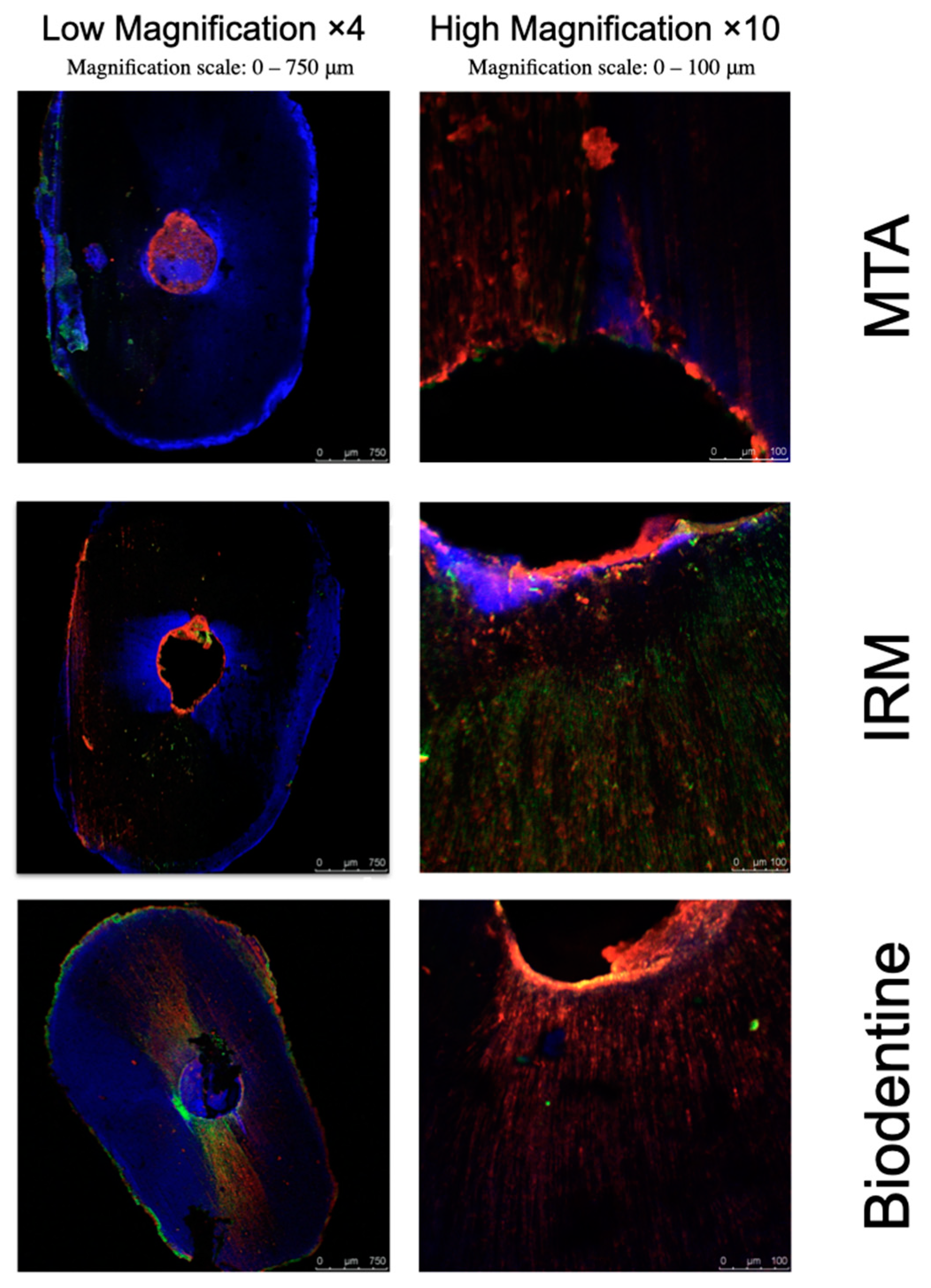
  • (n = 15): Retrograde 3 mm preparations filled with fluorescently labeled MTA (ProRoot; Dentsply Tulsa Dental, Johnson City, TN, USA).
  • (n = 15): Retrograde 3 mm preparations filled with fluorescently labeled IRM (IRM; Dentsply, Mannheim, Germany).
  • (n = 15): Retrograde 3 mm preparations filled with fluorescently labeled Biodentine (Biodentine; Septodont, Saint-Maur-des-Fossés, France).
  • (n = 5): Retrograde 3 mm preparations, not filled (Positive control).
  • (n = 5): Teeth without retrograde preparation or filling. The whole external root surface including the apical foramen was sealed with 2 layers of nail varnish (Negative control) [12,13].
  • (n = 5): Like Group 1 but without ensuing bacterial contamination (MTA negative control).
  • (n = 5): Like Group 2 but without ensuing bacterial contamination (IRM negative control).
  • (n = 5): Like Group 3 but without ensuing bacterial contamination (Biodentine negative control).
All specimens were then stored at 100% humidity and 37 °C for 24 h to allow for setting of the retrograde materials.

2.2. The Experimental Model

To avoid bacterial penetration across lateral canals or gaps in the cementum, 2 layers of nail varnish (Lilliput Nagellack; Wiesbaden, Germany) were applied to the external surfaces of the roots, without covering the prepared apical areas [13]. Then, the roots were placed in a previously described experimental model [2] (Figure 1a).
The roots were placed in 1.5 mL plastic Eppendorf tubes (disposable scintillation vials; Sigma-Aldrich, St. Louis, MO) and then placed in a glass vial (clear glass vial; Sigma-Aldrich, St. Louis, MO, USA) tightly through the rubber cap. The interfaces between the Eppendorf, the root, and the rubber cap were sealed using cyanoacrylate adhesive (Krazy Glue; Columbus, OH, USA).

2.3. Simulation of Enterococcus Faecalis Bacterial Infection

Ethylene oxide gas was used to sterilize the roots [14]. Then, a growth medium for streptomycin-resistant T2 strain E. faecalis bacteria (ATCC 29212, Manassas, VA, USA) was autoclaved. Since E. faecalis is resistant to streptomycin sulfate, 0.5 mg/mL streptomycin sulfate (Sigma-Aldrich; St. Louis, MO, USA) was then added to avoid possible contamination by other bacterial species [15].
The roots were then coronally filled with E. faecalis bacterial suspension using a pipette and incubated at 100% humidity and 37 °C. The bacterial suspension was freshly prepared and replaced every day up to 21 days [2] (Figure 1b).

2.4. Preparing the Roots for the Assessments

Following 21 days of incubation, the roots were fixed in a self-cure acrylic repair material (UNIFAST Trad; GC, St. Alsip, IL, USA). Then, in order to prepare samples of root dentin axial slices for the assessments, 3 mm sections of the root ends were cut vertically to the long axis of the root, using water cooling and a 500 rpm diamond saw (Isomet; Buehler, Lake Bluff, IL, USA). [2]. In order to validate the model, one slice from each tooth was scanned in Environmental SEM (ESEM). The ESEM slices were viewed in environmental “wet” mode using a Philips XL30 ESEM-Feg (FEI/Philpips Electron Otpics, Eindhoven, The Netherlands) (working conditions: 5 °C, 2.9–5.9 torr gas pressure, 80% relative humidity, 6–9 kV). Five interesting spots on each sample were selected.
The prepared samples were stained using a Live and Dead Bacterial Viability kit (L-7012 Molecular Probes; Eugene, OR, USA) [2], which contains two distinct vials of the two component dyes (propidium iodide and SYTO 9 in a 1-to-1 mixture) for the staining of the evaluated bacteria. The excitation/emission maxima of the dyes were 480–500 nm for the SYTO 9 (staining live bacteria in green), 490–635 nm for propidium iodide (staining dead bacteria in red) [7], and 365–440 nm for the Alexa Fluor 350 (retrograde filling material stained in blue) [16].

2.5. Confocal Laser Microscopy Assessment

Following the staining of the bacteria and the filling materials, their fluorescence was assessed by CLSM (Leica TCS SP5; Leica Microsystems CMS, Wetzlar, Germany). The evaluations were performed at the mesial, distal, buccal, and lingual areas of the root dentin axial slices. The red, green, and blue fluorescence was displayed using single-channel and simultaneous triple-channel imaging [2,16], producing images of the bacteria and of the fillings at a resolution of 1024 × 1024 pixels.
The images were then evaluated using dedicated software (LAS AF, version 2.6.0.7266; Leica Microsystems CMS, Wetzlar, Germany). The slices were assessed by a × 4 lens, and the extent of fluorescence staining within the buccal, lingual, mesial, and distal areas of the slices was assessed [2,6]. The following measurements were performed to assess the invasion of the bacteria into the dental tubuli (penetration depth and viability), and to assess the influence of the retrograde filling on the bacterial invasion (Figure 2):
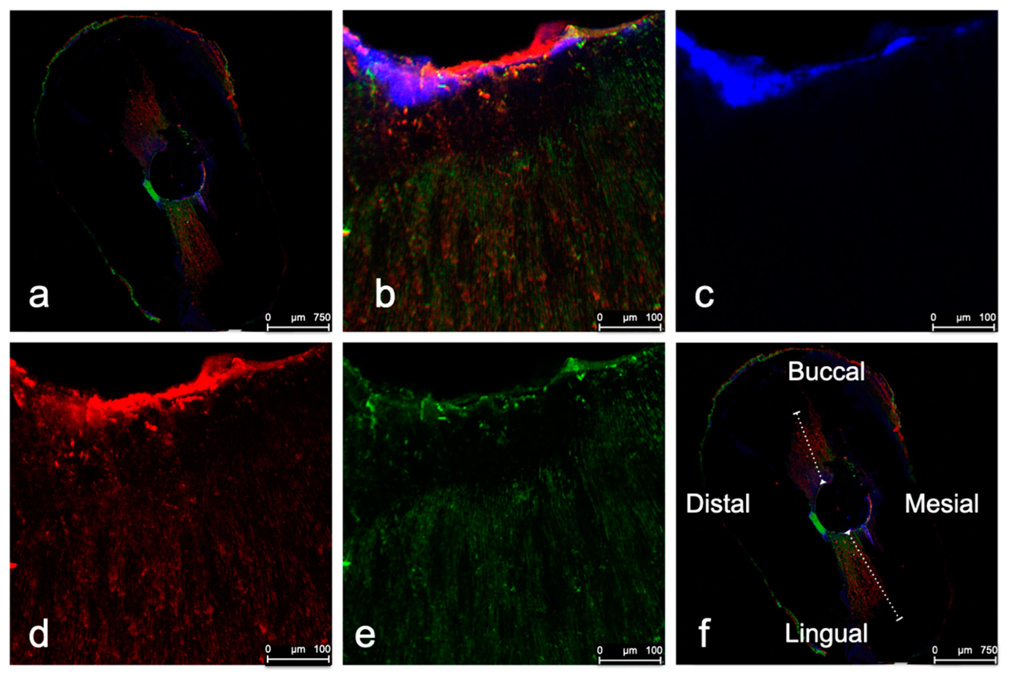
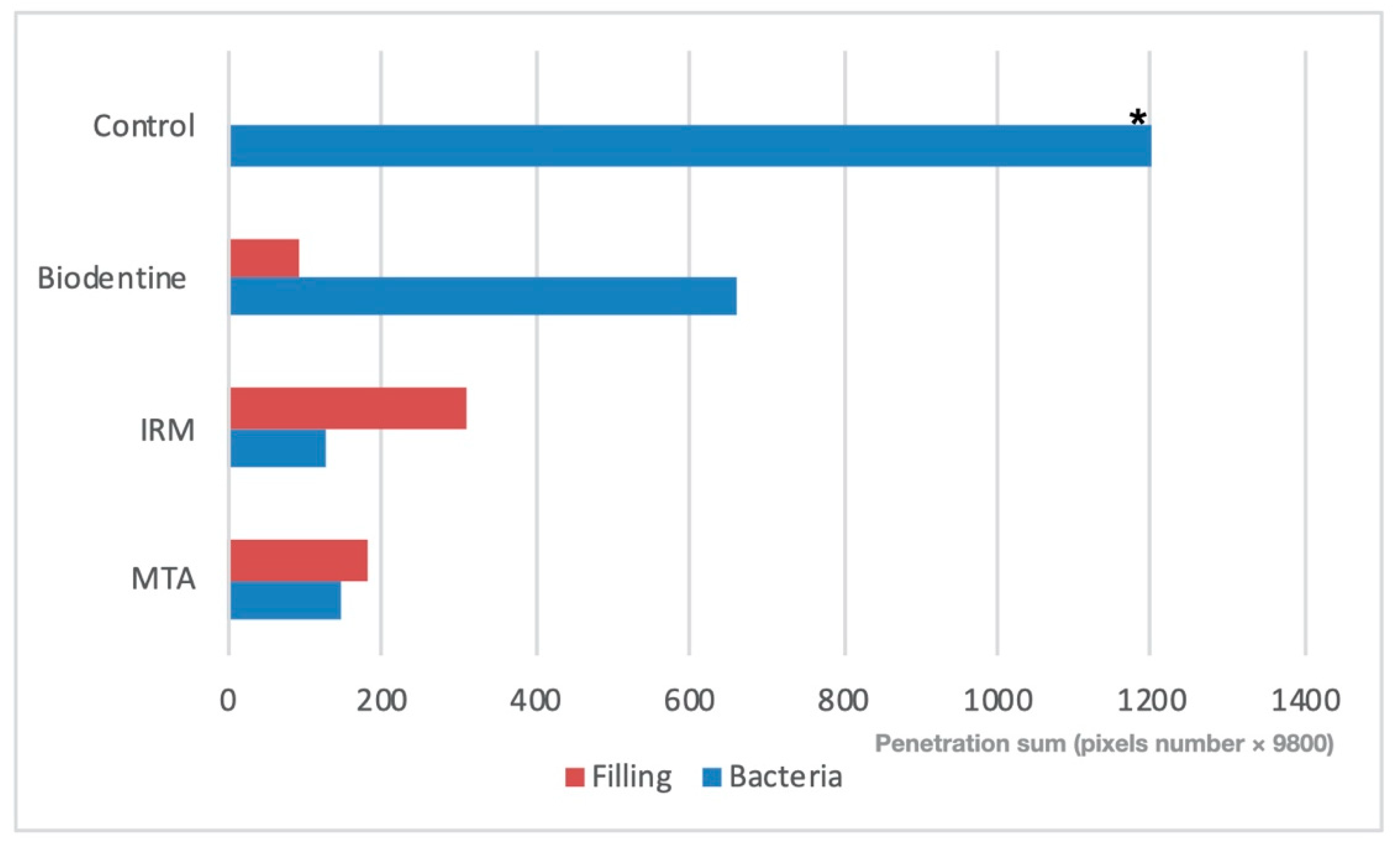
  • The depths of bacterial invasion and filling penetrations within the tubuli were measured at the buccal, lingual, mesial, and distal areas of the root dentin axial slices, defining the root canal wall as the beginning point (Figure 2f).
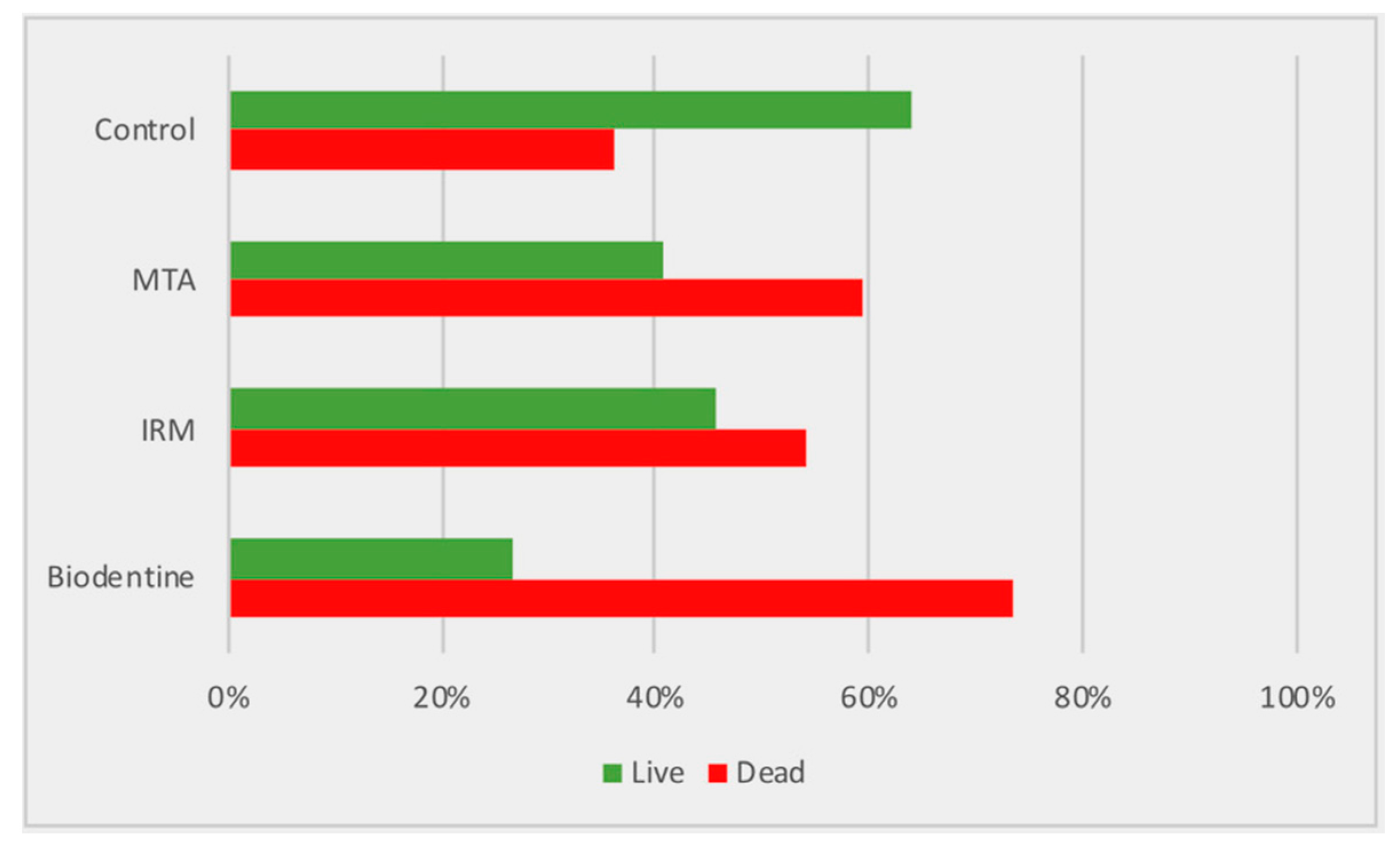
  • The bacterial viability was calculated as the proportions of live and dead bacteria.
  • The correlation between the filling material type and penetration depth, and that between the bacterial invasion depth and viability, was determined.

2.6. Statistical Evaluation

The results were statistically analyzed using SPSS software (SPSS version 22; SPSS Inc., Chicago, IL, USA). One-way ANOVA tests were performed to assess the fluorescence at the buccal, lingual, mesial, and distal areas and to evaluate the level of fluorescence staining within each area (in the same group and between the different groups), the depth of bacterial penetration into the dentinal tubules, and the depth of filling penetration into the tubules. Pearson’s Chi-squared test was performed to check for dependency between the bacterial viability and the filling material used. Chi-squared tests with Yates’ continuity correction were performed to evaluate the bacterial viability effect difference between the materials. p < 0.05 was considered statistically significant.

3. Results

Bacterial fluorescence was not detected in the negative control specimens, and fluorescence was detected in all the specimens of the positive control group.
The extents of the stained areas (bacteria and filling) were larger in the lingual and buccal areas compared to the distal and mesial areas. However, the differences between these areas was not statistically significant between the different retrograde filling material groups (p = 0.094) (Figure 2a,f).
Looking at all groups, the filling material and bacterial penetration depths within the tubuli were variable, with minimal and maximal filling penetration depths of 11 and 957 μm, respectively (mean of 130 ± 158 μm), and minimal and maximal bacterial penetration depths of 9 and 1480 μm, respectively (mean of 167 ± 317 μm) (Table 1). In addition, a negative correlation was found between the depth of filling material penetration and the bacterial penetration, where greater penetration depth of the filling material into the tubuli was associated with significantly shallower bacterial penetration depth (live and dead bacteria combined) (p = 0.015).
When comparing the filling and bacterial penetration depths between the filling groups, in the Biodentine group, bacteria penetrated deeper within the tubuli, while the filling penetration depths were shallower in comparison to the other materials (IRM, MTA) (p = 0.004) (Table 1).
Generally, more dead bacteria were detected compared to live bacteria in all the experimental groups (p = 0.003), without significant differences between the filling materials (p = 0.087) (Figure 3).
However, a significant negative correlation was found between the depth of filling material penetration and the penetration depth of live bacteria, where deeper filling penetration was associated with shallower penetration depths of live bacteria (p = 0.024) (Figure 4 and Figure 5).

4. Discussion

Following endodontic surgery, bacteria may still invade a retrofilled root canal and deep into the dentinal tubules [1,17], potentially leading to an inflammatory reaction when bacterial by-products invade the surrounding periradicular tissues [18]. These bacteria form biofilms, which are complex ecological communities that use diverse mechanisms to secure themselves against a harsh environment, including the immune system and antibacterial agents [19].
Gram-positive facultative anaerobes are the most commonly detected bacteria in endodontically treated teeth presenting with persistent endodontic infections [2]. Among them, E. faecalis plays a major role in bacterial biofilm invasion and is considered a suitable model for assessing root canal bacterial invasion [2,8].
Many microscopic, microbiological, and histological experimental models have been used to assess bacterial invasion within endodontically treated root canals [20,21]. The most common traditional experimental model is the two-chamber bacterial leakage model, which is used to assess the diffusion of bacteria from an upper chamber and over the retrograde filling to a suspension placed in the lower chamber. However, it has been reported that this traditional model suffers from many inherent limitations, such as a lack of proper control groups and histological evaluation, and that its reliability is compromised [22,23,24].
Additional ex vivo models to simulate and assess bacterial invasion, such as the traditional dye-penetration model [25] or the high-pressure replica technique used to assess the pore morphologies and apical leakage of sealers in retrofilled teeth [26], are restricted because in these traditional models indirect evaluations are used, incapable of demonstrating the real routes of bacterial invasion, and because of the absence of proper histological controls in these models [22]. Furthermore, under the adverse conditions of a filled root canal, bacteria can increase their stress tolerance by entering a dormant state where they are viable but not culturable [5,27], thus challenging the efficacy of traditional culture-based studies [5,19,27].
The authors of a recent study established a novel and reliable experimental model that enabled them to trace and quantify biofilm invasion into the dentinal tubules [2]. With this CLSM-based model, the actual routes of bacterial invasion are tracked histologically, and negative and positive histological controls are used to verify the suitability of the experimental model. Furthermore, the true viability of the proliferating bacteria, even when they enter a dormant, not-culturable state, can be assessed [5,19,27]. As a result, using CLSM together with live/dead bacterial staining techniques offers information regarding both the magnitude of the dentin infection and the vitality of the proliferating bacteria within the infected tubuli in situ [2,8], thus enabling a better understanding of the microbiological–pathological course after endodontic surgical procedures [2].
In the current study, we used this CLSM-based experimental model to assess the invasion of bacterial biofilms into the dentinal tubules. Furthermore, bacterial invasion was assessed in the presence of fluorescently labeled retrograde filling materials. The use of CLSM to assess the penetration of the retrograde filling materials into the tubuli [6,28], while assessing the depth of the bacterial invasion into the tubuli and their viability, provides a comprehensive view of the microbial–pathological process following surgery. Bacterial fluorescence was not detected in the negative control group, while fluorescence was detected in the positive control group specimens, confirming the reliability of the experimental model.
The rationale of a retrograde filling material is to inhibit bacterial biofilm growth and the outflow of bacterial byproducts and toxins into the surrounding periradicular tissues [29]. In addition, the root-end filling is expected to entomb any remaining bacteria beneath the filling in a way that would eventually result in bacterial death [30]. However, studies found bacterial biofilm at the interfaces between the filling and the canals’ dentin walls, and also deep in the dentinal tubules [7,15]. In order to provide a comprehensive view and analysis of the relationships between the proliferating biofilm and the filling material, in the current study, different filling materials were fluorescently labeled in order to evaluate and understand the influence of the filling materials and their penetration depths into the tubuli on bacterial invasion within the tubuli.
The same as in a previous study [2], in the current study, we found a pattern of bacterial invasion and filling penetration in which the bucco-lingual direction was preferred compared to the mesio-distal direction, regardless of the type of filling material. This finding was also supported by previous investigations [2,23,24,31]. The fact that both the fillings and the bacteria penetrated deeper in the bucco-lingual direction may be associated to an anatomical–physiological phenomenon, “the butterfly effect”, which means a butterfly-like appearance observed on the cross sections of roots that are associated with higher sclerosis down the tubuli at the distal and mesial sides of the root canal [24]. This phenomenon is commonly detected in single-rooted human teeth in a broad range of ages [32,33]. Similarly, in a study by Rechenberg et al. [24], histological observations revealed that the penetration of microorganisms might predominantly happen through tubular aspects of the dentin, whereas atubular or sclerotic dentin and the interfaces between dentin and sealer remained bacteria-tight [24]. Thus, it seems that while a two-chamber model is not suitable for the evaluation of bacterial leakage, it reliably reflects the invasion of bacteria into the tubuli [2,23,24].
The bacterial invasion depths into the dentinal tubules were variable, reaching as deep as 1480 μm (with a mean of 167 μm), which corroborates the findings of a previous study [2]. Peters et al. [34] evaluated the viable endodontic bacteria in the root dentin of infected teeth presenting with apical periodontitis using culturing methods. They found that in the majority of the roots, bacteria were identified deep within the dentin near the cementum. They also found that an anaerobic culturing method is more sensitive than histology in identifying these bacteria in the dentin. Within that scope, confocal microscopy seems to be a promising technique to assess bacterial penetration within the tubuli because it enables us to evaluate live and dead bacteria, as well as bacteria in dormant, not-culturable states [5,19,27]. Thus, CLSM is capable of assessing the actual level of bacterial invasion within the tubuli in situ [2].
In the current study, bacterial penetration into the dentinal tubules was significantly influenced by the type of filling material and its penetration depth into the dentinal tubules. For all materials, it was found that deeper filling penetration within the tubuli was significantly related with shallower bacterial penetration. This finding confirms that an appropriate retrograde filling material may inhibit bacterial penetration across the dentin into the periradicular tissues of teeth following root end resection and filling [35]. The fact that the Biodentine fillings penetrated less, and their associated bacterial penetration depths were deeper compared to the MTA and IRM groups (p < 0.05), may suggest that the type of root end filling material and its ability to penetrate into the tubuli may play a significant part in the risk of bacterial invasion following endodontic surgery (Figure 4).
Biodentine is a material based on calcium silicate that has several applications, including as a root-end filling material used in endodontic surgical procedures [36]. The material is prepared by the silicate-based cement technique with some adjustments aimed to improve its physical properties and handling. It had been claimed that Biodentine has a fast setting time due to its increased particle size, the addition of calcium chloride to its liquid constituent, and its decreased liquid content [35,37]. However, its bigger particle size may also explain its shallower penetration depth into the dentinal tubules compared with the other materials and its lesser ability to prevent bacterial invasion deep into the dentinal tubules as compared to the other evaluated materials.
Generally, in the presence of a filling material, significantly more dead bacteria than live bacteria were detected within the dental tubules (p = 0.003, Figure 3), without significant differences between the filling materials (p = 0.087). Nevertheless, deeper filling penetration was associated with shallower penetration depths of live bacteria (p = 0.015) (Figure 4). The fact that in the present investigation the viability of the proliferating bacteria was influenced by the filling material penetration depth, rather than by the filling material type, may seemingly contradict the results of other studies that focused on the direct antimicrobial properties of different retrograde filling materials by assessing their spectra of affected bacteria, effectiveness periods, and antimicrobial mechanisms [37,38,39,40,41]. Torabinejad et al. [38] reported that MTA has no effect on any of the strict anaerobic bacteria and some antibacterial effect on several of the facultative bacteria, whereas other materials such as IRM possess antibacterial properties on both groups of bacteria [38]. Additional studies have reported that immediately after its setting, IRM has antibacterial effects on E. faecalis and sustains these capabilities for at least one day [39], while others [40] reported identical effects in root-end fillings. Others claimed that Biodentine possesses antibacterial capabilities because of its relatively high pH that has an inhibitory effect on bacteria, and because of alkalization of the ecosystem that leads to the decontamination of neighboring soft and hard tissues [37,41].
However, in addition to the direct antibacterial effect of the retrograde filling material, its capability to penetrate and to entomb the bacteria is responsible for the prevention of bacterial invasion [1]. Our findings suggest that the antibacterial properties of the retrograde filling material are related to its penetration capacity deep into the tubuli.
In the current study we used an ex vivo model to simulate an in vivo clinical scenario. It should be noted that the experimental methods used to assess biofilm formation do not accurately simulate true in vivo conditions; therefore, the methods’ ability to provide clinically relevant information is limited. However, in the current study, we used representative surfaces to grow the biofilm—ex vivo dentin tissue samples—which are more likely to provide information that is relevant to true in vivo endodontic infections [42].

5. Conclusions

Given the limitations of this ex vivo model, this study enables better understanding of the microbiological–pathological course after endodontic surgical procedures. It was found that even with retrograde fillings, bacteria invade deep into the dental tubules (up to 1480 μm). This invasion is variable and is affected by the root tubular anatomy and by the root-end filling material and its actual penetration depth into the dentinal tubules. The ability of a filling material to penetrate deep into the dental tubules is associated with lesser penetration of the bacteria and reduced viability of the invading bacteria. Additional clinical studies are indicated to elucidate the clinical implications of bacterial invasion into retrofilled root ends following endodontic surgery. In addition, clinical and experimental assessments of retrograde filling materials should take into consideration not only the direct antimicrobial effect of the filling material but also its chemical and physical properties that affect its penetration capacity deep into the tubuli.

Author Contributions

E.R., S.E. and I.T. designed the study; S.E., S.H.-Y. and L.J. performed the laboratory experiments; E.R. and S.E. collected the data; E.R., S.E. and I.T. analyzed the data and prepared the tables; E.R., S.E., H.S. and I.T. wrote and reviewed the manuscript. All authors have read and agreed to the published version of the manuscript.

Funding

This study was supported by the Ernst & Tova Turnheim Clinical Research Fund in Dentistry.

Acknowledgments

The authors deny any conflicts of interest related to this study.

Conflicts of Interest

The authors declare no conflict of interest.

References

  1. Gartner, A.H.; Dorn, S.O. Advances in endodontic surgery. Dent. Clin. N. Am. 1992, 36, 357–378. [Google Scholar] [PubMed]
  2. Tsesis, I.; Elbahary, S.; Venezia, N.B.; Rosen, E. Bacterial colonization in the apical part of extracted human teeth following root-end resection and filling: A confocal laser scanning microscopy study. Clin. Oral Investig. 2018, 22, 267–274. [Google Scholar] [CrossRef] [PubMed]
  3. Gundam, S.; Patil, J.; Venigalla, B.S.; Yadanaparti, S.; Maddu, R.; Gurram, S.R. Comparison of marginal adaptation of mineral trioxide aggregate, glass ionomer cement and intermediate restorative material as root-end filling materials, using scanning electron microscope: An in vitro study. J. Conserv. Dent. 2014, 17, 566–570. [Google Scholar] [CrossRef] [PubMed] [Green Version]
  4. Nepal, M.; Shubham, S.; Tripathi, R.; Khadka, J.; Kunwar, D.; Gautam, V.; Gautam, N. Spectrophotometric analysis evaluating apical microleakage in retrograde filling using GIC, MTA and biodentine: An in-vitro study. BMC Oral Health 2020, 20, 37. [Google Scholar] [CrossRef]
  5. Heinrich, K.; Leslie, D.J.; Jonas, K. Modulation of bacterial proliferation as a survival strategy. Adv. Appl. Microbiol. 2015, 92, 127–171. [Google Scholar] [CrossRef]
  6. Kuci, A.; Alacam, T.; Yavas, O.; Ergul-Ulger, Z.; Kayaoglu, G. Sealer penetration into dentinal tubules in the presence or absence of smear layer: A confocal laser scanning microscopic study. J. Endod. 2014, 40, 1627–1631. [Google Scholar] [CrossRef]
  7. Aziz, A.; Parmar, D.; McNaughton, A.; Tompkins, G.R. Bacterial viability determination in a dentinal tubule infection model by confocal laser scanning microscopy. Methods Mol. Biol. 2010, 666, 141–150. [Google Scholar] [CrossRef]
  8. Zapata, R.O.; Bramante, C.M.; de Moraes, I.G.; Bernardineli, N.; Gasparoto, T.H.; Graeff, M.S.; Campanelli, A.P.; Garcia, R.B. Confocal laser scanning microscopy is appropriate to detect viability of Enterococcus faecalis in infected dentin. J. Endod. 2008, 34, 1198–1201. [Google Scholar] [CrossRef]
  9. Schneider, S.W. A comparison of canal preparations in straight and curved root canals. Oral Surg. Oral Med. Oral Pathol. 1971, 32, 271–275. [Google Scholar] [CrossRef]
  10. Wu, M.K.; R’Oris, A.; Barkis, D.; Wesselink, P.R. Prevalence and extent of long oval canals in the apical third. Oral Surg. Oral Med. Oral Pathol. Oral Radiol. Endod. 2000, 89, 739–743. [Google Scholar] [CrossRef] [Green Version]
  11. Roane, J.B.; Sabala, C.L.; Duncanson, M.G., Jr. The “balanced force” concept for instrumentation of curved canals. J. Endod. 1985, 11, 203–211. [Google Scholar] [CrossRef]
  12. Kytridou, V.; Gutmann, J.L.; Nunn, M.H. Adaptation and sealability of two contemporary obturation techniques in the absence of the dentinal smear layer. Int. Endod. J. 1999, 32, 464–474. [Google Scholar] [CrossRef] [PubMed]
  13. Adamo, H.L.; Buruiana, R.; Schertzer, L.; Boylan, R.J. A comparison of MTA, Super-EBA, composite and amalgam as root-end filling materials using a bacterial microleakage model. Int. Endod. J. 1999, 32, 197–203. [Google Scholar] [CrossRef]
  14. Brosco, V.H.; Bernardineli, N.; Torres, S.A.; Consolaro, A.; Bramante, C.M.; de Moraes, I.G.; Ordinola-Zapata, R.; Garcia, R.B. Bacterial leakage in obturated root canals-part 2: A comparative histologic and microbiologic analyses. Oral Surg. Oral Med. Oral Pathol. Oral Radiol. Endod. 2010, 109, 788–794. [Google Scholar] [CrossRef] [PubMed]
  15. Fuss, Z.; Weiss, E.I.; Shalhav, M. Antibacterial activity of calcium hydroxide-containing endodontic sealers on Enterococcus faecalis in vitro. Int. Endod. J. 1997, 30, 397–402. [Google Scholar] [CrossRef]
  16. Panchuk-Voloshina, N.; Haugland, R.P.; Bishop-Stewart, J.; Bhalgat, M.K.; Millard, P.J.; Mao, F.; Leung, W.Y.; Haugland, R.P. Alexa dyes, a series of new fluorescent dyes that yield exceptionally bright, photostable conjugates. J. Histochem. Cytochem. 1999, 47, 1179–1188. [Google Scholar] [CrossRef] [PubMed]
  17. Ørstavik, D. Endodontic filling materials. Endod. Top. 2014, 31, 53–67. [Google Scholar] [CrossRef]
  18. Kim, S.G. Variations in Outcome of Endodontic Surgery; Tsesis, I., Ed.; Springer: Berlin/Heidelberg, Germany, 2014. [Google Scholar] [CrossRef]
  19. Rosen, E.; Kolodkin-Gal, I.; Tsesis, I. Challenges in the Eradication of Enterococcus faecalis and its Implications on Health. Curr. Oral Health Rep. 2018. [Google Scholar] [CrossRef]
  20. Love, R.M.; Jenkinson, H.F. Invasion of dentinal tubules by oral bacteria. Crit. Rev. Oral Biol. Med. 2002, 13, 171–183. [Google Scholar] [CrossRef] [Green Version]
  21. Langeland, K. Tissue response to dental caries. Endod. Dent. Traumatol. 1987, 3, 149–171. [Google Scholar] [CrossRef]
  22. De-Deus, G. Research that matters—Root canal filling and leakage studies. Int. Endod. J. 2012, 45, 1063–1064. [Google Scholar] [CrossRef] [PubMed]
  23. Rechenberg, D.K.; De-Deus, G.; Zehnder, M. Potential systematic error in laboratory experiments on microbial leakage through filled root canals: Review of published articles. Int. Endod. J. 2011, 44, 183–194. [Google Scholar] [CrossRef] [PubMed]
  24. Rechenberg, D.K.; Thurnheer, T.; Zehnder, M. Potential systematic error in laboratory experiments on microbial leakage through filled root canals: An experimental study. Int. Endod. J. 2011, 44, 827–835. [Google Scholar] [CrossRef] [PubMed]
  25. Vertucci, F.J.; Beatty, R.G. Apical leakage associated with retrofilling techniques: A dye study. J. Endod. 1986, 12, 331–336. [Google Scholar] [CrossRef]
  26. Kielbassa, A.M.; Uchtmann, H.; Wrbas, K.T.; Bitter, K. In vitro study assessing apical leakage of sealer-only backfills in root canals of primary teeth. J. Dent. 2007, 35, 607–613. [Google Scholar] [CrossRef]
  27. Rosen, E.; Tsesis, I.; Elbahary, S.; Storzi, N.; Kolodkin-Gal, I. Eradication of Enterococcus faecalis Biofilms on Human Dentin. Front. Microbiol. 2016, 7, 2055. [Google Scholar] [CrossRef]
  28. Kim, Y.; Kim, B.S.; Kim, Y.M.; Lee, D.; Kim, S.Y. The Penetration Ability of Calcium Silicate Root Canal Sealers into Dentinal Tubules Compared to Conventional Resin-Based Sealer: A Confocal Laser Scanning Microscopy Study. Materials (Basel) 2019, 12, 531. [Google Scholar] [CrossRef] [Green Version]
  29. Dorn, S.O.; Gartner, A.H. Retrograde filling materials: A retrospective success-failure study of amalgam, EBA, and IRM. J. Endod. 1990, 16, 391–393. [Google Scholar] [CrossRef]
  30. Peters, L.B.; Wesselink, P.R.; Moorer, W.R. The fate and the role of bacteria left in root dentinal tubules. Int. Endod. J. 1995, 28, 95–99. [Google Scholar] [CrossRef]
  31. Weis, M.V.; Parashos, P.; Messer, H.H. Effect of obturation technique on sealer cement thickness and dentinal tubule penetration. Int. Endod. J. 2004, 37, 653–663. [Google Scholar] [CrossRef] [Green Version]
  32. Vasiliadis, L.; Darling, A.I.; Levers, B.G. The amount and distribution of sclerotic human root dentine. Arch. Oral Biol. 1983, 28, 645–649. [Google Scholar] [CrossRef]
  33. Russell, A.A.; Chandler, N.P.; Hauman, C.; Siddiqui, A.Y.; Tompkins, G.R. The butterfly effect: An investigation of sectioned roots. J. Endod. 2013, 39, 208–210. [Google Scholar] [CrossRef] [PubMed]
  34. Peters, L.B.; Wesselink, P.R.; Buijs, J.F.; van Winkelhoff, A.J. Viable bacteria in root dentinal tubules of teeth with apical periodontitis. J. Endod. 2001, 27, 76–81. [Google Scholar] [CrossRef] [PubMed]
  35. Ma, X.; Li, C.; Jia, L.; Wang, Y.; Liu, W.; Zhou, X.; Johnson, T.M.; Huang, D. Materials for retrograde filling in root canal therapy. Cochrane Database Syst. Rev. 2016, 12, CD005517. [Google Scholar] [CrossRef]
  36. Torabinejad, M.; Parirokh, M.; Dummer, P.M.H. Mineral trioxide aggregate and other bioactive endodontic cements: An updated overview—Part II: Other clinical applications and complications. Int. Endod. J. 2018, 51, 284–317. [Google Scholar] [CrossRef]
  37. Camilleri, J.; Sorrentino, F.; Damidot, D. Investigation of the hydration and bioactivity of radiopacified tricalcium silicate cement, Biodentine and MTA Angelus. Dent. Mater. 2013, 29, 580–593. [Google Scholar] [CrossRef]
  38. Torabinejad, M.; Hong, C.U.; Pitt Ford, T.R.; Kettering, J.D. Antibacterial effects of some root end filling materials. J. Endod. 1995, 21, 403–406. [Google Scholar] [CrossRef]
  39. Slutzky, H.; Slutzky-Goldberg, I.; Weiss, E.I.; Matalon, S. Antibacterial properties of temporary filling materials. J. Endod. 2006, 32, 214–217. [Google Scholar] [CrossRef]
  40. Chong, B.S.; Owadally, I.D.; Pitt Ford, T.R.; Wilson, R.F. Antibacterial activity of potential retrograde root filling materials. Endod. Dent. Traumatol. 1994, 10, 66–70. [Google Scholar] [CrossRef]
  41. Bani, M.; Sungurtekin-Ekci, E.; Odabas, M.E. Efficacy of Biodentine as an Apical Plug in Nonvital Permanent Teeth with Open Apices: An In Vitro Study. BioMed Res. Int. 2015, 2015, 359275. [Google Scholar] [CrossRef] [Green Version]
  42. Roberts, A.E.; Kragh, K.N.; Bjarnsholt, T.; Diggle, S.P. The Limitations of In Vitro Experimentation in Understanding Biofilms and Chronic Infection. J. Mol. Biol. 2015, 427, 3646–3661. [Google Scholar] [CrossRef] [PubMed]
Figure 1. (a): Illustration of the tested model. (b): Scanning electron microscopy (SEM) images demonstrating bacterial penetration into the dentinal tubules.
Applsci 10 06996 g001
Figure 2. (af): Demonstrating confocal laser scanning microscopy (CLSM) images (magnification scale; a: 0–750 μm, b–f: 0–100 μm), from the positive control group) of the E. faecalis bacterial invasion into the dentinal tubules. Live bacteria are presented in green (e), dead bacteria are presented in red (d), and the retrograde filling is presented in blue (c). The bacteria and the filling materials can be seen at high (be) and low (a,f) magnification. The minimal, maximal, and mean depths of bacterial and filling penetration within the tubuli were assessed by defining the canal wall as the beginning point (f).
Applsci 10 06996 g002
Figure 3. Average percentages of detected dead bacteria (red) and live bacteria (green). There were significantly more dead bacteria than live bacteria in all the experimental groups (Chi-squared, p = 0.003).
Applsci 10 06996 g003
Figure 4. Fluorescence penetration amounts for the different groups (presented as the number of pixels × 9800) (one-way ANOVA).
Applsci 10 06996 g004
Figure 5. CLSM images presenting the detected fluorescence of the different experimental groups following 21 days of bacterial contamination. Dead (red) and live (green) bacteria within the tubuli, and the filling material (blue) are demonstrated.
Applsci 10 06996 g005
Table 1. Presents the penetration depths (in μm) of the retrograde filling materials and bacteria within the tubuli. The minimal and maximal bacterial penetration depths into the dentinal tubules were 1 and 1480 μm, respectively, with a mean of 167 μm. The minimal and maximal filling penetration depths into the dentinal tubules were 0 and 957 μm, respectively, with a mean of 130 μm. In the Biodentine group, in comparison to the other materials (mineral trioxide aggregate (MTA), intermediate restorative material (IRM)), bacteria penetrated deeper into the dentin (b: one-way ANOVA, p = 0.0021), while the filling depth was lower (a: one-way ANOVA, p = 0.04). In the control group, bacteria penetrated deeper into the dentin in comparison to other groups (c: one-way ANOVA, p = 0.00018).
Filling Material PenetrationBacterial Penetration Group
SDMedianMaxMinSDMedianMaxMin
1981836445815814650033MTA
158311957113421299969IRM
15593 a535 a32451663 b1480160Biodentine
----1891200 c1610890 cControl

