Article

Evaluation of Antioxidant Activity, Toxicity, and Phenolic Profile of Aqueous Extracts of Chamomile (Matricaria chamomilla L.) and Sage (Salvia officinalis L.) Prepared at Different Temperatures

by
Nefeli S. Sotiropoulou
,
Stiliani F. Megremi
and
Petros Tarantilis
*
Department of Food Science and Human Nutrition, School of Food and Nutritional Sciences, Agricultural University of Athens, 11855 Athens, Greece
*
Author to whom correspondence should be addressed.
Appl. Sci. 2020, 10(7), 2270; https://doi.org/10.3390/app10072270
Submission received: 19 February 2020 / Revised: 19 March 2020 / Accepted: 21 March 2020 / Published: 26 March 2020
(This article belongs to the Special Issue Antioxidants in Natural Products)

Abstract

:
Chamomile and sage are common herbs that are mostly used as infusions due to their beneficial properties. The aims of this study were to determine the total phenolic content, antioxidant activity, and potential toxicity of chamomile and sage aqueous extracts prepared at three different temperatures (25, 80, 100 °C) and finally, to detect their phenolic profiles at the optimum temperature. In order to measure the total phenolic content and antioxidant capacity, Folin–Ciocalteu and 2,2-diphenyl-1-picrylhydrazyl hydrate (DPPH) assays were applied, respectively. The extraction temperature at 80 °C was the optimum, with maximal antioxidant activity and the highest total phenolic content for both herbs. Luminescence-based assay demonstrated that all the examined aqueous extracts possessed toxicity towards Vibrio fischeri. Microtox assay demonstrated no correlation with the other two assays, which were positively correlated. The major phenolics of chamomile were rutin trihydrate, ferulic acid, chlorogenic acid, and apigenin-7-O-glucoside; and major phenolics of sage were rosmarinic acid, salvianolic acid K, and luteolin-7-O-glucuronide, as defined by LC-MS of aqueous extracts at 80 °C. It can be concluded that the extraction of herbal aqueous extracts at 80 °C can provide significant bioactive and antioxidant compounds, but their consumption must be in moderation.

1. Introduction

Nowadays, herbal infusions are placed in most consumed beverages [1]. This consumption is increasing worldwide due to their significant role in the human diet as a source of antioxidants. Herbal infusions contain various bioactive phenolic compounds, which are considered beneficial for human health [2]. Herbal infusions are consumed traditionally, without safety. Since “safety” and “natural” are not synonymous and some phenolics have shown toxicity, it is essential to estimate and know their potential toxicity [3,4]. The modern and cost-effective Vibrio fischeri bioluminescence inhibition test has proved to be a reliable and sensitive method for the evaluation of acute toxicity of herbal infusions [5,6].
Chamomile (Matricaria chamomilla L., syn: Matricaria recutita) is a medicinal herb and one of the most consumed single-ingredient herbal tea [7], considered as a source of important chemicals with bioactive properties [8]. Chamomile is consumed as an infusion (with over a million cups per day), due to its healing benefits according to traditional medicine but also for its antioxidant, anti- nociceptive, and anticancer activities [9,10]. These benefits are partly due to the phenolic content [7], specifically the subfamily of flavonoids are the most responsible for its high antioxidant activity [2].
Sage (Salvia officinalis L.) is considered as one of the most popular herbs consumed widely and traditionally as an herbaceous infusion [11,12]. The incorporation of sage infusion in the daily diet can provide considerable benefits such as anti-mycotic [11], anti-carcinogenic [13,14], antidiabetic, antimicrobial [14], anti-inflammatory, and anti-proliferative [15]. In addition to these effects, sage infusion exhibits antiradical activity which correlates strongly with their high level of total phenolic content [11,13].
In general, preparation of infusion differs, depending on the tradition of each region where it is prepared with water at different temperatures. Habitually, according to the traditional use, the water temperature in household conditions of infusion preparation usually ranges from 80 to 100 °C [16,17]. However, the application of water at low temperature is also nominated in comparison with boiling water due to the feasibility to eliminate or diminish toxic components in the final obtained infusion [14]. Furthermore, there is no universal procedure for herbal infusion preparation that is proper for the extraction of antioxidant compounds [18]. There are a few works reporting the influence of extraction temperature on bioactivity [17,19] and acute toxicity [5] of herbal aqueous extracts.
There are no data, particularly for the preparation of chamomile and sage infusions, evaluating antioxidant properties and toxicity based on the extraction temperature. Therefore, the objective of this study is to evaluate the total phenolic content, antioxidant activity, and toxicity towards V. fischeri at three different temperatures (25, 80, and 100 °C) using in vitro spectrometric assays in order to define the optimum temperature. In addition, determination of phenolic profile of the optimum extract was obtained.

2. Materials and Methods

2.1. Chemicals

The following chemicals were purchased from Sigma-Aldrich (St. Louis, MO, USA): 2,2-diphenyl-1-picrylhydrazyl hydrate (DPPH), Folin–Ciocalteau phenol reagent, caffeic acid, and rosmarinic acid. Methanol (LC-MS grade), water (LC-MC grade), and sodium carbonate were purchased from Fischer Scientific (Loughborough, UK). Formic acid and 6-hydroxy-2,5,7,8-tetramethyl chroman-2-carboxylic acid (Trolox) were obtained from Panreac (Barcelona, Spain) and Acros Organics (Morris Plains, NJ, USA), respectively. Standard compounds chlorogenic acid, kaempheride, gallic acid, salvianolic acid B, 7-O-glucosides of apigenin, and luteolin were supplied by Extrasynthese (Genay, France); whereas, rutin trihydrate, myricitrin, ferulic acid, and p-coumaric acid were supplied by Fluka (Steinheim, Germany). All authentic compounds had an average purity of 95%. Syringe filters (25 mm, CA membrane 0.45 μm) were purchased from Macherey-Nagel (Dükel, Germany). For the acute toxicity test, the stock reagents: test organisms Vibrio fischeri, formerly known as Photobacterium phosphoreum (NRRL, No B-11177), diluent (sterile 2% sodium chloride), reconstitution solution, and osmotic adjusting solution 22% sodium chloride (OAS) were obtained from Strategic Diagnostic INC (Newark, DE, USA).

2.2. Plant Material

Two herbs, commonly consumed as infusions, were studied: Chamomile (M. chamomilla) and sage (S. officinalis), belonging to different botanical families, Asteraceae and Lamiaceae respectively. Plant nomenclature follows Euro+Med (2006-). Voucher specimens were accessioned for each herb and were deposited in the Laboratory of Chemistry, Agricultural University of Athens under the ascension numbers MC1 and SO2. Dry plant materials of chamomile and sage were obtained from Elis, Western Greece and Thessaloniki, Central Macedonia, respectively, by local Greek producers. Each species is endemic of each region. In the case of sage, leaves were used, while for chamomile flowering parts were used. The plant materials were ground to a powder in a mechanic grinder before the extraction and were stored in a cool place in the dark.

2.3. Sample Preparation

In order to simulate preparation of infusion to traditional household procedure, 2 g of dry plant material were extracted in 200 mL of distilled water for 10 min. Extraction was performed at 3 different temperatures (25, 80, and 100 °C) using a hotplate (Heidolph, MR 3001, Sigma-Aldrich). The extraction temperature at 25 °C was chosen to exclude the negative effect of temperature. The extraction temperature at 80 °C was chosen because it is recommended for foliar herbs. The extraction temperature at 100 °C was chosen since it is the most common temperature for preparation of infusions. For each temperature, extractions were completed in triplicate. Each extract was filtered using filter paper. Aqueous extracts were further filtered using a syringe CA filter, cooled at room temperature, and stored at −22 °C, until LC analysis and bioactivity assays were performed.

2.4. Determination of Total Phenolic Content

Total phenolic content (TPC) in aqueous herbal extracts was determined spectrophotometrically according to the modified Folin–Ciocalteu assay as previously described [20,21]. A calibration curve was performed with aqueous solutions of gallic acid in concentrations 300–2300 μΜ, in triplicate. Briefly, at well plates to a volume of 25 μL of tested sample, 125 μL Folin–Ciocalteu’s reagent and 1500 μL of distilled water were added. After 3 min, 375 μL 20% Na2CO3 were also added and diluted to 2.5 mL with distilled water. Standard solutions of gallic acid and a control solution containing distilled water were treated the same way and all well plates were stored in the dark for 2 h. By the end of the storage period, each sample was transferred with a cuvette and measured at 725 nm using a spectrophotometer reader (V-1200, VMR International Europe BVBA, Leuven, Belgium). Quantification of total phenolic content in each sample was obtained by interpolation of the absorbance against the calibration curve of gallic acid. Three individual preparations of each aqueous extract were subjected to determination of TPC and values were expressed as mg gallic acid equivalents per mL of each aqueous extract (GAE/mL).

2.5. Determination of Antioxidant Capacity

Determination of antioxidant capacity of aqueous herbal extracts was performed by a simple assay using the stable 2,2-diphenyl-1-picrylhydrazyl (DPPH) radical. The extracts were analyzed according to previously reported methods [22,23]. In brief, the samples (30 μL) were mixed with 3 mL of methanolic DPPH solution (4% w/v) into well plates. After incubation at room temperature for 60 min, the absorption of the reaction mixture was measured at 517 nm using a spectrophotometer (V-1200). Each sample was analyzed in triplicate. The results were calculated and expressed as the percentage of reduction (inhibition) of the DPPH (I %), which is described by following expression: I = 100 × (Acontrol − Asample)/Acontrol, where Acontrol is the initial absorbance and Asample is the value of the absorbance after the reaction. Finally, Trolox was used as a standard compound and the inhibition of DPPH by aqueous extracts was finally expressed as μg Trolox equivalents (TE) per mL of sample. A Trolox calibration curve was prepared with concentration range of 2 to 60 mΜ Trolox.

2.6. Microtox Assay

Acute toxicity was estimated by determining the bioluminescence inhibition of the marine Gram-negative bacterium V. fischeri (strain NRRL B-11177) after 15 min exposure to the different aqueous herbal extracts. Preparation and reconstitution of freeze-dried bacterium were carried out according to the device protocols, using a special reconstitution solution for rehydration. Optimum conditions of analysis were set by adjusting osmolality to 2% and pH at 6–8. Microtox M500 analyzer (AZUR Environmental Company) was used for measuring the light emission of bacterium V. fischeri in contact with each sample. The analysis was held according to the Basic Test Protocol (81.9%) and the data were obtained using the Microtox Omni Software. Toxicity estimations were expressed as the effective concentration (EC50), which is designated as the 50% of light inhibition from of sample. Each value was presented as EC50 mg of dry plant material/mL of aqueous extract.

2.7. Statistical Analysis

All the experiments were performed using three independent preparations and all the assays were carried out in triplicate. The medians and ranges were presented, and non-parametric methods were applied, since the data did not fit to a normal distribution. Medians values were evaluated using the Kruskal–Wallis test to determine the significance of difference medians, followed by p-value calculation, which was set at p < 0.05. To elucidate the possible correlation between the studied assays, the assays were subjected to Spearman’s correlation analysis. All statistical analyses were carried out using Statgraphics software (17.2.0.0, Statistical Graphics Corp., Rockville, MD, USA).

2.8. LC-MS Analysis of Phenolic Compounds

Qualitative and quantitative (%) characterization of herbal aqueous extracts were performed on a Shimadzu LC-MS-2010A equipped with a LC-10ADvp binary pump, a DGU-14A degasser, a SIL-10ADvp auto sampler, a SPD-M10Avp Photo Diode Array Detector (DAD), and a quadrupole mass detector (MSD) with an electron spray ion source (MS-ESI, ElectroSpray Ionization, negative mode). Separation was achieved using a reversed phase column Supelco Discovery HS C18 (250 mm × 4 mm, 5 μm) (Bellefonte, PA, USA) at 25 °C. The mobile phase consisted of water +0.1% formic acid (A) and methanol (B), that were used in the following gradient elution: 75% A, 25% B; 2 min 75% A, 25% B; 40 min 10% A, 90% B; 45 min 10% A, 90% B; 50 min 75% A, 25% B; 60 min 75% A, 25% B (60 min duration). The flow rate was set to 0.4 mL/min and the detection wavelengths to 260, 280, and 330 nm. The injection volume was 20 μL and was performed for each of the three repetitions. The processing of analysis and chromatographs was carried out using Lab Solutions software (Shimatzu version 3.40.307). The relative percentages of the individual phenolic components were qualitatively determined from relative peak areas (%). Phenolic compounds were identified according to the corresponding spectral characteristics: molecular ion, mass spectra, characteristic fragmentation, and retention time. Identification of most compounds in the samples was achieved by comparison of their spectral characteristics with the standard compounds, while some of them were identified tentatively.

3. Results and Discussion

3.1. Total Phenolic Content

The first inquiry of the study focuses on the impact of the extraction temperature on the total phenolic content of the studied herbal aqueous extracts. Total phenolic content of chamomile and sage aqueous extracts affected by extraction temperature are presented in Table 1.
Among the studied chamomile extracts, the aqueous extract at 80 °C presented higher total phenolic content in comparison with the extract at 100 °C, which contained the half-value, whereas at 25 °C did not detect phenolic content. Previously published data also reported that in cases of chamomile infusions at different temperatures total phenolic content was decreased in the following order: 80 °C > 100 °C > 60 °C [1]. In contrast to the obtained results in this investigation, another study reported that chamomile infused at 100 °C exhibited significantly higher total phenolic content than chamomile infused at 80 °C, but also reported that aqueous chamomile extracts had maximum total phenol concentration and minimum turbidity when extracted at 90 °C for 20 min [24]. Moreover, the obtained value of total phenolic content of chamomile aqueous extract at 80 °C (0.165 mg GAE/mL) was higher in comparison with the results of other literature sources’ reports on chamomile aqueous extracts prepared at 100 °C with 0.102 mg GAE/mL, 0.115 mg GAE/mL, and 0.123 mg GAE/mL, respectively [25,26,27].
Concerning sage aqueous extracts, the extraction temperature had an effect on total phenolic content, showing that the most abundant content (between the temperatures 25, 80, and 100 °C) was obtained at 80 °C. In comparative studies, aqueous extracts of sage prepared at 100 °C had also demonstrated less total phenolic content in relation to the tested aqueous extract of sage at 80 °C [13,25]. In addition, extraction with hot water (80 °C) differs significantly from other sage infusion preparation techniques in total phenolic content [5]. Dent at al. concluded that total phenolic content in water extracts of sage increased slightly with the increase of extraction temperature [12]. Sage aqueous extract yielded higher total phenolic content at 25 °C than at 100 °C. Water as a polar solvent at room temperature can extract polar compounds. Moreover, due to the decrease of water polarity at higher temperatures, its capability to dissolve polar compounds is reduced [18]. In all the cases, increasing the extraction temperature from 25 °C to 80 °C caused extracts yielded with higher phenolic content. These results were explained by Dent at al. who also studied sage aqueous extracts, reporting that the mass fraction of total polyphenols significantly depends on the extraction temperature [12]. An earlier research paper demonstrated that an increase in water temperature also causes a reduction in surface tension and viscosity, so the diffusion rate and the rate of mass transfer during extraction was increased [28]. Lim and Murtijaya also mentioned that cool water extracted significantly less polyphenols than boiling water from dried Phyllanthus amarus plant material [29].
A previous study on the effect of extraction temperature on polyphenol yield of papaya leaves aqueous extracts showed that the extraction yield of polyphenols increased when the extraction temperature increased from 50 to 70 °C. The yield however decreased when the temperature was raised to 100 °C, a result that may be linked to thermally-induced decomposition [30]. Vuong et al. explained the effect of elevated temperatures on the decrease of total phenolic content by trigger competing processes (decomposition and epimerization) [31]. Additionally, the vaporization of water at boiling point, by increasing the temperature, affects the ability of extraction of some phenolic compounds [32].
All these explanations could justify the above results and the fact that greater total phenolic content was demonstrated at higher temperatures, but not at boiling temperature. Concerning the different values of the two herbs, the obtained results are in accordance with previous ones, which demonstrated higher total phenolic content of sage infusion in comparison with chamomile infusion [25]. As a result, the extraction of total phenolic compounds from aqueous extracts of herbs was significantly affected by the temperature of extraction, but also by the species of plant material.

3.2. Antioxidant Capacity

The second inquiry of our study addresses the contribution of the extraction temperature on antioxidant capacity of the chamomile and sage aqueous extracts. The main conclusions are provided above and presented in Table 1.
Antioxidant activity of chamomile extracts increased when the extraction temperature increased from 80 to 100 °C. However, at 25 °C antioxidant capacity was not observed. This is similar to the current work studying water extracts of Matricaria flos, where the antioxidant activity decreased at the highest temperature [19]. Horžić et al. also reported that chamomile infusion at 80 °C reached higher antioxidant capacity than at 100 °C [1]. The absence of antioxidant activity in the aqueous extract of chamomile at 25 °C was due to the fact that the intense heat was able to release cell wall phenolics or bound phenolics by breaking down cellular constituents, thus causing more polyphenols to be extracted [33].
In the case of sage aqueous extracts, antioxidant capacity was increased from 25 to 80 °C, but between 80 and 100 °C there was a reduction. Hence the highest antioxidant capacity was obtained at 80 °C. The temperature of extraction procedure appears to affect total phenolic content, obtaining higher activity at 85 °C in comparison with room temperature at sage aqueous extracts [5]. Ollanketo et al. focused on the effectiveness of the extraction of antioxidative compounds from sage water extracts and found that higher temperatures, when water becomes significantly less polar, affect the extraction and thus antioxidant activity [32].
For the temperature-dependent antioxidant capacity, the best extraction determined in this study was at 80 °C. Intense thermal treatment is responsible for a significant loss of antioxidants, as most of these compounds are relatively unstable. In accordance to this result, a research by Vuong et al. also indicated that the antioxidant activity of the of papaya leaf extracts increased when the extraction temperature increased to 70 °C, and subsequently decreased when the extraction temperature exceeded 90 °C [30].
In agreement with the differentiation of antioxidant activity between chamomile and sage, in comparative studies with aqueous extracts, sage also showed higher antioxidant capacity in relation to chamomile [25], which contained a low content of antioxidants [16].
According to the above results of aqueous herbal extracts antioxidant activity, a temperature-dependent extraction relationship was identified, which is also dependent on the species of the plant material.

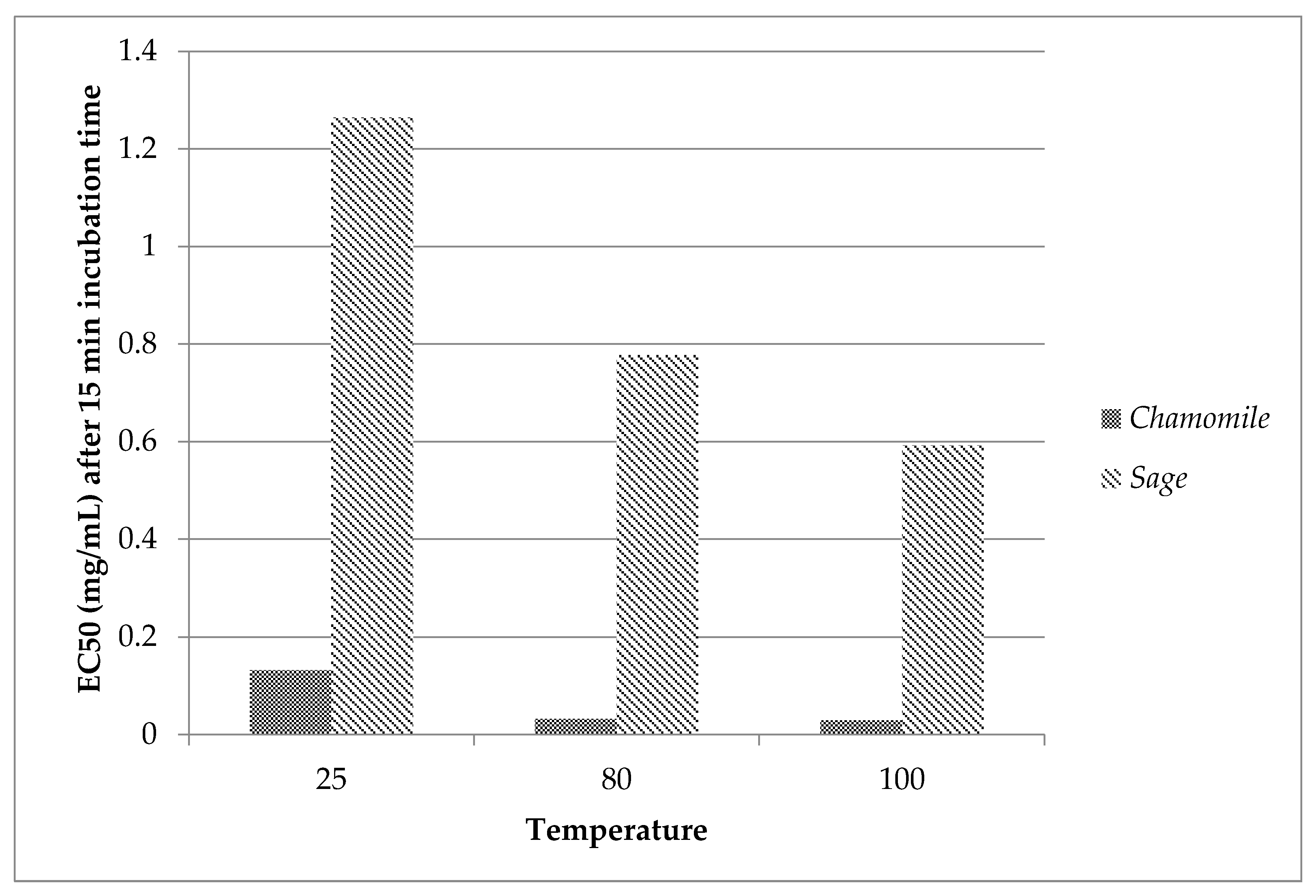
3.3. Evaluation of Acute Toxicity towards Vibrio fischeri

Another topic of this research was the evaluation of toxicity towards V. fischeri using bioluminescence inhibition of chamomile and sage aqueous extracts performed at the three temperatures. The results are presented in Figure 1.
The EC50 of extracts towards the bioluminescence photobacterium V. fischeri ranged between 0.032 and 1.264 mg/mL. Chamomile aqueous extracts showed a higher capacity to inhibit bioluminescence of V. fischeri than sage aqueous extracts. Specifically, the obtained values of EC50 for chamomile extracts at 80 and 100 °C indicated high toxicity whereas the aqueous extract at 25 °C exerted lower toxicity. In the case of sage aqueous extracts, toxicity towards V. fischeri increased as the extraction temperature increased.
There are no studies reporting bacterial toxicity to the bioluminescent bacteria V. fischeri from chamomile extracts, since most studies examined other herbal species [6,24]. However, scarce literature exists for toxicity towards V. fischeri of sage aqueous extracts associated with bioluminescence bioassay. In a previous study reporting inhibition of bioluminescence from sage aqueous extracts at different extraction temperatures, the data showed lower values than the obtained values, which could be explained by the differences in growing conditions, regions, and sage plan genotype, but indicated the same trend concerning the temperature [5]. The experimental values of sage infusion at boiled water showed good agreement with those estimated by Sotiropoulou et al. who reported the EC20 of inhibition [25].
Some phenolic compounds are toxic to V. fischeri, revealing the bacterial bioassay as sensitive to monitoring their toxicity [4]. In a previous study, the main compound of chamomile extract, ferulic acid, as shown in the Τable 2 below, was found to be toxic using the Microtox biotest [26]. Concerning sage infusions, synergistic action between water soluble compounds and volatile compounds towards V. fischeri has been presented. Moreover, this study reported that toxicity cannot be attributed to rosmarinic acid [5], which is the main compound of sage aqueous extract, according to Table 2.
To conclude, the extraction temperature of herbal aqueous extracts affected the toxicity towards V. fischeri. Moreover, besides the different values EC50 of the examined aqueous extracts, all of them exerted a toxic effect on V. fischeri, showing antimicrobial capacity and potent self-defense.

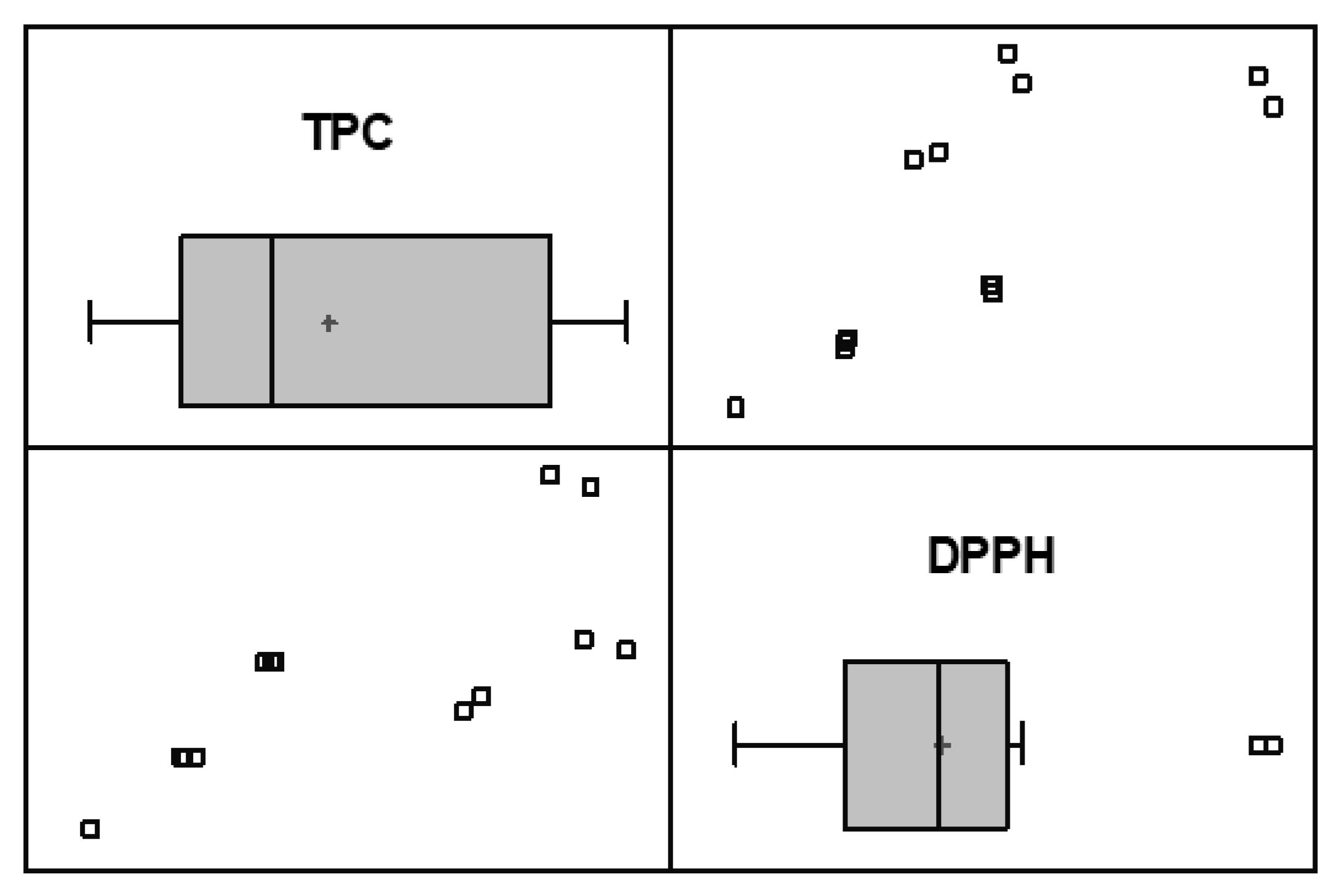
3.4. Statistical Data and Correlation between Examined Assays

The results showed that at all tested assays differences among treatments were not considered to be significant. The total phenolic content indicated a strong correlation with antioxidant capacity of chamomile and sage aqueous extracts prepared at all temperatures, as described by Spearman’s correlation coefficient (r = 0.90). The correlation is presented in Figure 2. In addition, there was a statistically significant relationship between the two assays, considering that the p-value was less than 0.05.
Reportedly, there is a relationship between total polyphenols concentration and DPPH radical-scavenging activity in chamomile and sage teas (correlation factor 0.950) [27]. This correlation is also in line with Jimenez-Zamora et al., who studied antioxidant capacity and total phenolic content of chamomile and sage as infusions [28]. Other studies on herbal aqueous extracts also indicated that antioxidant activity was positively correlated with total phenolic content [5,13,18,29,30,31,32,33]. However, according to Rivas Romero et al., the total content of antioxidants of an herbal extract, including aqueous extracts of chamomile and sage, is not bi-univocally related to its antioxidant capacity, because the distinct components of the extracts can have very different antioxidant capacities [16]. Moreover, total antioxidant activity of tea herbs is linked not only to the presence of phenols, but also to the presence of other free radical scavenging compounds [34].
In contrast, acute toxicity of herbal aqueous extracts is not correlated to their total phenolic content and antioxidant capacity. Similar results for herbal infusions previously indicated no association of bioluminescence inhibition either with total phenolic content or with antioxidant activity [5].

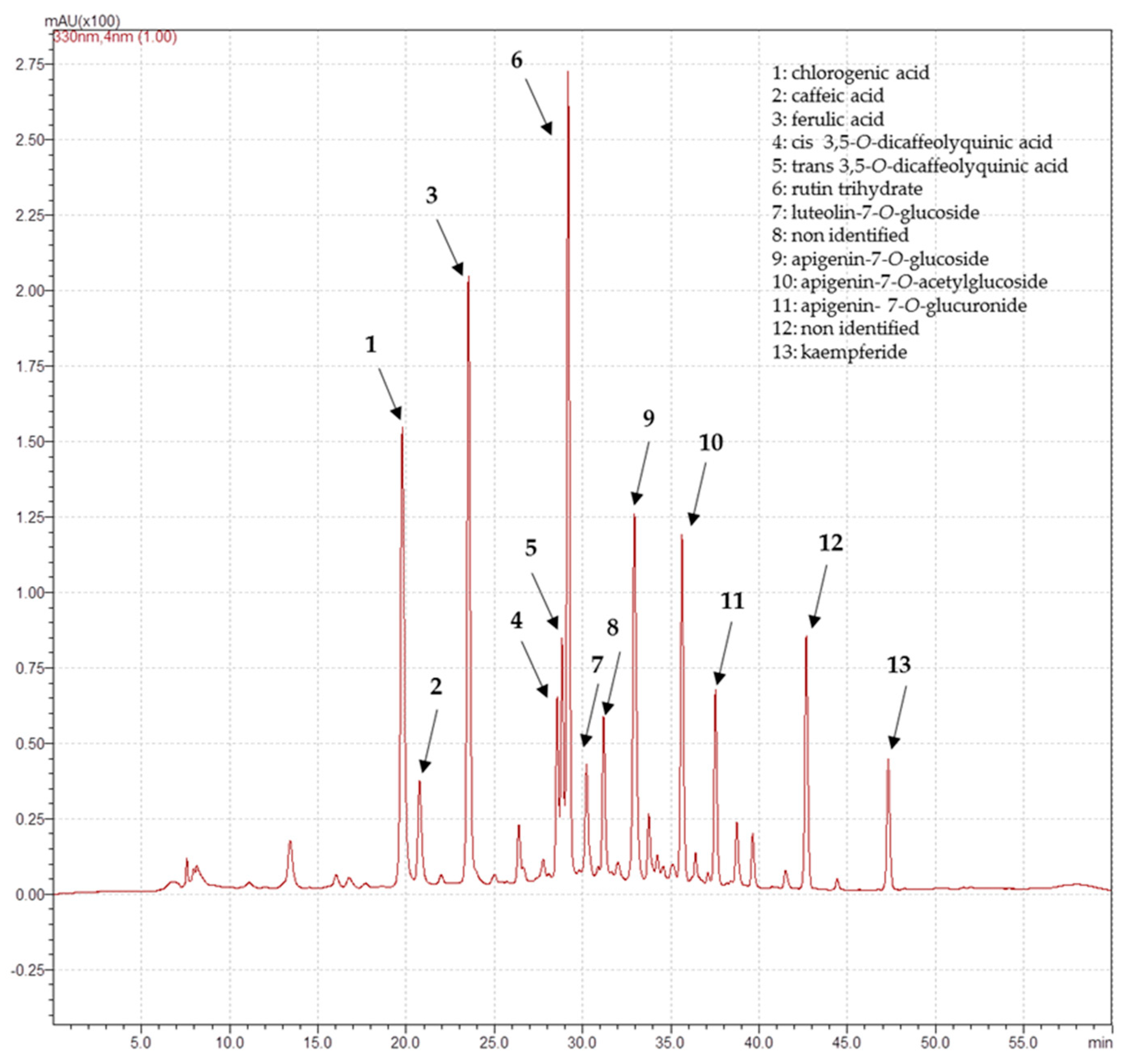
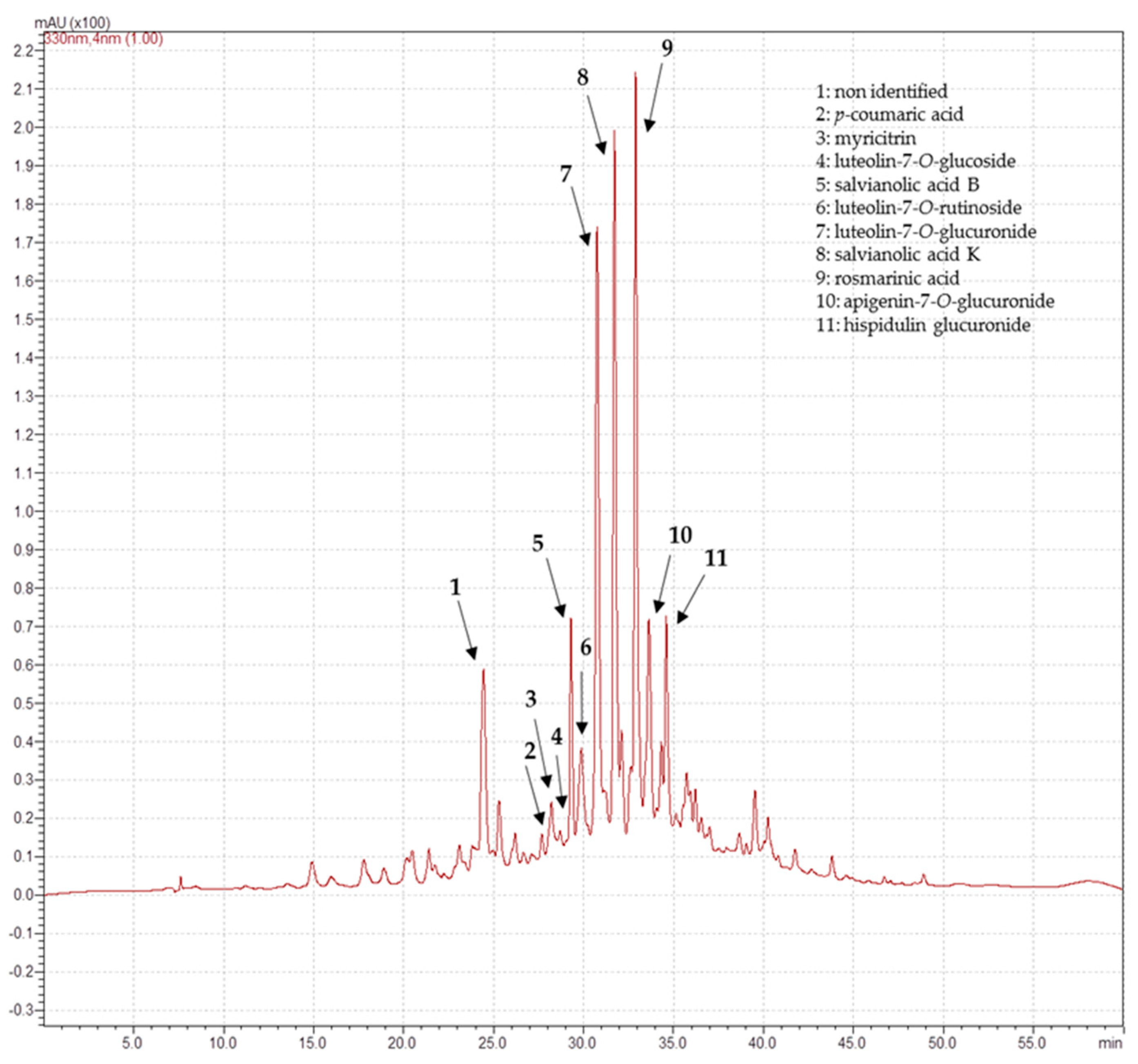
3.5. Determination of Phenolic Compounds

The analyses to determine the phenolic composition of chamomile and sage aqueous infusions were performed at 80 °C, which was found to be the optimal temperature concerning bioactivity. In Table 2, the phenolic compounds of both aqueous extracts are presented.
Thirteen main phenolic compounds were detected in chamomile aqueous extracts and 11 were identified. Chamomile extract contained six flavonoids including flavone glycosides (apigenin-7-O-glucoside and luteolin-7-O-glucoside) and flavonols (kaempferide and rutin trihydrate). Phenolic acids as caffeic, chlorogenic, cis-3,5-O-dicaffeoylquinic, trans-3,5-O-dicaffeoylquinic, and ferulic acid were also detected in the chamomile infusion, as shown on Figure 3. Compounds 4 and 5 yielded both the same ion [M-H] at m/z 515, which allowed tentative identification as cis and trans isomers of 3,5-O-dicaffeoylquinic acids, respectively. These phenolic acids presented similar fragmentation pattern to the ones previously reported by Caleja et al. Compounds 10 and 11 were identified as apigenin-7-O-acetyl glucoside and the other derivative apigenin 7-O-glucuronide, by comparison of mass spectrums with literature data [18,35]. Rutin trihydrate, ferulic acid, chlorogenic acid, and apigenin-7-O-glucoside were the dominant compounds of chamomile infusion. Apigenin-7-O-acetyl glucoside and trans-3,5-O-dicaffeoylquinic acid followed. At lower and similar amounts, apigenin 7-O-glucuronide, cis-3,5-O-dicaffeoylquinic acid, caffeic acid, kaempferide, and luteolin-7-O-glucoside were identified.
Flavone glycosides (apigenin-7-O-glucoside and luteolin-7-O-glucoside), chlorogenic acid, caffeic and ferulic acid derivatives were previously identified in chamomile water extracts [18]. Moreover, the presence of apigenin-7-glucoside as the main flavonoid of aqueous chamomile extracts was described by different authors [10,36,37]. Luteolin-7-O-glucoside, chlorogenic acid, and dicaffeoylquinic acids have also been detected in aqueous extracts (infusions, decoctions) of chamomile flower heads and leafy flowering stems [8]. Rutin (trihydrate) (quercetin-3-O-rutinoside), as far as we know, has not been cited in aqueous extracts of chamomile, although other derivatives of quercetin were found in chamomile infusions [18,35,38]; although other studies detected rutin at aqueous chamomile obtained by supercritical extraction [19,39]. The decoctions of M. recutita presented significant contents of caffeic acid, 7-O-glucosides of apigenin, and luteolin [35]. Raal with co-authors also investigated chamomile infusions. The major phenolic compounds in the chamomile infusions were also chlorogenic acid, dicaffeoylquinic acids, apigenin glycoside and acetyl glucoside, ferulic acid glycoside, and at low content glucoside of luteolin [38]. Remarkable contents of chlorogenic and caffeic acid were found in chamomile infusions, whilst ferulic acid was found at low contents. Moreover, glucosides (7-O) of apigenin and luteolin were identified [32]. Another study also examined chamomile aqueous extract at 80 °C, and demonstrated ferulic, caffeic, and p-coumaric acids as the main compounds, followed by lower content of chlorogenic acid [1].
In the case of sage aqueous extracts, 11 phenolic compounds were found. Up to 10 different phenolic compounds, consisting of four phenolic acids and six flavonoids, were identified in sage aqueous extract, as presented in Figure 4. The flavonoids were composed of myricitrin, apigenin-7-O-glucuronide, hispidulin glucoside, and luteolin derivatives (7-O-glucoside, 7-O-glucuronide, and 7-O-rutinoside). Phenolic acids determined in sage aqueous extract were p-coumaric, rosmarinic, salvianolic B and K acids. Compound 6 was assigned as luteolin-7-O-rutinoside, following the criteria reported by Cvetkovikj et al. Phenolic compound 7 at retention time 30.75 presented a pseudomolecular ion [M-H] at m/z 461, allowing its tentative identification as a luteolin-7-O-glucuronide as previously reported [11]. Compounds 8 ([M-H] at m/z 555) and 10 ([M-H] at m/z 445) were tentatively assigned as salvianolic acid K and apigenin-7-O-glucuronide, respectively, based on the spectroscopic data described by Cvetkovikj and colleagues [40]. Compound 11 was identified as hispidulin glucoside by comparison of its UV and mass spectrums with literature data [11,40]. The major compounds determined in sage aqueous extract were rosmarinic acid, salvianolic acid K, and luteolin-7-O-glucuronide. Apigenin-7-O-glucuronide, salvianolic acid B, hispidulin glucoside, and luteolin-7-O-rutinoside were less abundant phenolic compounds, followed by myricitrin, luteolin-7-O-glucoside, and p-coumaric acid at lower percentages (Table 2).
The presence of both compounds, rosmarinic acid and luteolin-7-O-glucoronide, has been reported in other studies [11,13,40] as the main phenolics in infusion of S.officinalis. Luteolin-7-O-glucoside and salvianolic acid were also detected in significant amounts [11]. Our findings are generally in agreement with the study of Kaliora et al., which reported that rosmarinic acid is the dominant compound in the infusion of S.officinalis; p-coumaric, rutinoside, and glucoside of luteolin were also detected [13]. Cvetkovikj et al. found rosmarinic acid to be the component with the highest content and variability in all analyzed infusions of S. officinalis, S. fruticosa, and S. pomifera. Moreover, salvianolic acid K, luteolin, and apigenin derivatives were the most frequently detected components and occurred in most of the examined Salvia populations [40]. According to many studies, rosmarinic acid is the most abundant phenolic compound in infusions of Salvia species [13,40].

4. Conclusions

The effect of extraction temperature on the bioactivity of herbal infusions is very important. This study provides results regarding total phenolic content, antioxidant capacity, and toxicity of aqueous extracts of chamomile and sage at different extraction temperatures. All the examined assays of bioactivity indicated influence of the extraction temperature. The highest total phenolic content and maximum antioxidant capacity of aqueous extracts of chamomile and sage at tested temperatures (25, 80, and 100 °C) were achieved at 80 °C. Toxicity of both herbal aqueous extracts, causing bioluminescence inhibition of V. fischeri, was increased along with extraction temperature. Chamomile showed higher capacity to inhibit bioluminescence in comparison with sage. For all the aqueous herbal extracts, total phenolic content was significantly correlated with antioxidant activity; however, there was no link between toxicity and these two examined assays. The analyses of phenolic profiles of both aqueous extracts at 80 °C confirmed the presence of different flavonoids and phenolic compounds. The dominant compounds of chamomile aqueous extract were rutin trihydrate, ferulic acid, chlorogenic acid, and apigenin7-O-glucoside. Rosmarinic acid, salvianolic acid K, and luteolin-7-O-glucuronide were detected as the main phenolic compounds of sage aqueous extract. Eventually, the effect of not only the extraction temperature but also other parameters such as time of extraction and amount of solvent play an important role in the evaluation of the beneficial properties and for monitoring the safety of herbal infusions. Overall, and despite the need for further studies to elucidate the optimal infusion preparation of the tested herbal extracts, it is possible to conclude that aqueous extracts of chamomile and sage at 80 °C could be regarded as health-promoting antioxidant beverages but their use should be moderated to ensure safe consumption.

Author Contributions

Conceptualization, N.S.S.; investigation, N.S.S. and S.F.M.; writing—original draft preparation, N.S.S.; writing—review and editing, N.S.S., S.F.M., and P.T.; supervision, P.T. All authors have read and agreed to the published version of the manuscript.

Funding

This research received no external funding.

Acknowledgments

The authors want to express their sincere thanks to Greek farms: Aroma farms (Elis, Greece) and LAV (Thessaloniki, Greece) for donating the studied plant materials of chamomile and sage, respectively.

Conflicts of Interest

The authors declare no conflicts of interest.

References

  1. Horžić, D.; Komes, D.; Belščak, A.; Ganić, K.K.; Iveković, D.; Karlović, D. The composition of polyphenols and methylxanthines in teas and herbal infusions. Food Chem. 2009, 115, 441–448. [Google Scholar] [CrossRef]
  2. Da Silva Pinto, M. Tea: A new perspective on health benefits. Food Res. Int. 2013, 53, 558–567. [Google Scholar] [CrossRef]
  3. Ekor, M. The growing use of herbal medicines: Issues relating to adverse reactions and challenges in monitoring safety. Front. Neurol. 2014, 4, 177. [Google Scholar] [CrossRef] [PubMed] [Green Version]
  4. Abbas, M.; Adil, M.; Ehtisham-ul-Haque, S.; Munir, B.; Yameen, M.; Ghaffar, A.; Shar, G.A.; Asif Tahir, M.; Iqbal, M. Vibrio fischeri bioluminescence inhibition assay for ecotoxicity assessment: A review. Sci. Total Environ. 2018, 626, 1295–1309. [Google Scholar] [CrossRef] [PubMed]
  5. Skotti, E.; Anastasaki, E.; Kanellou, G.; Polissiou, M.; Tarantilis, P.A. Total phenolic content, antioxidant activity and toxicity of aqueous extracts from selected Greek medicinal and aromatic plants. Ind. Crops Prod. 2014, 53, 46–54. [Google Scholar] [CrossRef] [Green Version]
  6. Conforti, F.; Ioele, G.; Statti, G.A.; Marrelli, M.; Ragno, G.; Menichini, F. Antiproliferative activity against human tumor cell lines and toxicity test on Mediterranean dietary plants. Food Chem. Toxicol. 2008, 46, 3325–3332. [Google Scholar] [CrossRef]
  7. McKay, D.L.; Blumberg, J.B. A Review of the Bioactivity and Potential Health Benefits of Chamomile Tea (Matricaria recutita L.). Phyther. Res. 2006, 20, 519–530. [Google Scholar] [CrossRef]
  8. Guimarães, R.; Barros, L.; Dueñas, M.; Calhelha, R.C.; Carvalho, A.M.; Santos-Buelga, C.; Queiroz, M.J.R.P.; Ferreira, I.C.F.R. Infusion and decoction of wild German chamomile: Bioactivity and characterization of organic acids and phenolic compounds. Food Chem. 2013, 136, 947–954. [Google Scholar] [CrossRef]
  9. Srivastava, J.K.; Gupta, S. Health Promoting Benefits of Chamomile in the Elderly Population. In Complementary and Alternative Therapies and the Aging Population; Academic Press: Cambridge, MA, USA, 2009; pp. 135–158. ISBN 9780123742285. [Google Scholar]
  10. Guzelmeric, E.; Ristivojević, P.; Vovk, I.; Milojković-Opsenica, D.; Yesilada, E. Quality assessment of marketed chamomile tea products by a validated HPTLC method combined with multivariate analysis. J. Pharm. Biomed. Anal. 2017, 132, 35–45. [Google Scholar] [CrossRef]
  11. Martins, N.; Barros, L.; Santos-Buelga, C.; Henriques, M.; Silva, S.; Ferreira, I.C.F.R. Evaluation of bioactive properties and phenolic compounds in different extracts prepared from Salvia officinalis L. Food Chem. 2014, 170, 378–385. [Google Scholar] [CrossRef] [Green Version]
  12. Dent, M.; Dragović-Uzelac, V.; Penić, M.; Brñić, M.; Bosiljkov, T.; Levaj, B. The effect of extraction solvents, temperature and time on the composition and mass fraction of polyphenols in dalmatian wild sage (Salvia officinalis L.) extracts. Food Technol. Biotechnol. 2013, 51, 84–91. [Google Scholar]
  13. Kaliora, A.C.; Kogiannou, D.A.A.; Kefalas, P.; Papassideri, I.S.; Kalogeropoulos, N. Phenolic profiles and antioxidant and anticarcinogenic activities of Greek herbal infusions; Balancing delight and chemoprevention? Food Chem. 2014, 142, 233–241. [Google Scholar] [CrossRef] [PubMed]
  14. Farzaneh, V.; Carvalho, I.S. A review of the health benefit potentials of herbal plant infusions and their mechanism of actions. Ind. Crops Prod. 2015, 65, 247–258. [Google Scholar] [CrossRef]
  15. Giacometti, J.; Bursać Kovačević, D.; Putnik, P.; Gabrić, D.; Bilušić, T.; Krešić, G.; Stulić, V.; Barba, F.J.; Chemat, F.; Barbosa-Cánovas, G.; et al. Extraction of bioactive compounds and essential oils from mediterranean herbs by conventional and green innovative techniques: A review. Food Res. Int. 2018, 113, 245–262. [Google Scholar] [CrossRef]
  16. Rivas Romero, M.P.; Estévez Brito, R.; Rodríguez Mellado, J.M.; González-Rodríguez, J.; Ruiz Montoya, M.; Rodríguez-Amaro, R. Exploring the relation between composition of extracts of healthy foods and their antioxidant capacities determined by electrochemical and spectrophotometrical methods. LWT 2018, 95, 157–166. [Google Scholar] [CrossRef]
  17. Harbourne, N.; Marete, E.; Jacquier, J.C.; O’Riordan, D. Stability of phytochemicals as sources of anti-inflammatory nutraceuticals in beverages-A review. Food Res. Int. 2013, 50, 480–486. [Google Scholar] [CrossRef]
  18. Cvetanović, A.; Švarc-Gajić, J.; Mašković, P.; Savić, S.; Nikolić, L. Antioxidant and biological activity of chamomile extracts obtained by different techniques: Perspective of using superheated water for isolation of biologically active compounds. Ind. Crops Prod. 2015, 65, 582–591. [Google Scholar] [CrossRef]
  19. Cvetanović, A.; Švarc-Gajić, J.; Zeković, Z.; Jerković, J.; Zengin, G.; Gašić, U.; Tešić, Ž.; Mašković, P.; Soares, C.; Fatima Barroso, M.; et al. The influence of the extraction temperature on polyphenolic profiles and bioactivity of chamomile (Matricaria chamomilla L.) subcritical water extracts. Food Chem. 2019, 271, 328–337. [Google Scholar] [CrossRef] [Green Version]
  20. Singleton, V.L.; Orthofer, R.; Lamuela-Raventós, R.M. Analysis of total phenols and other oxidation substrates and antioxidants by means of folin-ciocalteu reagent. Methods Enzymol. 1998, 299, 152–178. [Google Scholar]
  21. Huang, D.; Boxin, O.U.; Prior, R.L. The chemistry behind antioxidant capacity assays. J. Agric. Food Chem. 2005, 53, 1841–1856. [Google Scholar] [CrossRef]
  22. Surveswaran, S.; Cai, Y.Z.; Corke, H.; Sun, M. Systematic evaluation of natural phenolic antioxidants from 133 Indian medicinal plants. Food Chem. 2007, 102, 938–953. [Google Scholar] [CrossRef]
  23. Foti, M.C.; Daquino, C.; Geraci, C. Electron-Transfer Reaction of Cinnamic Acids and Their Methyl Esters with the DPPH. Radical in Alcoholic Solutions. J. Org. Chem. 2004, 69, 2309–2314. [Google Scholar] [CrossRef] [PubMed]
  24. Kováts, N.; Gölöncsér, F.; Ács, A.; Refaey, M. Quantification of the antibacterial properties of Artemisia absinthium, A. vulgaris, Chrysanthemum leucanthemum and Achillea millefolium using the Vibrio fischeri bacterial bioassay. Acta Bot. Hung. 2010, 25, 137–144. [Google Scholar] [CrossRef]
  25. Sotiropoulou, N.D.; Kokkini, M.K.; Megremi, S.P.; Daferera, D.J.; Skotti, E.P.; Kimbaris, A.C. Determination of ɑ -and β-thujone in Wormwood and Sage Infusions of Greek flora and Estimation of their Average Toxicity. Curr. Res. Nutr Food Sci. 2016, 4, 152–160. [Google Scholar] [CrossRef]
  26. Mierzejewska, E.; Baran, A.; Urbaniak, M. Biodegradation Potential and Ecotoxicity Assessment in Soil Extracts Amended with Phenoxy Acid Herbicide (2,4-D) and a Structurally-Similar Plant Secondary Metabolite (Ferulic Acid). Bull. Environ. Contam. Toxicol. 2019, 104, 200–205. [Google Scholar] [CrossRef]
  27. Aoshima, H.; Hirata, S.; Ayabe, S. Antioxidative and anti-hydrogen peroxide activities of various herbal teas. Food Chem. 2007, 103, 617–622. [Google Scholar] [CrossRef]
  28. Jiménez-Zamora, A.; Delgado-Andrade, C.; Rufián-Henares, J.A. Antioxidant capacity, total phenols and color profile during the storage of selected plants used for infusion. Food Chem. 2016, 199, 339–346. [Google Scholar] [CrossRef]
  29. Lim, Y.Y.; Murtijaya, J. Antioxidant properties of Phyllanthus amarus extracts as affected by different drying methods. LWT-Food Sci. Technol. 2007, 40, 1664–1669. [Google Scholar] [CrossRef]
  30. Herrera, T.; Aguilera, Y.; Rebollo-Hernanz, M.; Bravo, E.; Benítez, V.; Martínez-Sáez, N.; Arribas, S.M.; del Castillo, M.D.; Martín-Cabrejas, M.A. Teas and herbal infusions as sources of melatonin and other bioactive non-nutrient components. LWT-Food Sci. Technol. 2018, 89, 65–73. [Google Scholar] [CrossRef]
  31. Ho, S.C.; Wu, S.P.; Lin, S.M.; Tang, Y.L. Comparison of anti-glycation capacities of several herbal infusions with that of green tea. Food Chem. 2010, 122, 768–774. [Google Scholar] [CrossRef]
  32. Kogiannou, D.A.A.; Kalogeropoulos, N.; Kefalas, P.; Polissiou, M.G.; Kaliora, A.C. Herbal infusions; their phenolic profile, antioxidant and anti-inflammatory effects in HT29 and PC3 cells. Food Chem. Toxicol. 2013, 61, 152–159. [Google Scholar] [CrossRef] [PubMed]
  33. Stagos, D.; Portesis, N.; Spanou, C.; Mossialos, D.; Aligiannis, N.; Chaita, E.; Panagoulis, C.; Reri, E.; Skaltsounis, L.; Tsatsakis, A.M.; et al. Correlation of total polyphenolic content with antioxidant and antibacterial activity of 24 extracts from Greek domestic Lamiaceae species. Food Chem. Toxicol. 2012, 50, 4115–4124. [Google Scholar] [CrossRef] [PubMed]
  34. Gerolis, L.G.L.; Lameiras, F.S.; Krambrock, K.; Neves, M.J. Effect of gamma radiation on antioxidant capacity of green tea, yerba mate, and chamomile tea as evaluated by different methods. Radiat. Phys. Chem. 2017, 130, 177–185. [Google Scholar] [CrossRef]
  35. Caleja, C.; Barros, L.; Antonio, A.L.; Ciric, A.; Barreira, J.C.M.; Sokovic, M.; Oliveira, M.B.P.P.; Santos-Buelga, C.; Ferreira, I.C.F.R. Development of a functional dairy food: Exploring bioactive and preservation effects of chamomile (Matricaria recutita L.). J. Funct. Foods 2015, 16, 114–124. [Google Scholar] [CrossRef] [Green Version]
  36. Harbourne, N.; Jacquier, J.C.; O’Riordan, D. Optimisation of the extraction and processing conditions of chamomile (Matricaria chamomilla L.) for incorporation into a beverage. Food Chem. 2009, 115, 15–19. [Google Scholar] [CrossRef]
  37. Guzelmeric, E.; Vovk, I.; Yesilada, E. Development and validation of an HPTLC method for apigenin 7-O-glucoside in chamomile flowers and its application for fingerprint discrimination of chamomile-like materials. J. Pharm. Biomed. Anal. 2015, 107, 108–118. [Google Scholar] [CrossRef]
  38. Raal, A.; Orav, A.; Püssa, T.; Valner, C.; Malmiste, B.; Arak, E. Content of essential oil, terpenoids and polyphenols in commercial chamomile (Chamomilla recutita L. Rauschert) teas from different countries. Food Chem. 2012, 131, 632–638. [Google Scholar] [CrossRef]
  39. Cvetanović, A.; Švarc-Gajić, J.; Zeković, Z.; Gašić, U.; Tešić, Ž.; Zengin, G.; Mašković, P.; Mahomoodally, M.F.; Đurović, S. Subcritical water extraction as a cutting edge technology for the extraction of bioactive compounds from chamomile: Influence of pressure on chemical composition and bioactivity of extracts. Food Chem. 2018, 266, 389–396. [Google Scholar] [CrossRef]
  40. Cvetkovikj, I.; Stefkov, G.; Acevska, J.; Stanoeva, J.P.; Karapandzova, M.; Stefova, M.; Dimitrovska, A.; Kulevanova, S. Polyphenolic characterization and chromatographic methods for fast assessment of culinary Salvia species from South East Europe. J. Chromatogr. A 2013, 1282, 38–45. [Google Scholar] [CrossRef]
Figure 1. Concentration of chamomile and sage aqueous extracts that causes 50% of V. fischeri luminescence inhibition, presented as medians.
Applsci 10 02270 g001
Figure 2. Box plots and scatterplots, presented as medians and ranges, concerning Folin–Ciocalteu (total phenolic content (TPC)) and 2,2-diphenyl-1-picrylhydrazyl hydrate (DPPH) assays.
Applsci 10 02270 g002
Figure 3. Phenolic profile characterization by LC-MS of chamomile aqueous extract obtained at 80 °C.
Applsci 10 02270 g003
Figure 4. Phenolic profile characterization by LC-MS of sage aqueous extract obtained at 80 °C.
Applsci 10 02270 g004
Table 1. Total phenolic content and antioxidant activity of chamomile and sage aqueous extracts obtained at different temperatures.
Temperature of ExtractionHerb
ChamomileSage
Total phenolic content expressed as mg of gallic acid equivalents (GAE)/mL of extract
MedianRangeMedianRange
25 °CNd1Nd10.2220.019
80 °C 0.1650.0070.2070.018
100 °C0.0410.0060.0790.005
Antioxidant activity expressed as ug Trolox equivalents (TE)/ mL of extract
MedianRangeMedianRange
25 °CNd1Nd16.390.331
80 °C4.620.61611.91.80
100 °C2.530.0225.840.022
1 Nd: Not detected.
Table 2. LC-DAD-MS characteristics and quantification (%) of phenolic compounds identified in chamomile and sage aqueous extracts prepared at 80 °C.
Rt (min)λmax (nm)Molecular Ion [M H]
(m/z)
Proposed CompoundChamomile %Sage %
19.78327353Chlorogenic acid12.6 ± 0.2
20.75222, 298, 324179Caffeic acid2.9 ± 0.5
23.53242, 323193Ferulic acid13.3 ± 0.4
24.44325 Ni2 5.7 ± 0.2
27.68235, 309163p-Coumaric acid 0.42 ± 0.05
28.21231, 256, 352463Myricitrin 1.6 ± 0.4
28.55220, 326515cis 3,5-O-Dicaffeolyquinic acid13.1 ± 0.2
28.69255, 349447Luteolin-7-O-glucoside2.5 ± 0.10.37 ± 0.02
28.83220, 328515trans 3,5-O-Dicaffeolyquinic acid14.4 ± 0.05
29.17255, 356609Rutin trihydrate15.1 ± 0.1
29.30253, 286, 308717Salvianolic acid B 4.1 ± 0.4
29.87268, 337593Luteolin-7-O-rutinoside1 3.2 ± 0.4
30.75348461Luteolin- 7-O-glucuronide1 15 ± 1
31.19229, 328 Ni23.1 ± 0.2
31.72236, 286, 326555Salvianolic acid K1 15.4 ± 0.4
32.89243, 289, 330359Rosmarinic acid 15 ± 1
32.94242, 267, 338431Apigenin-7-O-glucoside9.1 ± 0.1
33.62 445Apigenin- 7-O-glucuronide13.8 ± 0.65 ± 0.3
33.74235, 322 Ni21.3 ± 0.1
34.59229, 273, 332475Hispidulin glucuronide1 4.5 ± 0.4
35.62238, 267, 332473Apigenin-7-O-acetylglucoside16.9 ± 0.2
42.66242, 254, 312 Ni25.2 ± 0.3
47.32266, 324, 365299Kaempferide2.8 ± 0.3
1 Tentative identification. 2 Ni: Not identified.

