Fragaria vesca L. Extract: A Promising Cosmetic Ingredient with Antioxidant Properties

 , , , , , ,
  and
and
Abstract
:1. Introduction
2. Materials and Methods
2.1. Materials
2.2. Methods
2.2.1. Fragaria vesca Extract Preparation
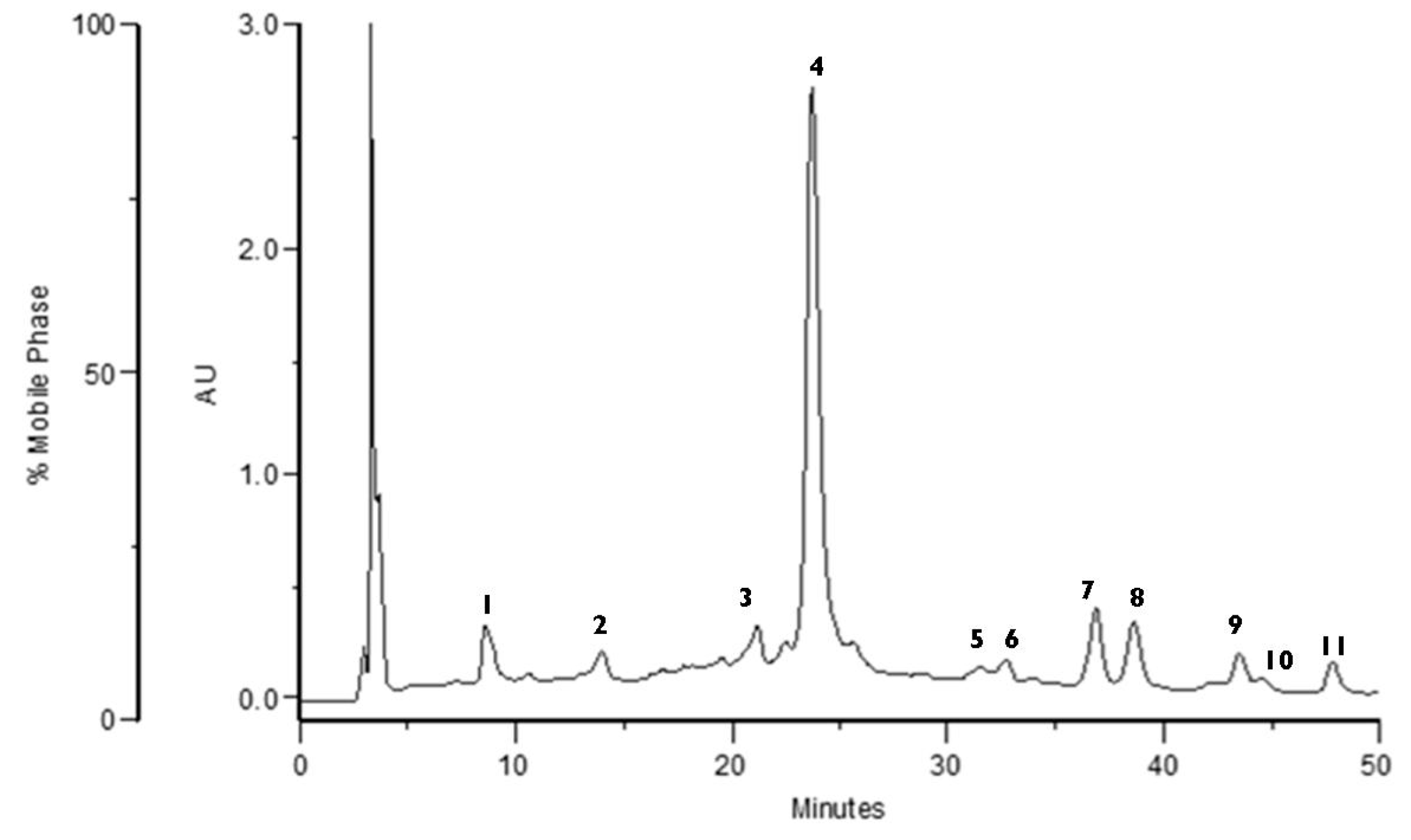
2.2.2. High-Performance Liquid Chromatography (HPLC)
2.2.3. Tyrosinase Inhibition
2.2.4. Preparation of Topical Formulation: F. vesca Based Hydrogel
2.2.5. Physicochemical Characterization and Stability of Topical Formulations
2.2.6. Total Tannins Content
2.2.7. In Vitro Cell Culture Assays
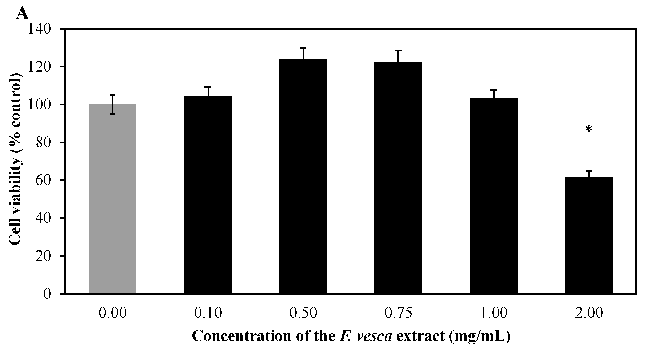
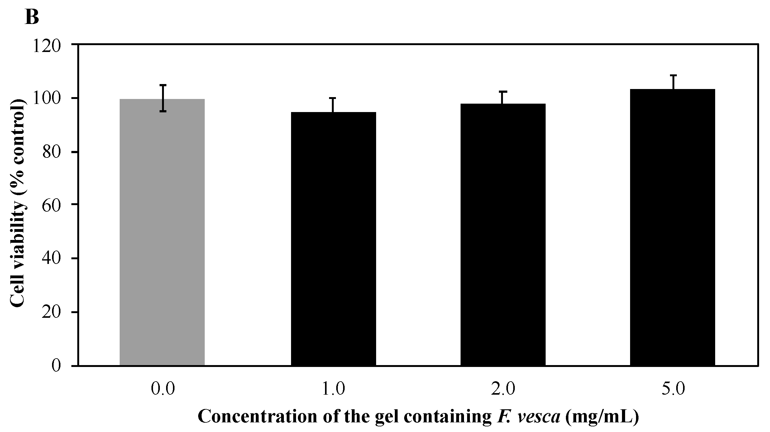
Cytotoxicity Studies
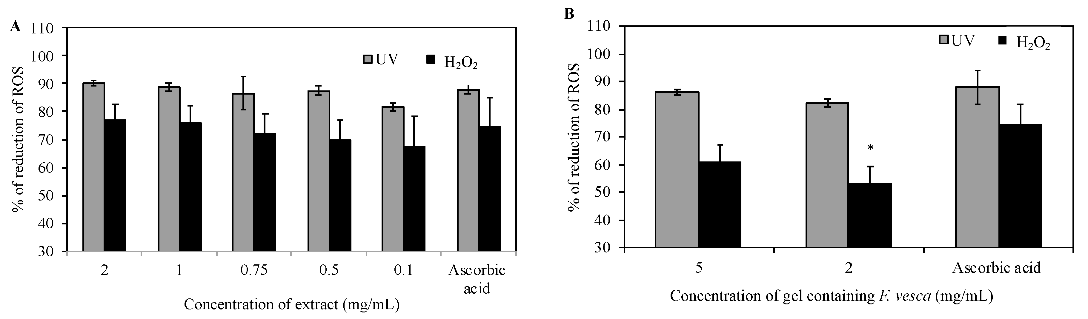
Reactive Oxygen Species (ROS) Production Measurement
2.2.8. In Vivo Safety and Efficacy Tests
Simple Patch Test
Human Repeat Insult Patch Test (HRIPT)
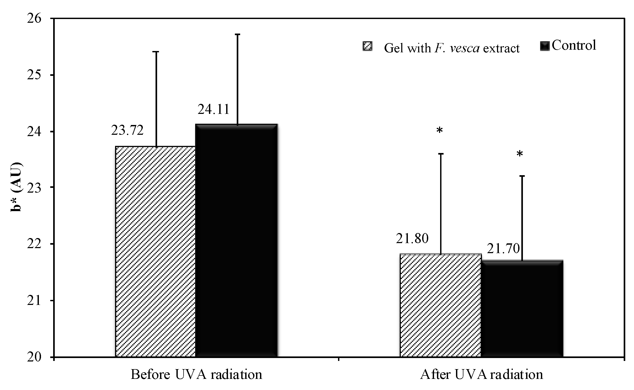
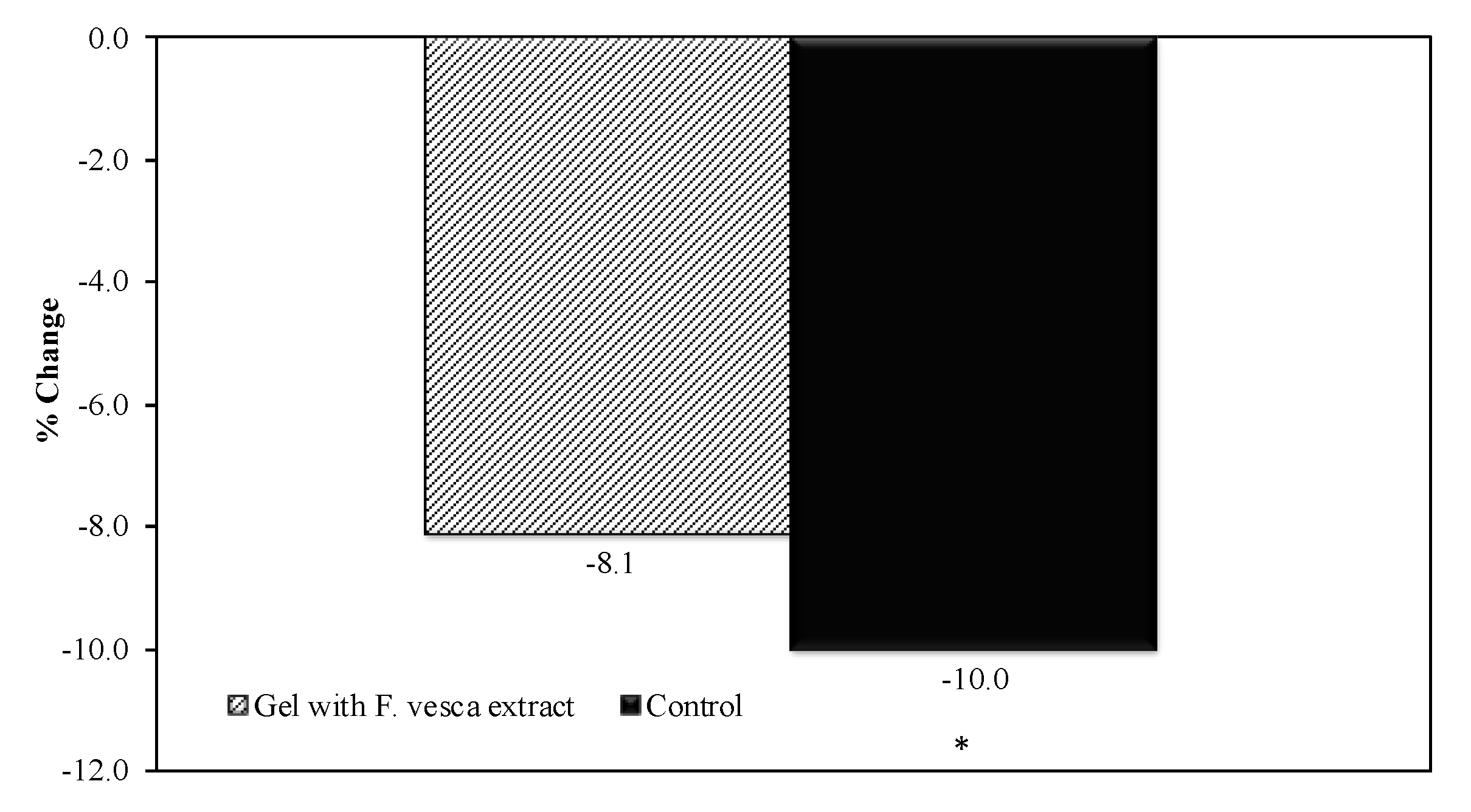
Antioxidant Efficacy
2.2.9. Statistical Analysis
3. Results
3.1. Characterization of the Extract
3.2. Evaluation of Tyrosinase Inhibitory Activity of the Extract
3.3. F. vesca Based Hydrogel Stability Studies
3.4. Texture Profile Analysis of the Formulation
3.5. Total Tannin Content in Extract and Hydrogel Formulation
3.6. Cytotoxicity Studies
3.7. Reactive Oxygen Species (ROS) Production Measurement
3.8. F. vesca Based Hydrogel Safety Tests
3.9. Antioxidant Efficacy
4. Conclusions
Author Contributions
Funding
Acknowledgments
Conflicts of Interest
References
- Fabricant, D.S.; Farnsworth, N.R. The value of plants used in traditional medicine for drug discovery. Environ. Health Perspect. 2001, 109 (Suppl. S1), 69–75. [Google Scholar]
- Dias, D.A.; Urban, S.; Roessner, U. A historical overview of natural products in drug discovery. Metabolites 2012, 2, 303–336. [Google Scholar] [CrossRef] [Green Version]
- Vázquez, C.V.; Rojas, M.G.V.; Ramírez, C.A.; Chávez-Servín, J.L.; García-Gasca, T.; Ferriz Martínez, R.A.; García, O.P.; Rosado, J.L.; López-Sabater, C.M.; Castellote, A.I.; et al. Total phenolic compounds in milk from different species. Design of an extraction technique for quantification using the Folin–Ciocalteu method. Food Chem. 2015, 176, 480–486. [Google Scholar]
- Ivanov, I.; Petkova, N.; Denev, P.; Pavlov, A. Polyphenols Content and Antioxidant Activities in Infusion and Decoction Extracts Obtained from Fragaria vesca L. Leaves. Scien. Bull. Ser. F Biotechnol. 2015, 19, 145–148. [Google Scholar]
- Liberal, J.; Francisco, V.; Costa, G.; Figueirinha, A.; Amaral, M.T.; Marques, C.; Girão, H.; Lopes, M.C.; Cruz, M.T.; Batista, M.T. Bioactivity of Fragaria vesca leaves through inflammation, proteasome and autophagy modulation. J. Ethnopharmacol. 2014, 158, 113–122. [Google Scholar] [CrossRef]
- Yildirim, A.B.; Turker, A.U. Effects of regeneration enhancers on micropropagation of Fragaria vesca L. and phenolic content comparison of field-grown and in vitro-grown plant materials by liquid chromatography-electrospray tandem mass spectrometry (LC–ESI-MS/MS). Sci. Hortic. 2014, 169, 169–178. [Google Scholar] [CrossRef]
- Kanodia, L.; Das, S. A comparative study of analgesic property of whole plant and fruit extracts of Fragaria vesca in experimental animal models. BJP 2009, 4. [Google Scholar] [CrossRef] [Green Version]
- Sharma, M.; Pandey, G. Some anticancer medicinal plants of foreign origin. Curr. Sci. 2009, 96, 779–783. [Google Scholar]
- Mudnic, I.; Modun, D.; Brizic, I.; Vukovic, J.; Generalic, I.; Katalinic, V.; Bilusic, T.; Ljubenkov, I.; Boban, M. Cardiovascular effects in vitro of aqueous extract of wild strawberry (Fragaria vesca, L.) leaves. Phytomedicine 2009, 16, 462–469. [Google Scholar] [CrossRef]
- Cunha, A.; Silva, A.; Roque, O.; Cunha, E. Plantas e produtos vegetais em cosmética e dermatologia, 2nd ed.; Fundação Calouste Gulbenkian: Lisbon, Portugal, 2008. [Google Scholar]
- Kubota, M.; Hosoya, T.; Fukumoto, S.; Miyagi, T.; Kumazawa, S. Anti-melanogenic compounds in Rubus croceacanthus. J. Berry Res. 2014, 4, 127–135. [Google Scholar] [CrossRef] [Green Version]
- Ortiz-Ruiz, C.V.; Berna, J.; Tudela, J.; Varon, R.; Garcia-Canovas, F. Action of ellagic acid on the melanin biosynthesis pathway. J. Dermatol. Sci. 2016, 82, 115–122. [Google Scholar] [CrossRef]
- Draelos, Z.; Dahl, A.; Yatskayer, M.; Chen, N.; Krol, Y.; Oresajo, C. Dyspigmentation, skin physiology, and a novel approach to skin lightening. J. Cosmet. Dermatol. 2013, 12, 247–253. [Google Scholar] [CrossRef]
- Shimogaki, H.; Tanaka, Y.; Tamai, H.; Masuda, M. In vitro and in vivo evaluation of ellagic acid on melanogenesis inhibition. Int. J. Cosmet. Sci. 2000, 22, 291–303. [Google Scholar] [CrossRef]
- Piwowarski, J.P.; Kiss, A.K. C-glucosidic Ellagitannins from Lythri herba (European Pharmacopoeia): Chromatographic Profile and Structure Determination. Phytochem. Anal. 2013, 24, 336–348. [Google Scholar] [CrossRef]
- Masamoto, Y.; Ando, H.; Murata, Y.; Shimoishi, Y.; Tada, M.; Takahata, K. Mushroom Tyrosinase Inhibitory Activity of Esculetin Isolated from Seeds of Euphorbia lathyris L. Biosci. Biotechnol. Biochem. 2003, 67, 631–634. [Google Scholar] [CrossRef]
- Ali, S.M.; Yosipovitch, G. Skin pH: From basic science to basic skin care. Acta Derm. Venereol. 2013, 93, 261–269. [Google Scholar] [CrossRef] [Green Version]
- Lambers, H.; Piessens, S.; Bloem, A.; Pronk, H.; Finkel, P. Natural skin surface pH is on average below 5, which is beneficial for its resident flora. Int. J. Cosmet. Sci. 2006, 28, 359–370. [Google Scholar] [CrossRef]
- Vitorino, C.; Alves, L.; Antunes, F.E.; Sousa, J.J.; Pais, A.A. Design of a dual nanostructured lipid carrier formulation based on physicochemical, rheological, and mechanical properties. J. Nanopart. Res. 2013, 15, 1993. [Google Scholar] [CrossRef]
- Şenyiğit, T.; Tekmen, I.; Sönmez, Ü.; Santi, P.; Özer, Ö. Deoxycholate hydrogels of betamethasone-17-valerate intended for topical use: In vitro and in vivo evaluation. Int. J. Pharm. 2011, 403, 123–129. [Google Scholar] [CrossRef]
- European Pharmacopoeia Online 9.0. Available online: http://online6.edqm.eu/ep900/ (accessed on 15 January 2020).
- Marto, J.; Ascenso, A.; Gonçalves, L.M.; Gouveia, L.F.; Manteigas, P.; Pinto, P.; Oliveira, E.; Almeida, A.J.; Ribeiro, H.M. Melatonin-based pickering emulsion for skin’s photoprotection. Drug Deliv. 2016, 23, 1594–1607. [Google Scholar] [CrossRef] [Green Version]
- Jaiswal, R.; Kiprotich, J.; Kuhnert, N. Determination of the hydroxycinnamate profile of 12 members of the Asteraceae family. Phytochemistry 2011, 72, 781–790. [Google Scholar] [CrossRef]
- Vrhovsek, U.; Guella, G.; Gasperotti, M.; Pojer, E.; Zancato, M.; Mattivi, F. Clarifying the Identity of the Main Ellagitannin in the Fruit of the Strawberry, Fragaria vesca and Fragaria ananassa Duch. J. Agric. Food Chem. 2012, 60, 2507–2516. [Google Scholar] [CrossRef]
- Tulipani, S.; Mezzetti, B.; Capocasa, F.; Bompadre, S.; Beekwilder, J.; de Vos, C.H.R.; Capanoglu, E.; Bovy, A.; Battino, M. Antioxidants, Phenolic Compounds, and Nutritional Quality of Different Strawberry Genotypes. J. Agric. Food Chem. 2008, 56, 696–704. [Google Scholar] [CrossRef]
- Buendía, B.; Gil, M.I.; Tudela, J.A.; Gady, A.L.; Medina, J.J.; Soria, C.; López, J.M.; Tomás-Barberán, F.A. HPLC-MS Analysis of Proanthocyanidin Oligomers and Other Phenolics in 15 Strawberry Cultivars. J. Agric. Food Chem. 2010, 58, 3916–3926. [Google Scholar] [CrossRef]
- Goenka, S.; Ceccoli, J.; Simon, S.R. Anti-melanogenic activity of ellagitannin casuarictin in B16F10 mouse melanoma cells. Nat. Prod. Res. 2019, 1–6. [Google Scholar] [CrossRef]
- Quideau, S.; Scientific, W. Chemistry and Biology of Ellagitannins: An Underestimated Class of Bioactive Plant Polyphenols; World Scientific Publishing Co., Inc.: Hackensack, NJ, USA, 2009; ISBN 978-981-279-741-4. [Google Scholar]
- SCCS; Degen, G.H. Opinion of the Scientific Committee on Consumer safety (SCCS)—Opinion on the safety of the use of α-arbutin in cosmetic products. Regul. Toxicol. Pharmacol. 2016, 74, 75–76. [Google Scholar]
- Hatano, Y.; Man, M.-Q.; Uchida, Y.; Crumrine, D.; Scharschmidt, T.C.; Kim, E.G.; Mauro, T.M.; Feingold, K.R.; Elias, P.M.; Holleran, W.M. Maintenance of an Acidic Stratum Corneum Prevents Emergence of Murine Atopic Dermatitis. J. Investig. Dermatol. 2009, 129, 1824–1835. [Google Scholar] [CrossRef] [Green Version]
- Fluhr, J.W.; Kao, J.; Ahn, S.K.; Feingold, K.R.; Elias, P.M.; Jain, M. Generation of Free Fatty Acids from Phospholipids Regulates Stratum Corneum Acidification and Integrity. J. Investig. Dermatol. 2001, 117, 44–51. [Google Scholar] [CrossRef] [Green Version]
- Deuschle, V.C.K.N.; Deuschle, R.A.N.; Bortoluzzi, M.R.; Athayde, M.L. Physical chemistry evaluation of stability, spreadability, in vitro antioxidant, and photo-protective capacities of topical formulations containing Calendula officinalis L. leaf extract. Braz. J. Pharm. Sci. 2015, 51, 63–75. [Google Scholar] [CrossRef] [Green Version]
- Wang, W.; Li, F.; Yu, J.; Navard, P.; Budtova, T. Influence of substitution on the rheological properties and gelation of hydroxyethyl cellulose solution in NaOH–water solvent. Carbohydr. Polym. 2015, 124, 85–89. [Google Scholar] [CrossRef]
- Hurler, J.; Engesland, A.; Poorahmary Kermany, B.; Škalko-Basnet, N. Improved texture analysis for hydrogel characterization: Gel cohesiveness, adhesiveness, and hardness. J. Appl. Polym. Sci. 2012, 125, 180–188. [Google Scholar] [CrossRef]
- Jones, D.S.; Woolfson, A.D.; Brown, A.F. Textural analysis and flow rheometry of novel, bioadhesive antimicrobial oral gels. Pharm. Res. 1997, 14, 450–457. [Google Scholar] [CrossRef]
- Žugić, A.; Đorđević, S.; Arsić, I.; Marković, G.; Živković, J.; Jovanović, S.; Tadić, V. Antioxidant activity and phenolic compounds in 10 selected herbs from Vrujci Spa, Serbia. Ind. Crop. Prod. 2014, 52, 519–527. [Google Scholar] [CrossRef]
- Dyduch-Siemińska, M.; Najda, A.; Dyduch, J.; Gantner, M.; Klimek, K. The Content of Secondary Metabolites and Antioxidant Activity of Wild Strawberry Fruit (Fragaria vesca L.). J. Anal. Methods Chem. 2015, 2015, 831238. [Google Scholar]
- Do, H.; Park, H.-J.; Sohn, E.-H.; Kim, B.-O.; Um, S.H.; Kwak, J.-H.; Moon, E.-Y.; Rhee, D.-K.; Pyo, S. Ethanol induces cell cycle arrest and triggers apoptosis via Sp1-dependent p75NTR expression in human neuroblastoma cells. Cell Biol. Toxicol. 2013, 29, 365–380. [Google Scholar] [CrossRef]
- Marques, P.; Marto, J.; Gonçalves, L.M.; Pacheco, R.; Fitas, M.; Pinto, P.; Serralheiro, M.L.M.; Ribeiro, H. Cynara scolymus L.: A promising Mediterranean extract for topical anti-aging prevention. Ind. Crop. Prod. 2017, 109, 699–706. [Google Scholar] [CrossRef]
- Barreira, J.C.M.; Rodrigues, S.; Carvalho, A.M.; Ferreira, I.C.F.R. Development of hydrosoluble gels with Crataegus monogyna extracts for topical application: Evaluation of antioxidant activity of the final formulations. Ind. Crop. Prod. 2013, 42, 175–180. [Google Scholar] [CrossRef]


| Ingredients | Quantitative Composition (%, w/w) |
|---|---|
| F. vesca Extract | 2.0 |
| Hydroxyethylcellulose (HEC) | 2.5 |
| Ethanol (96%) | 22.5 |
| Purified Water | 73.0 |
| Time (Days) | Hardness (g) | Compressibility (g.sec) | Adhesiveness (g.sec) | Cohesiveness | Elasticity |
|---|---|---|---|---|---|
| D0 | 19.201 ± 0.65 | 23.808 ± 0.4 | −23.953 ± 0.54 | 0.853 ±0.04 | 0.998 ± 0.08 |
| D28 (S1) | 37.515 ± 0.47 | 47.118 ± 0.25 | −36.157 ± 0.47 | 0.725 ± 0.06 | 0.918 ± 0.02 |
| D28 (S2) | 21.865 ± 0.24 | 22.504 ± 0.42 | −24.552 ± 0.61 | 0.973 ± 0.04 | 0.941 ± 0.03 |
| D28 (S3) | 40.289 ± 0.27 | 42.134 ± 0.4 | −40.967 ± 0.28 | 0.877 ± 0.03 | 0.955 ± 0.05 |
| D28 (S4) | 22.309 ± 0.39 | 23.367 ± 0.64 | −30.453 ± 0.22 | 1.038 ± 0.04 | 0.995 ± 0.07 |
© 2020 by the authors. Licensee MDPI, Basel, Switzerland. This article is an open access article distributed under the terms and conditions of the Creative Commons Attribution (CC BY) license (http://creativecommons.org/licenses/by/4.0/).
Share and Cite
Couto, J.; Figueirinha, A.; Batista, M.T.; Paranhos, A.; Nunes, C.; Gonçalves, L.M.; Marto, J.; Fitas, M.; Pinto, P.; Ribeiro, H.M.; et al. Fragaria vesca L. Extract: A Promising Cosmetic Ingredient with Antioxidant Properties. Antioxidants 2020, 9, 154. https://doi.org/10.3390/antiox9020154
Couto J, Figueirinha A, Batista MT, Paranhos A, Nunes C, Gonçalves LM, Marto J, Fitas M, Pinto P, Ribeiro HM, et al. Fragaria vesca L. Extract: A Promising Cosmetic Ingredient with Antioxidant Properties. Antioxidants. 2020; 9(2):154. https://doi.org/10.3390/antiox9020154
Chicago/Turabian StyleCouto, Joana, Artur Figueirinha, Maria Teresa Batista, António Paranhos, Carla Nunes, Lídia Maria Gonçalves, Joana Marto, Manuel Fitas, Pedro Pinto, Helena Margarida Ribeiro, and et al. 2020. "Fragaria vesca L. Extract: A Promising Cosmetic Ingredient with Antioxidant Properties" Antioxidants 9, no. 2: 154. https://doi.org/10.3390/antiox9020154