


Synthesis and Evaluation of Rutin–Hydroxypropyl β-Cyclodextrin Inclusion Complexes Embedded in Xanthan Gum-Based (HPMC-g-AMPS) Hydrogels for Oral Controlled Drug Delivery

Abstract
:1. Introduction
2. Materials and Methods
2.1. Materials
2.2. Preparation of Rutin Inclusion Complexes with HP-βCD (RIC)
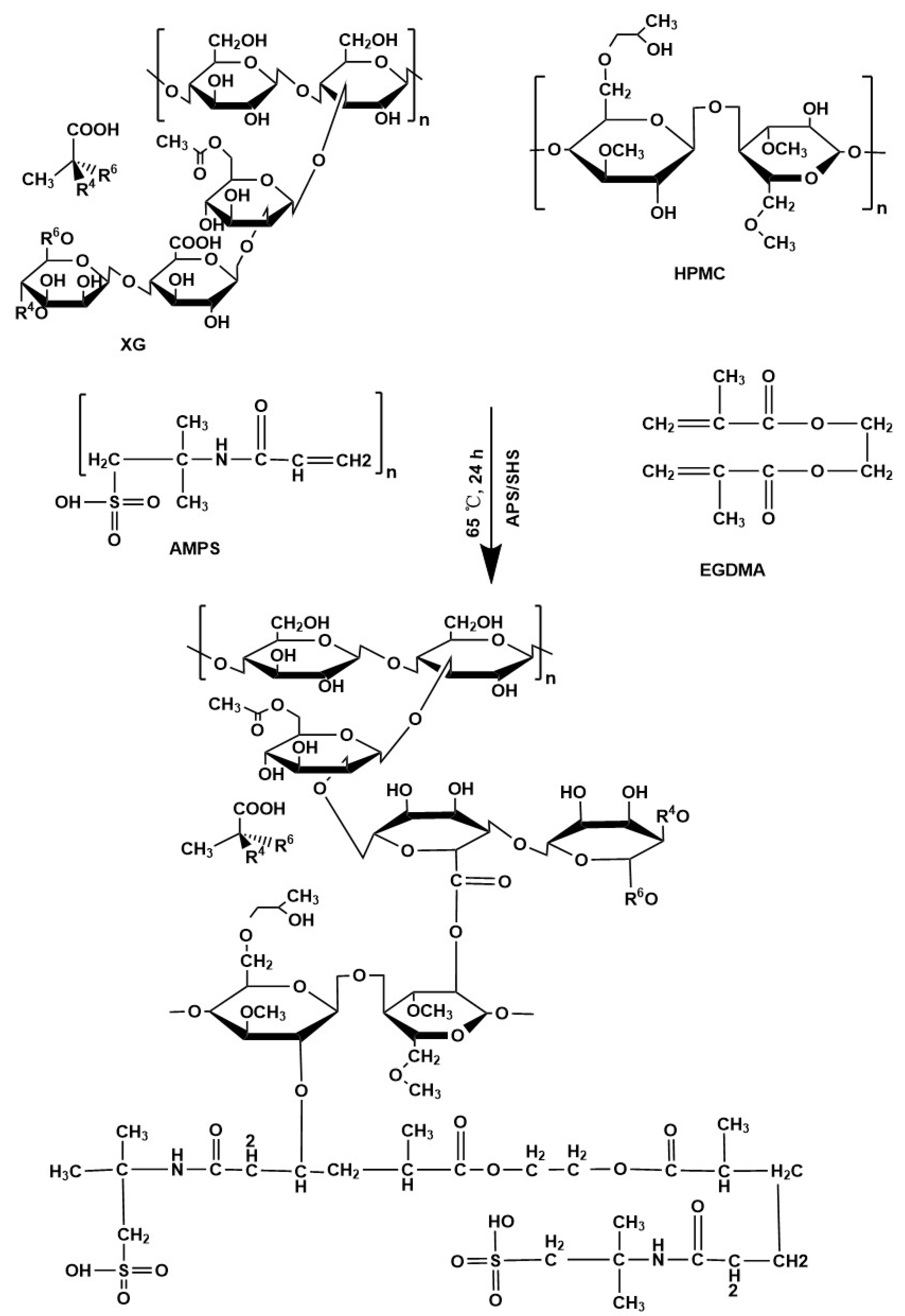
2.3. Synthesis of Xanthan Gum-Based (HPMC-g-AMPS) Hydrogels
RIC Loading in Xanthan Gum-Based (HPMC-g-AMPS) Hydrogels
2.4. Characterization
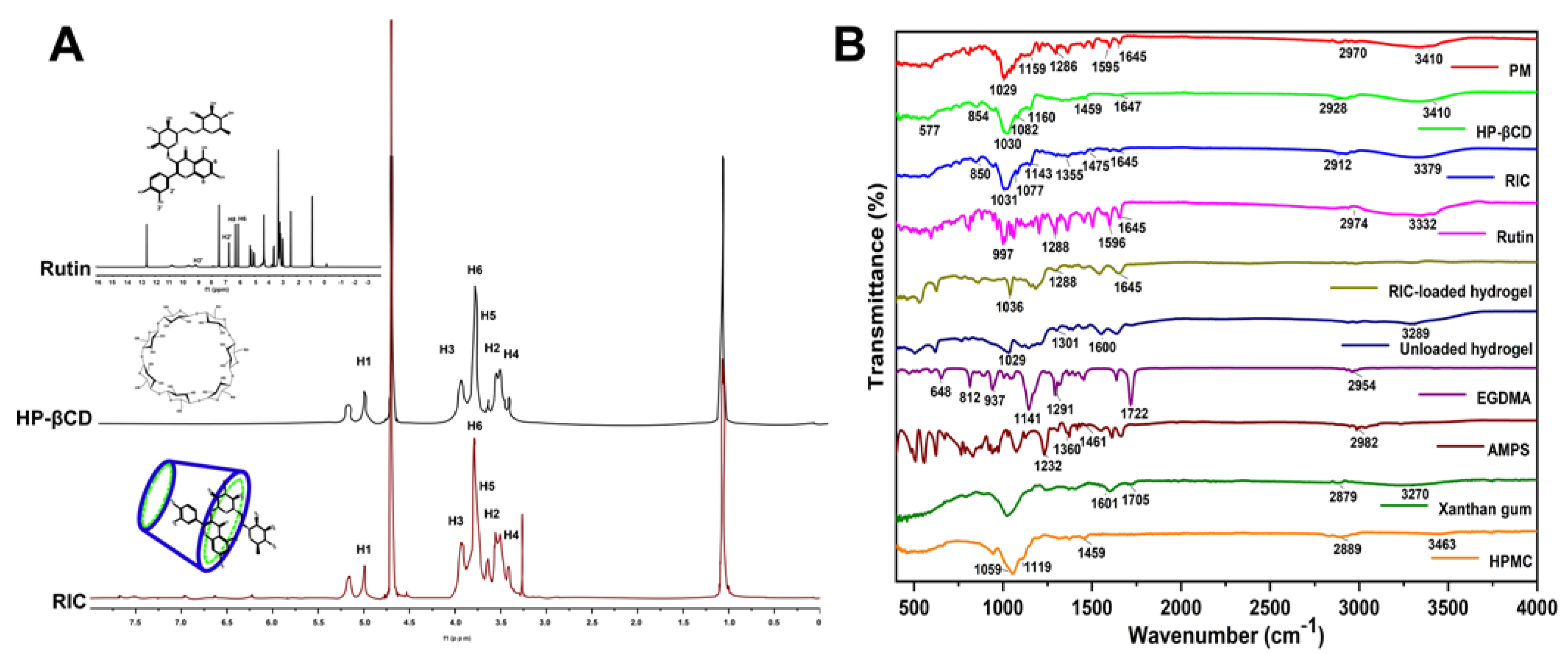
2.4.1. 1H NMR and Fourier Transform Infrared Spectroscopy (FTIR)
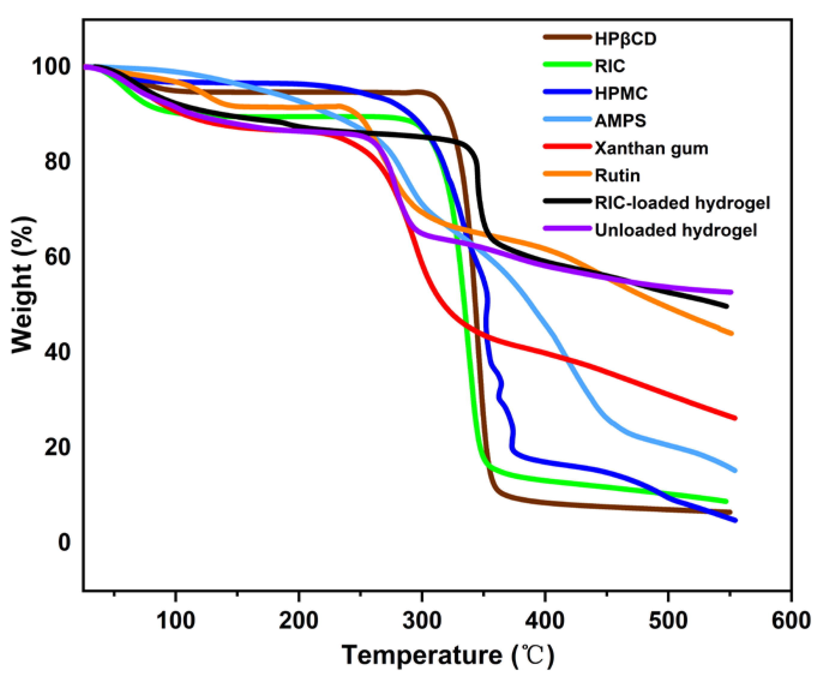
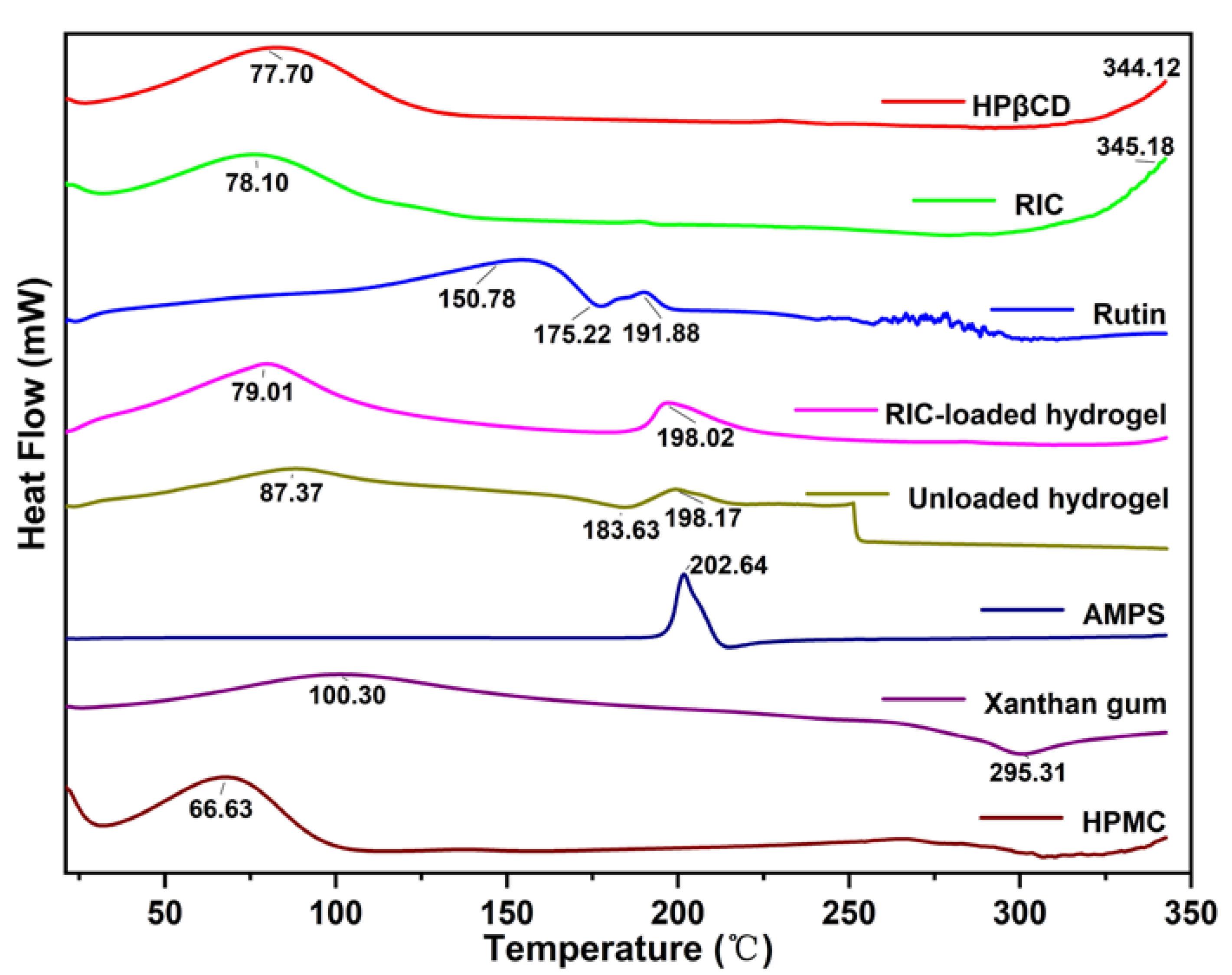
2.4.2. Thermal Analysis (TGA and DSC)
2.4.3. X-ray Diffraction (XRD)
2.4.4. Morphological Analysis
2.4.5. Mechanical Properties Analysis
2.4.6. Sol–Gel Study
2.4.7. Porosity Study
2.4.8. Biodegradation Study
2.5. Characteristics of the Synthesized Hydrogel Polymer Network
2.5.1. Diffusion Coefficient
2.5.2. Polymer Volume Fraction (V2,s)
2.5.3. Average Molecular Weight between Crosslinks (Mc)
2.5.4. Solvent Interaction Parameters (χ)
2.5.5. Crosslinking Units (N)
2.6. Hydrogels Swelling Study
2.7. In Vitro Release Study and Kinetics Data Modeling
2.8. Antioxidant Studies
2.8.1. DPPH Antioxidant Activity
2.8.2. ABTS Antioxidant Activity
2.9. Antibacterial Study
2.10. Statistical Analysis
3. Results and Discussion
3.1. 1H NMR and FTIR Analysis
3.2. TGA Analysis
3.3. Differential Scanning Calorimetry (DSC)
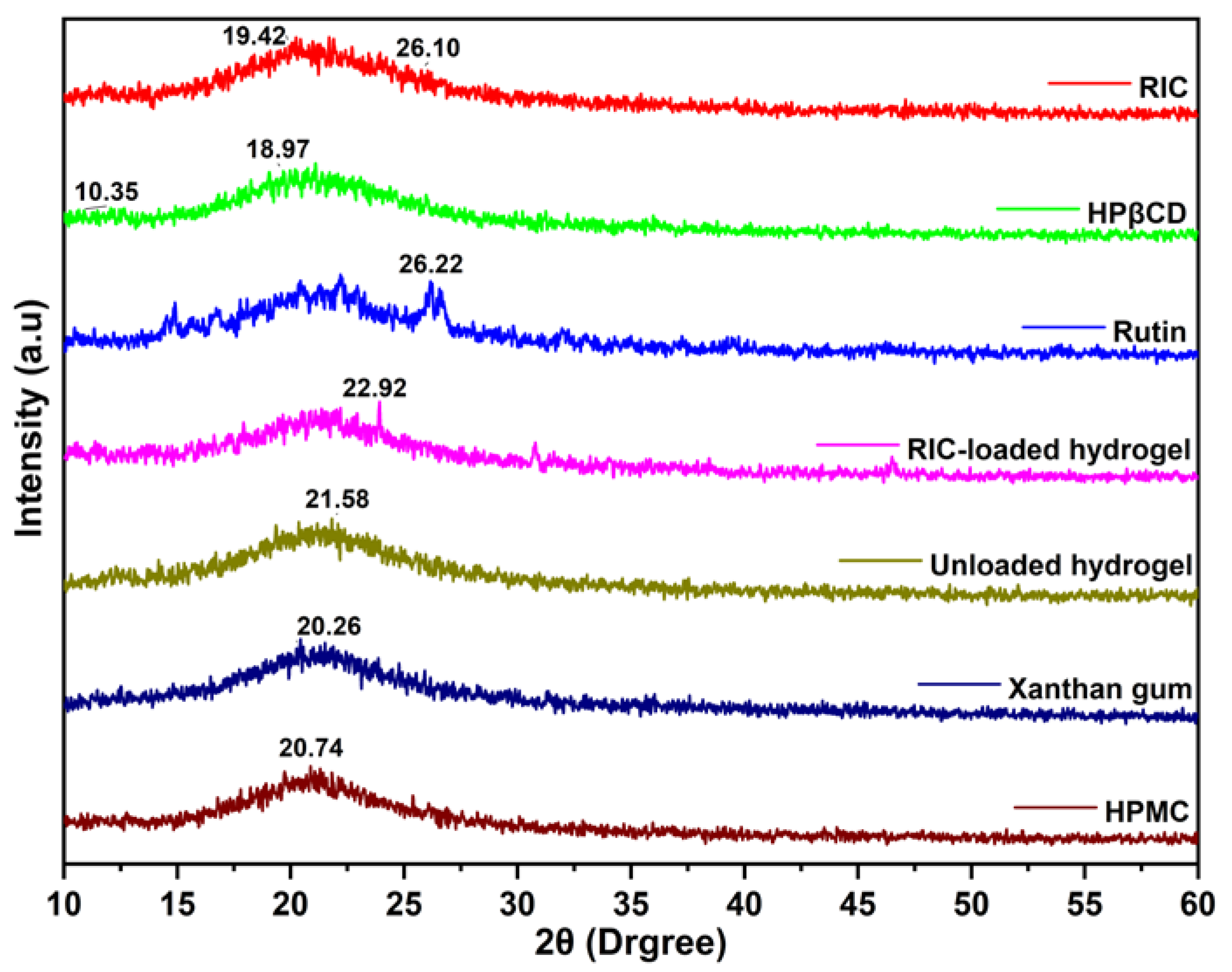
3.4. XRD Analysis
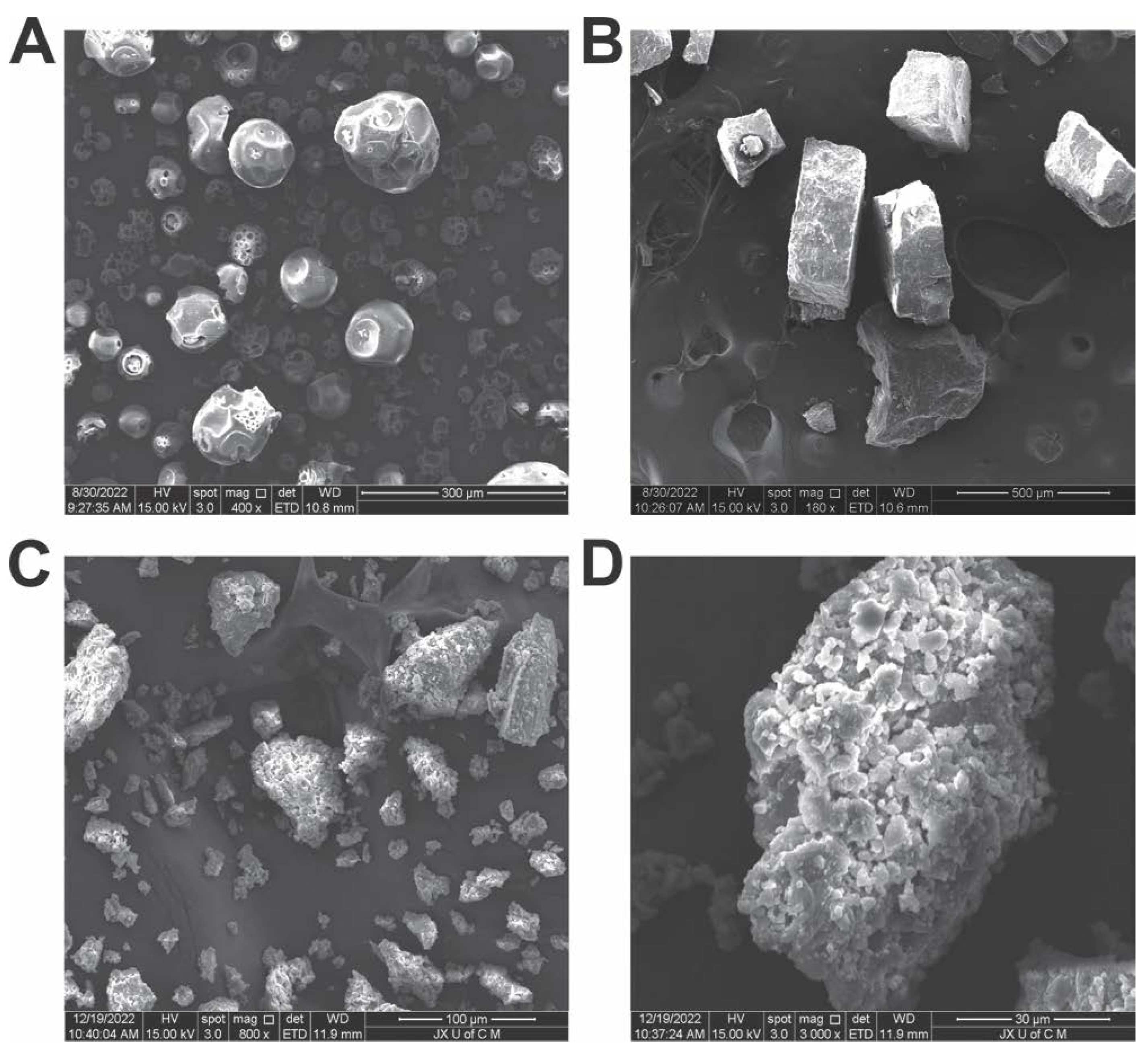
3.5. SEM Analysis
3.6. Mechanical Properties Analysis
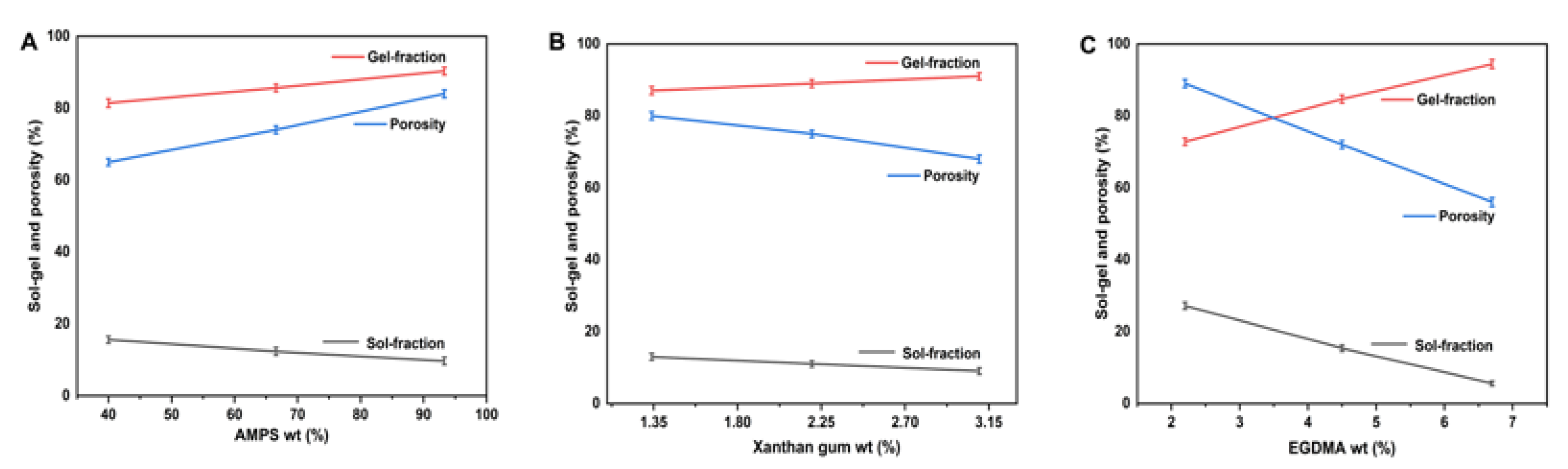
3.7. Sol–Gel Analysis
3.8. Porosity Study
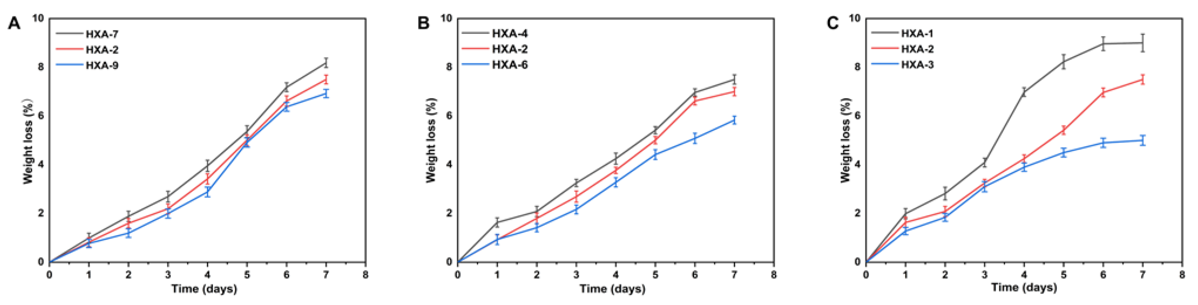
3.9. Biodegradation Analysis
3.10. Structural Parameters of Hydrogels
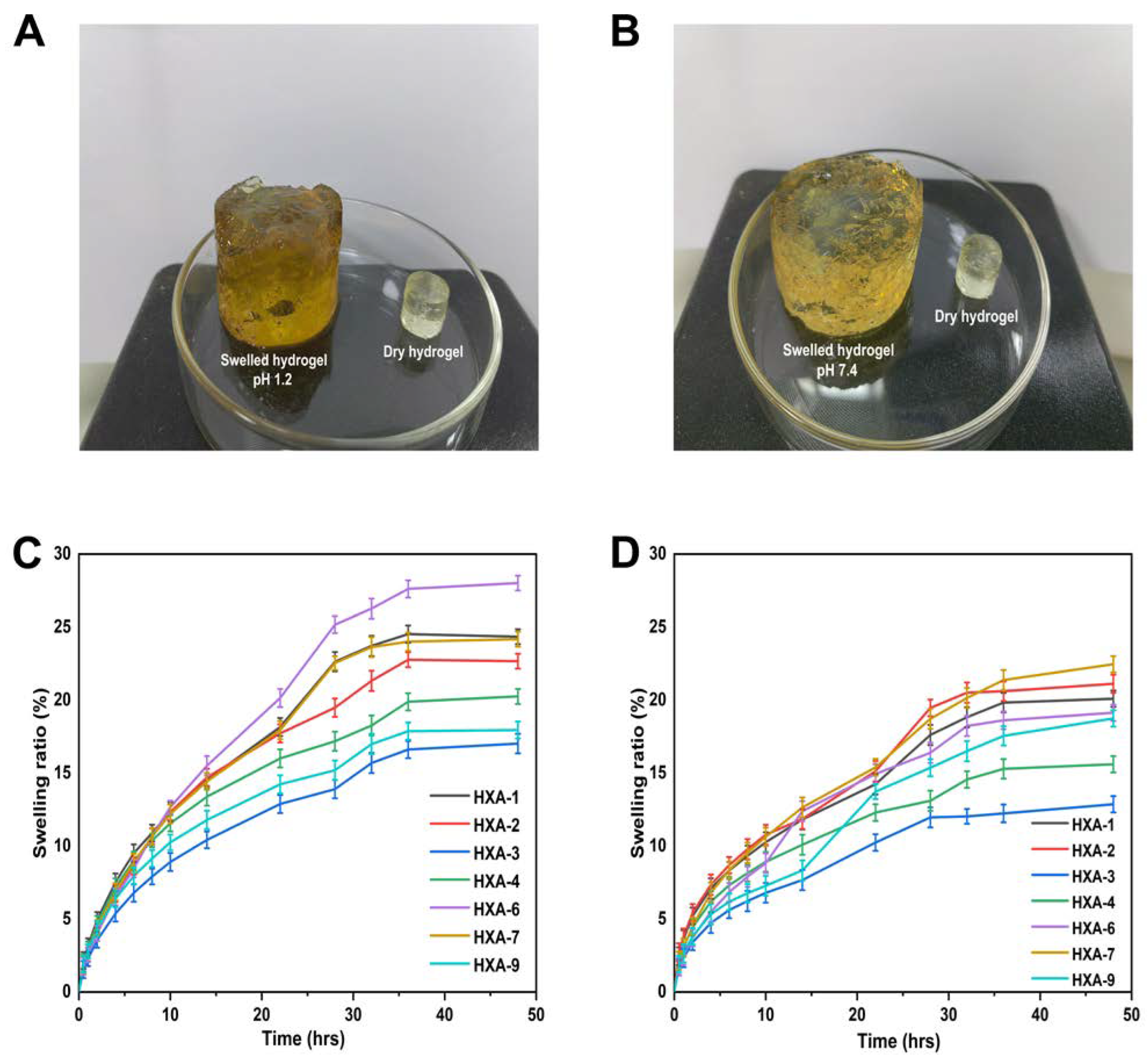
3.11. Swelling Behavior
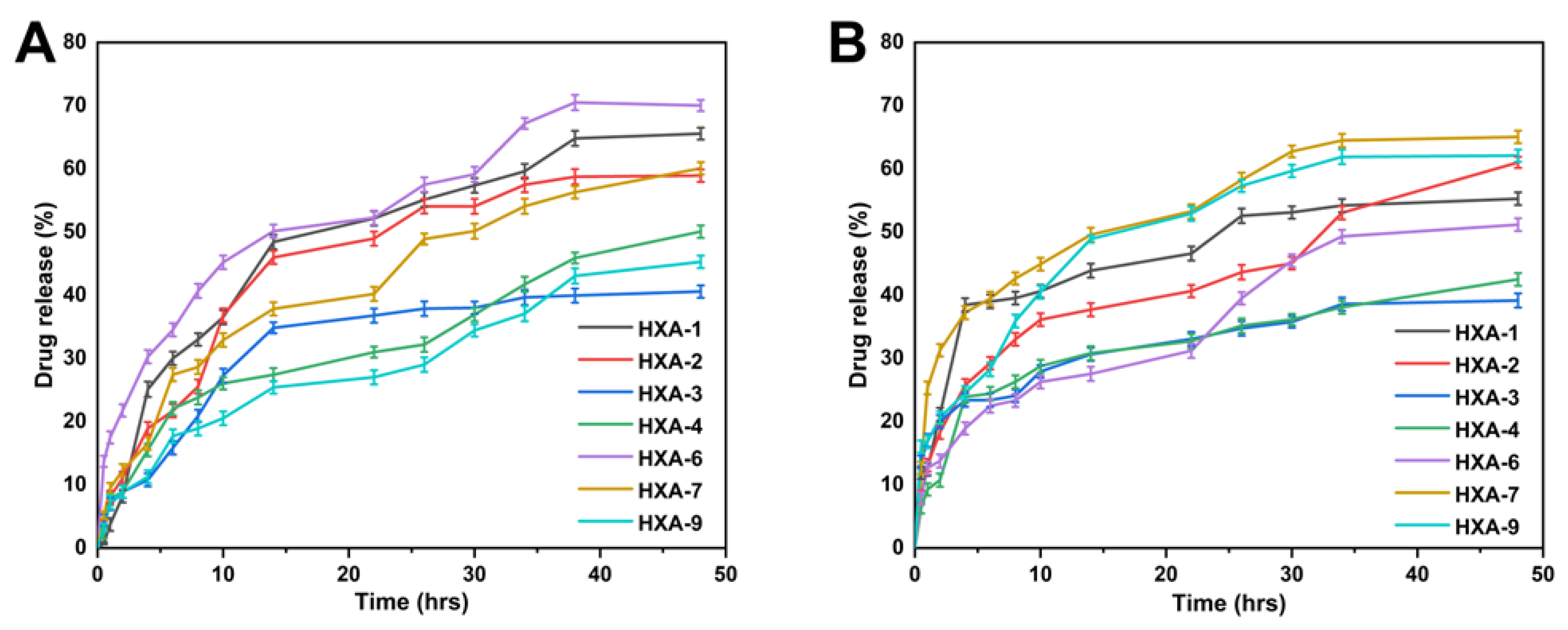
3.12. Drug Release Behaviour and Kinetics Modelling
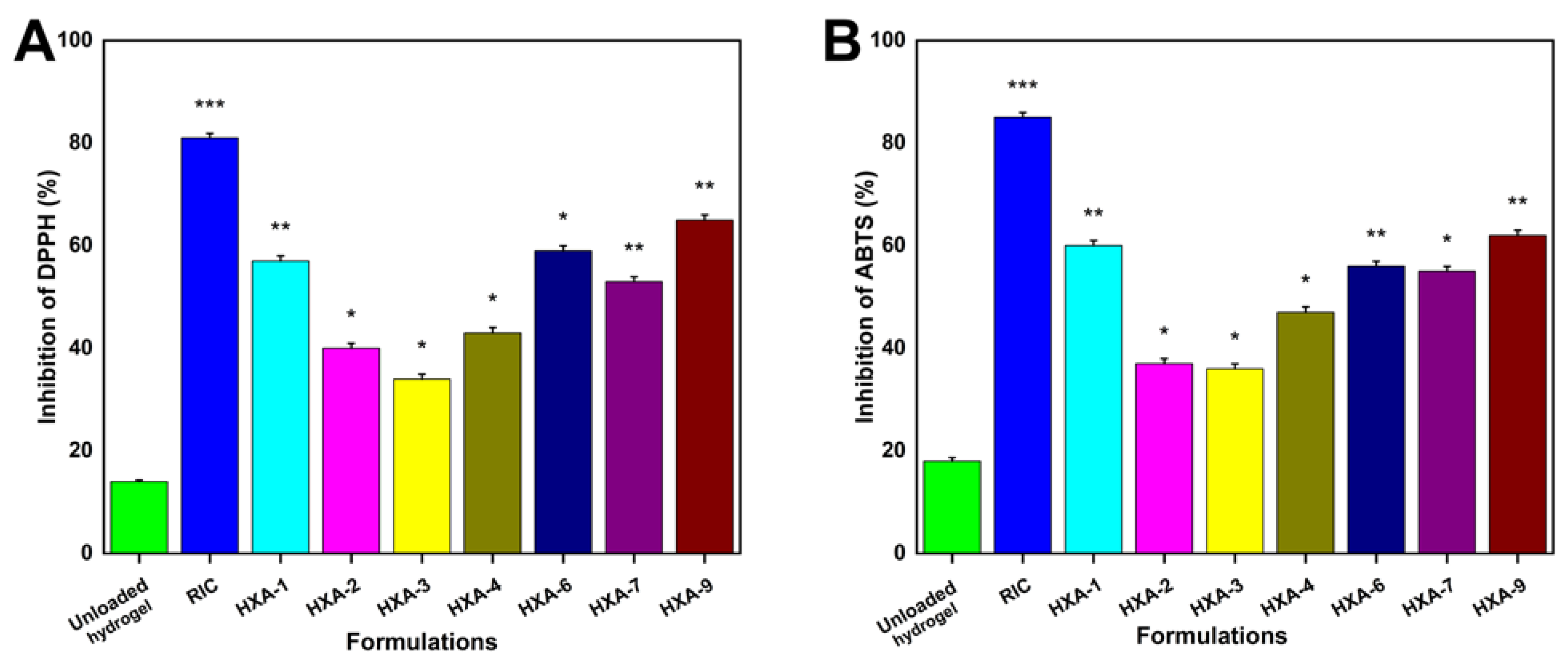
3.13. Antioxidation Analysis
3.14. Antibacterial Study
4. Conclusions
Author Contributions
Funding
Institutional Review Board Statement
Informed Consent Statement
Data Availability Statement
Acknowledgments
Conflicts of Interest
References
- Naeem, A.; Ming, Y.; Pengyi, H.; Jie, K.Y.; Yali, L.; Haiyan, Z.; Shuai, X.; Wenjing, L.; Ling, W.; Xia, Z.M.; et al. The fate of flavonoids after oral administration: A comprehensive overview of its bioavailability. Crit. Rev. Food Sci. Nutr. 2021, 62, 6169–6186. [Google Scholar] [CrossRef] [PubMed]
- Taldaev, A.; Terekhov, R.; Nikitin, I.; Zhevlakova, A.; Selivanova, I. Insights into the Pharmacological Effects of Flavonoids: The Systematic Review of Computer Modeling. Int. J. Mol. Sci. 2022, 23, 6023. [Google Scholar] [CrossRef] [PubMed]
- Li, C.; Chen, L.; McClements, D.; Peng, X.; Qiu, C.; Long, J.; Ji, H.; Zhao, J.; Zhou, X.; Jin, Z. Preparation and Characterization of Rutin–Loaded Zein–Carboxymethyl Starch Nanoparticles. Foods 2022, 11, 2827. [Google Scholar] [CrossRef] [PubMed]
- Colucci-D’Amato, L.; Cimaglia, G. Ruta graveolens as a potential source of neuroactive compounds to promote and restore neural functions. J. Tradit. Complement. Med. 2020, 10, 309–314. [Google Scholar] [CrossRef]
- Ganeshpurkar, A.; Saluja, A. The pharmacological potential of rutin. Saudi Pharm. J. 2017, 25, 149–164. [Google Scholar] [CrossRef] [Green Version]
- Nguyen, T.A.; Liu, B.; Zhao, J.; Thomas, D.S.; Hook, J.M. An investigation into the supramolecular structure, solubility, stability and antioxidant activity of rutin/cyclodextrin inclusion complex. Food Chem. 2013, 136, 186–192. [Google Scholar] [CrossRef]
- Kızılbey, K. Optimization of Rutin-Loaded PLGA Nanoparticles Synthesized by Single-Emulsion Solvent Evaporation Method. ACS Omega 2019, 4, 555–562. [Google Scholar] [CrossRef]
- Mujtaba, M.; Hassan, K.; Imran, M. Chitosan-alginate nanoparticles as a novel drug delivery system for rutin. Int. J. Adv. Biotechnol. Res. 2018, 9, 1895–1903. [Google Scholar]
- Yang, C.-Y.; Hsiu, S.-L.; Wen, K.-C.; Lin, S.-P.; Tsai, S.-Y.; Hou, Y.-C.; Chao, P.-D. Bioavailability and metabolic pharmacokinetics of rutin and quercetin in rats. J. Food Drug Anal. 2005, 13, 5. [Google Scholar] [CrossRef]
- Manach, C.; Williamson, G.; Morand, C.; Scalbert, A.; Rémésy, C. Bioavailability and bioefficacy of polyphenols in humans. I. Review of 97 bioavailability studies. Am. J. Clin. Nutr. 2005, 81, 230S–242S. [Google Scholar] [CrossRef] [Green Version]
- Lauro, M.R.; Torre, M.L.; Maggi, L.; De Simone, F.; Conte, U.; Aquino, R.P. Fast- and Slow-Release Tablets for Oral Administration of Flavonoids: Rutin and Quercetin. Drug Dev. Ind. Pharm. 2002, 28, 371–379. [Google Scholar] [CrossRef] [PubMed]
- Franco, P.; De Marco, I. Formation of Rutin–β-Cyclodextrin Inclusion Complexes by Supercritical Antisolvent Precipitation. Polymers 2021, 13, 246. [Google Scholar] [CrossRef] [PubMed]
- Carneiro, S.; Duarte, F.C.; Heimfarth, L.; Quintans, J.S.; Quintans-Júnior, L.; Júnior, V.V.; de Lima, Á.N. Cyclodextrin–drug inclusion complexes: In vivo and in vitro approaches. Int. J. Mol. Sci. 2019, 20, 642. [Google Scholar] [CrossRef] [PubMed] [Green Version]
- Hu, Y.; Qiu, C.; Qin, Y.; Xu, X.; Fan, L.; Wang, J.; Jin, Z. Cyclodextrin–phytochemical inclusion complexes: Promising food materials with targeted nutrition and functionality. Trends Food Sci. Technol. 2021, 109, 398–412. [Google Scholar] [CrossRef]
- Muñoz-Shugulí, C.; Vidal, C.P.; Cantero-López, P.; Lopez-Polo, J. Encapsulation of plant extract compounds using cyclodextrin inclusion complexes, liposomes, electrospinning and their combinations for food purposes. Trends Food Sci. Technol. 2020, 108, 177–186. [Google Scholar] [CrossRef]
- Liu, Z.; Ye, L.; Xi, J.; Wang, J.; Feng, Z.-G. Cyclodextrin polymers: Structure, synthesis, and use as drug carriers. Prog. Polym. Sci. 2021, 118, 101408. [Google Scholar] [CrossRef]
- Braga, S. Cyclodextrin superstructures for drug delivery. J. Drug Deliv. Sci. Technol. 2022, 75, 103650. [Google Scholar] [CrossRef]
- Mahjoubin-Tehran, M.; Kovanen, P.T.; Xu, S.; Jamialahmadi, T.; Sahebkar, A. Cyclodextrins: Potential therapeutics against atherosclerosis. Pharmacol. Ther. 2020, 214, 107620. [Google Scholar] [CrossRef]
- Gould, S.; Scott, R. 2-Hydroxypropyl-β-cyclodextrin (HP-β-CD): A toxicology review. Food Chem. Toxicol. 2005, 43, 1451–1459. [Google Scholar] [CrossRef]
- Wangsawangrung, N.; Choipang, C.; Chaiarwut, S.; Ekabutr, P.; Suwantong, O.; Chuysinuan, P.; Techasakul, S.; Supaphol, P. Quercetin/Hydroxypropyl-β-Cyclodextrin Inclusion Complex-Loaded Hydrogels for Accelerated Wound Healing. Gels 2022, 8, 573. [Google Scholar] [CrossRef]
- Miyake, K.; Arima, H.; Hirayama, F.; Yamamoto, M.; Horikawa, T.; Sumiyoshi, H.; Noda, S.; Uekama, K. Improvement of Solubility and Oral Bioavailability of Rutin by Complexation with 2-Hydroxypropyl-β-cyclodextrin. Pharm. Dev. Technol. 2000, 5, 399–407. [Google Scholar] [CrossRef] [PubMed]
- Gothoskar, A.; Joshi, A.; Joshi, N. Pulsatile drug delivery systems: A review. Drug Deliv. Technol. 2004, 4, 1–11. [Google Scholar]
- Layek, B.; Mandal, S. Natural polysaccharides for controlled delivery of oral therapeutics: A recent update. Carbohydr. Polym. 2020, 230, 115617. [Google Scholar] [CrossRef] [PubMed]
- Banker, G.S. Pharmaceutical Applications of Controlled Release: An Overview of the Past, Present, and Future. In Medical Applications of Controlled Release; CRC Press: Boca Raton, FL, USA, 2019; pp. 1–34. [Google Scholar] [CrossRef]
- Bruneau, M.; Bennici, S.; Brendle, J.; Dutournie, P.; Limousy, L.; Pluchon, S. Systems for stimuli-controlled release: Materials and applications. J. Control. Release 2018, 294, 355–371. [Google Scholar] [CrossRef]
- Onaciu, A.; Munteanu, R.A.; Moldovan, A.I.; Moldovan, C.S.; Berindan-Neagoe, I. Hydrogels Based Drug Delivery Synthesis, Characterization and Administration. Pharmaceutics 2019, 11, 432. [Google Scholar] [CrossRef] [PubMed] [Green Version]
- Narayanaswamy, R.; Torchilin, V.P. Hydrogels and Their Applications in Targeted Drug Delivery. Molecules 2019, 24, 603. [Google Scholar] [CrossRef] [Green Version]
- Hoare, T.R.; Kohane, D.S. Hydrogels in drug delivery: Progress and challenges. Polymer 2008, 49, 1993–2007. [Google Scholar] [CrossRef] [Green Version]
- Khalid, I.; Ahmad, M.; Minhas, M.U.; Barkat, K. Synthesis and evaluation of chondroitin sulfate based hydrogels of loxoprofen with adjustable properties as controlled release carriers. Carbohydr. Polym. 2017, 181, 1169–1179. [Google Scholar] [CrossRef]
- Auriemma, G.; Russo, P.; Del Gaudio, P.; García-González, C.; Landín, M.; Aquino, R. Technologies and formulation design of polysaccharide-based hydrogels for drug delivery. Molecules 2020, 25, 3156. [Google Scholar] [CrossRef]
- Cortes, H.; Caballero-Florán, I.; Mendoza-Muñoz, N.; Escutia-Guadarrama, L.; Figueroa-González, G.; Reyes-Hernández, O.; Carmen, M.G.-D.; Varela-Cardoso, M.; González-Torres, M.; Florán, B. Xanthan gum in drug release. Cell. Mol. Biol. 2020, 66, 199–207. [Google Scholar] [CrossRef]
- Zang, Z.; Zhao, S.; Yang, M.; Yu, C.; Ouyang, H.; Chen, L.; Zhu, W.; Liao, Z.-G.; Naeem, A.; Guan, Y. Blood chemical components analysis of honeysuckle and formulation of xanthan gum/starch-based (PVA-co-AA) hydrogels for controlled release. Arab. J. Chem. 2022, 15, 104312. [Google Scholar] [CrossRef]
- Khanum, H.; Ullah, K.; Murtaza, G.; Khan, S.A. Fabrication and in vitro characterization of HPMC-g-poly(AMPS) hydrogels loaded with loxoprofen sodium. Int. J. Biol. Macromol. 2018, 120, 1624–1631. [Google Scholar] [CrossRef] [PubMed]
- Ohara, T.; Kitamura, S.; Kitagawa, T.; Terada, K. Dissolution mechanism of poorly water-soluble drug from extended release solid dispersion system with ethylcellulose and hydroxypropylmethylcellulose. Int. J. Pharm. 2005, 302, 95–102. [Google Scholar] [CrossRef]
- Won, D.-H.; Kim, M.-S.; Lee, S.; Park, J.-S.; Hwang, S.-J. Improved physicochemical characteristics of felodipine solid dispersion particles by supercritical anti-solvent precipitation process. Int. J. Pharm. 2005, 301, 199–208. [Google Scholar] [CrossRef] [PubMed]
- Ali, A.E.; El-Rehiem, H.A.A.; Hegazy, E.A.; Ghobashy, M. Characterization and Potential Application of Electro-Active Acrylamido-2-methyl Propane Sulfonic Acid/Acrylic Acid Copolymer Prepared by Ionizing Radiation. J. Macromol. Sci. Part A 2007, 44, 91–98. [Google Scholar] [CrossRef]
- Sri, K.V.; Kondaiah, A.; Ratna, J.V.; Annapurna, A. Preparation and Characterization of Quercetin and Rutin Cyclodextrin Inclusion Complexes. Drug Dev. Ind. Pharm. 2007, 33, 245–253. [Google Scholar] [CrossRef] [PubMed]
- Guan, Y.; Yu, C.; Zang, Z.; Wan, X.; Naeem, A.; Zhang, R.; Zhu, W. Chitosan/xanthan gum-based (Hydroxypropyl methylcellulose-co-2-Acrylamido-2-methylpropane sulfonic acid) interpenetrating hydrogels for controlled release of amorphous solid dispersion of bioactive constituents of Pueraria lobatae. Int. J. Biol. Macromol. 2023, 224, 380–395. [Google Scholar] [CrossRef] [PubMed]
- Bueno, V.B.; Bentini, R.; Catalani, L.H.; Petri, D.F.S. Synthesis and swelling behavior of xanthan-based hydrogels. Carbohydr. Polym. 2013, 92, 1091–1099. [Google Scholar] [CrossRef] [Green Version]
- Verma, N.; Purohit, M.; Equbal, D.; Dhiman, N.; Singh, A.; Kar, A.; Shankar, J.; Tehlan, S.; Patnaik, S. Targeted smart pH and thermoresponsive N, O-carboxymethyl chitosan conjugated nanogels for enhanced therapeutic efficacy of doxorubicin in MCF-7 breast cancer cells. Bioconjugate Chem. 2016, 27, 2605–2619. [Google Scholar] [CrossRef]
- Choudhary, S.; Sharma, K.; Sharma, V.; Kumar, V. Grafting Polymers. In Reactive and Functional Polymers Volume Two: Modification Reactions, Compatibility and Blends; Gutiérrez, T., Ed.; Springer International Publishing: Cham, Switzerland, 2020; pp. 199–243. [Google Scholar]
- Rodríguez-Rodríguez, R.; García-Carvajal, Z.; Jiménez-Palomar, I.; Jiménez-Avalos, J.; Espinosa-Andrews, H. Development of gelatin/chitosan/PVA hydrogels: Thermal stability, water state, viscoelasticity, and cytotoxicity assays. J. Appl. Polym. Sci. 2019, 136, 47149. [Google Scholar] [CrossRef]
- Bozoğlan, B.K.; Duman, O.; Tunç, S. Preparation and characterization of thermosensitive chitosan/carboxymethylcellulose/scleroglucan nanocomposite hydrogels. Int. J. Biol. Macromol. 2020, 162, 781–797. [Google Scholar] [CrossRef] [PubMed]
- Sami, A.J.; Khalid, M.; Jamil, T.; Aftab, S.; Mangat, S.A.; Shakoori, A.; Iqbal, S. Formulation of novel chitosan guargum based hydrogels for sustained drug release of paracetamol. Int. J. Biol. Macromol. 2018, 108, 324–332. [Google Scholar] [CrossRef] [PubMed]
- Li, Z.; Jiang, X.; Huang, H.; Liu, A.; Liu, H.; Abid, N.; Ming, L. Chitosan/zein films incorporated with essential oil nanoparticles and nanoemulsions: Similarities and differences. Int. J. Biol. Macromol. 2022, 208, 983–994. [Google Scholar] [CrossRef] [PubMed]
- Chang, A.; Ye, Z.; Ye, Z.; Deng, J.; Lin, J.; Wu, C.; Zhu, H. Citric acid crosslinked sphingan WL gum hydrogel films supported ciprofloxacin for potential wound dressing application. Carbohydr. Polym. 2022, 291, 119520. [Google Scholar] [CrossRef]
- Tapdiqov, S.; Taghiyev, D.; Zeynalov, N.; Safaraliyeva, S.; Fatullayeva, S.; Hummetov, A.; Raucci, M.; Mustafayev, M.; Jafarova, R.; Shirinova, K. Cumulative release kinetics of levothyroxine-Na pentahydrate from chitosan/arabinogalactane based pH sensitive hydrogel and it’s toxicology. React. Funct. Polym. 2022, 178, 105334. [Google Scholar] [CrossRef]
- Yu, C.; Chen, X.; Zhu, W.; Li, L.; Peng, M.; Zhong, Y.; Naeem, A.; Zang, Z.; Guan, Y. Synthesis of Gallic Acid-Loaded Chitosan-Grafted-2-Acrylamido-2-Methylpropane Sulfonic Acid Hydrogels for Oral Controlled Drug Delivery: In Vitro Biodegradation, Antioxidant, and Antibacterial Effects. Gels 2022, 8, 806. [Google Scholar] [CrossRef]
- Jalil, A.; Khan, S.; Naeem, F.; Haider, M.; Sarwar, S.; Riaz, A.; Ranjha, N. The structural, morphological and thermal properties of grafted pH-sensitive interpenetrating highly porous polymeric composites of sodium alginate/acrylic acid copolymers for controlled delivery of diclofenac potassium. Des. Monomers Polym. 2017, 20, 308–324. [Google Scholar] [CrossRef] [Green Version]
- Jayaramudu, T.; Ko, H.-U.; Kim, H.; Kim, J.; Kim, J. Swelling behavior of polyacrylamide–cellulose nanocrystal hydrogels: Swelling kinetics, temperature, and pH effects. Materials 2019, 12, 2080. [Google Scholar] [CrossRef] [Green Version]
- Vigata, M.; Meinert, C.; Hutmacher, D.W.; Bock, N. Hydrogels as Drug Delivery Systems: A Review of Current Characterization and Evaluation Techniques. Pharmaceutics 2020, 12, 1188. [Google Scholar] [CrossRef]
- Xu, Z.; Liu, G.; Li, Q.; Wu, J. A novel hydrogel with glucose-responsive hyperglycemia regulation and antioxidant activity for enhanced diabetic wound repair. Nano Res. 2022, 15, 5305–5315. [Google Scholar] [CrossRef]
- Lorz, L.R.; Yoo, B.C.; Kim, M.-Y.; Cho, J.Y. Anti-Wrinkling and Anti-Melanogenic Effect of Pradosia mutisii Methanol Extract. Int. J. Mol. Sci. 2019, 20, 1043. [Google Scholar] [CrossRef] [PubMed] [Green Version]
- Abbasi, A.R.; Sohail, M.; Minhas, M.U.; Khaliq, T.; Kousar, M.; Khan, S.; Hussain, Z.; Munir, A. Bioinspired sodium alginate based thermosensitive hydrogel membranes for accelerated wound healing. Int. J. Biol. Macromol. 2020, 155, 751–765. [Google Scholar] [CrossRef] [PubMed]
- Song, S.; Gao, K.; Niu, R.; Yi, W.; Zhang, J.; Gao, C.; Yang, B.; Liao, X. Binding behavior, water solubility and in vitro cytotoxicity of inclusion complexes between ursolic acid and amino-appended β-cyclodextrins. J. Mol. Liq. 2019, 296, 111993. [Google Scholar] [CrossRef]
- Kfoury, M.; Landy, D.; Ruellan, S.; Auezova, L.; Greige-Gerges, H.; Fourmentin, S. Determination of formation constants and structural characterization of cyclodextrin inclusion complexes with two phenolic isomers: Carvacrol and thymol. Beilstein J. Org. Chem. 2016, 12, 29–42. [Google Scholar] [CrossRef] [Green Version]
- Lu, A.; Petit, E.; Li, S.; Wang, Y.; Su, F.; Monge, S. Novel thermo-responsive micelles prepared from amphiphilic hydroxypropyl methyl cellulose-block-JEFFAMINE copolymers. Int. J. Biol. Macromol. 2019, 135, 38–45. [Google Scholar] [CrossRef]
- Ding, C.; Zhang, M.; Li, G. Preparation and characterization of collagen/hydroxypropyl methylcellulose (HPMC) blend film. Carbohydr. Polym. 2015, 119, 194–201. [Google Scholar] [CrossRef]
- Said, M.; Haq, B.; Al Shehri, D.; Rahman, M.M.; Muhammed, N.S.; Mahmoud, M. Modification of Xanthan Gum for a High-Temperature and High-Salinity Reservoir. Polymers 2021, 13, 4212. [Google Scholar] [CrossRef]
- Hu, J.; Dai, H.; Zeng, Y.; Yang, Y.; Wang, H.; Zhu, X.; Li, L.; Zhou, G.; Chen, R.; Guo, L. A Cross-Linker-Based Poly(Ionic Liquid) for Sensitive Electrochemical Detection of 4-Nonylphenol. Nanomaterials 2019, 9, 513. [Google Scholar] [CrossRef] [Green Version]
- Neto, C.M.S.; Lima, F.C.; Morais, R.P.; de Andrade, L.R.M.; de Lima, R.; Chaud, M.V.; Pereira, M.M.; Júnior, R.L.C.D.A.; Cardoso, J.C.; Zielińska, A.; et al. Rutin-Functionalized Multi-Walled Carbon Nanotubes: Molecular Docking, Physicochemistry and Cytotoxicity in Fibroblasts. Toxics 2021, 9, 173. [Google Scholar] [CrossRef]
- Gera, S.; Pooladanda, V.; Godugu, C.; Challa, V.S.; Wankar, J.; Dodoala, S.; Sampathi, S. Rutin nanosuspension for potential management of osteoporosis: Effect of particle size reduction on oral bioavailability, in vitro and in vivo activity. Pharm. Dev. Technol. 2020, 25, 971–988. [Google Scholar] [CrossRef]
- Sun, C.; Cao, J.; Wang, Y.; Chen, J.; Huang, L.; Zhang, H.; Wu, J.; Sun, C. Ultrasound-mediated molecular self-assemble of thymol with 2-hydroxypropyl-β-cyclodextrin for fruit preservation. Food Chem. 2021, 363, 130327. [Google Scholar] [CrossRef] [PubMed]
- Enawgaw, H.; Tesfaye, T.; Yilma, K.T.; Limeneh, D.Y. Synthesis of a Cellulose-Co-AMPS Hydrogel for Personal Hygiene Applications Using Cellulose Extracted from Corncobs. Gels 2021, 7, 236. [Google Scholar] [CrossRef] [PubMed]
- Ma, D.; Djemai, A.; Gendron, C.; Xi, H.; Smith, M.; Kogan, J.; Li, L. Development of a HPMC-based controlled release formulation with hot melt extrusion (HME). Drug Dev. Ind. Pharm. 2013, 39, 1070–1083. [Google Scholar] [CrossRef] [PubMed]
- Kazachenko, A.; Vasilieva, N.; Borovkova, V.; Fetisova, O.; Issaoui, N.; Malyar, Y.; Elsuf’ev, E.; Karacharov, A.; Skripnikov, A.; Miroshnikova, A. Food xanthan polysaccharide sulfation process with sulfamic acid. Foods 2021, 10, 2571. [Google Scholar] [CrossRef] [PubMed]
- Zheng, J.; Wang, B.; Xiang, J.; Yu, Z. Controlled Release of Curcumin from HPMC (Hydroxypropyl Methyl Cellulose) Co-Spray-Dried Materials. Bioinorg. Chem. Appl. 2021, 2021, 7625585. [Google Scholar] [CrossRef]
- Hani, U.; Hg, S.; Osmani, R.A.M.; Srivastava, A.; Varma, N.S.K. Development of a Curcumin Bioadhesive Monolithic Tablet for Treatment of Vaginal Candidiasis. Iran. J. Pharm. Res. 2016, 15, 23–34. [Google Scholar] [CrossRef]
- Ashames, A.; Ullah, K.; Al-Tabakha, M.; Khan, S.A.; Hassan, N.; Mannan, A.; Ikram, M.; Buabeid, M.; Murtaza, G. Development, characterization and In-vitro evaluation of guar gum based new polymeric matrices for controlled delivery using metformin HCl as model drug. PLoS ONE 2022, 17, e0271623. [Google Scholar] [CrossRef]
- Feroz, S.; Dias, G. Hydroxypropylmethyl cellulose (HPMC) crosslinked keratin/hydroxyapatite (HA) scaffold fabrication, characterization and in vitro biocompatibility assessment as a bone graft for alveolar bone regeneration. Heliyon 2021, 7, e08294. [Google Scholar] [CrossRef]
- Hu, X.; Wang, K.; Yu, M.; He, P.; Qiao, H.; Zhang, H. Characterization and Antioxidant Activity of a Low-Molecular-Weight Xanthan Gum. Biomolecules 2019, 9, 730. [Google Scholar] [CrossRef] [Green Version]
- Li, Y.; He, Z.-D.; Zheng, Q.-E.; Hu, C.; Lai, W.-F. Hydroxypropyl-β-cyclodextrin for Delivery of Baicalin via Inclusion Complexation by Supercritical Fluid Encapsulation. Molecules 2018, 23, 1169. [Google Scholar] [CrossRef] [Green Version]
- He, J.; Guo, F.; Lin, L.; Chen, H.; Chen, J.; Cheng, Y.; Zheng, Z.-P. Investigating the oxyresveratrol β-cyclodextrin and 2-hydroxypropyl-β-cyclodextrin complexes: The effects on oxyresveratrol solution, stability, and antibrowning ability on fresh grape juice. LWT 2019, 100, 263–270. [Google Scholar] [CrossRef]
- Gholamali, I.; Hosseini, S.N.; Alipour, E.; Yadollahi, M. Preparation and Characterization of Oxidized Starch/CuO Nanocomposite Hydrogels Applicable in a Drug Delivery System. Starch-Stärke 2018, 71, 1800118. [Google Scholar] [CrossRef]
- Park, D.; Haam, S.; Lee, T.; Kim, H.; Kim, W. Chemoenzymatic synthesis of sugar-containing biocompatible hydrogels: Crosslinked poly (β-methylglucoside acrylate) and poly (β-methylglucoside methacrylate). J. Biomed. Mater. Res. Part A 2004, 71, 497–507. [Google Scholar] [CrossRef]
- Chen, C.; Peng, Z.; Gu, J.; Peng, Y.; Huang, X.; Wu, L. Exploring Environmentally Friendly Biopolymer Material Effect on Soil Tensile and Compressive Behavior. Int. J. Environ. Res. Public Health 2020, 17, 9032. [Google Scholar] [CrossRef] [PubMed]
- Nguyen, K.D.; Trang, T.T.C.; Kobayashi, T. Chitin-halloysite nanoclay hydrogel composite adsorbent to aqueous heavy metal ions. J. Appl. Polym. Sci. 2018, 136, 47207. [Google Scholar] [CrossRef]
- Akhlaq, M.; Maryam, F.; Elaissari, A.; Ullah, H.; Adeel, M.; Hussain, A.; Ramzan, M.; Ullah, O.; Danish, M.Z.; Iftikhar, S.; et al. Pharmacokinetic evaluation of quetiapine fumarate controlled release hybrid hydrogel: A healthier treatment of schizo-phrenia. Drug Deliv. 2018, 25, 916–927. [Google Scholar] [CrossRef] [Green Version]
- Liu, C.; Tripathi, A.; Gao, W.; Tsavalas, J. Crosslinking in Semi-Batch Seeded Emulsion Polymerization: Effect of Linear and Non-Linear Monomer Feeding Rate Profiles on Gel Formation. Polymers 2021, 13, 596. [Google Scholar] [CrossRef]
- Naeem, A.; Yu, C.; Zhu, W.; Chen, X.; Wu, X.; Chen, L.; Zang, Z.; Guan, Y. Gallic Acid-Loaded Sodium Alginate-Based (Polyvinyl Alcohol-Co-Acrylic Acid) Hydrogel Membranes for Cutaneous Wound Healing: Synthesis and Characterization. Molecules 2022, 27, 8397. [Google Scholar] [CrossRef]
- Abu Fara, D.; Dadou, S.M.; Rashid, I.; Al-Obeidi, R.; Antonijevic, M.D.; Chowdhry, B.Z.; Badwan, A. A Direct Compression Matrix Made from Xanthan Gum and Low Molecular Weight Chitosan Designed to Improve Compressibility in Controlled Release Tablets. Pharmaceutics 2019, 11, 603. [Google Scholar] [CrossRef] [Green Version]
- Suhail, M.; Khan, A.; Rosenholm, J.; Minhas, M.; Wu, P.-C. Fabrication and Characterization of Diclofenac Sodium Loaded Hydrogels of Sodium Alginate as Sustained Release Carrier. Gels 2021, 7, 10. [Google Scholar] [CrossRef] [PubMed]
- Suhail, M.; Wu, P.-C.; Minhas, M.U. Using Carbomer-Based Hydrogels for Control the Release Rate of Diclofenac Sodium: Preparation and In Vitro Evaluation. Pharmaceuticals 2020, 13, 399. [Google Scholar] [CrossRef] [PubMed]
- Hasan, M.; Uddin, M.; Zabin, N.; Shakil, M.; Alam, M.; Achal, F.; Begum, A.; Hosney, M.; Hossen, M.; Hasan, M. Fabrication and Characterization of Chitosan-Polyethylene Glycol (Ch-Peg) Based Hydrogels and Evaluation of Their Potency in Rat Skin Wound Model. Int. J. Biomater. 2021, 2021, 4877344. [Google Scholar] [CrossRef] [PubMed]
- Malik, N.S.; Ahmad, M.; Minhas, M.U.; Tulain, R.; Barkat, K.; Khalid, I.; Khalid, Q. Chitosan/Xanthan Gum Based Hydrogels as Potential Carrier for an Antiviral Drug: Fabrication, Characterization, and Safety Evaluation. Front. Chem. 2020, 8, 50. [Google Scholar] [CrossRef] [Green Version]
- Suhail, M.; Shih, C.-M.; Liu, J.-Y.; Hsieh, W.-C.; Lin, Y.-W.; Minhas, M.U.; Wu, P.-C. Synthesis, Characterization, In-Vitro and In-Vivo Evaluation of Ketorolac Tromethamine-Loaded Hydrogels of Glutamic Acid as Controlled Release Carrier. Polymers 2021, 13, 3541. [Google Scholar] [CrossRef]
- Hanna, D.H.; Lotfy, V.F.; Basta, A.H.; Saad, G.R. Comparative evaluation for controlling release of niacin from protein- and cellulose-chitosan based hydrogels. Int. J. Biol. Macromol. 2020, 150, 228–237. [Google Scholar] [CrossRef]
- Saleh, A.; ElFayoumi, H.M.; Youns, M.; Barakat, W. Rutin and orlistat produce antitumor effects via antioxidant and apoptotic actions. Naunyn-Schmiedeberg’s Arch. Pharmacol. 2018, 392, 165–175. [Google Scholar] [CrossRef]
- Li, Q.; Qiu, Y.; Mao, M.; Lv, J.; Zhang, L.; Li, S.; Li, X.; Zheng, X. Antioxidant Mechanism of Rutin on Hypoxia-Induced Pulmonary Arterial Cell Proliferation. Molecules 2014, 19, 19036–19049. [Google Scholar] [CrossRef] [Green Version]
- Yang, Y.; Ye, H.; Zhao, C.; Ren, L.; Wang, C.; Georgiev, M.I.; Xiao, J.; Zhang, T. Value added immunoregulatory polysaccharides of Hericium erinaceus and their effect on the gut microbiota. Carbohydr. Polym. 2021, 262, 117668. [Google Scholar] [CrossRef]


| Formulation | HPMC (g) | Xanthan Gum (g) | APS/SHS (g) | AMPS (g) | EGDMA (g) |
|---|---|---|---|---|---|
| HXA-1 | 0.5 | 0.5 | 0.3/0.3 | 20 | 0.5 |
| HXA-2 | 0.5 | 0.5 | 0.3/0.3 | 20 | 1 |
| HXA-3 | 0.5 | 0.5 | 0.3/0.3 | 20 | 1.5 |
| HXA-4 | 0.5 | 0.5 | 0.3/0.3 | 12 | 1 |
| HXA-5 | 0.5 | 0.5 | 0.3/0.3 | 20 | 1 |
| HXA-6 | 0.5 | 0.5 | 0.3/0.3 | 28 | 1 |
| HXA-7 | 0.5 | 0.3 | 0.3/0.3 | 20 | 1 |
| HXA-8 | 0.5 | 0.5 | 0.3/0.3 | 20 | 1 |
| HXA-9 | 0.5 | 0.7 | 0.3/0.3 | 20 | 1 |
| Protons | Δ(free) | Δ(complex) | Δδ |
|---|---|---|---|
| HP-βCD | |||
| H1 | 4.982 | 4.996 | 0.014 |
| H2 | 3.550 | 3.552 | 0.002 |
| H3 | 3.933 | 3.946 | 0.013 |
| H4 | 3.509 | 3.496 | −0.013 |
| H5 | 3.643 | 3.658 | 0.015 |
| H6 | 3.793 | 3.772 | −0.021 |
| F. Codes | Thickness (mm) | TS (N/mm) | EAB (%) | RIC Loaded per 1 g Hydrogel (g) |
|---|---|---|---|---|
| HXA-1 | 1.425 | 0.356 | 31.3 | 0.365 |
| HXA-2 | 1.334 | 0.668 | 59.1 | 0.451 |
| HXA-3 | 1.298 | 1.117 | 79.9 | 0.235 |
| HXA-4 | 1.255 | 0.774 | 66.7 | 0.249 |
| HXA-5 | 1.334 | 0.668 | 59.1 | 0.451 |
| HXA-6 | 1.415 | 0.651 | 60.1 | 0.334 |
| HXA-7 | 1.554 | 0.548 | 58.6 | 0.482 |
| HXA-8 | 1.334 | 0.668 | 59.1 | 0.451 |
| HXA-9 | 1.610 | 0.801 | 67.4 | 0.322 |
| F. Codes | V2,s | χ | Mc | Mr | N | D × 10−5 (cm2 s−1) |
|---|---|---|---|---|---|---|
| HXA-1 | 0.041 ± 0.003 | 0.514 ± 0.041 | 4406.1 ± 1.509 | 231.99 ± 0.002 | 37.98 ± 0.98 | 0.309 ± 0.061 |
| HXA-2 | 0.044 ± 0.005 | 0.515 ± 0.018 | 3369.4 ± 2.067 | 231.22 ± 0.001 | 29.14 ± 1.02 | 0.219 ± 0.023 |
| HXA-3 | 0.058 ± 0.006 | 0.520 ± 0.005 | 2357.5 ± 0.765 | 230.48 ± 0.002 | 20.45 ± 0.97 | 0.011 ± 0.001 |
| HXA-4 | 0.049 ± 0.004 | 0.516 ± 0.031 | 3157.9 ± 1.251 | 244.91 ± 0.003 | 25.83 ± 1.07 | 0.041 ± 0.003 |
| HXA-5 | 0.044 ± 0.005 | 0.515 ± 0.018 | 2298.9 ± 1.367 | 231.22 ± 0.001 | 29.14 ± 0.69 | 0.219 ± 0.023 |
| HXA-6 | 0.035 ± 0.008 | 0.511 ± 0.016 | 3577.4 ± 2.004 | 224.82 ± 0.007 | 31.82 ± 1.16 | 0.028 ± 0.007 |
| HXA-7 | 0.041 ± 0.006 | 0.514 ± 0.011 | 4563.2 ± 1.669 | 224.01 ± 0.002 | 40.74 ± 1.21 | 0.672 ± 0.052 |
| HXA-8 | 0.041 ± 0.003 | 0.514 ± 0.041 | 4406.1 ± 1.509 | 231.99 ± 0.002 | 37.98 ± 0.98 | 0.309 ± 0.061 |
| HXA-9 | 0.044 ± 0.005 | 0.515 ± 0.018 | 3369.4 ± 2.067 | 231.22 ± 0.001 | 29.14 ± 1.02 | 0.219 ± 0.023 |
| F. Codes | pH | Zero Order | First Order | Higuchi Model | Korsmeyer–Peppas Model | ||||
|---|---|---|---|---|---|---|---|---|---|
| Ko (h−1) | r2 | K1 (h−1) | r2 | K2 (h−1) | r2 | r2 | n | ||
| HXA-1 | 1.2 | 0.913 | 0.9492 | 0.011 | 0.9666 | 5.227 | 0.9951 | 0.9954 | 0.453 |
| 7.4 | 0.647 | 0.9450 | 0.008 | 0.9566 | 3.725 | 0.9907 | 0.9910 | 0.413 | |
| HXA-2 | 1.2 | 1.001 | 0.9386 | 0.013 | 0.9616 | 5.755 | 0.9923 | 0.9932 | 0.455 |
| 7.4 | 0.772 | 0.9456 | 0.009 | 0.9597 | 4.445 | 0.9914 | 0.9917 | 0.412 | |
| HXA-3 | 1.2 | 0.793 | 0.9446 | 0.010 | 0.9605 | 4.562 | 0.9957 | 0.9964 | 0.427 |
| 7.4 | 0.619 | 0.9317 | 0.007 | 0.9446 | 3.612 | 0.9905 | 0.9931 | 0.361 | |
| HXA-4 | 1.2 | 0.626 | 0.9274 | 0.007 | 0.9416 | 3.628 | 0.9915 | 0.9952 | 0.401 |
| 7.4 | 0.541 | 0.9236 | 0.006 | 0.9358 | 3.156 | 0.9886 | 0.9934 | 0.364 | |
| HXA-5 | 1.2 | 1.001 | 0.9386 | 0.013 | 0.9616 | 5.755 | 0.9923 | 0.9932 | 0.455 |
| 7.4 | 0.772 | 0.9456 | 0.009 | 0.9597 | 4.445 | 0.9914 | 0.9917 | 0.412 | |
| HXA-6 | 1.2 | 1.217 | 0.9644 | 0.017 | 0.9846 | 6.851 | 0.9946 | 0.9952 | 0.536 |
| 7.4 | 0.876 | 0.9007 | 0.011 | 0.9120 | 5.048 | 0.9406 | 0.9405 | 0.417 | |
| HXA-7 | 1.2 | 0.970 | 0.9512 | 0.012 | 0.9699 | 5.553 | 0.9955 | 0.9958 | 0.471 |
| 7.4 | 0.786 | 0.9383 | 0.010 | 0.9546 | 4.512 | 0.9861 | 0.9865 | 0.441 | |
| HXA-8 | 1.2 | 1.001 | 0.9386 | 0.013 | 0.9616 | 5.755 | 0.9923 | 0.9932 | 0.455 |
| 7.4 | 0.772 | 0.9456 | 0.009 | 0.9597 | 4.445 | 0.9914 | 0.9917 | 0.412 | |
| HXA-9 | 1.2 | 0.607 | 0.9287 | 0.007 | 0.9422 | 3.521 | 0.9917 | 0.9953 | 0.395 |
| 7.4 | 0.519 | 0.8716 | 0.006 | 0.8830 | 3.017 | 0.9258 | 0.9274 | 0.378 | |
Disclaimer/Publisher’s Note: The statements, opinions and data contained in all publications are solely those of the individual author(s) and contributor(s) and not of MDPI and/or the editor(s). MDPI and/or the editor(s) disclaim responsibility for any injury to people or property resulting from any ideas, methods, instructions or products referred to in the content. |
© 2023 by the authors. Licensee MDPI, Basel, Switzerland. This article is an open access article distributed under the terms and conditions of the Creative Commons Attribution (CC BY) license (https://creativecommons.org/licenses/by/4.0/).
Share and Cite
Naeem, A.; Yu, C.; Zang, Z.; Zhu, W.; Deng, X.; Guan, Y. Synthesis and Evaluation of Rutin–Hydroxypropyl β-Cyclodextrin Inclusion Complexes Embedded in Xanthan Gum-Based (HPMC-g-AMPS) Hydrogels for Oral Controlled Drug Delivery. Antioxidants 2023, 12, 552. https://doi.org/10.3390/antiox12030552
Naeem A, Yu C, Zang Z, Zhu W, Deng X, Guan Y. Synthesis and Evaluation of Rutin–Hydroxypropyl β-Cyclodextrin Inclusion Complexes Embedded in Xanthan Gum-Based (HPMC-g-AMPS) Hydrogels for Oral Controlled Drug Delivery. Antioxidants. 2023; 12(3):552. https://doi.org/10.3390/antiox12030552
Chicago/Turabian StyleNaeem, Abid, Chengqun Yu, Zhenzhong Zang, Weifeng Zhu, Xuezhen Deng, and Yongmei Guan. 2023. "Synthesis and Evaluation of Rutin–Hydroxypropyl β-Cyclodextrin Inclusion Complexes Embedded in Xanthan Gum-Based (HPMC-g-AMPS) Hydrogels for Oral Controlled Drug Delivery" Antioxidants 12, no. 3: 552. https://doi.org/10.3390/antiox12030552