Article

Hesperidin Protects SH−SY5Y Neuronal Cells against High Glucose−Induced Apoptosis via Regulation of MAPK Signaling

by
Chaemoon Lim
1,†,
Ao Xuan Zhen
2,†,
Sungwoo Ok
1,
Pincha Devage Sameera Madushan Fernando
2,
Herath Mudiyanselage Udari Lakmini Herath
2,
Mei Jing Piao
3,
Kyoung Ah Kang
3 and
Jin Won Hyun
2,3,*
1
Department of Orthopedic Surgery, Jeju National University Hospital, College of Medicine, Jeju National University, Jeju 63241, Korea
2
Department of Biochemistry, College of Medicine, Jeju National University, Jeju 63243, Korea
3
Jeju Research Center for Natural Medicine, Jeju National University, Jeju 63243, Korea
*
Author to whom correspondence should be addressed.
These authors contributed equally to this work.
Antioxidants 2022, 11(9), 1707; https://doi.org/10.3390/antiox11091707
Submission received: 6 July 2022 / Revised: 18 August 2022 / Accepted: 26 August 2022 / Published: 30 August 2022

Abstract

:
Neurodegenerative diseases are associated with neuronal cell death through apoptosis. Apoptosis is tightly associated with the overproduction of reactive oxygen species (ROS), and high glucose levels contribute to higher oxidative stress in diabetic patients. Hesperidin, a natural active compound, has been reported to scavenge free radicals. Only a few studies have explored the protective effects of hesperidin against high glucose−induced apoptosis in SH−SY5Y neuronal cells. Glucose stimulated neuronal cells to generate excessive ROS and caused DNA damage. In addition, glucose triggered endoplasmic reticulum stress and upregulated cytoplasmic as well as mitochondrial calcium levels. Hesperidin inhibited glucose−induced ROS production and mitigated the associated DNA damage and endoplasmic reticulum stress. The downregulation of antiapoptotic protein Bcl−2 following glucose treatment was reversed by a hesperidin treatment. Furthermore, hesperidin repressed the glucose−induced Bcl−2−associated X protein, cleaved caspase−9, and cleaved caspase−3. Hesperidin also suppressed the glucose−induced phosphorylation of extracellular signal−regulated kinase and c−Jun N−terminal kinase. The current results confirmed that hesperidin could protect neuronal cells against glucose−induced ROS. Mechanistically, hesperidin was shown to promote cell viability via attenuation of the mitogen−activated protein kinase signaling pathway.

1. Introduction

The prevalence of diabetes mellitus (DM) and its complications are increasing worldwide, which imposes great pressure on patients and medical systems [1]. While neurons have a constant high demand for glucose, diabetic hyperglycemia is known to induce neuronal damage via intracellular glucose metabolism [2]. Diabetic neuropathy is the most frequent complication of DM and the most important reason for foot ulcers as well as nontraumatic amputations [3]. Patients with diabetic neuropathy have a compromised quality of life due to pain, disability, and hospitalization [4]. Further, the cost of treatment for diabetic neuropathy increases annually, in parallel to DM incidence and prevalence [1]. In recent years, many researchers have worked to elucidate the pathogenesis of diabetic neuropathy and develop novel treatments based on the pathogenic mechanism.
Hyperglycemia is an established factor that induces oxidative stress and endoplasmic reticulum (ER) stress, leading to extensive injury in the peripheral nervous system with various types of cells (such as Schwann cells, sensory neurons, and myelinated axons) [5,6]. Among these, oxidative stress−induced damage within the mitochondria−rich axons drives their degeneration or neuronal apoptosis [7]. Oxidative stress−induced apoptosis plays a central role in several neurodegenerative diseases, such as Huntington’s, Alzheimer’s, and Parkinson’s diseases [8]. Similarly, calcium homeostasis and associated mitochondrial dysfunction play an important role in the pathogenesis of diabetes [9]. One study has suggested that increasing the level of antioxidant enzymes in mitochondria, such as glutathione, may be a therapeutic strategy for neurodegenerative disorders caused by oxidative stress, and can be assessed by mitochondrial membrane potential and cytochrome c release, among other techniques [10].
Flavonoids are a group of natural hydroxylated phenolic compounds found in citrus fruits, vegetables, and tea [11]. Their free radical scavenging capacity makes flavonoids potential therapeutic compounds against oxidative stress−driven disease [12,13]. Further, the antioxidant effect of flavonoids in diabetes has been reported in many studies [14,15]. Recently, hesperidin, a flavanone glycoside, has been identified as a neuroprotective compound against Parkinson’s disease or Alzheimer’s disease [16,17]. Clinical and in vivo studies have shown that hesperidin protects against neurodegeneration via the upregulation of intracellular antioxidant defenses [18]. The SH−SY5Y neuroblastoma cell line is most widely used for in vitro studies of Parkinson’s disease [19]. As the protective mechanism of hesperidin against high glucose−induced neuronal cell damage remains unclear, we examined its antioxidant and cytoprotective effects against hyperglycemia−induced apoptosis in SH−SY5Y cells as well as the underlying mechanism of action.

2. Materials and Methods

2.1. Reagents and Chemicals

Hesperidin (Figure 1a), glucose, N−acetylcysteine (NAC), 2′,7′−dichlorodihydrofluorescein diacetate (H2DCFDA), avidin−tetramethyl−rhodamine isothiocyanate (TRITC) conjugate, and actin were obtained from Sigma−Aldrich (St. Louis, MO, USA). 5,5−Dimethyl−1−pyrroline−N−oxide (DMPO) was purchased from Cayman Chemical (Ann Arbor, MI, USA). Fluo−4 acetoxymethyl ester (Fluo−4 AM) and Rhod−2 AM were purchased from Molecular Probes (Eugene, OR, USA). Hoechst 33342 was provided by Biomol GmbH (Hamburg, Germany). Inhibitors of U0126 and SP600125 were provided from Merck KGaA (Darmstadt, Germany) and Tocris (Ellisville, MO, USA), respectively. Primary antibodies against phospho−H2A.X (Ser139), C/EBP homologous protein (CHOP), caspase−9, caspase−3, phospho−extracellular signal−regulated kinase (ERK), phospho−c−Jun N−terminal kinase (JNK), and JNK were purchased from Cell Signaling Technology (Beverly, MA, USA). Primary antibodies against Bcl−2, Bcl−2−associated X protein (Bax), phospho−protein kinase R−like ER kinase (PERK), phospho−eukaryotic initiation factor 2 alpha (eIF2α), and ERK2 were purchased from Santa Cruz Biotechnology (Dallas, TX, USA). Anti−IgG secondary antibodies were purchased from Invitrogen (Rockford, IL, USA). All other chemicals and reagents used were of analytical grade.

2.2. Cell Culture

SH−SY5Y neuronal cells were seeded in Dulbecco’s modified Eagle’s medium supplemented with 10% fetal bovine serum and 1% antibiotic−antimycotic (Life Technologies Co., Grand Island, NY, USA). Cells were cultured at 37 °C in a humidified atmosphere containing 5% CO2. For assaying the effects of high glucose on SH−SY5Y cells, cells were cultured in the above medium containing 50 mM glucose with or without hesperidin.

2.3. Cell Viability

The 3−(4,5−dimethylthiazol−2−yl)−2,5−diphenyltetrazolium bromide (MTT; Sigma−Aldrich) assay was performed to test the cytotoxic effect of hesperidin on cells, which was determined by detecting the mitochondrial dehydrogenase−mediated cleavage of the tetrazolium salt in viable cells [20]. Cells were cultured in a 24−well plate at a concentration of 1 × 10⁵ cells/mL for 16 h. The cells in the medium with or without glucose at 50 mM were treated with hesperidin at different concentrations, U0126, or SP600125 for 3 days. MTT stock solution was added for 4 h to yield formazan crystals and dissolved in dimethyl sulfoxide. Finally, the absorbance was measured at 540 nm using a scanning multi−well spectrophotometer (Sunrise, Tecan, Maennedorf, Switzerland).

2.4. Analysis of Superoxide and Hydroxyl Radicals

Superoxide radicals were generated by xanthine and xanthine oxidase, and hydroxyl radicals were generated via the Fenton reaction (FeSO4 + H2O2). The DMPO signals of O2 and OH⋅ were detected using a JES−FA200 electron spin resonance (ESR) spectrometer (JEOL Ltd., Tokyo, Japan), with the parameters set as previously described [21].

2.5. Intracellular Reactive Oxygen Species (ROS) Measurement

The ability of hesperidin to inhibit high glucose−induced intracellular ROS production was investigated using H2DCFDA. Cells were seeded in a 96−well plate at 1 × 10⁵ cells/mL and treated with hesperidin (10, 20, 40, 60, 80, or 100 μM) or NAC (1 mM) for 16 h after seeding. After 30 min, 50 mM glucose was added to each well. The cells were cultured for an additional 30 min and the fluorescence of H2DCFDA was detected using a GloMax®−Multi Base Instrument (Promega, Madison, WI, USA). Similarly, cells stained with H2DCFDA were detected via FV1200 confocal microscopy (Olympus, Tokyo, Japan) and flow cytometry (Becton Dickinson, Mountain View, CA, USA) [22].

2.6. 8−Oxoguanine (8−oxoG) Detection

Cells were cultured under high glucose in a chamber slide at 1.5 × 10⁵ cells/mL. The 8−oxoG modification was observed under a confocal microscope using the avidin−TRITC conjugate [23].

2.7. Western Blot Analysis

Cells were exposed to 50 mM glucose on different days or 20 μM hesperidin and 50 mM glucose simultaneously for three days. Total protein was obtained from the harvested cells and separated via SDS−PAGE, where proteins were transferred to the membrane. The membranes were incubated with primary antibodies against phospho−H2A.X, phospho−PERK, phospho−eIF2α, CHOP, Bcl−2, Bax, caspase−9, caspase−3, phospho−ERK, ERK2, phospho−JNK, JNK, and actin, followed by incubation with secondary antibodies for another 1 h. Bands were visualized using an Amersham ECL western blotting detection reagent (Cytiva, Buckinghamshire, UK).

2.8. Cellular and Mitochondrial Calcium Level Detection

As previously described, we used Fluo−4 AM and Rhod−2 AM to detect total calcium and mitochondrial calcium levels in neuronal cells, respectively [24]. Cells were seeded into 6−well plates and incubated with Fluo−4 AM or Rhod−2 AM for 30 min. The fluorescence of intracellular and mitochondrial Ca2+ was measured using flow cytometry [25].

2.9. Hoechst 33342 Staining

Hoechst 33342, a nuclear−specific dye, was used to evaluate apoptotic bodies to stain SH−SY5Y cell nuclei. Cells were cotreated with hesperidin and glucose for 3 days. Hoechst 33342 was then added to each group, which was further maintained for 10 min in the dark. To observe the nuclear condensation, apoptotic nuclei were observed under an Olympus 100W Mercury Power Supply (Olympus, Tokyo, Japan) [26].

2.10. Statistical Analysis

All experiments were performed in triplicates. Data are presented as the mean ± standard error. Means were compared using an analysis of variance (ANOVA), followed by Tukey’s test. p < 0.05 was considered significant.

3. Results

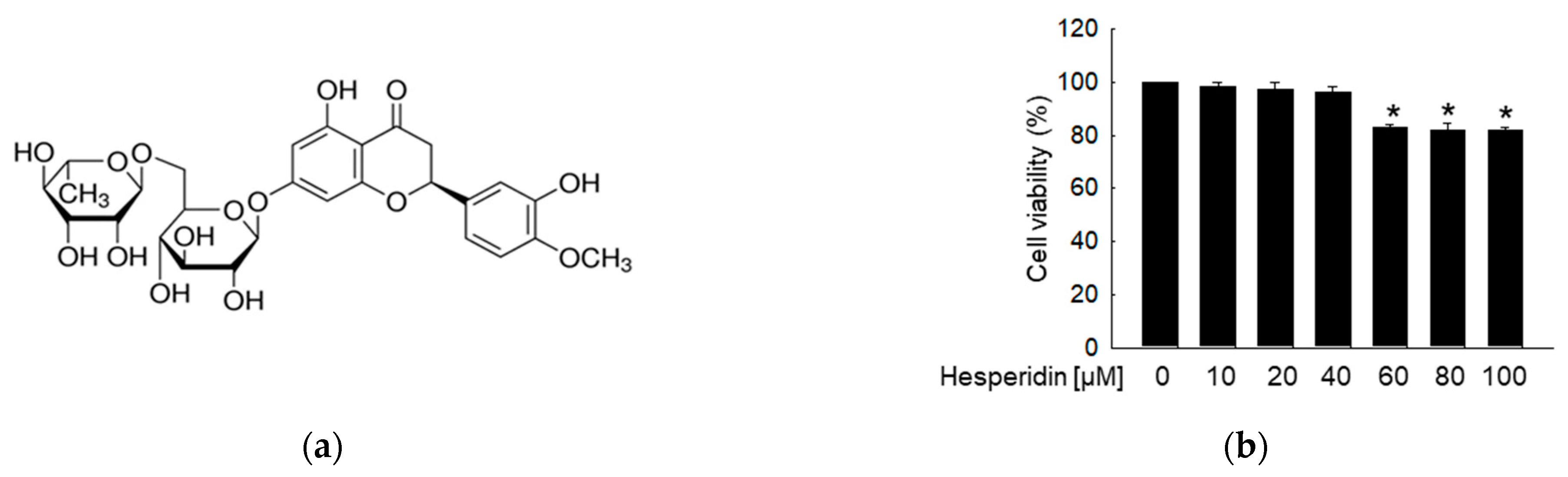
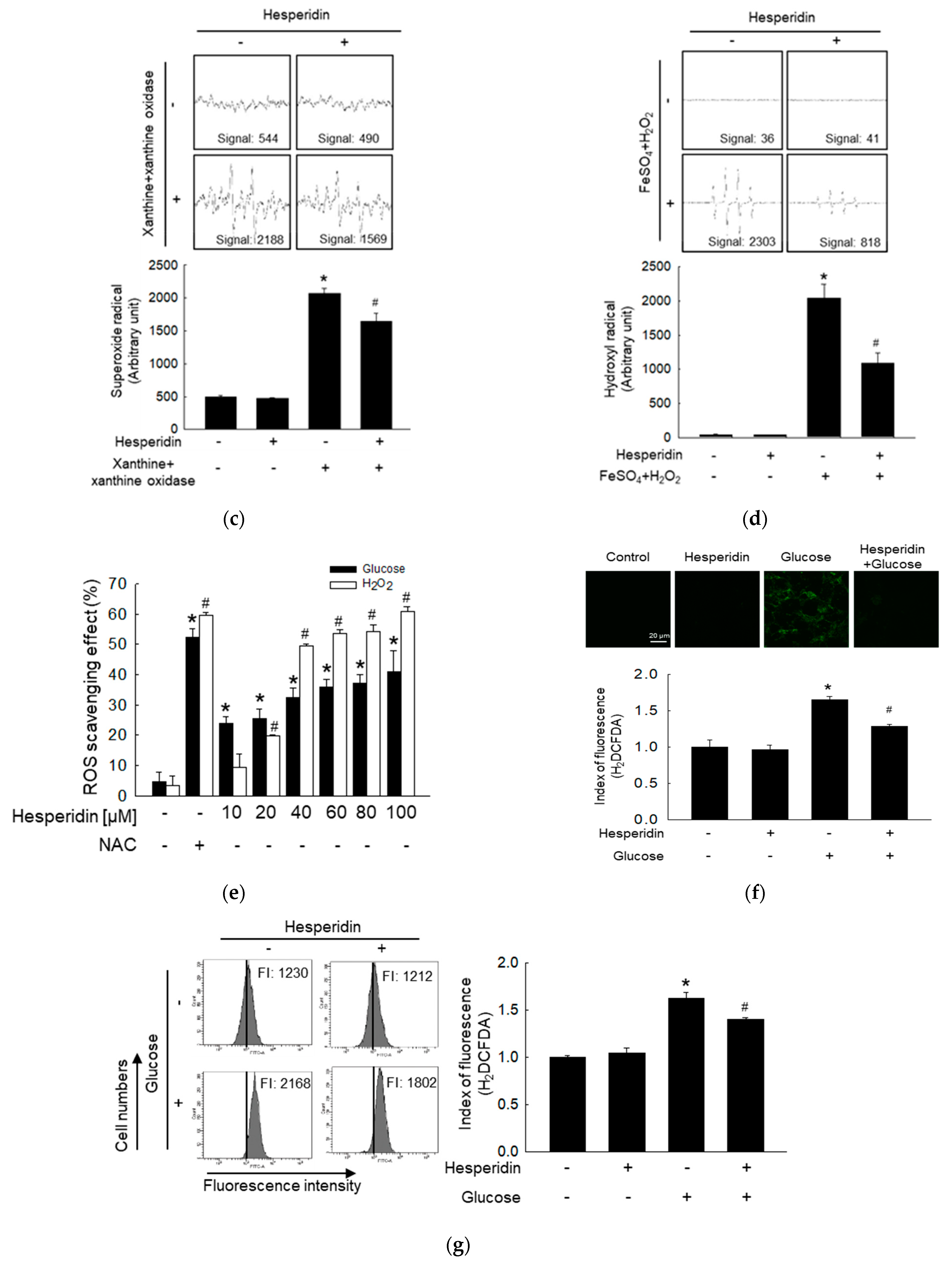
3.1. Hesperidin Protects Neuronal Cells from High Glucose−Induced Oxidative Stress

In the MTT assay, hesperidin showed no cytotoxicity at doses below 40 μM (Figure 1b). During glucose metabolism within mitochondria, ROS induced by high glucose was assessed via superoxide and hydroxyl radicals [27]. The superoxide radical scavenging ability of hesperidin was detected at 20 μM, with significant suppression of superoxide radical levels (Figure 1c). Similarly, 20 μM hesperidin significantly reduced hydroxyl radicals (Figure 1d). We then confirmed the intracellular ROS scavenging ability of hesperidin in SH−SY5Y neuronal cells, as high glucose levels, like H2O2, induced high levels of ROS, which were inhibited by hesperidin in a dose−dependent manner (Figure 1e). Based on results from the cell viability, ESR, and ROS scavenging tests, 20 μM hesperidin was found to be the optimal concentration in this study. According to a previous study on high glucose−induced cytotoxicity and apoptosis, glucose at 50 mM significantly inhibited the cell viability of SH−SY5Y and induced apoptosis after 2 days [28]. Thus, 50 mM was set as the high glucose concentration in our assays. To confirm the ROS scavenging ability of hesperidin, we subjected cells to confocal microscopy and flow cytometry analyses. Both results indicated that the high levels of ROS induced by glucose in SH−SY5Y cells were inhibited under hesperidin treatment (Figure 1f,g). Taken together, hesperidin protected neuronal cells from high glucose−induced ROS, and we selected 20 μM as the optimal concentration for subsequent cellular experiments.

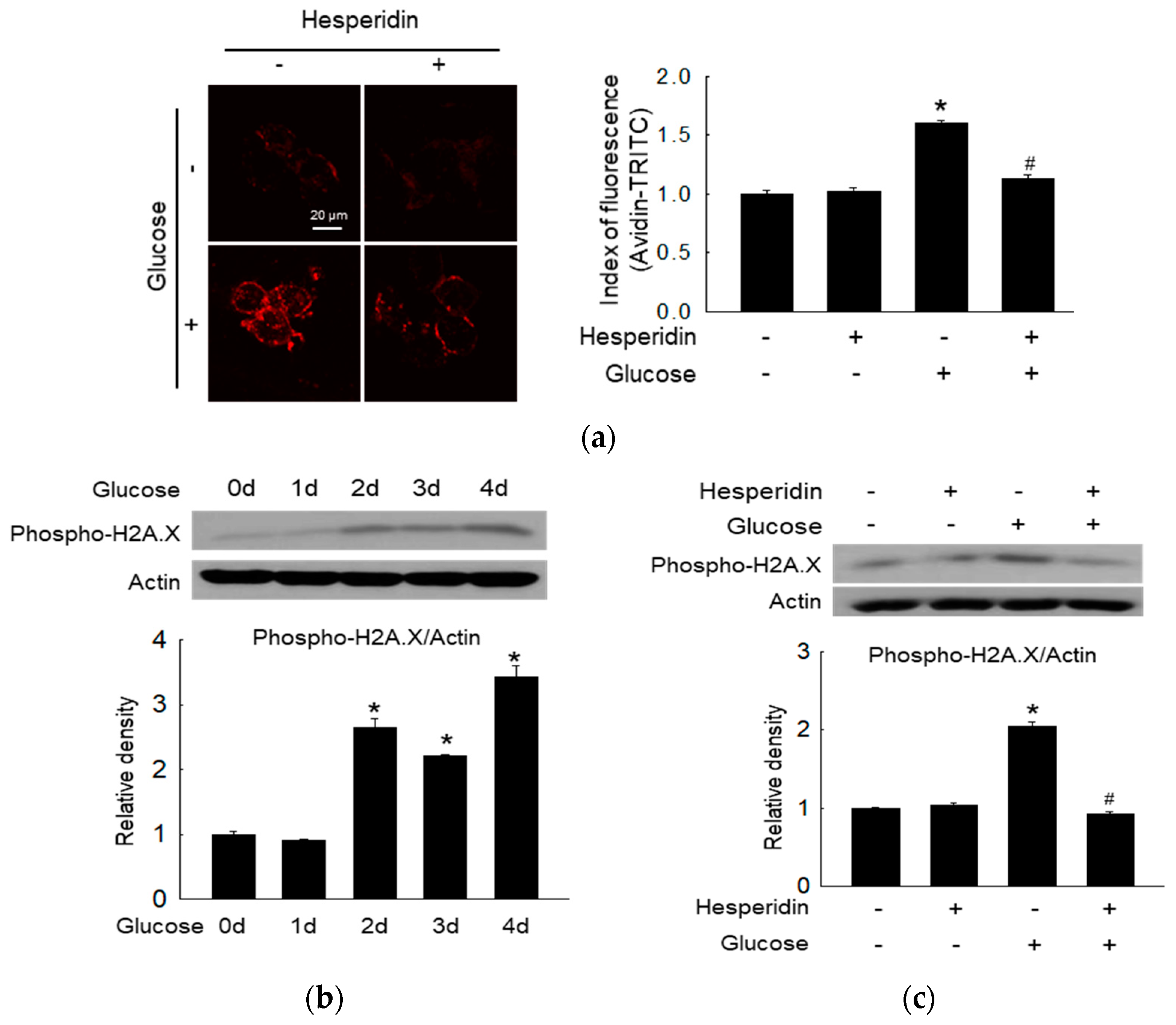
3.2. Hesperidin Ameliorated Glucose−Induced DNA Damage

Oxidative stress promotes neurodegenerative disease via the disruption of the DNA structure [29]. Glucose metabolism is associated with increased levels of 8−oxoG in cells, which hesperidin alleviated (Figure 2a). The phosphorylation of histone H2A.X, a specific DNA damage marker, was induced by glucose treatment in a time−dependent manner (Figure 2b). However, hesperidin reversed the glucose−induced upregulation of phospho−H2A.X (Figure 2c). Thus, hesperidin protected neuronal cells from glucose−induced DNA damage.

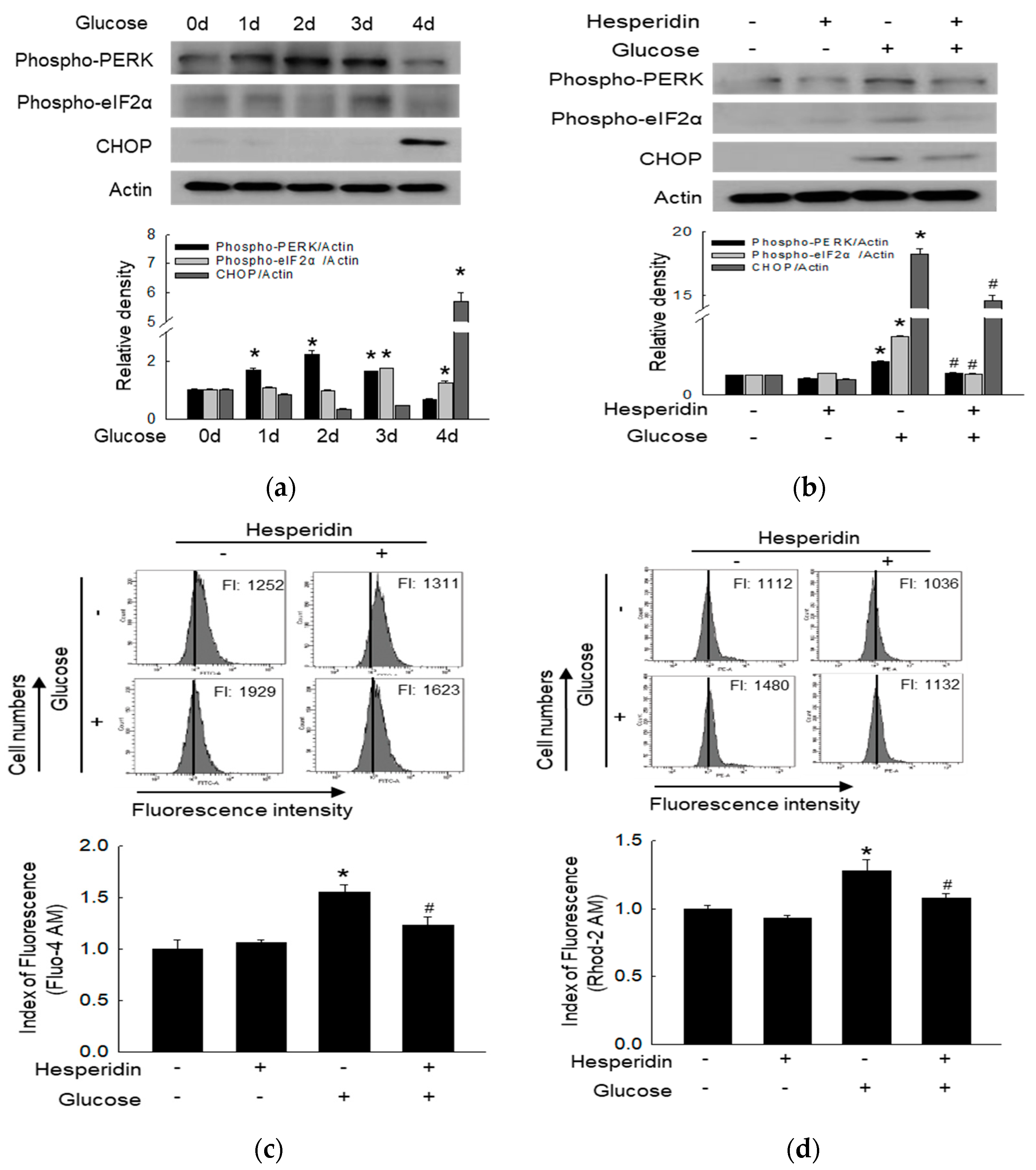
3.3. Hesperidin Suppressed Glucose−Induced ER Stress Response

ROS can also promote neuronal injury via ER stress [30]. Glucose upregulated the protein levels of phospho−PERK, phospho−eIF2α, and CHOP over time, which belong to the ER stress (Figure 3a). Hesperidin suppressed these ER stress-associated factors (Figure 3b). Cytoplasmic calcium levels were upregulated under treatment with glucose, which was once again reversed through hesperidin pretreatment (Figure 3c). Furthermore, hesperidin restored mitochondrial calcium homeostasis (Figure 3d). Taken together, hesperidin suppressed high glucose−induced ER stress and maintained cellular calcium levels.

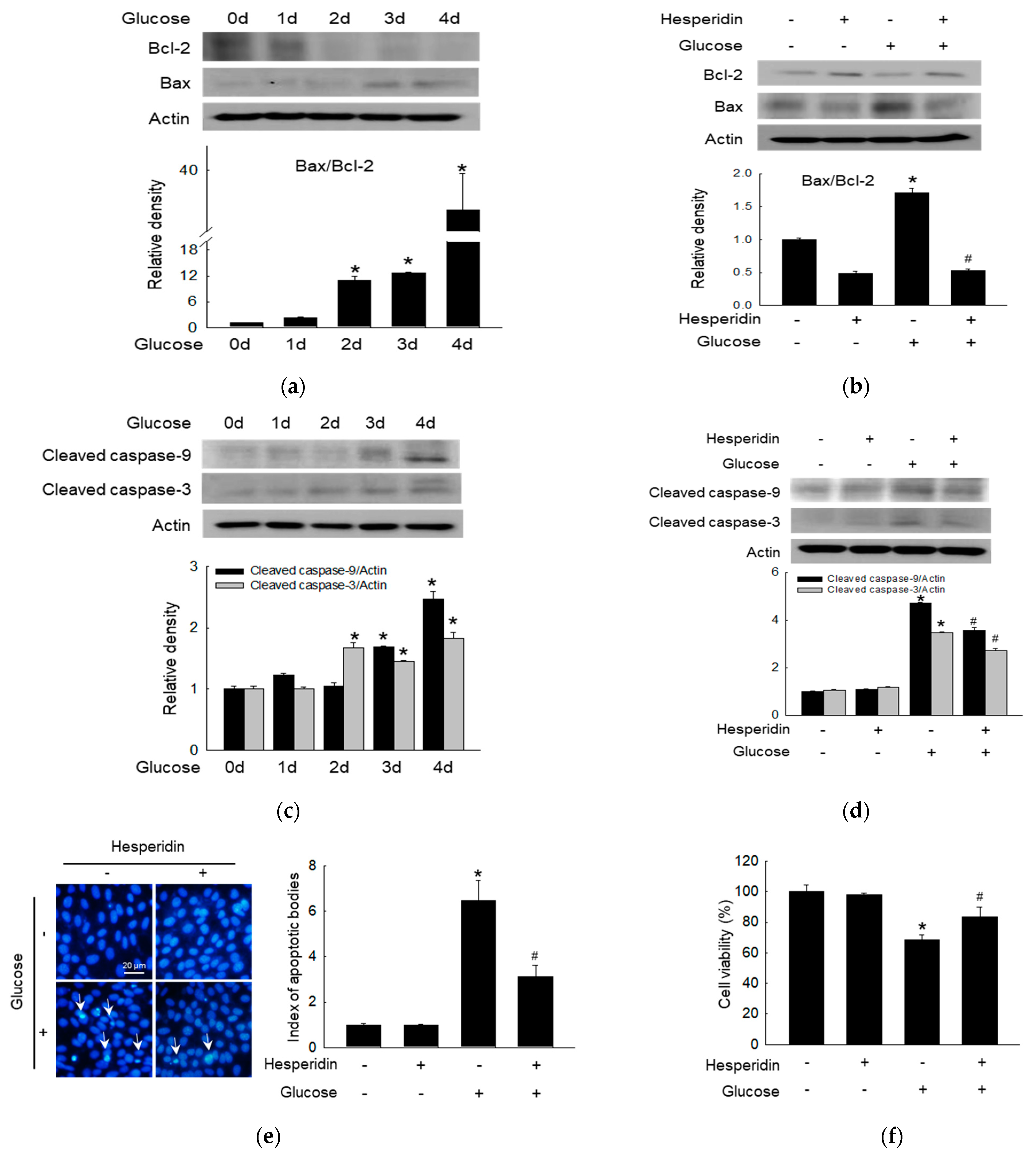
3.4. Hesperidin Protected Neuronal Cells against Glucose−Induced Apoptosis

The Bcl−2 was suppressed, while Bax was upregulated by glucose in a time−dependent manner, which represented antiapoptotic proteins and proapoptotic proteins, respectively (Figure 4a). However, hesperidin treatment reversed these expression changes (Figure 4b). Apoptosis effectors caspase−9 and caspase−3 were activated by glucose over time (Figure 4c), which was suppressed by the treatment with hesperidin (Figure 4d). The inhibition apoptotic effect of hesperidin was confirmed through the observation of glucose−induced apoptotic bodies via Hoechst 33342 staining, which was reversed by hesperidin (Figure 4e). Finally, glucose suppressed cell viability, once again reversed by hesperidin treatment (Figure 4f). Taken together, hesperidin suppressed glucose−induced neuronal apoptosis.

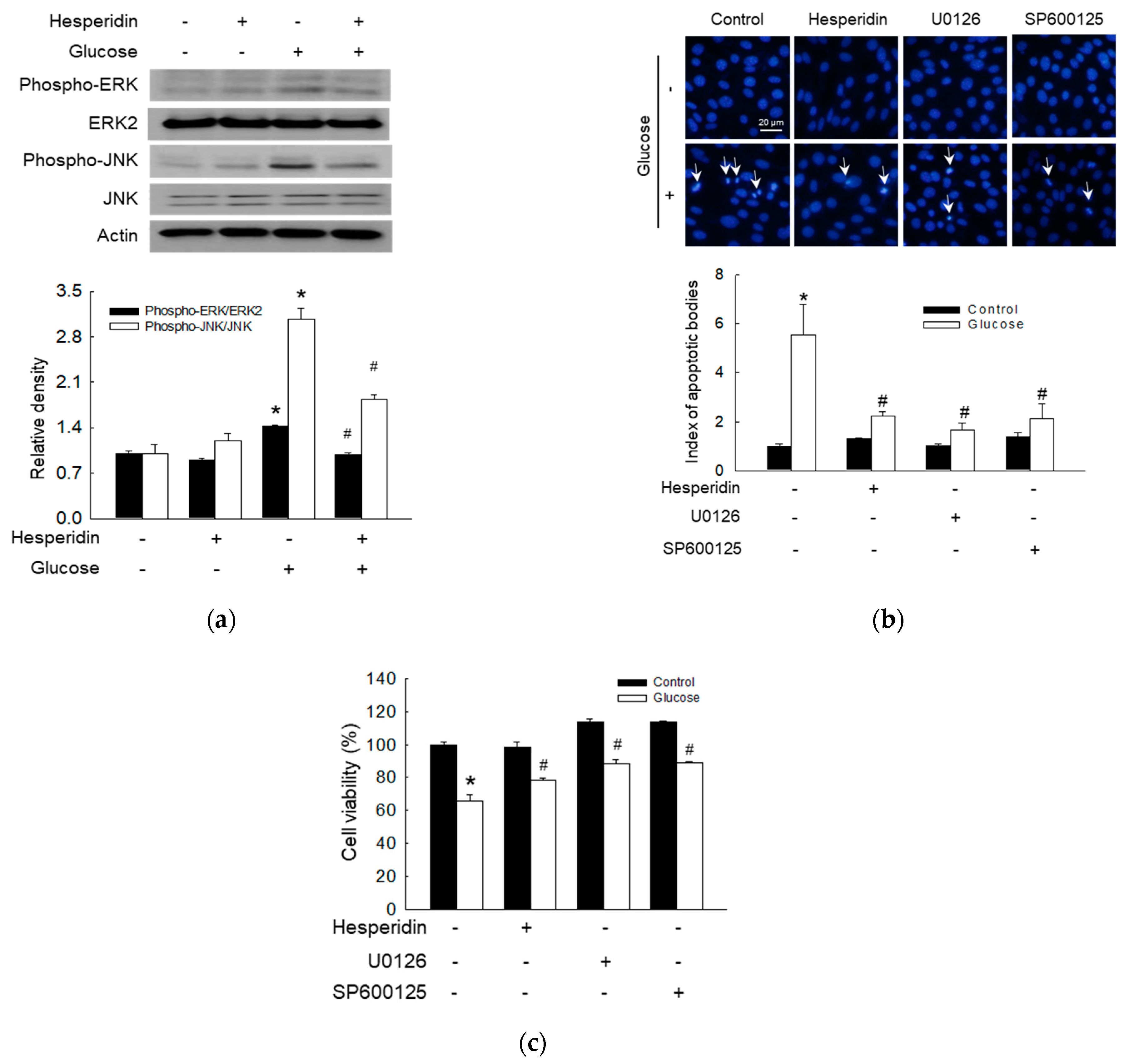
3.5. Hesperidin Protected Neuronal Cells via Attenuation of ERK/JNK Signaling Pathway

A recent study reported the activation of mitogen−activated protein kinases (MAPKs) in SH−SY5Y cells undergoing H2O2−induced apoptosis [31]. Hesperidin suppressed phospho−ERK and phospho−JNK, which were stimulated by glucose (Figure 5a). Cells cultured in the presence of glucose as well as mitogen−activated protein kinase/ERK kinase (MEK, the upstream of ERK) [31] and JNK inhibitors (U0126 and SP600125, respectively) exhibited a lower index of apoptotic bodies than those in the glucose alone group, but similar to that in the hesperidin and glucose group (Figure 5b). After treatment with MEK and JNK inhibitors (U0126 and SP600125, respectively), the glucose−induced reduction in cell viability was enhanced, yet treatment with hesperidin reversed it (Figure 5c). Taken together, hesperidin protected against apoptosis and promoted cell viability via inhibition of the ERK and JNK MAPK signaling pathways.

4. Discussion

Hyperglycemia−induced oxidative stress is a critical mechanism in the pathogenesis of diabetic neuropathy. A previous study revealed that high glucose inhibited neural cell differentiation via oxidative stress and ER stress, causing neural tube defects in maternal diabetic patients [32]. Increased oxidative stress has an adverse effect on the metabolism of the peripheral nervous system [33], promoting axon degeneration in the early stage of diabetes [34]. Interestingly, increasing doses of free radical accumulation stimulate the hermetic response (a low dose stimulation and a high dose inhibition) of the intrinsic cellular redox homeostasis system (including free radical damage and decreased energy production), resulting in neurodegeneration during the aging process, and notably, this dose−response relationship may modulate the neuroprotective effects [35,36]. Hormesis provides a novel insight into assessing the therapeutic potentiality of pharmaceutical drug candidates based on their neuroprotective abilities. Herein, to explore new treatment strategies for neurodegenerative diseases, we assessed hesperidin known for its antioxidative effects. Hesperidin has diverse biological activities, including antioxidative and anti−inflammatory effects, mediated via radical scavenging and the promotion of antioxidant defense [37]. Hesperidin was shown to alleviate retinal and plasma abnormalities in diabetic rats via inhibition of ROS [38]. In the present study, SH−SY5Y cells were cultured under high glucose to mimic nerve cells’ response to high glucose. High glucose upregulated intracellular ROS, which were significantly reduced by hesperidin treatment. In addition, ROS can trigger the cellular DNA damage response, which is intimately linked to the manifestation of neurodegenerative disorders [39]. Hesperidin was shown to modulate the high glucose−induced DNA damage response. These properties of hesperidin are expected to be beneficial against diabetic neuropathy.
Increased ROS and compromised endogenous antioxidant defense can disturb protein folding, leading to the accumulation of unfolded proteins [40]. These in turn activate PERK localized on the ER membrane by triggering its homodimerization and autophosphorylation. PERK activation leads to eIF2α phosphorylation, which in turn promotes CHOP expression. CHOP then activates the action of cytochrome c release and expression of proapoptotic proteins, such as Bax, while inhibiting antiapoptotic proteins, such as Bcl−2 [41]. Herein, we confirmed that a high glucose level upregulated the phosphorylation of PERK and eIF2α as well as its downstream signaling protein CHOP. However, hesperidin treatment significantly reduced the activation of the PERK/eIF2α/CHOP axis. Cellular calcium overload together with ROS contributes to high levels of mitochondrial calcium, which leads to cell death via mitochondrial dysfunction, observed in several neural diseases [42]. The upregulation of mitochondrial glutathione can prevent neurons from free radicals (such as nitric oxide) induced damage and has important implications for liver diseases and neurodegenerative disorders [10,43]. Moreover, the mitochondrial membrane potential is a determinant of cell apoptosis and calcium homeostasis [44]. Our results showed that hesperidin could restore calcium homeostasis, which may be beneficial for inhibiting ER stress−induced diabetic neuropathy.
High glucose−induced ROS can activate Bax and inhibit Bcl−2, triggering cell apoptosis [45]. The apoptosome complex can cleave caspase−3, one of the effector caspases, leading to the degradation of cellular proteins and subsequent apoptosis [46]. Moreover, increased cleavage of caspase−9 and caspase−3 has been detected in neurons and Schwann cells from 12−month diabetic rats under high glucose [47]. In addition, hesperidin inhibited high glucose−induced apoptosis by regulating mitochondria−related proteins, including the reduction of Bcl−2 expression, induction of Bax expression, as well as the cleavage of caspase−9 and −3 in SH−SY5Y neuronal cells. Thus, hesperidin has the potential to suppress neuronal cell death.
The MAPK pathway has been reported to participate in cell proliferation, senescence, and apoptosis [48]. The other two studies also showed that a high glucose level triggered DNA damage response and dysregulated MAPK via excessive ROS in hyperglycemia, and an unfolded protein response in ER stress could regulate phosphorylation of JNK causing apoptosis [5,49]. In this study, a high glucose level significantly increased the levels of phospho−ERK and phospho−JNK, indicating that the ERK and JNK pathways were activated. However, hesperidin treatment significantly reduced both levels. Furthermore, hesperidin, an ERK inhibitor, and a JNK inhibitor increased cell viability by suppressing apoptosis, indicating that hesperidin protects neural cells from glucose−induced cytotoxicity via attenuation of the MAPK signaling pathway.

5. Conclusions

In conclusion, hesperidin markedly inhibited high glucose−induced ROS production through its antioxidant effect in SH−SY5Y neuronal cells (Figure 6). By scavenging ROS, hesperidin effectively protected SH−SY5Y neuronal cells against oxidative injury, ER stress, and apoptosis. And hesperidin inhibited the activation of ERK and JNK, which induced by oxidative stress, and recovered MAPK signaling-reduced cell viability. Our study suggests that hesperidin is a promising biomolecular for diabetic neuropathy treatment.

Author Contributions

Conceptualization, C.L., A.X.Z. and J.W.H.; investigation, C.L., A.X.Z., S.O., P.D.S.M.F. and H.M.U.L.H.; Writing—original draft preparation, C.L. and A.X.Z.; Writing—review and editing, C.L., A.X.Z., P.D.S.M.F., H.M.U.L.H., M.J.P., K.A.K. and J.W.H. All authors have read and agreed to the published version of the manuscript.

Funding

This work was funded by a research grant from Jeju National University Hospital in 2021.

Institutional Review Board Statement

Not applicable.

Informed Consent Statement

Not applicable.

Data Availability Statement

The data presented in this study are available on request from the corresponding author.

Conflicts of Interest

The authors declare no conflict of interest.

References

  1. Armstrong, D.G.; Swerdlow, M.A.; Armstrong, A.A.; Conte, M.S.; Padula, W.V.; Bus, S.A. Five year mortality and direct costs of care for people with diabetic foot complications are comparable to cancer. J. Foot Ankle Res. 2020, 13, 16. [Google Scholar] [CrossRef] [PubMed]
  2. Koepsell, H. Glucose transporters in brain in health and disease. Pflug. Arch. Eur. J. Physiol. 2020, 472, 1299–1343. [Google Scholar]
  3. Megallaa, M.H.; Ismail, A.A.; Zeitoun, M.H.; Khalifa, M.S. Association of diabetic foot ulcers with chronic vascular diabetic complications in patients with type 2 diabetes. Diabetes Metab. Syndr. 2019, 13, 1287–1292. [Google Scholar] [CrossRef] [PubMed]
  4. Deng, L.; Du, C.; Song, P.; Chen, T.; Rui, S.; Armstrong, D.G.; Deng, W. The role of oxidative stress and antioxidants in diabetic wound healing. Oxid. Med. Cell. Longev. 2021, 2021, 8852759. [Google Scholar] [CrossRef]
  5. Bahar, E.; Kim, H.; Yoon, H. ER stress−mediated signaling: Action potential and Ca2+ as key players. Int. J. Mol. Sci. 2016, 17, 1558. [Google Scholar] [CrossRef]
  6. Rosenberger, D.C.; Blechschmidt, V.; Timmerman, H.; Wolff, A.; Treede, R.D. Challenges of neuropathic pain: Focus on diabetic neuropathy. J. Neural. Transm. 2020, 127, 589–624. [Google Scholar]
  7. Federico, A.; Cardaioli, E.; Da Pozzo, P.; Formichi, P.; Gallus, G.N.; Radi, E. Mitochondria, oxidative stress and neurodegeneration. J. Neurol. Sci. 2012, 322, 254–262. [Google Scholar]
  8. Radi, E.; Formichi, P.; Battisti, C.; Federico, A. Apoptosis and oxidative stress in neurodegenerative diseases. J. Alzheimers Dis. 2014, 42 (Suppl. 3), S125–S152. [Google Scholar] [CrossRef]
  9. Fernyhough, P.; McGavock, J. Mechanisms of disease: Mitochondrial dysfunction in sensory neuropathy and other complications in diabetes. Handb. Clin. Neurol. 2014, 126, 353–377. [Google Scholar]
  10. Drake, J.; Sultana, R.; Aksenova, M.; Calabrese, V.; Butterfield, D.A. Elevation of mitochondrial glutathione by gamma−glutamylcysteine ethyl ester protects mitochondria against peroxynitrite−induced oxidative stress. J. Neurosci. Res. 2003, 74, 917–927. [Google Scholar]
  11. Ren, J.; Lu, Y.; Qian, Y.; Chen, B.; Wu, T.; Ji, G. Recent progress regarding kaempferol for the treatment of various diseases. Exp. Ther. Med. 2019, 18, 2759–2776. [Google Scholar] [PubMed]
  12. Amić, D.; Davidović−Amić, D.; Bešlo, D.; Trinajstić, N. Structure−radical scavenging activity relationships of flavonoids. Croat. Chem. Acta 2003, 76, 55–61. [Google Scholar]
  13. Soobrattee, M.A.; Neergheen, V.S.; Luximon−Ramma, A.; Aruoma, O.I.; Bahorun, T. Phenolics as potential antioxidant therapeutic agents: Mechanism and actions. Mutat. Res. 2005, 579, 200–213. [Google Scholar] [CrossRef]
  14. Nicolle, E.; Souard, F.; Faure, P.; Boumendjel, A. Flavonoids as promising lead compounds in type 2 diabetes mellitus: Molecules of interest and structure−activity relationship. Curr. Med. Chem. 2011, 18, 2661–2672. [Google Scholar] [CrossRef] [PubMed]
  15. Ola, M.S.; Al−Dosari, D.; Alhomida, A.S. Role of oxidative stress in diabetic retinopathy and the beneficial effects of flavonoids. Curr. Pharm. Des. 2018, 24, 2180–2187. [Google Scholar] [CrossRef] [PubMed]
  16. Thenmozhi, A.J.; Raja, T.R.; Janakiraman, U.; Manivasagam, T. Neuroprotective effect of hesperidin on aluminium chloride induced Alzheimer’s disease in Wistar rats. Neurochem. Res. 2015, 40, 767–776. [Google Scholar] [CrossRef]
  17. Tamilselvam, K.; Braidy, N.; Manivasagam, T.; Essa, M.M.; Prasad, N.R.; Karthikeyan, S.; Thenmozhi, A.J.; Selvaraju, S.; Guillemin, G.J. Neuroprotective effects of hesperidin, a plant flavanone, on rotenone−induced oxidative stress and apoptosis in a cellular model for Parkinson’s disease. Oxid. Med. Cell. Longev. 2013, 2013, 102741. [Google Scholar] [CrossRef]
  18. Hajialyani, M.; Hosein Farzaei, M.; Echeverría, J.; Nabavi, S.M.; Uriarte, E.; Sobarzo−Sánchez, E. Hesperidin as a neuroprotective agent: A review of animal and clinical evidence. Molecules 2019, 24, 648. [Google Scholar] [CrossRef]
  19. Xicoy, H.; Wieringa, B.; Martens, G.J. The SH−SY5Y cell line in Parkinson’s disease research: A systematic review. Mol. Neurodegener. 2017, 12, 10. [Google Scholar]
  20. Kim, H.; Choi, J.; Lee, H.; Park, J.; Yoon, B.I.; Jin, S.M.; Park, K. Skin corrosion and irritation test of nanoparticles using reconstructed three−dimensional human skin model, EpiDermTM. Toxicol. Res. 2016, 32, 311–316. [Google Scholar]
  21. Cha, J.W.; Piao, M.J.; Kim, K.C.; Yao, C.W.; Zheng, J.; Kim, S.M.; Hyun, C.L.; Ahn, Y.S.; Hyun, J.W. The polyphenol chlorogenic acid attenuates UVB−mediated oxidative stress in human HaCaT keratinocytes. Biomol. Ther. 2014, 22, 136–142. [Google Scholar] [CrossRef] [PubMed]
  22. Zhen, A.X.; Piao, M.J.; Hyun, Y.J.; Kang, K.A.; Ryu, Y.S.; Cho, S.J.; Kang, H.K.; Koh, Y.S.; Ahn, M.J.; Kim, T.H.; et al. Purpurogallin protects keratinocytes from damage and apoptosis induced by ultraviolet B radiation and particulate matter 2.5. Biomol. Ther. 2019, 27, 395–403. [Google Scholar] [CrossRef] [PubMed]
  23. Piao, M.J.; Ahn, M.J.; Kang, K.A.; Ryu, Y.S.; Hyun, Y.J.; Shilnikova, K.; Zhen, A.X.; Jeong, J.W.; Choi, Y.H.; Kang, H.K.; et al. Particulate matter 2.5 damages skin cells by inducing oxidative stress, subcellular organelle dysfunction, and apoptosis. Arch. Toxicol. 2018, 92, 2077–2091. [Google Scholar] [PubMed]
  24. Wang, H.; Zhang, J.; Hu, S.H.; Tan, S.Z.; Zhang, B.; Zhou, H.M.; Peng, R.Y. Real−time microwave exposure induces calcium efflux in primary hippocampal neurons and primary cardiomyocytes. Biomed. Environ. Sci. 2018, 31, 561–571. [Google Scholar] [PubMed]
  25. Hyun, Y.J.; Piao, M.J.; Kang, K.A.; Zhen, A.X.; Fernando, P.D.S.M.; Kang, H.K.; Ahn, Y.S.; Hyun, J.W. Effect of fermented fish oil on fine particulate matter−induced skin aging. Mar. Drugs 2019, 17, 61. [Google Scholar] [CrossRef] [PubMed]
  26. Cho, S.J.; Kang, K.A.; Piao, M.J.; Ryu, Y.S.; Fernando, P.D.S.M.; Zhen, A.X.; Hyun, Y.J.; Ahn, M.J.; Kang, H.K.; Hyun, J.W. 7,8−Dihydroxyflavone protects high glucose−damaged neuronal cells against oxidative stress. Biomol. Ther. 2019, 27, 85–91. [Google Scholar] [CrossRef]
  27. Nishikawa, T.; Edelstein, D.; Du, X.L.; Yamagishi, S.; Matsumura, T.; Kaneda, Y.; Yorek, M.A.; Beebe, D.; Oates, P.J.; Hammes, H.P.; et al. Normalizing mitochondrial superoxide production blocks three pathways of hyperglycaemic damage. Nature 2000, 404, 787–790. [Google Scholar] [CrossRef]
  28. Liu, Y.; Sun, L.; Ma, Y.; Wei, B.; Gao, M.; Shang, L. High glucose and bupivacaine-induced cytotoxicity is mediated by enhanced apoptosis and impaired autophagy via the PERK-ATF4-CHOP and IRE1-TRAF2 signaling pathways. Mol. Med. Rep. 2019, 20, 2832–2842. [Google Scholar] [CrossRef]
  29. Singh, A.; Kukreti, R.; Saso, L.; Kukreti, S. Oxidative stress: A key modulator in neurodegenerative diseases. Molecules 2019, 24, 1583. [Google Scholar] [CrossRef]
  30. Chen, T.; Zhu, J.; Wang, Y.H.; Hang, C.H. ROS−mediated mitochondrial dysfunction and ER stress contribute to compression−induced neuronal injury. Neuroscience 2019, 416, 268–280. [Google Scholar] [CrossRef]
  31. Sugimoto, M.; Ko, R.; Goshima, H.; Koike, A.; Shibano, M.; Fujimori, K. Formononetin attenuates H2O2−induced cell death through decreasing ROS level by PI3K/Akt−Nrf2−activated antioxidant gene expression and suppressing MAPK−regulated apoptosis in neuronal SH−SY5Y cells. Neurotoxicology 2021, 85, 186–200. [Google Scholar] [CrossRef] [PubMed]
  32. Chen, X.; Shen, W.B.; Yang, P.; Dong, D.; Sun, W.; Yang, P. High glucose inhibits neural stem cell differentiation through oxidative stress and endoplasmic reticulum stress. Stem. Cells Dev. 2018, 27, 745–755. [Google Scholar] [CrossRef] [PubMed]
  33. Bhatti, J.S.; Sehrawat, A.; Mishra, J.; Sidhu, I.S.; Navik, U.; Khullar, N.; Kumar, S.; Bhatti, G.K.; Reddy, P.H. Oxidative stress in the pathophysiology of type 2 diabetes and related complications: Current therapeutics strategies and future perspectives. Free Radic. Biol. Med. 2022, 184, 114–134. [Google Scholar] [PubMed]
  34. Cheng, Y.; Liu, J.; Luan, Y.; Liu, Z.; Lai, H.; Zhong, W.; Yang, Y.; Yu, H.; Feng, N.; Wang, H.; et al. Sarm1 gene deficiency attenuates diabetic peripheral neuropathy in mice. Diabetes 2019, 68, 2120–2130. [Google Scholar] [CrossRef]
  35. Calabrese, V.; Cornelius, C.; Dinkova−Kostova, A.T.; Calabrese, E.J.; Mattson, M.P. Cellular stress responses, the hormesis paradigm, and vitagenes: Novel targets for therapeutic intervention in neurodegenerative disorders. Antioxid. Redox Signal. 2010, 13, 1763–1811. [Google Scholar]
  36. Perluigi, M.; Di Domenico, F.; Giorgi, A.; Schininà, M.E.; Coccia, R.; Cini, C.; Bellia, F.; Cambria, M.T.; Cornelius, C.; Butterfield, D.A.; et al. Redox proteomics in aging rat brain: Involvement of mitochondrial reduced glutathione status and mitochondrial protein oxidation in the aging process. J. Neurosci. Res. 2010, 88, 3498–3507. [Google Scholar] [CrossRef]
  37. Parhiz, H.; Roohbakhsh, A.; Soltani, F.; Rezaee, R.; Iranshahi, M. Antioxidant and anti−inflammatory properties of the citrus flavonoids hesperidin and hesperetin: An updated review of their molecular mechanisms and experimental models. Phytother. Res. 2015, 29, 323–331. [Google Scholar]
  38. Shi, X.; Liao, S.; Mi, H.; Guo, C.; Qi, D.; Li, F.; Zhang, C.; Yang, Z. Hesperidin prevents retinal and plasma abnormalities in streptozotocin−induced diabetic rats. Molecules 2012, 17, 12868–12881. [Google Scholar] [CrossRef]
  39. Madabhushi, R.; Pan, L.; Tsai, L.H. DNA damage and its links to neurodegeneration. Neuron 2014, 83, 266–282. [Google Scholar]
  40. O’Brien, P.D.; Hinder, L.M.; Sakowski, S.A.; Feldman, E.L. ER stress in diabetic peripheral neuropathy: A new therapeutic target. Antioxid. Redox Signal. 2014, 21, 621–633. [Google Scholar] [CrossRef]
  41. Cheng, W.C.; Wen, Y.; Chiu, Y.S.; Chou, C.H.; Lim, C.J.; Lin, S.H.; Chang, J.M.; Lin, C.C. Pendulone induces apoptosis via the ROS−mediated ER−stress pathway in human non−small cell lung cancer cells. Toxicol. Vitr. 2022, 81, 105346. [Google Scholar] [CrossRef] [PubMed]
  42. Godoy, J.A.; Rios, J.A.; Picón−Pagès, P.; Herrera−Fernández, V.; Swaby, B.; Crepin, G.; Vicente, R.; Fernández−Fernández, J.M.; Muñoz, F.J. Mitostasis, calcium and free radicals in health, aging and neurodegeneration. Biomolecules 2021, 11, 1012. [Google Scholar] [CrossRef] [PubMed]
  43. Mancuso, C.; Pani, G.; Calabrese, V. Bilirubin: An endogenous scavenger of nitric oxide and reactive nitrogen species. Redox Rep. 2006, 11, 207–213. [Google Scholar] [CrossRef] [PubMed]
  44. Calabrese, V.; Mancuso, C.; Calvani, M.; Rizzarelli, E.; Butterfield, D.A.; Stella, A.M. Nitric oxide in the central nervous system: Neuroprotection versus neurotoxicity. Nat. Rev. Neurosci. 2007, 8, 766–775. [Google Scholar] [CrossRef] [PubMed]
  45. Glory, A.; Bettaieb, A.; Averill−Bates, D.A. Mild thermotolerance induced at 40°C protects cells against hyperthermia−induced pro−apoptotic changes in Bcl−2 family proteins. Int. J. Hyperth. 2014, 30, 502–512. [Google Scholar] [CrossRef]
  46. Eskandari, E.; Eaves, C.J. Paradoxical roles of caspase−3 in regulating cell survival, proliferation, and tumorigenesis. J. Cell Biol. 2022, 221, e202201159. [Google Scholar]
  47. Cheng, C.; Zochodne, D.W. Sensory neurons with activated caspase−3 survive long−term experimental diabetes. Diabetes 2003, 52, 2363–2371. [Google Scholar] [CrossRef]
  48. Su, Z.; Han, S.; Jin, Q.; Zhou, N.; Lu, J.; Shangguan, F.; Yu, S.; Liu, Y.; Wang, L.; Lu, J.; et al. Ciclopirox and bortezomib synergistically inhibits glioblastoma multiforme growth via simultaneously enhancing JNK/p38 MAPK and NF−κB signaling. Cell Death Dis. 2021, 12, 251. [Google Scholar] [CrossRef]
  49. Wang, K.; Fu, X.Y.; Fu, X.T.; Hou, Y.J.; Fang, J.; Zhang, S.; Yang, M.F.; Li, D.W.; Mao, L.L.; Sun, J.Y.; et al. DSePA antagonizes high glucose−induced neurotoxicity: Evidences for DNA damage−mediated p53 phosphorylation and MAPKs and AKT pathways. Mol. Neurobiol. 2016, 53, 4363–4374. [Google Scholar] [CrossRef]
Figure 1. Hesperidin protected SH−SY5Y neuronal cells against high glucose−induced oxidative stress. (a) Chemical structure of hesperidin. (b) Cell viability was assessed using the MTT assay. * p < 0.05 compared to control cells. (c) The superoxide radical was examined via ESR. * p < 0.05, # p < 0.05 compared to control and xanthine + xanthine oxidase, respectively. (d) The hydroxyl radical was examined via ESR. * p < 0.05, # p < 0.05 compared to control and FeSO4 + H2O2, respectively. (e) The ROS were determined using spectrometry after H2DCFDA staining. * p < 0.05, # p < 0.05 compared to glucose− and H2O2−exposed cells, respectively. (f,g) The ROS were analyzed using confocal microscopy and flow cytometry after H2DCFDA staining. * p < 0.05, # p < 0.05 compared to control cells and glucose−exposed cells, respectively.
Antioxidants 11 01707 g001aAntioxidants 11 01707 g001b
Figure 2. Hesperidin relieved high glucose−induced DNA damage in SH−SY5Y cells. (a) 8−OxoG was assessed using confocal microscopy after staining of avidin−TRITC. (b,c) Phospho−H2A.X was measured via western blotting. Actin was regarded as a loading control. The ‘d’ stands for ‘day’ after cells were treated with glucose. * p < 0.05 and # p < 0.05 compared to control cells and glucose−exposed cells, respectively.
Antioxidants 11 01707 g002
Figure 3. Hesperidin mitigated high glucose−induced ER stress in SH−SY5Y neuronal cells. (a,b) Phospho−PERK, phospho−eIF2α, and CHOP were detected via western blot. (c) ER calcium levels were analyzed by Fluo−4 AM dye. (d) Mitochondrial calcium levels were measured by Rhod−2 AM dye. * p < 0.05 and # p < 0.05 compared to control cells and glucose−exposed cells, respectively.
Antioxidants 11 01707 g003
Figure 4. Hesperidin protected SH−SY5Y neuronal cells against high glucose−induced apoptosis. (a,b) Bcl−2 and Bax were detected via western blot. (c,d) Cleaved caspase−9 and cleaved caspase−3 were detected via western blot. (e) Hoechst 33342 dye was used for the observation of apoptotic bodies. (f) Cell viability was detected via MTT assays. * p < 0.05 and # p < 0.05 compared to control cells and glucose−exposed cells, respectively.
Antioxidants 11 01707 g004
Figure 5. Hesperidin protected SH−SY5Y neuronal cells against high glucose−induced cell death via the ERK and JNK signaling pathways. (a) Phospho−ERK, ERK2, phospho−JNK, and JNK were measured via western blot. (b) Apoptotic bodies were detected using Hoechst 33342. Arrow indicates apoptotic body. (c) Cell viability was detected via MTT assay. * p < 0.05 and # p < 0.05 compared to control cells and glucose−exposed cells, respectively.
Antioxidants 11 01707 g005
Figure 6. Schematic diagram of hesperidin’s protective effects against high glucose−induced SH−SY5Y neuronal cell death. Glucose−induced ROS in neuronal cells cause DNA damage and ER stress, in turn triggering apoptosis. However, hesperidin suppresses ROS levels and protects cells from apoptosis through inhibition of the MAPK signaling pathway.
Antioxidants 11 01707 g006
Publisher’s Note: MDPI stays neutral with regard to jurisdictional claims in published maps and institutional affiliations.

