Article

Antimicrobial Peptides (AMP) in the Cell-Free Culture Media of Xenorhabdus budapestensis and X. szentirmaii Exert Anti-Protist Activity against Eukaryotic Vertebrate Pathogens including Histomonas meleagridis and Leishmania donovani Species

1
Department of Genetics, Institute of Biology, Eötvös Loránd University, Pázmány Péter. sétány 1C, H-1117 Budapest, Hungary
2
Clinic for Poultry and Fish Medicine, Department for Farm Animals and Veterinary Public Health, University of Veterinary Medicine (Vetmeduni Vienna), 1210 Vienna, Austria
3
Agribiotechnology and Precision Breeding for Food Security National Laboratory, Department of Microbiology and Applied Biotechnology, Institute of Genetics and Biotechnology, Hungarian University of Agriculture and Life Sciences, Páter Károly utca 1, H-2100 Gödöllő, Hungary
4
Autovakcina Kft., H-1171 Budapest, Hungary
5
Institute of Physiology and Nutrition, Georgikon Campus, Hungarian University of Agriculture and Life Sciences (MATE), Deák Ferenc utca 16, H-8360 Keszthely, Hungary
6
Department of Microbiology and Infectious Diseases, University of Veterinary Medicine, H-1143 Budapest, Hungary
7
First Department of Pathology and Experimental Cancer Research, Semmelweis University, H-1085 Budapest, Hungary
8
USDA-ARS & Department of Entomology, The Ohio State University, 13416 Claremont Ave, Cleveland, OH 44130, USA
9
Department of Soil, Plant and Food Sciences, University of Bari “Aldo Moro”, Via Amendola 165/A, 70126 Bari, Italy
10
Division of Infectious Diseases, Department of Internal Medicine, The Ohio State University, Columbus, OH 43210, USA
*
Authors to whom correspondence should be addressed.
Antibiotics 2023, 12(9), 1462; https://doi.org/10.3390/antibiotics12091462
Submission received: 31 July 2023 / Revised: 1 September 2023 / Accepted: 5 September 2023 / Published: 19 September 2023
(This article belongs to the Special Issue Potential of Antimicrobial Peptides for an Exciting Future)

Abstract

:
Anti-microbial peptides provide a powerful toolkit for combating multidrug resistance. Combating eukaryotic pathogens is complicated because the intracellular drug targets in the eukaryotic pathogen are frequently homologs of cellular structures of vital importance in the host organism. The entomopathogenic bacteria (EPB), symbionts of entomopathogenic–nematode species, release a series of non-ribosomal templated anti-microbial peptides. Some may be potential drug candidates. The ability of an entomopathogenic–nematode/entomopathogenic bacterium symbiotic complex to survive in a given polyxenic milieu is a coevolutionary product. This explains that those gene complexes that are responsible for the biosynthesis of different non-ribosomal templated anti-microbial protective peptides (including those that are potently capable of inactivating the protist mammalian pathogen Leishmania donovanii and the gallinaceous bird pathogen Histomonas meleagridis) are co-regulated. Our approach is based on comparative anti-microbial bioassays of the culture media of the wild-type and regulatory mutant strains. We concluded that Xenorhabdus budapestensis and X. szentirmaii are excellent sources of non-ribosomal templated anti-microbial peptides that are efficient antagonists of the mentioned pathogens. Data on selective cytotoxicity of different cell-free culture media encourage us to forecast that the recently discovered “easy-PACId” research strategy is suitable for constructing entomopathogenic-bacterium (EPB) strains producing and releasing single, harmless, non-ribosomal templated anti-microbial peptides with considerable drug, (probiotic)-candidate potential.

1. Introduction

Anti-microbial peptides (AMPs) are any polyamide, or even their bio-co-polymer with esters, thioesters, or otherwise modified backbone, that can be made on a contemporary chemical peptide synthesizer [1]. This provides an option of taking advantage of using the remarkable toolkit of quantitative structure–activity relation (QSAR) [2,3] in designing novel anti-microbial-active sister molecules of any newly discovered natural drug-candidate AMP molecule. As for their biosynthesis, peptides of anti-microbial potential can be either ribosomal encoded (RP) single gene products [4] or enzymatically biosynthesized non-ribosomal templated peptides (NRP) [5], (NR-AMPs) [6]. Considering that there are already >100 peptide-based drugs that have been of clinical use (see [7]), an lookout at that field may be advisable. As for history, we refer to the review of Zasloff [8], who outlined those discoveries that can be considered as milestones, like cecropins [9], defensins [10,11], maganins [12], and add the proline-rich AMP (PrAMP family) [13,14] to the still incomplete list. Many derivatives of discovered natural AMPs have been chemically synthesized and or modified, and there are several excellent reviews [15,16,17,18,19] beyond the scope of this paper.
The two selected targets here are Leishmania [20], a mammalian pathogen, transmittable by insects like cave-dwelling sand flies [21], and Histomonas meleagridis, the etiological agent of the life-threatening histomonosis (syn. blackhead disease) of poultry species such as turkeys, chickens, and pheasants, first described in 1893 [22,23], and have been studied in detail until the mid of last century [24].
In the actual study we aimed to investigate if any of those biosynthetic NR-AMPs, like fabclavine [25,26], phenazine [27], and others, produced by entomopathogenic–nematode symbiont bacteria (EPB) we have been studying [28,29] were suitable as anti-protozoal drug candidates against the intracellular parasite Leishmania or the extracellular parasite H. meleagridis. The arguments for choosing in vitro EPB liquid cultures as appropriate sources of efficient AMPs for this purpose are that obligate bacterial symbionts of entomopathogenic–nematode species synthesize and release several non-ribosomal hybrid peptides [30] with large target specificities. These mainly serve to provide well-balanced pathobiome conditions for this symbiosis in polyxenic soil and cadaver environments [31]. These peptides are also considered potential sources of potent natural anti-microbial compounds [30]. The motivation for choosing in vitro EPB liquid cultures as a source of efficient AMPs for this purpose is the proven activity on Gram-positive [32,33] and Gram-negative [34] pathogenic bacteria, oomycetes, and different plant pathogens [35,36] in our previous studies. Since we have reasons to believe that some of the autoclaved Xenorhabdus cultures may not be harmful or toxic when added as a food supplement, while some anti-microbial ingredients retain active [6], we consider the potential for probiotic applications, at least on an experimental level.
The eukaryotic pathogens studied here include the Leishmania donovanii species, kinetoplastid protozoa [37], and the causative agent of human and canine leishmaniasis [38,39,40].
Leishmania are intracellular parasites that target professional phagocytes (macrophages and dendritic cells) [41] and human infection leads to diverse forms of disease from singular or diffuse cutaneous lesions (cutaneous leishmaniasis), invasive destruction of mucous membranes (mucocutaneous leishmaniasis), or dissemination to the liver, spleen, and bone marrow (visceral leishmaniasis) [42,43]. While mainly relegated to endemic foci in poor rural areas in the third world, the infections have a global impact due to human migration, climate change, and anthropogenic disturbance, causing significant worldwide health and economic burdens [44]. There are currently no approved vaccines available [45], leaving control of leishmaniasis reliant on chemotherapy [46]. The mainstays of therapy are antimonial derivatives [47] of amphotericin B (AmB), a polyene macrolide antibiotic derived from actinomycetes [48], which can be renally toxic and are not widely available in poor rural areas. Increasingly, there are issues with leishmanial infections that are resistant or refractory to antimony and amphotericin therapy. Miltefosine is the first oral agent approved for the treatment of leishmaniasis; however, there have been descriptions of miltefosine resistance [46]. There is an obvious need for the development of newer, less toxic agents to combat these infections.
The other chosen pathogen, H. meleagridis, belongs to the Dientamoebidae, order Tritrichomanidida. Infections in turkeys may cause nearly 100% mortality whereas outbreaks in chickens, peafowl, quail, and pheasants are more often marked by morbidity and subsequent recovery. H. meleagridgridis is carried by the eggs of the cecal worm Heterakis gallinarum, enabling them to survive for long periods in the soil as a source of infection [22,23,24]. In the EU and USA, there are currently no drugs available for the treatment of blackhead disease [22,24]. (As for molecular taxonomy and identification, the 5.8S, ITS-1, and ITS-2 rRNA regions were first sequenced [49]. Since then, the complete, annotated Histomonas reference genome sequence became available [50]).
Since the introduction of effective drugs into the market in the middle of the previous century, there was no practical need for further research. Following the recent ban of available drugs, research programs with new profiles were set up in various places focusing on different features of the parasite and the disease.
Consequently, the poultry industry works without approved prophylactics, therapeutics, or vaccines to combat histomonosis [51].
Recently, numerous chemical and botanical compounds were also tested for their efficacy against H. meleagridis, with varying outcomes. One of the explanations for these half-failures/half-successes may be the complicated in vitro Histomonas culturing technique. In this context, it is of high importance that H. meleagridis relies on live bacteria and Escherichia coli strongly supports the growth of H. meleagridis in a monoxenic culture without influencing its pathogenicity [52]. In the previously mentioned study, a clonal cultures of H. meleagridis was further optimized to obtain a monoxenic culture in a liquid medium [52]. For this, the fecal flora was exchanged for defined bacterial strains by selective destruction of the initial bacteria with various antibiotics, keeping the flagellate alive. E. coli was found to strongly support the growth of the parasite, whereas Salmonella enterica serovar Typhimurium and Pseudomonas aeruginosa were less efficient. Whether the special feature of the parasite’s intricate interplay with bacteria in vitro and in vivo can be considered mutualistic or a predator-prey one is an open question [50,51,52,53,54,55,56,57,58,59,60]. (We are shared about it). The option of the monoxenic culturing technique may open a door to preliminary attempts to protect birds with cultured attenuated histomonads, forecasting the possibility of vaccination [56].
After banning all previously used prophylactic and therapeutic drugs against H. meleagridis in the USA and the European Union, benzimidazoles were tested but found inactive [61]. Similar half-successes were reported about various herbal substances, some of which showed good efficacy in vitro but failed in vivo [62]. Reduced sensitivity of H. meleagridis to nitarsone in vitro and in vivo was also shown [63].
A publication talked about that, identical monoxenic settings for cultures of the same H. meleagridis clonal strain, in its virulent low-passage and attenuated high-passage form, enabled a comparative analysis of parasite characteristics [64], but the conclusions severely contradict a previously cited, unambiguously reliable publication [59]. This debate is out of scope of the present paper.
Herein, we compared the spectrum of in vitro anti-microbial/-prozozoal activity of cell-free conditioned culture media (CFCM) from three EPB species, together with testing against Leishmania donovanii, (together with Trypanosoma cruzi, as an expectedly negative control), Histomonas meleagridis, and also against a panel of clinical bacterial and fungal isolates, as positive controls. Considering the roles of the bacterial symbiont in the EPN/EPB symbiosis, we suppose that ani-protozoal compounds must coexist with other anti-microbial compounds in the CFCM of EPBs. Therefore, for reliable bioassays, we should use appropriate controls. Two main aspects should be taken into consideration: (A) To be able to distinguish between the specified antiprotist activity and general cytotoxicity of the future drug-candidate anti-microbial compounds in the CFCM. (B) To be able to identify the special antiprotist compounds. In this paper, we describe the very first steps toward these goals. For (A), one has to consider that the lack of uniformity regarding the choice of cell types for cytotoxicity assays may lead to incomparable and inconclusive data. In vitro assays relying solely on non-phagocytic cell models may not represent a realistic result, as the effect of an anti-leishmanial agent should ideally be presented based on its cytotoxicity profile against reticuloendothelial system cells [65]. In the Leishmania studies, we used the macrophage cell line J774A.1 [66,67,68], as a cytopathogenicity control test organism (see Section 2 Materials and Methods). In the Histomonas studies, we used the permanent chicken liver cells (LMH leghorn male hepatoma) cell line [6,69,70] as a cytopathogenicity control test organism (see Section 2 Materials and Methods). For (B), one has to take into consideration the following points: each NR-AMP-producing EPB cell is in the so-called primary (10) phase, or Phase 1 [71]. In other terms, the phenotypic precondition for antibiotic production (as well as the capability of symbiosis) of the EPB species is the primary phase [30,72,73]. For NR-AMP production, it means that in a liquid culture of the primary (10) cells (of each of the studied EPB species, strain, and isolate), more than one biosynthetic AMP is present simultaneously. Each expresses anti-microbial (maybe antiprotist) activity, so the anti-microbial (antiprotist) activity of a CFCM sample represents a cumulative activity. However, in an optimal test system, one would prefer to determine the anti-microbial activity of each of the single anti-microbial compounds one by one.
Since the discovery that the activity (the “switch-on”/“switch-off” states) of those group genes (operons, biosynthetic gene complexes, and BGCs), which are responsible for the primary-secondary phenotypic phase shift in each known EPB strain, not only work under the control of the versatile regulator hexA [74,75], but are also coordinately regulated at a higher level (via hfq-gene controlled sRNS/HexA-mRNA base pairing). More precisely, the Hfq-dependent sRNA, ArcZ, directly base-pairs with the HexA-encoding mRNA [76,77,78]; it is possible to construct double-mutant strains from each EPB species (or isolate) following the genuine strategy called easyPACId (easy promoter-activated compound identification) approach [79]. Each biosynthesizes and releases only one single NR-AMP molecule into the culture media. This discovery may be a starting point of the renaissance of Xenorhabdus anti-microbial peptide research [76,77,78,79,80,81,82,83]. We managed to reconstruct the “easyPACId” hfq deletion mutant strains from EMA and EMC (Boros et al., in preparation) and publish here some information, including data about their anti-microbial potential. In this paper, we give an account of the details of constructing our hfq-del mutants from our EMA and EMC [29] strains and provide their phenotypic descriptions. Our “easyPACId” strains will be available for cooperation with fellow scientists worldwide.

2. Materials and Methods

The plasmids [84,85,86,87,88,89,90,91,92] used in this study [77] are listed in Table A1 in Appendix A. Several different entomopathogenic bacteria (EPB) species were used as producers of anti-microbial compounds. Various clinical isolates of Gram-positive and Gram-negative bacteria served as test organisms, as did eukaryotic organisms (protozoa, fungi, and murine macrophages). Plasmids with a temperature-sensitive pSC101 replication system were produced in E. coli at 30 °C under antibiotic selection [84]. For cloning purposes, E. coli TG1 [85] was used as the host strain. For plasmid mobilization into Xenorhabdus, E. coli S17-1λ pir [86] was applied. E. coli strains were routinely grown in conventional Luria–Broth (LB) liquid media at 37 °C in the presence of the appropriate selective antibiotics.

2.1. NR-AMP-Producing EPB Strains, Test Organisms, and Bioassays of CFCMs

2.1.1. The Anti-Microbial Activity of EPB CFCMs Was Assessed against Trypanosomatid Protozoa

Leishmania donovanii, and T. cruzi, using both the native (i.e., centrifuged and sterile-filtered) and heat-sterilized (i.e., centrifuged, sterile-filtered, and autoclaved) CFCMs of liquid cultures of three species of anti-microbial-producing EPB strains, EMA, EMC, and EMK (Xenorhabdus budapestensis nov. DSM16342(T)) [28], X. szentirmaii nov. DSM16338(T)) [28], and X. innexii nov. DSM16337(T) [8,28,29] were bioassayed with promastigotes and epimastigotes of Leishmania amazonensis and T. cruzi (Brazil strain), respectively, as well as on clinical isolates of Gram-positive bacteria (methicillin-sensitive and -resistant Staphylococcus aureus) and Gram-negative bacteria (Francisella novicida, E. coli, Salmonella typhimurium, and P. aeruginosa), and with the yeast, Candida albicans. The J774 murine macrophage cells served as a control for measuring the general eukaryotic cytopathogenicity of the CFCMs [68] (see below and Table 1 for additional details).

2.1.2. The Anti-Protozoan Activity of EPB CFCMs

EPB CFCMS were assessed against the protists. H. meleagridis of both the native and heat-sterilized (as previously described) CFCMs from liquid cultures of two anti-microbial-producing EPB strains (Pl.-Yellow and Pl.-RED). They were both grown in Medium 199 containing Earle’s salts, L-glutamine, 25 mM HEPES, L-amino acids, 15% heat-inactivated fetal bovine serum (FBS) and 0.22% rice starch for 65 h at 30 °C, representing the type color colony variants (yellow and red) of the EPB species P. luminescens TT01 [93] distinguishable on MacConkey [94] agar plates.

2.2. Bioassays

2.2.1. Bioassays on Leishmania, T. cruzi, and Control Test Organisms

The bioassays were extended to clinical pathogenic protozoa using previously described routine methods [95]). The CFCM of EMA and EMC (stored at 4 °C for two years) were used for these experiments. Both native and autoclaved CFCM were tested in each bioassay. Samples of EMA-CFCM and EMC-CFCM were also fractionated using Centricon spin columns, as described by the manufacturer (Millipore, Sigma, Burlington, MA, USA), and fractions from <3 kDa and >3 kDa were assayed for activity. The bioassays were carried out in 96-hole tissue culture plates, in 200 µL volumes, containing 95 µL of the respective liquid media, 100 µL cell-free respective CFCM, and 5 µL of overnight LB cultures of the respective test organisms in duplicate. Briefly, colony count assays were used to measure activity against clinical isolates of bacteria and Candida. Overnight incubation in 25–100 microliters of a LB medium was followed by plating on a solid medium. MTT utilization assays were performed after overnight incubation of cultures of stationary phase pro- or epimastigotes, respectively, with CFCM (1:1 ratio) for 4 h for anti-leishmanial and -trypanosomal activity. Untreated controls were assayed in parallel with the LB medium alone. A reduction of CFUs (for bacteria or Candida) or MTT activity (for protozoa) of at least 80% was considered + for anti-microbial activity. Cytopathogenecity for mammalian cells was assessed using the macrophage cell line J774A.1 (TIB-67; ATCC, Manassas, VA, USA) in MTT assays and light microscopy. J774 cells [66,67] were grown in DMEM with high glucose (25 mM) and 1.5 g/L NaHCO3, 50,000 U/L penicillin 50 mg/L streptomycin, and 10% FCS (heat-inactivated). Cells were grown in bioreactor tubes in an incubator at 37 °C and 5% CO2. The J774 cells were passaged twice a week, similar to those described [68]. As for positive and negative controls, the CFCMs used in these experiments were only those that showed any kind of anti-microbial activity either against Gram-positive (B. subtilis) or Gram-negative (E. coli OP50) or Candida targets in previous experiments. The anti-microbial activities of each CFCM used in these experiments in Columbus (Ohio) had been determined in Wooster (Ohio, Klein Laboratory) right after they were obtained. Then, three years later, just before being used in Leishmania experiments in Columbus, that is, after a three-year store. No significant difference was detectable. In Columbus, each replicate was accompanied by the respective negative and positive control flasks, containing (i) only media, (ii) media + test organism without CFCM, and (iii) media plus CFCM only and without test organisms. The test organisms were naturally Leishmania, T. cruzii, Candida, MRSA, MSSA, Pseudomonas, E. coli, and J77, respectively.

2.2.2. Bioassays on H. meleagridis Grown in Monoxenic Culture with E. coli

Each of the CFCMs mentioned above was tested on Histomonas grown in an in vitro monoxenic culture with E. coli DH5α cells. The monoxenic culture of H. meleagridis Turkey/Austria/2922-C6/04 with E. coli DH5α was grown in Medium 199 containing Earle’s salts, L-glutamine, 25 mM HEPES, L-amino acids, 15% heat-inactivated fetal bovine serum FBS, and 0.22% rice starch for 72 h at 40 °C, as described [52,59,60]. Consequently, the anti-E.coli activities also had to be determined. In the first experiment (see Supplementary Material), the anti-histomonal tests were cell-free filtrates from cultures of P. luminescens TT01 yellow and red variants, prepared as described above and tested on E.coli that were then plated, On the resective table (Table 2 in the Section 3 Results) we can see qualitative data (“sigle colonies”, “cell layers”).
As described in detail in the Supplementary Material, the liquid medium of the protozoan culture was removed and replaced either by different CFCMs (in different concentrations) (Experimental groups), or fresh media (Control groups). The Histomonas cell numbers were determined. This experimental was set up for trying to distinguish between the reasons for the death of the CFCM-treated Histomonas cells: whether they died by direct anti-protozoal effects of the EPB-released (“CFCM-born”) AMPs, or by the indirect consequences of the death of their mutualistic prokaryote partner E. coli DH5@ cells.
The unchanged protozoan cultures and protozoan cultures in which the culture medium was replaced by fresh Medium 199 supplemented with 15% FBS were used as controls. Each of the different CFCM analyses was performed in triplicate. The cultures were incubated at 40 °C and evaluated every 24 h by counting the protozoan cells.

2.3. Construction and Bioassay of the Anti-Microbial Potential of hfq(del) Mutants of EMA and EMC

2.3.1. Construction of hfq(del) Mutants of EMA and EMC (for Details, see Appendix A)

The Δhfq mutant EMC strain was generated according to the one-step gene inactivation method [87], which was adapted to Photorhabdus [88] and Xenorhabdus. The protocol was as follows: The appropriately modified plasmids (Table A1 in Appendix A, for plasmids) were introduced into X. szentirmaii (EMC) by electroporation. Electrocompetent cells were prepared for the introduction of plasmids as follows: 1 mL of X. szentirmaii (EMC) culture grown at 30 °C in a LB broth supplemented with ampicillin (Ap) (150 μg/mL) to OD600~1.1–1.2, centrifuged for 30 s at 9000 rpm at 2 °C. Then, resuspended in an ice-cold wash SMG buffer (0.5 M sorbitol, 0.5 M mannitol, 10% glycerol) and centrifuged again. The washing step was repeated two times; then, the cells were resuspended in a 60 μL SMG buffer. Approximately 1 μg of plasmid DNA purified by the Qiagen Plasmid Midi Kit according to the manufacturer’s protocol was added to 40 μL of electrocompetent cells. Electroporation was carried out using 2 mm gap electroporation cuvettes and a BTX Electro Cell Manipulator 600 with the setting 129 Ohm, 24 kV/cm. Transformants were selected on LB plates supplemented with the appropriate antibiotics (kanamycin (Km)—120 μg/mL, streptomycin (Sm)—100 μg/mL, gentamicin (Gm)—50 μg/mL, and Ap—150 μg/mL). For the hfq KO recombination, EMC cells containing plasmid pBZs7 were grown in a LB broth supplemented with the appropriate antibiotics to a late logarithmic (OD600~1.1–1.2) phase; then, the culture was 2× diluted with a fresh LB broth. L-arabinose was added in a final concentration of 1% and grown further in a shaker for 1.5 h at 30 °C. The electro-competent cells were prepared as described above. The KO PCR fragment was amplified using a pKD4 template plasmid and the primers hfq EMCdelfor and hfq EMCdelrev. The amplicon was precipitated in 96% ethanol, dried, and resuspended in 10 mM Tris pH 8.0. For electroporation, approximately 1 μg of a purified DNA fragment was used as described. After electroporation, cells were grown for 5 h at 30 °C, then spread on LB agar plates supplemented with 120 μg/mL Km and incubated at 30 °C for 2 days. The Δhfq::KmR recombinant was tested by colony PCRs. For deletion of the KmR cassette from the Δhfq::KmR mutant, pBZs1 was electroporated. A SmRApRKmR transformant colony was grown overnight at 30 °C in a LB broth supplemented with Ap, spread on LB + Ap agar plates, and incubated for 24 h at 30 °C. Then, the colonies were individually tested for the loss of the KmR gene and the pBZs1 plasmid on LB + Ap, LB + Km, and LB + Sm plates. The KmS Δhfq strains were tested by colony PCR using the primer pair hfqseqfor–hfqseqrev. The Δhfq mutant EMA strain was generated according to [77]. The 769-bp upstream and the 617 bp downstream region of the hfq gene of EMA were amplified with primer pairs hfq_up-for-Bam-hfq_up-rev and hfq-EMA_down-for-hfq_down-rev-Sac, respectively. The two amplicons were assembled in the following PCR: The assembled amplicon was purified using the Illustra GFX PCR DNA and Gel Band Purification Kit (GE Healthcare), digested with BamHI and SacI, and ligated into the BamHI-SacI site of pBluescript II SK(+) (pBZS13). After sequencing, the BamHI-SacI fragment from pBZS13 was transferred into the BamHI-SacI site of the R6Kγ-based plasmid pBZs12, leading to the SacB-bearing mobilizable recombination vector pBZs17. The recombination vector pBZs17 was then mobilized into EMA from E. coli S17-1 λ pir. The ON culture of S17-1 λ pir/pBZs17 was 50× diluted with a fresh LB broth supplemented with Km and Sm and grown 3 h to OD600~0.5, while the EMA recipient was grown in LB + Ap ON to OD600~1.0. where 0.1 mL of donor and 1 mL of recipient cultures were mixed, centrifuged, and washed twice with 0.9% NaCl solution, then spread onto LB agar plates. After 4 h of incubation at 30 °C, the bacterial lawn was resuspended in a 3 mL 0.9% NaCl solution, and the cells were centrifuged and spread onto LB + KmAp agar plates. After four days of incubation at 30 °C, the Km-Gm-Ap-resistant recombinant colonies were tested by colony PCRs using primer pairs EMAhfqseqfor-trn5neo3 and pUCfor24-hfqseqrev. To gain Δhfq recombinants, the EMA::pBZs17 strain was grown for 18 h at 30 °C in LB + Ap broth, and 50, 100, and 200 μL of the culture was spread onto LB + Ap + 5% sucrose agar plates. After 48 h incubation at 30 °C, the individual SacR colonies were tested for KmS and GmS phenotypes; then, the appropriate colonies were tested by colony PCR using primers EMAhfqseqfor and hfqseqrev. To verify the hfq deletion, the amplicon was sequenced.

2.3.2. Bioassay of the Anti-Microbial Activities of Wild-Type and hfq(del) Mutants of EMA and EMC

The bioassays were carried out in 96-well (8 rows and 12 columns) tissue culture plates, in 200 μL volumes, containing 95 µL Mueller–Hinton liquid media, 100 µL cell-free respective CFCM, and 5 µL of overnight LB cultures of the respective test organisms. Each test was carried out in duplicates in the following way: rows AB contained 100 µL wild-type CFCMs, 95 µL Mueller–Hinton (MH) media, and 5 µL of stationary phase-test organisms from 37 °C overnight cultures in each hole. Rows C and D contained 100 µL of the hfq-del mutant CFCMs of the same species (EMA or EMC), 95 µL Mueller–Hinton (MH) media, and 5 µL of stationary phase culture of the respective test organism (from 37 °C overnight cultures) in each hole. Row E: 100 µL wild-type CFCMs, 100 µL Mueller–Hinton liquid (MH) media. Row F: 100 µL of the hfq-del mutant CFCMs of the same species (EMA or EMC), and 100 µL of Mueller–Hinton (MH) media. Row G: 195 µL of 50% Mueller–Hinton (MH) media + 5 µL of stationary phase test organisms (from 37 °C overnight cultures) in each hole. Row H: Row G: 200 μL of 0% Mueller–Hinton (MH) media and 5 µL of stationary phase test organisms (from 37 °C overnight culture) in each hole.

2.4. Statistical Analysis

ANOVA was carried out using the respective propositions of SAS 9.4 (see Acknowledgment section). The significant differences (α = 0.05) between treatment means were assessed using the least significant difference (LSD).

3. Results

3.1. Results on Anti-Leishmanial Potential

Anti-leishmanial activities of the cell-free conditioned media (CFCM) of three species of the entomopathogenic–nematode–symbiont bacterium genus Xenorhabdus, X. budapestensis sp. nov (Lengyel) DSM16342 (T); X. szentirmaii sp. nov. (Lengyel) DSM16338 (T) and; X. innexii sp. nov., (Lengyel) DSM16336 (T), termed EMA, EMC, and EMK, respectively, were determined and compared with antibacterial and antifungal potential. Each of the three (EMA, EMC, and EMK) examined CFCM showed significant anti-leishmanial activities, but EMA and EMC exerted stronger anti-microbial potential in general than EMK. Data are summarized in Table 1.
Each of the three CFCMs strongly antagonized each of the tested clinical isolates of Gram-positive (methicillin-sensitive (MSSA), and -resistant (MRSA) St. aureus) and Gram-negative (F. novicida, E. coli, S. typhimurium, as well as P. aeruginosa) bacteria and a clinical isolate of C. albicans (Table 1).
As for the molecular sizes of the active natural fractions of the CFCM, interestingly, the anti-microbial activity against all microbes was retained in different size fractions of EMA (below 3 kDa) and EMC (above 3 kDa), indicating differences in the sizes of the active compounds in the different CFCMs. Autoclaved CFCM samples of EMA and EMC were as active as the native ones. As for EMK, only native CFCM samples were tested. The results demonstrated the thermostability of the active components in CFCM of EMA and EMC since autoclaving these samples did not affect the anti-microbial activity against any of the organisms. Interestingly, the anti-microbial activity of EMC, but not EMC, was susceptible to proteinase K (OK) inactivation. This suggests that the active compound (s) in EMC are proteinaceous, whereas in EMK, they are not. None of the CFCMs were toxic to J774 macrophages under the conditions in our study.

3.2. Antimicrobial Potential of EPB CFCM on the Prokaryotic and the Eukaryotic Partner of the H. meleagridis/E. coli Monoxenic Culture

Each CFCM was tested on H. meleagridis cells, which (as discovered by [26]) can in vitro be cultured as monoxenic culture with E. coli DH5α cells, only. Consequently, the anti-Gram-negative bacterial activities were also determined.

Antibacterial Potential of EPB CFCM on the Prokararyotic Partner

To see if the NR-AMP molecules were produced by strong AMP-producing EPB species, the anti-histomonal potential CFCMs from four different cultures were compared.
The Photorhabdus data are presented in Figure 1 (effects on Histomonas) and Table 2 (effects on Escherichia coli DH5α) cells. The results indicated that there is no difference between the anti-microbial/anti-histomonal potential of the colony color variants, as both exerted equally strong effects on both partners of the monoxenic culture. In the cultures supplemented with either the red or the yellow colony color variants of TT01, CFCM, the cell propagation ceased, and the Histomonas cells—earlier or later—died.
From Figure 1 and Table 2, we concluded that the tested CFCM of each of the two colony-color variants of the EPB P. luminescence exerted cytotoxic activity not only on the prokaryotic (E. coli DH5α) but also on the eukaryotic H. meleagridis Turkey/Austria/2922-C6/0 pathogen as well. Although thie experimental was set up was scheduled for trying to distinguish between the reasons for the death of the CFCM-treated Histomonas cells: whether they died by direct anti-protozoal effects of the EPB-released (”CFCM-born”) AMPs, or by the indirect consequences of the death of their mutualistic prokaryote partner E. coli DH5@ cells, these experimental results does not allow to take this kind of distintion.
The growth curve of the monoxenic culture from the non-supplemented (that is, only FKS-containing M199) control media (green) could be characterized as performing a perfect log phase after a moderate growth period. It starts at 24 h and reaches a peak between 48 and 72 h, after which it declines. The growth curve of the monoxenic culture grown in the “completely supplemented” M199 control media (blue) can be characterized by a log phase that starts right after the start of incubation (at 0 h), reaches a peak at 48 h, and then rapidly declines. Each of the exogenous EPB CFCM resulted in reduced Histomonas cells. The CFCM of the red variant acted immediately, while the yellow variants acted after 24 h. Even in the latter case, the size “growth peak” at 24 h was less than half of the 4 h value of the respective control cultures. As expected, the CFCM of each colony color variant almost totally killed all the cells of the EMA and monoxenic accompanying prokaryotic species E. coli DH5 alpha (Table 2).

3.3. Anti-Microbial Activities of the hfq(del) Mutants EMA and EMC

We have constructed hfq-del regulatory mutants similar to those of [79] (Bode et al., 2019) from both EMA and EMC strains. The relevant publication is in preparation and the method that we elaborated as a modified version of the original one is given in Appendix A. We then bioassayed the anti-microbial activities of the respective CFCMs on several targets. We intend to apply the recently discovered and ingenious method called easyPACId [k1] to identify antiprotozoal compounds in the future, similar to the work by the Turkish team of Prof. Selcuk Hazir and his associates at Menderes University in Ankara, Turkey [83]. This technique has been discovered and elaborated by Edna Bode and her associates in Frankfurt, Germany [79]. Briefly, ∆hfq mutants of two studied EPB bacterial species X. budapestensis and X. szentirmaii were constructed, and then, the CFCMs of the wt and hfq mutants were used to test different pathogen organisms. However, before obtaining that, we intend to forecast the limitations of the potential use of this approach. To learn how this influences the reproducibility of the method, we tested the anti-microbial potential of the hfq mutant of both EMA and EMC species, and the data are presented in Table 3, Table 4 and Table 5.
These data indicate that the extremely strong anti-Gram-positive potential of the EMA strain is likely, but probably not exclusively, under the control of the hfq-govern genetic regulation [76]. The anti-Gram-positive antibacterial potential of EMC is stronger than the anti-Gram-negative potential but, in comparison to that of the EMA, is weaker. Note that the EMC-resistant Gram-positive species are not the same as those resistant to the antimicrobials present in the CFCM of the hfq-del regulatory mutant of EMA. To see how the intraspecific variability modifies the picture, we compared the resistance/sensitivity patterns of different geographical isolations of the unambiguously EMA-sensitive Gram-positive bacterium species Paeibacillus larvae [96]. The P. larvae is a rapidly spreading deadly pathogen organism for honey bee larvae, so it is a significant pest of agricultural significance.
These data indicate that although the P. larvae species are uniformly and unambiguously sensitive to the anti-microbial(s) present in the CFCM of the EMA wild-type strain, there is a significant intraspecific variability concerning partly the resistance//sensitivity issue to EMC-produced antimicrobials, partly resistance/sensitivity issue to the antibiotic potential of EMA hfq-del regulatory mutants. These data are of key importance when drawing our conclusion concerning the usefulness and the limitation of the potential use of the easyPACId [79] approach in our future research. These data also confirm that the extremely strong anti-Gram-positive potential of the EMA strain likely, but probably not exclusively, is under the control of the hfq-govern genetic regulation [76]. As for the anti-histomonal studies, we sum them up in the following way.
In Figure 2, one can see the difference between the colonies of the wild type and the hfq-(the regulatory mutant of the X. szentirmaii DSM 16338 (EMC)). The spectacular physical difference is presented in the wild-type, left A, and the mutant, right B.

4. Discussion

When planning experiments aimed at applying AMPs against eukaryotic (for instance, anti-protozoal) pathogens, we have to take into consideration that the strategy of designing antiprotozoals to control eukaryotic parasites (pathogens) must be different from that of designing antimicrobials to control prokaryotic pathogens [97,98,99]. The targetable intracellular structures of a eukaryotic parasite (pathogen) biochemically might be similar to some intracellular structures of vital functions in the eukaryote host to be protected.
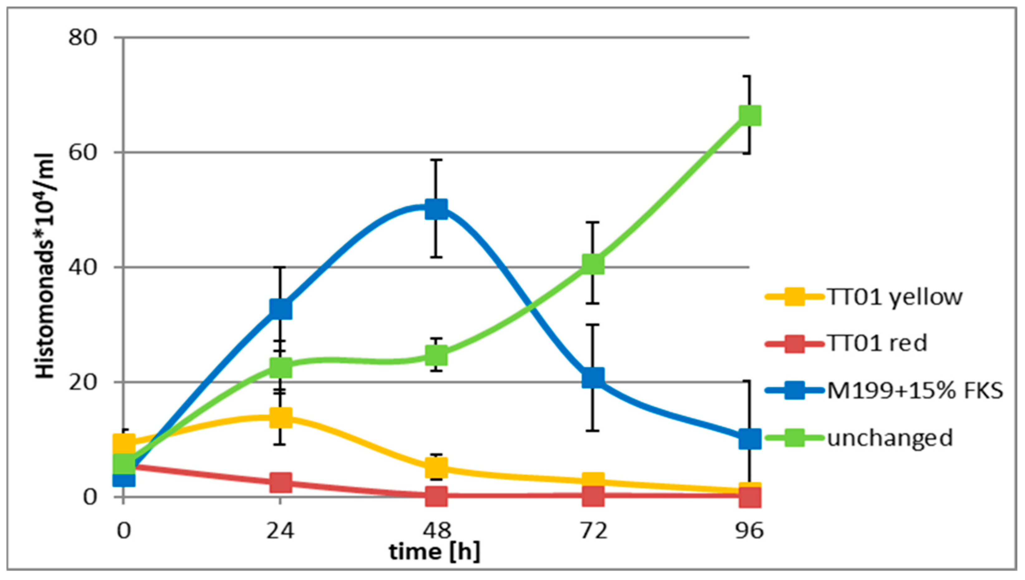
As for our H. meleagrididis exeriments, we worked in a monoxenic system where prokaryotic and eukaryotics cells coexisted. We found that both the prokaryotic and the eukaryotic cells died when EPB CFCM were added in th right concentration.
The question is, however, that what was the primary reason for the death of the H. meleagridis cells. They might die (1) because their procaryote mutualistic partner died; (2) they might be killed by a direct antiprotist activity of any (or more) AMP molecules of the EPB CFCMs; or (3) the two killing effects cumulated. Unfortunately, these published experiments cannot give an unambiguous answer to that question. We hope for the answer from our future experiments when we intend to use CFCMs from double (easyPACId) mutants (with hfq-deletion and exchanged (re-activated) promoter of only one AMP-responsible operon. We will search for some which is harmless to prokaryotic partner but kill H. meleagridis cells selectively). It is true, that the peptides killed the mutualistic (E. coli DH5@) bacteria, which itself could be considered as an explanation of the death of the H. meleagridis cells. It is also true, that from our experiments no proof supports the idea that histomonads were killed directly. But we also have evidence that CFCM contains active anti-protist active components as well, (see the Leishmania donovanii data from this study or the published results of the Bode group and others), so the only question is what of these compounds is capable of (in lucky case, selectively) killing H. meleagridis. This is what we intend to learn from our future easyPACId mutant experiments.
This paper is about the anti-protist potential of anti-microbial compounds produced by entomopathogenic bacterium species (EMA, EMC, and EMK Photorhabdus luminescens TT01) [6,28,29,93], the obligate symbionts of certain entomopathogenic–nematodes (EPN). In laboratory in vitro conditions they release their products into their conditioned culture media (CFCM) in vitro. The chemical nature of these antimicrobials is oligo-peptide. However they are non-ribosomal anti-microbial peptides (NR-AMPs), which means that they are enzymatically synthesized through more than one step in their respective biosynthetic pathways. Those genes encoding for the enzymes forming the respective biosynthetic pathway of given NR-AMP are clustered in a single operon [100], also called biosynthetic gene complex (BGC) [101]. One of the natural roles of the prokaryote partner in the EPN/EPB symbiosis is to protect the given monoxenic symbiotic association in a given polyxenic milieu, producing a chemical toolkit (e.g., a particular set of NR-AMPs) providing competitiveness against the prokaryotic and eukaryotic competitors, including protists. The biosynthesis of the given sets of these effective antimicrobials is synchronized by a unique, genuine hierarchical genetic regulation mechanism called primary-secondary phase shift [71] with the hfq gene on the top [74,75,76,77,78]. In the laboratory liquid culture conditions (as well as in the active symbiotic stage), these compounds are produced abundantly, providing an inexhaustible “gold mine” for the anti-microbial searching scientist. The bioassays of the CFCMs are the tools to find the best sources and optimize the culture conditions. We found that those strains (EMA and EMC) which were discovered and further developed not only by our international team of a “laboratory without walls” in those famous laboratories of Maxime Gualtieri in France and Helge Bode in Germany, proving that these two strains are probably best found so far. In addition, other laboratories are probably really excellent sources. The data presented here on anti-leishmanial and anti-histomonal potentials seem to confirm this conclusion. The next step is to find a way to obtain mutants, not “genetically manipulated”, but mutant strains which produce only single NR-AMP molecules instead of a bunch of them. The method to do that was recently discovered in the Bode laboratory [79,80,81,82,83]. We demonstrate here that we have started this project as well by constructing and characterizing hfq-del mutants from both EPB species EMA and EMC.
Our larger scope is to benefit from the existence of NR-AMP amongst the metabolites of our EPB bacteria. In other terms, our goals are to beat target pathogens, including prokaryotic pathogenic and zoonic bacteria, and eukaryotic parasites and pathogens of different taxa, which are important from the aspects of importance for animal and human health.
We are keenly interested in exploring whether the anti-microbial activity of NR-AMP of EMA and EMC can be developed as chemotherapeutic agents to treat protozoa such as the intracellular parasite Leishmania [37] and the extracellular avian pathogen H. gallinarum [97]. Both of these flagellated protozoa have been demonstrated to have unique and efficient resistance mechanisms to other anti-parasitic agents and require the development of additional effective agents. Leishmania spp. is particularly problematic in light of its sophisticated resistance mechanisms, but it is also an excellent model for analyzing the mechanisms of drug resistance in eukaryotes [98,99]. Our research conception is that we think that some Xenorhabdus CFCM molecules (i.e., phenazine, fabclavine, or other molecules) may be selectively capable of dysregulating eukaryotic cell functions and stimulating apoptosis in protozoa without affecting the host [100]. From an application point of view, the development of natural compounds as anti-protozoal requires several aspects to be taken into consideration: (A) the anti-protozoal potential; (B) the durability, thermotolerance, and bioavailability; and (C) cytotoxicity, and unwanted side effects, especially if target organisms are eukaryotic pathogens. We have demonstrated that Xenorhabdus CFCM has broad-spectrum anti-protozoal activity against several human microbial pathogens. The key issue is identifying the specific active molecules in a complex mixture of potential anti-microbial molecules. The CFCMs of all three Xenorhabdus species were all active against Leishmania promastigotes in our experiments, but interestingly, none were active against Trypanosoma cruzi epimastigotes. Other researchers [83] found that EMA and EMK CFCMs were equally efficient against both Leishmania and Trypanosoma. In that study, they found that X. budapestensis, X. cabanillasii, X. hominickii, X. indica, X. innexii, and X. stockiae supernatants caused 100% mortality at the highest tested concentration (10%) against the promastigote form of L. tropica. No differences occurred between this treatment group and the positive control (p > 0.05) using N-methyl meglumine. There are several possible explanations for the differences between our findings and those of the Turkish team. For instance, the two studies used different strains of T. cruzi. In addition, there are differences in the experimental methods used between the two studies. In the case of the differential susceptibility of L. amazoennsis and T. cruzi (Brazil strain), it is likely due to differences in the surface compositions between the two parasites that lead to differential disruption of the cell membrane and/or intracellular penetration to act on specific subcellular targets. We focus on EMA and EMC species. We compared the anti-microbial potential of each of the used hfq-del mutants before using them as the source of double mutants producing single NR-AMPs. Our data in Table 3, Table 4 and Table 5 show that the anti-microbial activities of the hfq-del mutants can have enhanced anti-microbial activity not present in the wild-type stains. This may not be quite surprising considering that approximately 7.5% of the total genes in Xenorhabdus bacteria are dedicated to secondary metabolite biosynthesis, and probably most of these products are NR-AMPs. With an average 2.4 Mb-genome size of Xenorhabdus and a 1 kb locus size with about 10 ORFs per operon, there are 2400 genes and 240 respective operons (BGS) per genome. It is comparable with that of Staphylococcus, which has about 225 operons. According to the data accumulated, only about a dozen of biosynthetic anti-microbials have been discovered. In our lab, we intend to work with about 12 operons, responsible for the biosynthesis of 12 NR-AMPs in EMC, and operons which are responsible for 3 NR_AMPs in EMA under hfq regulations.
However, we cannot rule out that there are several other operons encoding for the synthesis of additional anti-microbial compounds, which are not under hfq control. In the future, we intend to establish and analyze RNA-seq from our wild-type stains and their hfq mutants. We hypothesize that the situation is somewhat simpler since the hfq likely regulates a well-defined group of operons providing the genetic machinery for the so-called “primary-secondary phase shift” [72] (or “phenotypic phase variation”, the term of Professor S. A. Forst), which describes that only a relatively small set of natural products (NP) are anti-microbial synthetic genes. Toxicity studies using J774 macrophages [66,67] have indicated that only the CFCM of EMA was able to cause significant mortality of these cells. In contrast, the CFCMs of EMC or EMK had no effect. This experiment confirmed our previous unpublished observations that EMA exerts stronger cytotoxicity on a range of eukaryotic cells, which may cause limitations concerning future medical applications. However, this selective cytotoxicity may provide an option to select for and find specific CFCMs against different eukaryotic parasites of different animal and plant hosts. Interestingly, our studies with J774 cells and the CFCMs of EMA, EMC, and EMK did not show toxicity. These differences may be related to different experimental methods or differences in the bacterial and cell types used.
Overall, in light of the availability of few agents for the treatment of leishmaniasis and the increasing incidence of development resistance, there is a demand for new agents [100,101,102,103]. Moving forward, drug discovery will be based on complete genome sequencing of multiple strains, utilization of CRISPR/Cas9 technology for gene inactivation, in vivo, and bioluminescence-based imaging together with high-throughput analysis of compounds [44]. As for the results in the control (bacterium, fungi, and other strains), previous studies have identified several anti-microbial molecules in the CFCM of Xenorhabdus, including fabclavine [25,26]. This strange metabolite polypeptide/polyketide hybrid molecule is synthesized via non-ribosomal peptide synthesis. Many different-sized derivatives of fabclavines can be produced by X. szentirmaii [80] and can vary between entomopathogenic–nematode–symbiont (Xenorhabdus, Photorhabdus) bacterium species [80] (Wenski et al., 2019). These have a wide-ranging anti-microbial activity against various microbes [29]. Several other anti-microbial molecules are known to be produced by different EPB species and released into their CFCMs. Xenofuranone A and B [104], cabanillasin [105], PAX peptides [106], odilorhabdins [107], cyclic depsipeptides (xenematides, F and G), anti-oomycete peptides [108], novel anti-microbial peptides [109], xenortide [110], xenortides, rhabdopeptided/xenortide-like peptides [111], other rhabdopeptides [112], and szentiamid [113,114] have been discovered in the last ten years in different Xenorhabdus species.The proper approach is the easyPACId method, based on the latest information on the highest organization level of gene regulation in bacteria [115,116,117,118,119,120]. Whether any of them can be considered a potential anti-histomonal drug candidate will be learned if we had a chance to screen both the single mutant hfq-del stains of EMA and EMC.

5. Conclusions

Conclusions could be drawn from this study if we see the aims and the results along with the following aspects:
  • Perspectives of EPN/EPB symbiotic associations as abundant sources of AMPs, and we feel that the present publication provides some new idea into this direction
  • The comparative aspects of biosynthetic (non-ribosomal templated) and primary gene-product AMPs from drug potential aspects; and this publication drew the attention of the reade tosome complementary aspects.
  • The strategic aspects of controlling selected eukaryotic pathogens; we concluded the NR-AMPS have more option of becoming potential drugs to handle this issue.
  • The scientific and industrial aspects of the recently discovered hierarchical regulatory systems following signal integration provide great metabolic versatility that results in excellent adaptability and metabolic optimization in antibiotic-producing bacteria [99], like EPBs.
We would like to emphasize the actual importance of this research when concluding that the delineation of specific anti-microbial products in CFCM can now be achieved using the “easyPACId” approach (easy promoter-activated compound identification), which takes advantage of the alteration of regulation of coordinate gene clusters whose products are responsible for their biosynthesis [79]. We are convinced that this discovery may revolutionize the Xenorhabdus/Photorhabdus research, which in the age of multidrug resistance seems important. The point is that the operation of the different biosynthetic gene clusters BGCs (that is, operons) is synchronous, and their regulation is coordinated [76,77] by the gene encoding the RNA chaperone, Hfq [79]. The experimental system is based on the option of manipulation of the regulatory gene hfq [100,101,102,103,104] of the respective species. The regulatory mutant strain does not produce any biosynthetic AMP, but the respective individual promoters upstream of the respective biosynthetic gene cluster can be changed to inducible promotors, and this recombination will produce only a single respective AMP molecule. This provides an option of obtaining bacteria that produce only one single AMP molecule. The NR-AMP molecules have similar advantages to traditional antibiotics as the ribosomal templated AMPs, that is, the different pools of resistance genes with different motility patterns and collateral sensitivity, as well as molecular versatility. The advantage of the natural abundance in AMPs provided by the coevolutionary pattern of EPN/EPB symbiotic association may provide ammunition usable against MDR pathogens for a long time. This research line is further facilitated by the completed genome sequencing of several Xenorhabdus species, including X. szentirmaii [121] and X. budapestensis [122], making possible the identification of the promoter regions of each biosynthetic gene cluster (BGC, operon). Despite demonstrating the efficacy of vaccination in an experimental setting [55,58,123,124,125] no vaccine is yet commercially available. Therefore, and in addition to various other attempts, alternative chemotherapeutic solutions ares justified and the application potential of AMPs [126] seems a realistic idea.
Creating bacterial mutants that synthesize individual anti-microbial products into CFCM can facilitate their identification, purification, and bioassays of anti-microbial activity [82] and finally their potential application.

Supplementary Materials

The following supporting information can be downloaded at: https://www.mdpi.com/article/10.3390/antibiotics12091462/s1, Material S1: Histomonas Experiments.

Author Contributions

Conceptualization, A.F., M.H., B.S.M., A.S., T.V. and J.K.; investigation, A.F., Z.B., J.K., M.M.K. and P.G.; methodology, J.K., L.M., M.G.K. and E.T.; data curation, L.P.; formal analysis, C.H.; funding acquisition, K.D., M.G.K., T.V. and M.H.; project administration, T.V. and M.H.; resources, T.V. and M.H.; visualization, L.M.; Supervision, L.F., M.G.K., T.V. and M.H.; validation, C.H. and K.D.; writing—original draft, A.F. and M.G.K.; writing—review and editing, C.H., M.H., J.K., B.S.M., E.T., T.V., L.F. and L.M. All authors have read and agreed to the published version of the manuscript.

Funding

These project has partly been funded by Prof. Vellai, OTKA (Hungaria Research Fund) Grant Number: K132439 at the Department of Genetics, Eövös University Another source of funding: Dr. János Kiss, OTKA (Hungarian Research Fund) Grant Number: K 128203. The experiments in Gödöllő (J.K., Zs.B.) were also supported by RRF-2.3.1-21-2022-00007 from NKFIH (Nemzeti Kutatási, Fejlesztési és Innovációs Hivatal, https://nkfih.go, Hungary). The “EMA-EMC International Project” started at the Department of Genetics, Eötvös University; several other labs have been involved but have also been terminating here, although a significant part has been accomplished somewhere else. The Histomonas experiment was part of the Hungarian–Austrian international cooperation and supported by the TÁMOP-4.2.2/B-10/1-2010-0025 project based on the financial support of the State of Hungary and the EU. The CFCM preparations for Histomonas experiments were supported by the Hess–Dublecz project (recently a joint unit of the Hungarian University of Agriculture and Life Sciences (MATE). The Leishmania experiments were supported by the bench money of Bradford McGwire in Columbus, OH, USA.

Institutional Review Board Statement

Not applicable.

Informed Consent Statement

Not applicable.

Data Availability Statement

All of the data are available from the corresponding author. All data generated from this study are included in this article, and some information in the Supplementary Materials is strongly inseparable from information published in our previous paper [6].

Acknowledgments

A special thanks to special thanks to Jun Fu (Helmholtz International Lab for Anti-Infectives, Shandong University, Helmholtz Institute of Biotechnology, State Key Laboratory of Microbial Technology, Shandong University, Qingdao, China), Tao Sun and Jia Yin (Hunan Provincial Key Laboratory of Animal Intestinal Function and Regulation, College of Life Sciences, Hunan Normal University, Changsha, China) for providing us the plasmid pSC101-BAD-plu2934/2935/2936-gamma-amp. The use of these plasmids was essential for the success of getting hfq-del mutants from EMA and EMC. This work has been performed in a “laboratory without walls” that is within a frame of voluntary international research cooperation in Budapest. Despite his coauthrship, we thank the Head of the Department of Genetics at Eötvös University, for welcoming us back to the project and supporting it financially and morally as well as technically. (This project was born 20 years earlier in that department.). In Vienna, at the Clinic for Poultry and Fish Medicine, Department for Farm Animals and Veterinary Public Health, University of Veterinary Medicine (Vetmeduni, Vienna, Austria), we would like to express thanks and appreciation not only for their professional technical but also the intellectual help to the whole research team of the Hess laboratory, especially to Aziza Amin, and Irina Prokofieva in Keszthely, at the Institute of Physiology and Nutrition, Georgikon Campus of Hungarian University of Agriculture and Life Sciences, (MATE), Hungary, where the previous (and probably the future) in vivo feeding experiments have been going on. We express thanks and appreciation not only for their professional technical but also for the intellectual help of the Dublecz team. Many thanks for the professional and financial help in the molecular and bioinformatical work in the team of János Kiss (especially Mónika Szabó). We would like to express the invaluable help in the laboratories of Michael Klein, Joe Hogan, and Brad McGwire concerning the Leishmania work in Ohio.

Conflicts of Interest

The authors declare no conflict of interest. We sincerely consider those fellow scientists working in the same field as potential cooperators rather than competitors. Furthermore, the authors declare that the research was conducted without any commercial or financial relationships that could be construed as a potential conflict of interest.

Appendix A

Appendix A.1. Experimental Details of the Construction of hfq(del) Mutants of EMA and EMC Strains

The Δhfq mutant EMC strain was generated according to the one-step gene inactivation method [100]. which was adapted to Xenorhabdus. The λ Red recombinase was replaced by the Photorhabdus luminescence-derived homolog encoded by the Plu2934/Plu2935/Plu2936 operon [101]. The plasmid pSC101-BAD-plu2934/2935/2936-gamma-amp containing a thermosensitive pSC101-derived replication system and the Plu2934/Plu2935/Plu2936 operon with the λ Redγ gene under the control of ParaBAD was a kind gift from Jun Fu. Plasmids were introduced into X. szentirmaii (EMC) by electroporation. Electrocompetent cells were prepared for introduction of plasmids as follows: 1 mL of X. szentirmaii (EMC) culture grown at 30 °C in LB broth supplemented with Ap (150 μg/mL) to OD600~1.1–1.2 was centrifuged for 30 s at 9000 rpm at 2 °C, then resuspended in an ice-cold wash buffer SMG (0.5 M sorbitol, 0.5 M mannitol, 10% glycerol) and centrifuged again. The washing step was repeated two times; then, the cells were resuspended in a 60 μL SMG buffer.
Approximately 1 μg of plasmid DNA purified by the Qiagen Plasmid Midi Kit according to the manufacturer’s protocol was added to 40 μL electrocompetent cells. Electroporation was carried out using 2 mm gap electroporation cuvettes and BTX Electro Cell Manipulator 600 with the setting 129 ohm 24 kV/cm. Transformants were selected on LB plates supplemented with the appropriate antibiotics (Km—120 μg/mL, Sm—100 μg/mL, Gm—50 μg/mL, and Ap—150 μg/mL).
For the hfq KO recombination, EMC cells containing plasmid pBZs7 were grown in LB broth supplemented with Gm and Ap to a late logarithmic (OD600~1.1–1.2) phase, then the culture was 2× diluted with fresh LB broth, L-arabinose was added in a final concentration of 1% and grown further in a shaker for 1.5 h at 30 °C. The electro-competent cells were prepared as described above. The KO PCR fragment was amplified using pKD4 template plasmid and the primers hfq_EMCdelfor and hfq_EMCdelrev. The amplicon was precipitated in 96% ethanol, dried, and resuspended in 10 mM Tris pH 8.0. For electroporation, approximately 1 μg of purified DNA fragment was used as described. After electroporation, cells were grown for 5 h at 30 °C, then spread on LB agar plates supplemented with 120 μg/mL Km and incubated at 30 °C for 2 days. The Δhfq::KmR recombinant was tested by colony PCRs using the primer pairs hfqseqfor-trn5neo5, rn5neo3-hfqseqrev, and hfqseqfor-hfqseqrev.
For deletion of the KmR cassette from the Δhfq::KmR mutant, pBZs1 was electroporated. A SmRApRKmR transformant colony was grown overnight at 30 °C in LB broth supplemented with Ap, spread on LB + Ap agar plates, and incubated for 24 h at 30 °C; then, the colonies were individually tested for the loss of the KmR gene and the pBZs1 plasmid on LB + Ap, LB + Km, and LB + Sm plates. The KmS Δhfq strains were tested by colony PCR using the primer pair hfqseqfor–hfqseqrev.
The Δhfq mutant EMA strain was generated according to [77] (Aref 10) The 769 bp upstream and the 617 bp downstream region of the hfq gene of EMA were amplified with primer pairs hfq_upforBam-hfq_uprev and hfqEMA_downfor-hfq_downrevSac, respectively. The two amplicons were assembled in the following PCR:
Using 0.5 μL of the upstream and 2 μL of the downstream amplicons, 60 s denaturation at 94 °C followed by 15 cycles of 94 °C 20 s, 55 °C 30 s, and 72 °C for 2 min, then 0.5 μL of primers hfq_upforBam and hfq_downrevSac (10 μM each) were added and the cycling was continued for a further 35 cycles. The amplicon was purified using the Illustra GFX PCR DNA and Gel Band Purification Kit (GE Healthcare), digested with BamHI and SacI, and ligated into the BamHI-SacI site of pBluescript II SK(+) (pBZS13). After sequencing, the BamHI-SacI fragment from pBZS13 was transferred into the BamHI-SacI site of the R6Kγ-based plasmid pBZs12, leading to the SacB-bearing mobilizable recombination vector pBZs17.
pBZs17 was mobilized into EMA from E. coli S17-1 λ pir. The ON culture of S17-1 λ pir/pBZs17 was 50× diluted with fresh LB broth supplemented with Km and Sm and grown 3 h to OD600~0.5, while the EMA recipient was grown in LB + Ap ON to OD600~1.0. 0.1 mL of donor and 1 mL of recipient cultures were mixed, centrifuged, and washed twice with 0.9% NaCl solution, then spread onto LB agar plates. After 4 h incubation at 30 °C, the bacterial lawn was resuspended in 3 mL 0.9% NaCl solution, and the cells were centrifuged and spread onto LB + KmAp agar plates. After four days of incubation at 30 °C, the KmRGmRApR recombinant colonies were tested by colony PCRs using primer pairs EMAhfqseqfor–trn5neo3 and pUCfor24–hfqseqrev.
To gain Δhfq recombinants, the EMA::pBZs17 strain was grown for 18 h at 30 °C in LB + Ap broth, and 50, 100, and 200 μL of the culture was spread onto LB + Ap + 5% sucrose agar plates. After 48 h incubation at 30 °C, the individual SacR colonies were tested for KmS and GmS phenotypes; then, the appropriate colonies were tested by colony PCR using primers EMAhfqseqfor and hfqseqrev. To verify the hfq deletion, the amplicon was sequenced.
Restriction enzymes and DNA polymerases were purchased from Thermo Scientific. Colony PCRs were carried out using Dream Taq polymerase, while for the amplification of fragments for cloning or KO experiments, a Phusion polymerase was applied.
For maintaining the plasmids with a temperature-sensitive pSC101 replication system, growth at 30 °C with antibiotic selection was applied, while curing these plasmids was carried out by growth at 30 °C without antibiotic selection, followed by testing the resistance pattern of individual colonies for selecting plasmid-cured clones.
For cloning purposes, E. coli TG1 [3] was used as a host strain. For plasmid mobilization into Xenorhabdus, E. coli S17-1λ pir [86] was applied. E. coli strains were routinely grown in LB at 37 °C with appropriate antibiotic selection. Restriction enzymes and DNA polymerases were purchased from Thermo Scientific. Colony PCRs were carried out using Dream Taq polymerase, while the amplification of fragments for cloning or KO experiments, Phusion polymerase, was applied.
Table A1. Relevant features of plasmids used in this work.
NameRelevant FeaturesReferences
pBluescript II SK (+)pMB1-based ApR cloning vector.[89]
pKD4KmR,ApR r6kγ-based PCR template plasmid for one-step recombination gene-KO[2]
pBZs1SmR,CmR,ApS, derivative of pCP20 [1] containing also the oriT of RP4, thermo-inducible FLP recombinase expression (λ pR::FLP), temperature-sensitive pSC101 replication system, λ cI857, mobilization by S17-1λ pir host strainthis work
pBZS7KmR,ApS derivative of pSC101-BAD-plu2934/2935/2936-gamma-amp [88], L-ara-inducible expression of λ Red recombinase homolog from P. luminescens and λ Redγ protein, temperature-sensitive pSC101 replication systemthis work
pBZs12pJQ200-SK derivative, where the SspI-NheI fragment carrying the p15A origin of replication was replaced by the ClaI-NheI fragment of pSG76-K containing the KmR gene and R6Kγ origin of replication. KmR, GmR, RP4 oriT, R6Kγ oriV, SacBthis work
pBZs17pBZs12 derivative, containing the assembled upstream and downstream region of the hfq gene of EMA. KmR, GmR, RP4 oriT, R6Kγ oriV, SacBthis work
pSG76-KR6Kγ-based KmR replicon [90]
pJQ200-SKA SacB-bearing p15A-based vector. GmR, RP4 oriT, p15A oriV, SacB. [91]
Table A2. Relevant features of bacterial strains used in the molecular genetics section of this study.
NameRelevant FeaturesReferences
TG1supE hsdΔ5 thi Δ(lac-proAB) F’[traD36 proAB+ lacIq lacZΔM15][90]
S17-1 λpirS17-1 λpir, a λ lysogen derivative of S17-1 (pro thi recA hsdR (r m+) TpR SmR KmS [Ω RP4-2-Tc::Mu-Km::Tn7]) expressing Π protein from pir gene of R6K[91]
X. budapestensis DSM16342wt isolate[90]
X. szentirmaii DSM 16338wt isolate
Table A3. List of oligonucleotides used.
PrimerSequence (5′-3′)Reference
hfq_EMCdelforgaaccaaaaggttcttagttaaaacaacaaaataaggaaaatatagaatgGTGTAGGCTGGAGCTGCTTCthis work
hfq_EMCdelrevaagtctccctacctatgtttttatttacttcaggtatggtcactgatttaCATATGAATATCCTCCTTAGTTCthis work
hfqEMAseqfortgtggtcttatctcgcgggcthis work
hfqseqfortctcatgatgagatggtttatcgtgthis work
hfqseqrevgcacaggagaaacacctgcggthis work
trn5neo3tcgccttctatcgccttcttgathis work
trn5neo5aatagcctctccacccaagcggthis work
hfq_upforBamaaggatccactcctggttggcggaaccatgcthis work
hfq_uprevcaaagattgccccttagccattctatattttccttattttgthis work
hfqEMA_downforcaaaataaggaaaatatagaatggctaaggggcaatctttgatcgctgaataaatcagtgataatgcctggthis work
hfq_downrevSacaagagctctttgtcacgcagcatacggcggtcthis work
pUCfor24cgccagggttttcccagtcacgac[92]

References

  1. Ötvos, L., Jr.; Wade, J.D. Current challenges in peptide-based drug discovery. Front. Chem. 2014, 2, 62. [Google Scholar] [CrossRef]
  2. Karelson, M.; Lobanov, V.S.; Katritzky, A.R. Quantum-Chemical Descriptors in QSAR/QSPR Studies. Chem. Rev. 1996, 96, 1027–1044. [Google Scholar] [CrossRef]
  3. Gini, G. QSAR Methods. Methods Mol. Biol. 2022, 2425, 1–26. [Google Scholar] [CrossRef] [PubMed]
  4. Cammue, B.P.; De Bolle, M.F.; Schoofs, H.M.; Terras, F.R.; Thevissen, K.; Osborn, R.W.; Rees, S.B.; Broekaert, W.F. Gene-encoded anti-microbial peptides from plants. Ciba. Found. Symp. 1994, 186, 91–101. [Google Scholar] [CrossRef] [PubMed]
  5. Medema, M.H.; Kottmann, R.; Yilmaz, P.; Cummings, M.; Biggins, J.B.; Blin, K.; de Bruijn, I.; Chooi, Y.H.; Claesen, J.; Coates, R.C.; et al. Minimum Information about a Biosynthetic Gene cluster. Nat. Chem. Biol. 2015, 11, 625–631. [Google Scholar] [CrossRef] [PubMed]
  6. Fodor, A.; Vellai, T.; Hess, C.; Makrai, L.; Dublecz, K.; Pál, L.; Molnár, A.; Klein, M.G.; Tarasco, E.; Józsa, S.; et al. XENOFOOD—An Autoclaved Feed Supplement Containing Autoclavable Anti-microbial Peptides—Exerts Anticoccidial GI Activity, and Causes Bursa Enlargement, but Has No Detectable Harmful Effects in Broiler Cockerels despite In Vitro Detectable Cytotoxicity on LHM Cells. Pathogens 2023, 12, 458. [Google Scholar] [CrossRef]
  7. Sharma, K.; Sharma, K.K.; Sharma, A.; Jain, R. Peptide-based drug discovery: Current status and recent advances. Drug Discov. Today 2023, 28, 103464. [Google Scholar] [CrossRef]
  8. Zasloff, M. Anti-microbial Peptides of Multicellular Organisms: My Perspective. Adv. Exp. Med. Biol. 2019, 1117, 3–6. [Google Scholar] [CrossRef]
  9. Boman, H.G.; Steiner, H. Humoral immunity in Cecropia pupae. Curr. Top. Microbiol. Immunol. 1981, 94–95, 75–91. [Google Scholar] [CrossRef]
  10. Selsted, M.E.; Harwig, S.S.; Ganz, T.; Schilling, J.W.; Lehrer, R.I. Primary structures of three human neutrophil defensins. J. Clin. Investig. 1985, 76, 1436–1439. [Google Scholar] [CrossRef]
  11. Solanki, S.S.; Singh, P.; Kashyap, P.; Sansi, M.S.; Ali, S.A. Promising role of defensins peptides as therapeutics to combat against viral infection. Microb. Pathog. 2021, 155, 104930. [Google Scholar] [CrossRef] [PubMed]
  12. Zasloff, M. Magainins, a class of anti-microbial peptides from Xenopus skin: Isolation, characterization of two active forms, and partial cDNA sequence of a precursor. Proc. Natl. Acad. Sci. USA 1987, 84, 5449–5453. [Google Scholar] [CrossRef] [PubMed]
  13. Welch, N.G.; Li, W.; Hossain, M.A.; Separovic, F.; O’Brien-Simpson, N.M.; Wade, J.D. (Re)Defining the Proline-Rich Anti-microbial Peptide Family and the Identification of Putative New Members. Front. Chem. 2020, 8, 607769. [Google Scholar] [CrossRef]
  14. Mangano, K.; Klepacki, D.; Ohanmu, I.; Baliga, C.; Huang, W.; Brakel, A.; Krizsan, A.; Polikanov, Y.S.; Hoffmann, R.; Vázquez-Laslop, N.; et al. Inhibition of translation termination by the anti-microbial peptide Drosocin. Nat. Chem. Biol. 2023, 19, 1082–1090. [Google Scholar] [CrossRef]
  15. Mojsoska, B.; Jenssen, H. Peptides and Peptidomimetics for Anti-microbial Drug Design. Pharmaceuticals 2015, 8, 366–415. [Google Scholar] [CrossRef] [PubMed]
  16. Sierra, J.M.; Fusté, E.; Rabanal, F.; Vinuesa, T.; Viñas, M. An overview of anti-microbial peptides and the latest advances in their development. Expert Opin. Biol. Ther. 2017, 17, 663–676. [Google Scholar] [CrossRef] [PubMed]
  17. Gao, Y.; Fang, H.; Fang, L.; Liu, D.; Liu, J.; Su, M.; Fang, Z.; Ren, W.; Jiao, H. The Modification and Design of Anti-microbial Peptide. Curr. Pharm. Des. 2018, 24, 904–910. [Google Scholar] [CrossRef] [PubMed]
  18. Romero, S.M.; Cardillo, A.B.; Martínez Ceron, M.C.; Camperi, S.A.; Giudicessi, S.L. Temporins: An Approach of Potential Pharmaceutic Candidates. Surg. Infect. 2020, 21, 309–322. [Google Scholar] [CrossRef]
  19. Li, W.; Separovic, F.; O’Brien-Simpson, N.M.; Wade, J.D. Chemically modified and conjugated anti-microbial peptides against superbugs. Chem. Soc. Rev. 2021, 50, 4932–4973. [Google Scholar] [CrossRef] [PubMed]
  20. Chaoulitch, S.P. Leishmania donovani Laveran et Mesnil, 1903 parasite aussä les érythrocytes [Leishmania donovani Laveran and Mesnil 1903 also parasitic in erythrocytes]. Bull. Soc. Pathol. Exot. Fil. 1954, 47, 244–246. [Google Scholar]
  21. Dutra-Rêgo, F.; Freire, M.L.; Carvalho, G.M.L.; Andrade-Filho, J.D. Revisiting the cave-dwelling sand flies (Diptera, Psychodidae, Phlebotominae) from Brazil: Diversity and potential role in the transmission of Leishmania Ross, 1903 (Kinetoplastida: Trypanosomatidae). Med. Vet.-Entomol. 2022, 36, 408–423. [Google Scholar] [CrossRef] [PubMed]
  22. McDougald, L.R. Blackhead disease (Histomoniasis) in poultry: A critical review. Avian Dis. 2005, 49, 462–476. [Google Scholar] [CrossRef]
  23. McDougald, L.R. Intestinal protozoa important to poultry. Poult. Sci. 1998, 77, 1156–1158. [Google Scholar] [CrossRef] [PubMed]
  24. Hess, M.; Liebhart, D.; Bilic, I.; Ganas, P. Histomonas meleagridis—New insights into an old pathogen. Vet.-Parasitol. 2015, 208, 67–76. [Google Scholar] [CrossRef] [PubMed]
  25. Fuchs, S.W.; Sachs, C.C.; Kegler, C.; Nollmann, F.I.; Karas, M.; Bode, H.B. Neutral loss fragmentation pattern based screening for arginine-rich natural products in Xenorhabdus and Photorhabdus. Anal. Chem. 2012, 84, 6948–6955. [Google Scholar] [CrossRef] [PubMed]
  26. Fuchs, S.W.; Grundmann, F.; Kurz, M.; Kaiser, M.; Bode, H.B. Fabclavines: Bioactive peptide-polyketide-polyamino hybrids from Xenorhabdus. ChemBioChem 2014, 15, 512–516. [Google Scholar] [CrossRef]
  27. Shi, Y.-M.; Brachmann, A.O.; Westphalen, M.A.; Neubacher, N.; Tobias, N.J.; Bode, H.B. Dual phenazine gene clusters enable diversification during biosynthesis. Nat. Chem. Biol. 2019, 15, 331–339. [Google Scholar] [CrossRef]
  28. Lengyel, K.; Lang, E.; Fodor, A.; Szállás, E.; Schumann, P.; Stackebrandt, E. Description of four novel species of Xenorhabdus, family Enterobacteriaceae: Xenorhabdus budapestensis sp. nov., Xenorhabdus ehlersii sp. nov., Xenorhabdus innexi sp. nov., and Xenorhabdus szentirmaii sp. nov. Syst. Appl. Microbiol. 2005, 28, 115–122. [Google Scholar] [CrossRef]
  29. Fodor, A.; Gualtieri, M.; Zeller, M.; Tarasco, E.; Klein, M.G.; Fodor, A.M.; Haynes, L.; Lengyel, K.; Forst, S.A.; Furgani, G.M.; et al. Type Strains of Entomopathogenic–nematode-Symbiotic Bacterium Species, Xenorhabdus szentirmaii (EMC) and X. budapestensis (EMA), Are Exceptional Sources of Non-Ribosomal Templated, Large-Target-Spectral, Thermotolerant-Anti-microbial Peptides (by Both), and Iodinin (by EMC). Pathogens 2022, 11, 342. [Google Scholar] [CrossRef]
  30. Akhurst, R.J. Antibiotic Activity of Xenorhabdus spp., Bacteria symbiotically associated with insect pathogenic nematodes of the families Heterorhabditidae and Steinernematidae. J. Gen. Microbiol. 1982, 128, 3061–3065. [Google Scholar] [CrossRef]
  31. Ogier, J.-C.; Pagès, S.; Frayssinet, M.; Gaudriault, S. Entomopathogenic–nematode-associated microbiota: From monoxenic paradigm to pathobiome. Microbiome 2020, 8, 25. [Google Scholar] [CrossRef] [PubMed]
  32. Furgani, G.; Böszörményi, E.; Fodor, A.; Máthé-Fodor, A.; Forst, S.; Hogan, J.S.; Katona, Z.; Klein, M.G.; Stackebrandt, E.; Szentirmai, A.; et al. Xenorhabdus antibiotics: A comparative analysis and potential utility for controlling mastitis caused by bacteria. J. Appl. Microbiol. 2008, 104, 745–758. [Google Scholar] [CrossRef] [PubMed]
  33. Fodor, A.; Makrai, L.; Fodor, L.; Venekei, I.; Husvéth, F.; Pál, L.; Molnár, A.; Dublecz, K.; Pintér, C.; Józsa, S.; et al. Anti-Coccidiosis Potential of Autoclaveable Anti-microbial Peptides from Xenorhabdus budapestensis Resistant to Proteolytic (Pepsin, Trypsin) Digestion Based on In Vitro Studies. Microbiol. Res. J. Int. 2018, 22, 1–17. [Google Scholar] [CrossRef]
  34. Böszörményi, E.; Érsek, T.; Fodor, A.; Fodor, A.M.; Földes, L.; Hevesi, M.; Hogan, J.; Katona, Z.; Klein, M.; Kormány, A.; et al. Isolation and activity of Xenorhabdus anti-microbial compounds against the plant pathogens Erwinia amylovora and Phytophthora nicotianae. J. Appl. Microbiol. 2009, 107, 746–759. [Google Scholar] [CrossRef] [PubMed]
  35. Fodor, A.; Varga, I.; Hevesi, M.; Máthé-Fodor, A.; Racsko, J.; Hogan, J.A. Novel anti-microbial peptides of Xenorhabdus origin against multidrug resistant plant pathogens. In A Search for Antibacterial Agents; Bobbarala, V., Ed.; IntechOpen: London, UK, 2012; pp. 3–32. Available online: https://www.intechopen.com/books/2129 (accessed on 30 June 2012).
  36. Vozik, D.; Bélafi-Bakó, K.; Hevesi, M.; Böszörményi, E.; Fodor, A. Effectiveness of a Peptide-rich Fraction from Xenorhabdus budapestensis Culture against Fire Blight Disease on Apple Blossoms. Not. Bot. Hortic. Agrobot. 2015, 43, 547–553. Available online: https://www.notulaebotanicae.ro/ndex.php/index (accessed on 22 February 2022). [CrossRef]
  37. Fernández-Arévalo, A.; El Baidouri, F.; Ravel, C.; Ballart, C.; Abras, A.; Lachaud, L.; Tebar, S.; Lami, P.; Pratlong, F.; Gállego, M.; et al. The Leishmania donovani species complex: A new insight into taxonomy. Int. J. Parasitol. 2020, 50, 1079–1088. [Google Scholar] [CrossRef]
  38. McGwire, B.S.; Satoskar, A.R. Leishmaniasis: Clinical syndromes and treatment. QJM 2014, 107, 7–14. [Google Scholar] [CrossRef]
  39. Alvar, J.; Cañavate, C.; Molina, R.; Moreno, J.; Nieto, J. Canine leishmaniasis. Adv. Parasitol. 2004, 57, 1–88. [Google Scholar] [CrossRef]
  40. Dantas-Torres, F.; Miró, G.; Baneth, G.; Bourdeau, P.; Breitschwerdt, E.; Capelli, G.; Cardoso, L.; Day, M.J.; Dobler, G.; Ferrer, L.; et al. Canine Leishmaniasis Control in the Context of One Health. Emerg. Infect. Dis. 2019, 25, 1–4. [Google Scholar] [CrossRef]
  41. Walker, D.M.; Oghumu, S.; Gupta, G.; McGwire, B.S.; Drew, M.E.; Satoskar, A.R. Mechanisms of cellular invasion by intracellular parasites. Cell Mol. Life Sci. 2014, 71, 1245–1263. [Google Scholar] [CrossRef]
  42. Burza, S.; Croft, S.L.; Boelaert, M. Leishmaniasis. Lancet 2018, 392, 951–970. [Google Scholar] [CrossRef]
  43. Safavi, M.; Eshaghi, H.; Hajihassani, Z. Visceral Leishmaniasis: Kala-azar. Diagn. Cytopathol. 2021, 49, 446–448. [Google Scholar] [CrossRef] [PubMed]
  44. Altamura, F.; Rajesh, R.; Catta-Preta, C.M.C.; Moretti, N.S.; Cestari, I. The current drug discovery landscape for trypanosomiasis and leishmaniasis: Challenges and strategies to identify drug targets. Drug Dev. Res. 2022, 83, 225–252. [Google Scholar] [CrossRef] [PubMed]
  45. Mauel, J. Vaccination Against Leishmania Infections. Curr. Drug Targets-Immune Endocr. Metab. Disord. 2002, 2, 201–226. [Google Scholar] [CrossRef]
  46. Ibarra-Meneses, A.V.; Corbeil, A.; Wagner, V.; Beaudry, F.; Monte-Neto, R.L.D.; Fernandez-Prada, C. Exploring direct and indirect targets of current antileishmanial drugs using a novel thermal proteomics profiling approach. Front. Cell. Infect. Microbiol. 2022, 12, 954144. [Google Scholar] [CrossRef]
  47. Douanne, N.; Wagner, V.; Roy, G.; Leprohon, P.; Ouellette, M.; Fernandez-Prada, C. MRPA-independent mechanisms of antimony resistance in Leishmania infantum. Int. J. Parasitol. Drugs Drug Resist. 2020, 13, 28–37. [Google Scholar] [CrossRef] [PubMed]
  48. Golenser, J.; Domb, A. New formulations and derivatives of Amphotericin B for treatment of leishmaniasis. Mini-Rev. Med. Chem. 2006, 6, 153–162. [Google Scholar] [CrossRef]
  49. Lollis, L.; Gerhold, R.; McDougald, L.; Beckstead, R. Molecular Characterization of Histomonas meleagridis and Other Parabasalids in the United States Using the 5.8S, ITS-1, and ITS-2 rRNA Regions. J. Parasitol. 2011, 97, 610–615. [Google Scholar] [CrossRef]
  50. Palmieri, N.; de Jesus Ramires, M.; Hess, M.; Bilic, I. Complete genomes of the eukaryotic poultry parasite Histomonas meleagridis: Linking sequence analysis with virulence/attenuation. BMC Genom. 2021, 22, 753. [Google Scholar] [CrossRef]
  51. Beer, L.C.; Petrone-Garcia, V.M.; Graham, B.D.; Hargis, B.M.; Tellez-Isaias, G.; Vuong, C.N. Histomonosis in Poultry: A Comprehensive Review. Front. Vet.-Sci. 2022, 9, 880738. [Google Scholar] [CrossRef]
  52. Ganas, P.; Liebhart, D.; Glösmann, M.; Hess, C.; Hess, M. Escherichia coli strongly supports the growth of Histomonas meleagridis, in a monoxenic culture, without influence on its pathogenicity. Int. J. Parasitol. 2012, 42, 893–901. [Google Scholar] [CrossRef] [PubMed]
  53. Mitra, T.; Bilic, I.; Hess, M.; Liebhart, D. The 60S ribosomal protein L13 is the most preferable reference gene to investigate gene expression in selected organs from turkeys and chickens, in context of different infection models. Vet.-Res. 2016, 47, 105. [Google Scholar] [CrossRef] [PubMed]
  54. Kidane, F.A.; Bilic, I.; Mitra, T.; Wernsdorf, P.; Hess, M.; Liebhart, D. In situ hybridization to detect and localize signature cytokines of T-helper (Th) 1 and Th2 immune responses in chicken tissues. Vet.-Immunol. Immunopathol. 2016, 175, 51–56. [Google Scholar] [CrossRef]
  55. Mitra, T.; Kidane, F.A.; Hess, M.; Liebhart, D. Unravelling the Immunity of Poultry Against the Extracellular Protozoan Parasite Histomonas meleagridis Is a Cornerstone for Vaccine Development: A Review. Front. Immunol. 2018, 9, 2518. [Google Scholar] [CrossRef]
  56. Liebhart, D.; Ganas, P.; Sulejmanovic, T.; Hess, M. Histomonosis in poultry: Previous and current strategies for prevention and therapy. Avian Pathol. 2017, 46, 1–18. [Google Scholar] [CrossRef] [PubMed]
  57. Hauck, R.; Balczulat, S.; Hafez, H.M. Detection of DNA of Histomonas meleagridis and Tetratrichomonas gallinarum in German poultry flocks between 2004 and 2008. Avian Dis. 2010, 54, 1021–1025. [Google Scholar] [CrossRef]
  58. Lagler, J.; Schmidt, S.; Mitra, T.; Stadler, M.; Wernsdorf, P.; Grafl, B.; Hatfaludi, T.; Hess, M.; Gerner, W.; Liebhart, D. Comparative investigation of IFN-γ-producing T cells in chickens and turkeys following vaccination and infection with the extracellular parasite Histomonas meleagridis. Dev. Comp. Immunol. 2021, 116, 103949. [Google Scholar] [CrossRef]
  59. Gruber, J.; Ganas, P.; Hess, M. Long-term in vitro cultivation of Histomonas meleagridis coincides with the dominance of a very distinct phenotype of the parasite exhibiting increased tenacity and improved cell yields. Parasitology 2017, 144, 1253–1263. [Google Scholar] [CrossRef]
  60. Bilic, I.; Hess, M. Interplay between Histomonas meleagridis and Bacteria: Mutualistic or Predator–Prey? Trends Parasitol. 2020, 36, 232–235. [Google Scholar] [CrossRef]
  61. Hauck, R.; Hafez, H.M. Partial sequence of the beta-tubulin of Histomonas meleagridis and the activity of benzimidazoles against H. meleagridis in vitro. Parasitol. Res. 2009, 104, 1183–1189. [Google Scholar] [CrossRef]
  62. Grabensteiner, E.; Liebhart, D.; Arshad, N.; Hess, M. Antiprotozoal activities determined in vitro and in vivo of certain plant extracts against Histomonas meleagridis, Tetratrichomonas gallinarum and Blastocystis sp. Parasitol. Res. 2008, 103, 1257–1264. [Google Scholar] [CrossRef]
  63. Abraham, M.; McDougald, L.R.; Beckstead, R.B. Blackhead disease: Reduced sensitivity of Histomonas meleagridis to nitarsone in vitro and in vivo. Avian Dis. 2014, 58, 60–63. [Google Scholar] [CrossRef] [PubMed]
  64. Collins, J.B.; Jordan, B.; Vidyashankar, A.; Bishop, A.; Kaplan, R.M. Fenbendazole resistance in Heterakis gallinarum, the vector of Histomonas meleagridis, on a broiler breeder farm in South Carolina. Vet.-Parasitol. Reg. Stud. Rep. 2022, 36, 100785. [Google Scholar] [CrossRef] [PubMed]
  65. Brioschi, M.B.; Coser, E.M.; Coelho, A.C.; Gadelha, F.R.; Miguel, D.C. Models for cytotoxicity screening of antileishmanial drugs: What has been done so far? Int. J. Antimicrob. Agents 2022, 60, 106612. [Google Scholar] [CrossRef] [PubMed]
  66. Tominaga, T.; Suzuki, M.; Saeki, H.; Matsuno, S.; Tachibana, T.; Kudo, T. Establishment of an activated macrophage cell line, A-THP-1, and its properties. Tohoku J. Exp. Med. 1998, 186, 99–119. [Google Scholar] [CrossRef]
  67. Andreu, N.; Phelan, J.; de Sessions, P.F.; Cliff, J.M.; Clark, T.G.; Hibberd, M.L. Primary macrophages and J774 cells respond differently to infection with Mycobacterium tuberculosis. Sci. Rep. 2017, 7, 42225. [Google Scholar] [CrossRef]
  68. Geroldinger, G.; Rezk, M.; Idris, R.; Gruber, V.; Tonner, M.; Moldzio, R.; Staniek, K.; Monzote, L.; Gille, L. Techniques to study phagocytosis and uptake of Leishmania tarentolae by J774 macrophages. Exp. Parasitol. 2019, 197, 57–64. [Google Scholar] [CrossRef]
  69. Kawaguchi, T.; Nomura, K.; Hirayama, Y.; Kitagawa, T. Establishment and characterization of a chicken hepatocellular car-cinoma, cell line, LMH. Cancer Res. 1987, 47, 4460–4464. [Google Scholar]
  70. Amin, A.; Bilic, I.; Berger, E.; Hess, M. Trichomonas gallinae, in comparison to Tetratrichomonas gallinarum, induces distinctive cytopathogenic effects in tissue cultures. Vet.-Parasitol. 2012, 186, 196–206. [Google Scholar] [CrossRef]
  71. Forst, S.; Nealson, K. Molecular biology of the symbiotic-pathogenic bacteria Xenorhabdus spp. and Photorhabdus spp. Microbiol. Rev. 1996, 60, 21–43. [Google Scholar] [CrossRef]
  72. Akhurst, R.J. Morphological and functional dimorphism in Xenorhabdus spp., Bacteria symbiotically associated with the insect pathogenic nematodes Neoaplectana and Heterorhabditis. J. Gen. Microbiol. 1980, 121, 303–309. [Google Scholar] [CrossRef]
  73. Boemare, N.E.; Akhurst, R.J. Biochemical and physiological characterization of colony form variants in Xenorhabdus spp. (Enterobacteriaceae). Microbiology 1988, 134, 751–761. [Google Scholar] [CrossRef]
  74. Joyce, S.A.; Clarke, D.J. A hexA homologue from Photorhabdus regulates pathogenicity, symbiosis and phenotypic variation. Mol. Microbiol. 2003, 47, 1445–1457. [Google Scholar] [CrossRef] [PubMed]
  75. Langer, A.; Moldovan, A.; Harmath, C.; Joyce, S.A.; Clarke, D.J.; Heermann, R. HexA is a versatile regulator involved in the control of phenotypic heterogeneity of Photorhabdus luminescens. PLoS ONE 2017, 12, e0176535. [Google Scholar] [CrossRef]
  76. Tobias, N.J.; Mishra, B.; Gupta, D.K.; Sharma, R.; Thines, M.; Stinear, T.P.; Bode, H.B. Genome comparisons provide insights into the role of secondary metabolites in the pathogenic phase of the Photorhabdus life cycle. BMC Genom. 2016, 17, 537. [Google Scholar] [CrossRef]
  77. Tobias, N.J.; Heinrich, A.K.; Eresmann, H.; Wright, P.R.; Neubacher, N.; Backofen, R.; Bode, H.B. Photorhabdus-nematode symbiosis is dependent on hfq-mediated regulation of secondary metabolites. Environ. Microbiol. 2017, 19, 119–129. [Google Scholar] [CrossRef]
  78. Neubacher, N.; Tobias, N.J.; Huber, M.; Cai, X.; Glatter, T.; Pidot, S.J.; Stinear, T.P.; Lütticke, A.L.; Papenfort, K.; Bode, H.B. Symbiosis, virulence and natural-product biosynthesis in entomopathogenic bacteria are regulated by a small RNA. Nat. Microbiol. 2020, 5, 1481–1489. [Google Scholar] [CrossRef]
  79. Bode, E.; Heinrich, A.K.; Hirschmann, M.; Abebew, D.; Shi, Y.; Vo, T.D.; Wesche, F.; Shi, Y.; Grün, P.; Simonyi, S.; et al. Promoter Activation in Δhfq Mutants as an Efficient Tool for Specialized Metabolite Production Enabling Direct Bioactivity Testing. Angew. Chem. Int. Ed. 2019, 58, 18957–18963. [Google Scholar] [CrossRef]
  80. Wenski, S.L.; Kolbert, D.; Grammbitter, G.L.C.; Bode, H.B. Fabclavine biosynthesis in X. szentirmaii: Shortened derivatives and characterization of the thioester reductase FclG and the condensation domain-like protein FclL. J. Ind. Microbiol. Biotechnol. 2019, 46, 565–572. [Google Scholar] [CrossRef]
  81. Wenski, S.L.; Cimen, H.; Berghaus, N.; Fuchs, S.W.; Hazir, S.; Bode, H.B. Fabclavine diversity in Xenorhabdus bacteria. Beilstein J. Org. Chem. 2020, 16, 956–965. [Google Scholar] [CrossRef]
  82. Cimen, H.; Touray, M.; Gulsen, S.H.; Hazir, S. Natural products from Photorhabdus and Xenorhabdus: Mechanisms and impacts. Appl. Microbiol. Biotechnol. 2022, 106, 4387–4399. [Google Scholar] [CrossRef]
  83. Gulsen, S.H.; Tileklioglu, E.; Bode, E.; Cimen, H.; Ertabaklar, H.; Ulug, D.; Ertug, S.; Wenski, S.L.; Touray, M.; Hazir, C.; et al. Antiprotozoal activity of different Xenorhabdus and Photorhabdus bacterial secondary metabolites and identification of bioactive compounds using the easyPACId approach. Sci. Rep. 2022, 12, 10779. [Google Scholar] [CrossRef] [PubMed]
  84. Cherepanov, P.P.; Wackernagel, W. Gene disruption in Escherichia coli: TcR and KmR cassettes with the option of Flp-catalyzed excision of the antibiotic-resistance determinant. Gene 1995, 158, 9–14. [Google Scholar] [CrossRef] [PubMed]
  85. Gibson, T.J. Studies on the Epstein-Barr Virus Genome. Ph.D. Thesis, University of Cambridge, Cambridge, UK, 1984. [Google Scholar]
  86. Simon, R.; Priefer, U.; Pühler, A. A Broad Host Range Mobilization System for In Vivo Genetic Engineering: Transposon Mutagenesis in Gram Negative Bacteria. Bio/Technol. 1983, 1, 784–791. [Google Scholar] [CrossRef]
  87. Datsenko, K.A.; Wanner, B.L. One-step inactivation of chromosomal genes in Escherichia coli K-12 using PCR products. Proc. Natl. Acad. Sci. USA 2000, 97, 6640–6645. [Google Scholar] [CrossRef] [PubMed]
  88. Yin, J.; Zhu, H.; Xia, L.; Ding, X.; Hoffmann, T.; Hoffmann, M.; Bian, X.; Müller, R.; Fu, J.; Stewart, A.F.; et al. A new recombineering system for Photorhabdus and Xenorhabdus. Nucleic Acids Res. 2015, 43, e36. [Google Scholar] [CrossRef] [PubMed]
  89. Short, J.M.; Fernandez, J.M.; Sorge, J.A.; Huse, W.D. λ ZAP: A bacteriophage λ expression vector with in vivo excision properties. Nucleic Acids Res. 1988, 16, 7583–7600. [Google Scholar] [CrossRef]
  90. Pósfai, G.; Koob, M.D.; Kirkpatrick, H.A.; Blattner, F.R. Versatile insertion plasmids for targeted genome manipu-lations in bacteria: Isolation, deletion, and rescue of the pathogenicity island LEE of the Escherichia coli O157:H7 genome. J. Bacteriol. 1997, 179, 4426–4428. [Google Scholar] [CrossRef] [PubMed]
  91. Quandt, J.; Hynes, M.F. Versatile suicide vectors which allow direct selection for gene replacement in Gram-negative bacteria. Gene 1993, 127, 15–21. [Google Scholar] [CrossRef] [PubMed]
  92. Nagy, I.; Szabó, M.; Hegyi, A.; Kiss, J. Salmonella Genomic Island 1 requires a self-encoded small RNA for mobilization. Mol. Microbiol. 2021, 116, 1533–1551. [Google Scholar] [CrossRef]
  93. Waterfield, N.; Kamita, S.G.; Hammock, B.D.; Ffrench-Constant, R. The Photorhabdus Pir toxins are similar to a developmentally regulated insect protein but show no juvenile hormone esterase activity. FEMS Microbiol. Lett. 2005, 245, 47–52. [Google Scholar] [CrossRef] [PubMed]
  94. Rahaman, M.M.; Huq, I.; Dey, C.R. Superiority of MacConkey’s agar over salmonella-shigella agar for isolation of Shigella dysenteriae type 1. J. Infect. Dis. 1975, 131, 700–703. [Google Scholar] [CrossRef] [PubMed]
  95. McGwire, B.S.; Kulkarni, M.M. Interactions of anti-microbial peptides with Leishmania and trypanosomes and their functional role in host parasitism. Exp. Parasitol. 2010, 126, 397–405. [Google Scholar] [CrossRef] [PubMed]
  96. Ebeling, J.; Knispel, H.; Hertlein, G.; Fünfhaus, A.; Genersch, E. Biology of Paenibacillus larvae, a deadly pathogen of honey bee larvae. Appl. Microbiol. Biotechnol. 2016, 100, 7387–7395. [Google Scholar] [CrossRef]
  97. Gerbod, D.; Edgcomb, V.P.; Noel, C.; Zenner, L.; Wintjens, R.; Delgado-Viscogliosi, P.; Holder, M.E.; Sogin, M.L.; Viscogliosi, E. Phylogenetic position of the trichomonad parasite of turkeys, Histomonas meleagridis (Smith) Tyzzer, inferred from small subunit rRNA sequence1. J. Eukaryot. Microbiol. 2001, 48, 498–504. [Google Scholar] [CrossRef]
  98. Segovia, M. Leishmania gene amplification: A mechanism of drug resistance. Ann. Trop. Med. Parasitol. 1994, 88, 123–130. [Google Scholar] [CrossRef]
  99. Ponte-Sucre, A.; Gamarro, F.; Dujardin, J.-C.; Barrett, M.P.; López-Vélez, R.; García-Hernández, R.; Pountain, A.W.; Mwenechanya, R.; Papadopoulou, B. Drug resistance and treatment failure in leishmaniasis: A 21st century challenge. PLoS Neglected Trop. Dis. 2017, 11, e0006052. [Google Scholar] [CrossRef]
  100. Rochford, G.; Molphy, Z.; Kavanagh, K.; McCann, M.; Devereux, M.; Kellett, A.; Howe, O. Cu(ii) phenanthroline–phenazine complexes dysregulate mitochondrial function and stimulate apoptosis. Metallomics 2020, 12, 65–78. [Google Scholar] [CrossRef]
  101. Jacob, F.; Perrin, D.; Sanchez, C.; Monod, J. Operon: A group of genes with the expression coordinated by an operator. C. R. Hebd. Seances Acad. Sci. 1960, 250, 1727–1729. (In French) [Google Scholar]
  102. Lanois-Nouri, A.; Pantel, L.; Fu, J.; Houard, J.; Ogier, J.-C.; Polikanov, Y.S.; Racine, E.; Wang, H.; Gaudriault, S.; Givaudan, A.; et al. The Odilorhabdin Antibiotic Biosynthetic Cluster and Acetyltransferase Self-Resistance Locus Are Niche and Species Specific. mBio 2022, 13, e0282621. [Google Scholar] [CrossRef]
  103. Kazemi-Rad, E.; Mohebali, M.; Khadem-Erfan, M.B.; Saffari, M.; Raoofian, R.; Hajjaran, H.; Hadighi, R.; Khamesipour, A.; Rezaie, S.; Abedkhojasteh, H.; et al. Identification of antimony resistance markers in Leishmania tropica field isolates through a cDNA-AFLP approach. Exp. Parasitol. 2013, 135, 344–349. [Google Scholar] [CrossRef]
  104. Brachmann, A.O.; Forst, S.; Furgani, G.M.; Fodor, A.; Bode, H.B. Xenofuranones A and B: Phenylpyruvate Dimers from Xenorhabdus szentirmaii. J. Nat. Prod. 2006, 69, 1830–1832. [Google Scholar] [CrossRef]
  105. Houard, J.; Aumelas, A.; Noël, T.; Pages, S.; Givaudan, A.; Fitton-Ouhabi, V.; Villain-Guillot, P.; Gualtieri, M. Cabanillasin, a new antifungal metabolite, produced by entomopathogenic Xenorhabdus cabanillasii JM26. J. Antibiot. 2013, 66, 617–620. [Google Scholar] [CrossRef]
  106. Fuchs, S.W.; Proschak, A.; Jaskolla, T.W.; Karas, M.; Bode, H.B. Structure elucidation and biosynthesis of lysine-rich cyclic peptides in Xenorhabdus nematophila. Org. Biomol. Chem. 2011, 9, 3130–3132. [Google Scholar] [CrossRef] [PubMed]
  107. Racine, E.; Gualtieri, M. From worms to drug candidate: The story of odilorhabdins, a new class of anti-microbial agents. Front. Microbiol. 2019, 10, 2893. [Google Scholar] [CrossRef]
  108. Xi, X.; Lu, X.; Zhang, X.; Bi, Y.; Li, X.; Yu, Z. Two novel cyclic depsipeptides Xenematides F and G from the entomopathogenic bacterium Xenorhabdus budapestensis. J. Antibiot. 2019, 2, 736–743. [Google Scholar] [CrossRef]
  109. Xiao, Y.; Meng, F.; Qiu, D.; Yang, X. Two novel anti-microbial peptides purified from the symbiotic bacteria Xenorhabdus budapestensis NMC-10. Peptides 2012, 35, 253–260. [Google Scholar] [CrossRef] [PubMed]
  110. Reimer, D.; Nollmann, F.I.; Schultz, K.; Kaiser, M.; Bode, H.B. Xenortide Biosynthesis by Entomopathogenic Xenorhabdus nematophila. J. Nat. Prod. 2014, 77, 1976–1980. [Google Scholar] [CrossRef]
  111. Zhao, L.; Kaiser, M.; Bode, H.B. Rhabdopeptide/Xenortide-like peptides from Xenorhabdus innexi with terminal amines showing potent antiprotozoal activity. Org. Lett. 2018, 20, 5116–5120. [Google Scholar] [CrossRef]
  112. Bi, Y.; Gao, C.; Yu, Z. Rhabdopeptides from Xenorhabdus budapestensis SN84 and their nematicidal activities against Meloidogyne incognita. J. Agric. Food Chem. 2018, 66, 3833–3839. [Google Scholar] [CrossRef]
  113. Ohlendorf, B.; Simon, S.; Wiese, J.; Imhoff, J.F. Szentiamide, an N-formylated cyclic depsipeptide from Xenorhabdus szentirmaii DSM 16338T. Nat. Prod. Commun. 2011, 6, 1247–1250. [Google Scholar] [CrossRef] [PubMed]
  114. Nollmann, F.I.; Dowling, A.; Kaiser, M.; Deckmann, K.; Grösch, S.; Ffrench-Constant, R.; Bode, H.B. Synthesis of szentiamide, a depsipeptide from entomopathogenic Xenorhabdus szentirmaii with activity against Plasmodium falciparum. Beilstein J. Org. Chem. 2012, 8, 528–533. [Google Scholar] [CrossRef] [PubMed]
  115. Monteagudo-Cascales, E.; Santero, E.; Canosa, I. The Regulatory Hierarchy Following Signal Integration by the CbrAB Two-Component System: Diversity of Responses and Functions. Genes 2022, 13, 375. [Google Scholar] [CrossRef]
  116. Ishihama, A. Modulation of the nucleoid, the transcription apparatus, and the translation machinery in bacteria for stationary phase survival. Genes Cells 1999, 4, 135–143. [Google Scholar] [CrossRef]
  117. Vogel, J.; Luisi, B.F. Hfq and its constellation of RNA. Nat. Rev. Microbiol. 2011, 9, 578–589. [Google Scholar] [CrossRef]
  118. Cech, G.M.; Szalewska-Pałasz, A.; Kubiak, K.; Malabirade, A.; Grange, W.; Arluison, V.; Węgrzyn, G. The Escherichia coli Hfq Protein: An Unattended DNA-Transactions Regulator. Front. Mol. Biosci. 2016, 3, 36. [Google Scholar] [CrossRef] [PubMed]
  119. King, K.A.; Caudill, M.T.; Caswell, C.C. A comprehensive review of small regulatory RNAs in Brucella spp. Front. Vet.-Sci. 2022, 9, 1026220. [Google Scholar] [CrossRef]
  120. Ponath, F.; Hör, J.; Vogel, J. An overview of gene regulation in bacteria by small RNAs derived from mRNA 3′ ends. FEMS Microbiol. Rev. 2022, 46, fuac017. [Google Scholar] [CrossRef]
  121. Gualtieri, M.; Ogier, J.-C.; Pagès, S.; Givaudan, A.; Gaudriault, S. Draft genome sequence and annotation of the entomopathogenic bacterium Xenorhabdus szentirmaii Strain DSM16338. Genome Announc. 2014, 2, e00190-14. [Google Scholar] [CrossRef]
  122. Li, B.; Qiu, D.; Wang, S. Complete genome sequence data of Xenorhabdus budapestensis strain C72, a candidate biological control agent from China. Plant Dis. 2021, 105, 3276–3278. [Google Scholar] [CrossRef]
  123. Liebhart, D.; Windisch, M.; Hess, M. Oral vaccination of 1-day-old turkeys with in vitro attenuated Histomonas meleagridis protects against histomonosis and has no negative effect on performance. Avian Pathol. 2010, 39, 399–403. [Google Scholar] [CrossRef] [PubMed]
  124. Liebhart, D.; Hess, M. Spotlight on Histomonosis (blackhead disease): A re-emerging disease in turkeys and chickens. Avian Pathol. 2020, 49, 1–4. [Google Scholar] [CrossRef] [PubMed]
  125. Hatfaludi, T.; Rezaee, M.S.; Liebhart, D.; Bilic, I.; Hess, M. Experimental reproduction of histomonosis caused by Histomonas meleagridis genotype 2 in turkeys can be prevented by oral vaccination of day-old birds with a monoxenic genotype 1 vaccine candidate. Vaccine 2022, 40, 4986–4997. [Google Scholar] [CrossRef] [PubMed]
  126. Fosgerau, K.; Hoffmann, T. Peptide therapeutics: Current status and future directions. Drug Discov. Today 2015, 20, 122–128. [Google Scholar] [CrossRef]
Figure 1. Monoxenic culture: Histomonas meleagridis (Turkey/Austria/2922-C6/04) cells were co-cultured with E. coli DH5α cells not-supplemented M199 tissue culture media, which contained FKS (green, or n supplemented M199 tissue culture media (blue). The cells were viable and propagated up to 72h in (a) (green) or at least 48 h in (b) (blue) at 40 °C in the CFCM-free cultures. In those cultures, however, which were supplemented with the sterile filtered CFCM (either of the red or the yellow) TT01 colony-color variants, the cell propagation quickly ceased, both in the supplemented M199 media (red, earlier) and in the non-supplemented (but FKS-containing) M199 media, a little later, and then, the Histomonas cells died (data are given in the Supplementary Material).
Antibiotics 12 01462 g001
Figure 2. A colony of the hfq-del regulatory mutant (left, A), and that of the the wild-type (right, B) and of Xenorhabdus szentirmaii DSM16339 (EMC). The spectacular iodine crystals (the end product of the phenazine biosynthesis) are missing, indicating that the phenazine-operon encoded biosynthesis pathway is switched off in the hfq-deleted regulatory mutant.
Antibiotics 12 01462 g002
Table 1. Anti-microbial activities of 5-day mid-stationary-phase cell-free conditioned culture media (CFCM) of Xenorhabdus species *.
CFCM EMAEMCEMK
Pre-Treatment:NoneHeatPKCentriconNoneHeatPKCentriconNone
<3 kDa>3 kDa<3 kDa>3 kDa
Bacteria-Gram +
Staphylococcus aureus (MRSA)++++++++
Staphylococcus aureus (MSSA)++++++++
Bacteria-Gram −
Francisella novicida++++++++
Salmonella typhimurium++++++++
Escherichia coli++++++++
Pseudomonas aeruginosa++++++++
Protozoa
Leishmania amazonensis++++++++
Trypanosoma cruzi
Fungi
Candida albicans++++++++
Toxicity
J774 macrophages
* Notes: CFCM = 5-day mid-stationary-phase cell-free conditioned culture media of Xenorhabdus species; EMA = X budapestensis nov. DSM16342T; EMC = X. szentirmaii nov. DSM16338T; EMK = X. innexii nov. DSM16336T; Heat = autoclaved in standard conditions (15 min, 121 °C, 1 Atm) and then tested; PK = indicated CFCM was pre-incubated with proteinase K (1 mg/mL for 30 min at 37 °C) prior to testing for anti-microbial activity; Centricon = molecular size fractionation of indicated CFCM in centricon spin columns prior to testing anti-microbial activities; Toxicty = host toxicity, tested on determined microscopically and also determining MTT metabolism of J774-line murine macrophages (serving as checking for unspecific general cytotoxicity), exposed to the respective CM at 37 °C for 72 h; MS and MR: methicillin-sensitive and -resistant Staphylococcus aureus, respectively. Activity assays were performed on cultured cells of the respective bacterial and fungal microorganisms incubated in 25–100 microliter of a Luria–Bertani liquid medium overnight. Each was then plated on the appropriate medium to CFU reduction assays (concerning bacterial or fungal pathogens) or using MTT utilization for protozoa. +, anti-microbial activity with greater than 80% reduction in growth after CFCM incubation compared to untreated control cells; −, no anti-microbial activity observed.
Table 2. Effect of Photorhabdus CFCMs on the prokaryote partner of Histomonas meleagidis in monoxenic culture.
E. coli DH5 Alpha
SampleHours024487296
1TT01 Yellow Single coloniesSingle coloniesSingle coloniesSingle colonies
2TT01 YellowSingle coloniesSingle coloniesSingle coloniesSingle colonies
3TT01 YellowSingle coloniesSingle coloniesSingle coloniesSingle colonies
4TT01 RedSingle coloniesSingle coloniesSingle coloniesSingle colonies
5TT01 RedSingle coloniesSingle coloniesSingle coloniesSingle colonies
6TT01 RedSingle coloniesSingle coloniesSingle coloniesSingle colonies
7M199 + 15% MKSCell layerCell layerCell layerCell layer
8M199 + 15% MKSCell layerCell layerCell layerCell layer
9M199 + 15% MKSCell layerCell layerCell layerCell layer
10UnchangedCell layerCell layerCell layerCell layer
11UnchangedCell layerCell layerCell layerCell layer
12UnchangedCell layerCell layerCell layerCell layer
100 μL samples were taken from H. meleagridis/Esherichia coli monoxenic culture (grown in M199 media) and plated on MacConkey agar afterward, where only the E. coli cells can grow, while the H. meleagridis cells cannot.
Table 3. Anti-Gram-negative activities of CFCMs of wild-type n hfq (del) regulatory mutant cells of Xenorhabdus budapestensis DSM16342 and X. szentirmaii DSM 16338 (EMC).
TEST ORGANIS?EMA (WT)EMA (hfq-del)EMC (WT)EMC (hfq-del)
GRAM-NEGATIVE TARGETS
E. coli+
Klebsiella pneumoniae~50%+
Salmonella enterica+++
Pasteurella multocida++
Mannheimia haemolytica+++
Ps. aeruginosa VB (phytopathogenic)~50%++
Actinobacillus equuli+++
Enterobacter cloacae+/−++
Serratia marcescens+++
Bordetella bronchiseptica++
Each of the tested Gram-negative bacteria of veterinary significance was completely sensitive to one or more anti-microbial compounds in the CFCM of the wild-type X. budapestensis DSM16342 (EMA) strain. The majority of them (except for E.coli, Salmonella, and Bordetella) were resistant to the anti-microbial active compound(s) present in the CFCM of the hfq (del) regulatory mutant of the same species (EMA). All but E. coli and Klebsiella were sensitive to the anti-microbial ingredient present in the CFCM of the wild-type X. szentirmaii DSM 16338 (EMC), but each of them was fully resistant to the anti-microbial active compound(s) present in the CFCM of the hfq (del) regulatory mutant. Abbreviations: −: The test organism grew in the given CFCM, so the test organism is considered resistant to the antimicrobials in the CFCM present; +: The test organism did not succeed in the given CFCM, so the test organism is considered sensitive to the antimicrobials in the CFCM present; +/−: the test organism seemingly but hardly grew at least in one of the replicates, so the test organism is considered as somewhat resistant to the antimicrobials in the CFCM present. These data indicate that the strong anti-Gram-negative potential of the EMA strain likely, but probably not exclusively, is under the control of the hfq-govern genetic regulation [76]. The anti-Gram-positive potential of EMC is limited in comparison to the EMA. It is similarly true concerning the anti-Gram-positive antibacterial potential; see Table 3.
Table 4. Anti-Gram-positive activities of CFCMs of wild-type n hfq (del) regulatory mutant cells of Xenorhabdus budapestensis DSM16342 and X. szentirmaii DSM 16338 (EMC).
TEST ORGANISMSEMA (WT)EMA (hfq-del)EMC (WT)EMC (hfq-del)
GRAM-POSITIVE
Bacillus cereusNT
Staphylococcus pseudintermedius+NT
Staphylococcus aureus (MRSA)+NT
Staphylococcus hyicus NT
Streptococcus suis+NT
Streptococcus agalactiaeNT
Listeria monocytogenes+NT
Erysipelothrix rhusiopathiaeNT
Corynebacterium pseudotuberculosisNT
Rhodococcus equiNT
It can be seen that each of the tested (pathogenic and/or zoonic) Gram-negative bacteria was completely sensitive to one or more anti-microbial compounds in the CFCM of the wild-type Xenorhabdus budapestensis DSM16342 (EMA) strain. The majority of them (except for E.coli, Salmonella, and Bordetella) were resistant to the anti-microbial active compound(s) present in the CFCM of the hfq (del) regulatory mutant of the same species (EMA). All but E. coli and Klebsiella were sensitive to the anti-microbial ingredient present in the CFCM of the wild-type X. szentirmaii DSM 16338 (EMC), but each of them was fully resistant to the anti-microbial active compound(s) present in the CFCM of the hfq (del) regulatory mutant. Abbreviations: −: The test organism grew in the given CFCM, so the test organism is considered resistant to the antimicrobials in the CFCM present; +: The test organism did not succeed in the given CFCM, so the test organism is considered sensitive to the antimicrobials in the CFCM present; +/−: The test organism seemingly but hardly grew at least one in one of the replicates, so the test organism is considered as somewhat resistant to the antimicrobials in the CFCM present.
Table 5. Intraspecific variability within the Gram-positive Paenibacillus larvae to anti-microbial compounds of hfq deleted mutants.
TEST ORGANISMSEMA (WT)EMA (hfq-del)EMC (WT)EMC (hfq-del)
GRAM-POSITIVE
Paenibaillus larvae isolates
ML-234+++
ML-236+++
ML-237+++
ML-238++
ML-239+++
ML-248+
ML-250+
ML-252+
ML-263+
Abbreviations: +: The test bacterium grows, −: The test bacterium does not grow. Each of the tested geographical isolates of the Gram-positive (deadly pathogen for honeybee larvae) Paeibacillus larvae [96] bacterium species seems to be completely sensitive to (one or more) anti-microbial compounds present in the CFCM of wild-type Xenorhabdus budapestensis DSM16342—mans (EMA). Six of the nine isolates, however, also seem to be sensitive to the anti-microbial active compound(s) present in the CFCM of the hfq (del) regulatory mutant of the same species (EMA). About half of them (mainly those which are resistant to the antimicrobials of the hfq (del) regulatory EMA mutant) were also resistant to the wild-type EMC CFCM, but each of them is resistant to the antimicrobials present in the CFCM of the hfq/del, a regulatory mutant of EMC.
Disclaimer/Publisher’s Note: The statements, opinions and data contained in all publications are solely those of the individual author(s) and contributor(s) and not of MDPI and/or the editor(s). MDPI and/or the editor(s) disclaim responsibility for any injury to people or property resulting from any ideas, methods, instructions or products referred to in the content.

