Article

Bioactive Compounds from the Mushroom-Forming Fungus Chlorophyllum molybdites

1
Faculty of Agriculture, Shizuoka University, 836 Ohya, Suruga-ku, Shizuoka 422-8529, Japan
2
Research Institute for Mushroom Science, Shizuoka University, 836 Ohya, Suruga-ku, Shizuoka 422-8529, Japan
3
Graduate School of Integrated Science and Technology, Shizuoka University, 836 Ohya, Suruga-ku, Shizuoka 422-8529, Japan
4
Graduate School of Science and Technology, Shizuoka University, 836 Ohya, Suruga-ku, Shizuoka 422-8529, Japan
5
Research Institute of Green Science and Technology, Shizuoka University, 836 Ohya, Suruga-ku, Shizuoka 422-8529, Japan
6
Iwade Research Institute of Mycology Co., Ltd., Suehirocho 1-9, Tsu 514-0012, Japan
7
Department of Immunology, Mie University Graduate School of Medicine, Edobashi 2-174, Tsu 524-8507, Japan
*
Author to whom correspondence should be addressed.
Antibiotics 2023, 12(3), 596; https://doi.org/10.3390/antibiotics12030596
Submission received: 13 February 2023 / Revised: 13 March 2023 / Accepted: 14 March 2023 / Published: 16 March 2023

Abstract

:
A novel compound (1) along with two known compounds (2 and 3) were isolated from the culture broth of Chlorophyllum molybdites, and three known compounds (46) were isolated from its fruiting bodies. The planar structure of 1 was determined by the interpretation of spectroscopic data. By comparing the specific rotation of the compound with that of the analog compound, the absolute configuration of 1 was determined to be R. This is the first time that compounds 24 were isolated from a mushroom-forming fungus. Compound 2 showed significant inhibition activity against Axl and immune checkpoints (PD-L1, PD-L2). In the bioassay to examine growth inhibitory activity against the phytopathogenic bacteria Peptobacterium carotovorum, Clavibacter michiganensis and Burkholderia glumae, compounds 2 and 3 inhibited the growth of P. carotovorum and C. michiganensis. In the bioassay to examine plant growth regulatory activity, compounds 14 showed a significant regulatory activity on lettuce growth.

1. Introduction

The fruiting body of certain kinds of eukaryotic, non-photosynthetic and aerobic fungi is generally known as a mushroom. The mushroom-forming fungi produce spores, and the spores germinate and create mycelia. The mycelia eventually produce primordia, which grow into new whole mushrooms, and the life cycle continues. Based on the taxonomic classification, the mushroom-forming fungi are divided into two groups, Basidiomycetes and Ascomycetes [1,2]. There is an expression that says, “plants act as producers, animals as consumers, and fungi as restorers or decomposers”. In other words, plants create organic substances (carbohydrates) by photosynthesis and animals consume such plants. Then fungi, including mushroom-forming ones, play important roles in bringing the plants and animals back to the land. There are large differences in the structures of metabolites produced by mushroom-forming fungi compared to those produced by plants and animals, and biological activities indigenous to mushroom-forming fungi are often due to the differences [3,4]. Mushroom extracts and their secondary metabolites have been found to have various biological activities such as antioxidant, antimicrobial, antiobesity and immunomodulatory activities. The value of medicinal properties is increased, especially for their activities on cardiometabolic parameters, on the immune system and as anti-inflammatory and anticancer agents [5,6,7]. We also found nerve growth factor (NGF) stimulators from the fruiting bodies and mycelia of Hericium erinaceus, and named them hericenones C to H and erinacines A to I, which stimulate the biosynthesis of NGF and are considered to be effective against dementia [8]. There is a growing interest in the medicinal use of nutritional products derived from mushrooms. Mushroom extracts are increasingly available as dietary supplements, especially to increase immune function and anticancer activities [1].
Cancer is one of the leading causes of death worldwide, and according to the WHO, it accounted for nearly 10 million deaths in 2020 [9]. There are many reports demonstrating the beneficial effects of mushrooms on cancer treatment [10]. The polysaccharides (PLP) isolated from Phellinus linteus inhibit tumor growth and lung metastasis by stimulating the immune response and have no direct toxic effect on cancer cells [11]. Triterpenoids from Ganoderma lucidum shows anticancer properties [12]. β-d-glucans in G. lucidum shows anti-cancer effects by inhibiting cancer cells, protecting normal cells from free radicals and reducing damage to normal cells [13]. We also have reported the isolation and structure determination of an anti-tumor β-(1→6)-d-glucan-protein complex from the fruiting bodies of A. blazei [14].
Chlorophyllum molybdites belongs to the family Agaricaceae, which is a kind of poisonous mushroom, and is found in western Japan and the Tokai region throughout the rainy season in Japan. The fruiting bodies of this fungus have various biological properties, such as antimicrobial and antiplasmodial activities [15,16]. Chemical study of the fruiting bodies of this mushroom led to the isolation of anti-cancer steroids, pyrrolidine derivative alkaloid lepiotin B and a toxic protein molybdophyllysin [17,18,19]. We also have reported previously the purification and characterization of an N-glycolylneuraminic acid-specific lectin from the fruiting bodies [20].
During screening, we also found the receptor tyrosine kinase (Axl) and immune checkpoint inhibitory activities and anti-phytopathogenic-bacterial activity of extracts of the fruiting bodies of C. molybdites, and we attempted to find the active compounds from this mushroom.
As one of the most common and serious kinds of cancer, lung cancer is the leading cause of cancer death all over the world [21]. Axl and programmed death ligands 1 and 2 (PD-L1 and PD-L2) have been intensively studied in cancer treatment [22,23,24]. Activation of Axl signaling stimulates cell survival and increases the migration and invasion of cancer cells. The Axl pathway also enhances immune evasion in the tumor microenvironment cells. As a key factor in drug resistance and metastasis, Axl has been extensively implicated in the epithelial–mesenchymal plasticity of cancer cells [25,26,27,28]. On the other hand, programmed cell death-1 (PD-1) is activated upon binding to its ligands PD-L1 and PD-L2, which is an important inhibitory receptor expressed on the surface of activated T cells and B cells [29,30]. In cancer cells, the expression of PD-L1 and PD-L2 is a very important part of the mechanism contributing to the immune escape of cancer cell [31]. Several antibody-based or small-molecule Axl, PD-L1 and PD-L2 inhibitors have been developed and used in preclinical studies [29,32,33,34]. Recently, we have focused on Axl, PD-L1 and PD-L2 inhibitors and found their inhibitors from the mushrooms Leucopaxillus giganteus, Pleurocybella porrigens and Lepista luscina [35].
Plant diseases are one of the major causes of global total crop production losses. The severity of disease outbreaks caused by plant phytopathogenic fungi and bacteria has been steadily increasing over the past decades [36]. For example, fungal leaf spot of maize cause by Drechslera maydis in warm humid areas and leaf rust caused by Melampsora spp. in willow plantations are the most serious and destructive diseases [37,38]. Pesticides including bactericides and fungicides play an important role in plant disease management. However, pesticide residues pose a serious threat to environmental, biodiversity and human health due to their slow biodegradation [39]. Therefore, it is important to search for effective chemicals from natural sources to suppress phytopathogenic bacteria without environmental pollution. Recently, we found that anti-phytopathogenic-bacterial fatty acids were isolated from the mushrooms A. blazei [40].
On the other hand, C. molybdites is known to form fairy rings, and normally found in farmlands, lawns, etc. It is suitable for artificial cultivation [15]. Fairy rings are an interesting phenomenon in which the growth of grass is promoted and/or inhibited by the interaction between fungi and plants worldwide [41]. We discovered three plant growth regulators, 2-azahypoxanthine (AHX), 2-aza-8-oxohypoxanthine (AOH) and imidazole-4-carboxamide (ICA), as the fairy-ring-causing principles. Our study of fairy rings was covered in Nature and we named them “fairy chemicals” after the title of the article in the journal [42]. AHX and ICA were found from the culture broth of the fairy-ring-forming fungus Lepista sordida, and AOH was isolated from AHX-treated rice as a metabolite of AHX [43]. Recently, we also reported that AHX is a promising anti-angiogenic agent in retinal neovascularization by inhibiting the activation of hypoxia inducible factor [44]. AOH is effective as a cosmetic ingredient with a skin barrier function against water loss and skin lightening [45]. ICA inhibited the expression of Axl receptor tyrosine kinase and immune checkpoint molecules in melanoma cells in vitro and improved the therapeutic response to cisplatin in mouse melanoma xenografts in vivo [46]. Additionally, erinaceolactones A and B, erinachromanes A and B and erinaphenol A were isolated as plant growth inhibitors from the culture broth of Hericium erinaceus [2]. Plant growth regulators, armillariols A to C and sesquiterpene aryl esters, were isolated from the culture broth of Armillaria sp. They might play some roles in the Armillaria root disease [2]. We continue to search for the substances that interact between plants and fungi in C. molybdites.
Therefore, we attempted to find Axl and immune checkpoint inhibitors, anti-phytopathogenic-bacterial and plant-growth-regulating compounds from both the culture broth and fruiting bodies of C. molybdites. As a result, a novel compound (1) along with two known compounds (2 and 3) were isolated from the culture broth, and three known compounds (46) were isolated from the fruiting bodies. Here, we describe the isolation, structure determination and Axl immune checkpoint inhibitory activities of the compounds. In addition, we report anti-phytopathogenic-bacterial activity and plant growth regulation activity of these compounds.

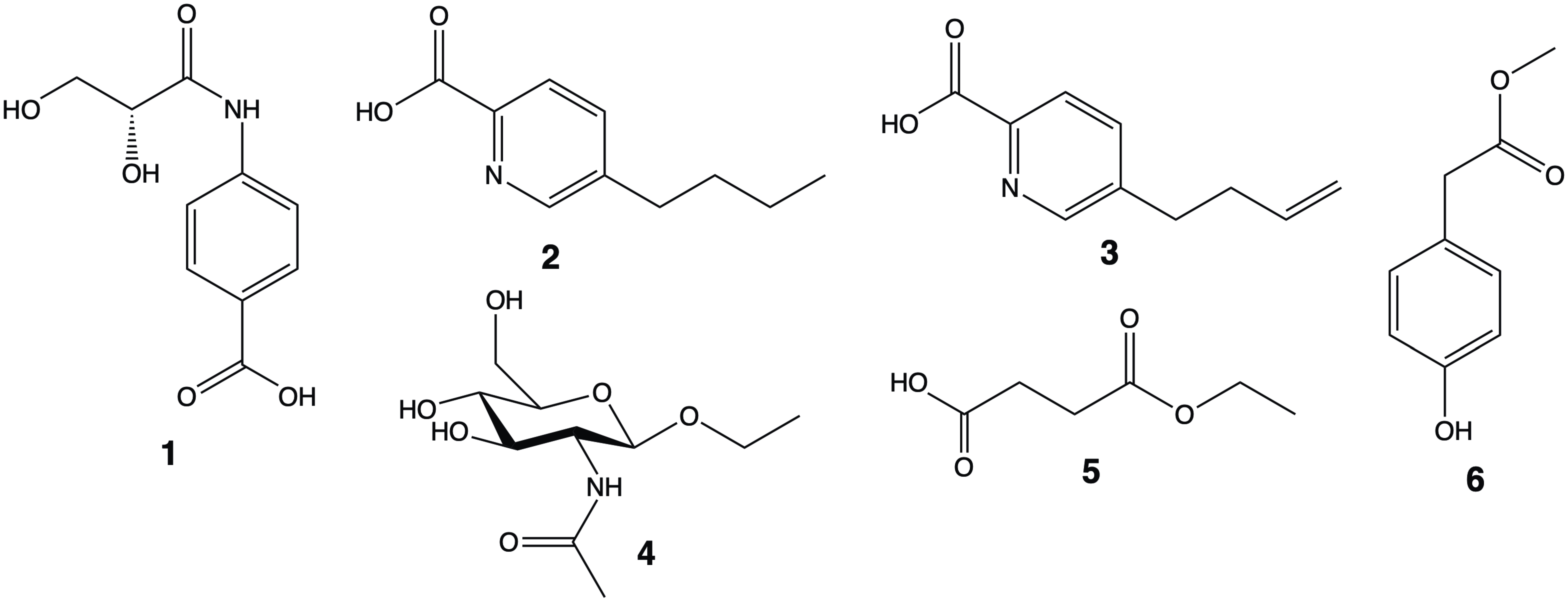
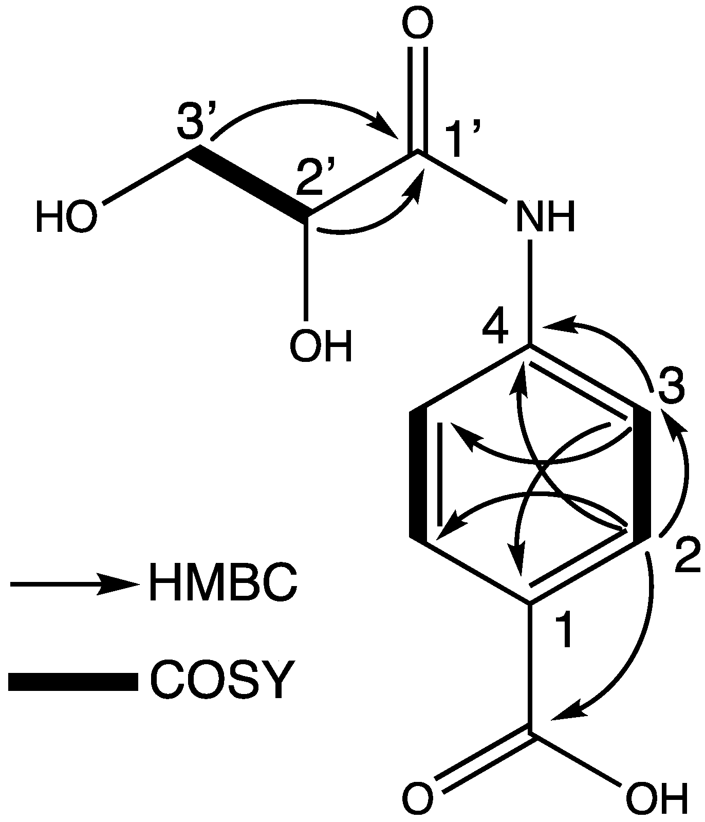
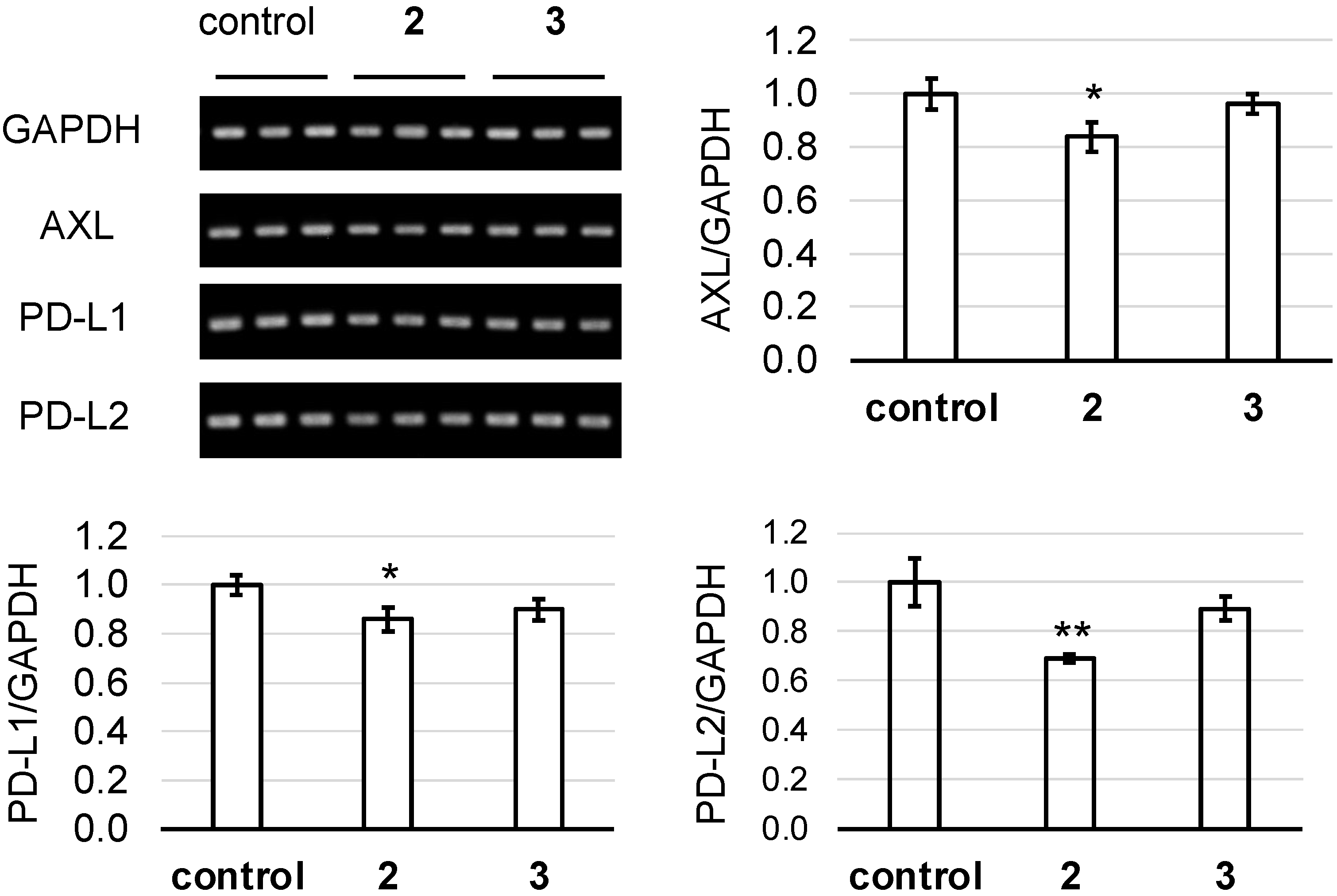
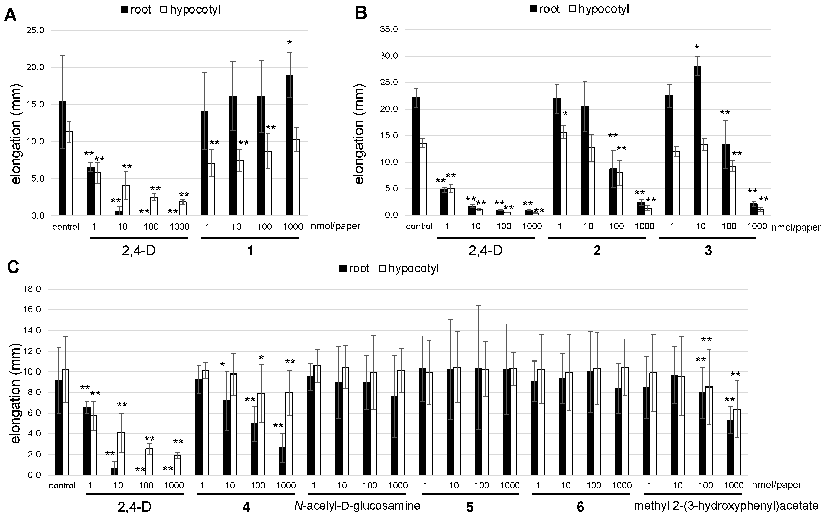
2. Results and Discussion

The culture broth of C. molybdites was partitioned between n-hexane and water, and then EtOAc and water, successively. The EtOAc-soluble parts were fractionated with repeated chromatography, and a novel compound (1) and two known compounds (2 and 3) were isolated (Figure 1). The fresh fruiting bodies of C. molybdites were extracted with EtOH and then with acetone. After the solutions were combined and concentrated, they were partitioned between n-hexane and water, EtOAc and water, and the water part concentrated under reduced pressure, and then extracted with EtOH, successively. The EtOAc-soluble part and the EtOH-soluble part were fractionated with repeated chromatography. As a result, three known compounds (46) were isolated (Figure 1).
Compound 1 was obtained as brown amorphous. The molecular formula was determined as C10H11NO5 with HRESIMS (m/z 224.0584 [M-H]; calcd for C10H10NO5, 224.0565), indicating the presence of six degrees of unsaturation in the molecule. The structure of 1 was elucidated through the interpretation of NMR spectra including DEPT, HMQC, COSY and HMBC (Figures S2–S5). The DEPT experiment indicated the presence of one methylene, five methines and four tetrasubstituted carbons including two carboxy groups (δC 171.0, 173.6). The complete assignment of all the protons and carbons was accomplished as shown in Table 1. The 4-aminobenzoic acid group was constructed based on the characteristic 1H and 13C NMR chemical shifts and coupling constants [1-COOH, δC 171.0; C-1, δC 129.5; C-2, 6, δH 7.96 (d, 8.4), δC 131.6; C-3, 5, δH 7.71 (d, 8.4), δC 120.4; C-4, δC 142.7), the HMBC correlations (H-2/C-3, C-4, C-6, 1-COOH; H-3/C-1, C-4, C-5) and the COSY correlations (H-2/H-3, H-5/H-6) (Figure 2). The 1H and 13C NMR chemical shifts [C-1′, δC 173.6; C-2′, δH 4.20 (dd, 4.6, 3.7), δC 74.6; C-3′, δH 3.80 (dd, 11.6, 4.6), 3.83 (dd, 11.6, 3.7), δC 65.3), the HMBC correlations (H-2′/C-1′, H-3′/C-1′), the COSY correlations (H-2′/H-3′) and the molecular formula suggested the presence of the 2,3-dihydroxypropanoate moiety and the amide bond (Figure 2). Hence, its planar structure was determined to be 4-(2,3-dihydroxypropanamido)benzoic acid. To determine the absolute configuration of 1, the specific rotation {[α]D23 +29 (c = 0.12, MeOH)} was compared with that of its analog, (R)-2,3-dihydroxy-N-(4-vinylphenyl)propenamide {[α]D20 +59.2 (c = 1.30, acetone)}, whose absolute configuration has been determined [47]. All the data allowed us to conclude that compound 1 was a novel compound, (R)-4-(2,3-dihydroxypropanamido)benzoic acid (Figure 1).
Compounds 2 and 3 were identified as fusaric acid and 9,10-dehydrofusaric acid by the comparison of their spectroscopic data with those reported [48]. Compounds 2 and 3 have been isolated from the culture filtrate of Fusarium nygamai, which showed a strong inhibition of root elongation of seedlings as well as wide chlorosis of tomato leaves rapidly evolving into necrosis [48]. Both the compounds were first isolated from mushroom-forming fungi. Compound 4 was identified as ethyl 2-acetylamino-2-deoxy-β-d-glucopyranoside, which was isolated from Aspergillus terreus and has growth-promoting activity for Lactobacillus bifidus var. pennsylvanicus [49]. This compound also was isolated from mushrooms for the first time. Compound 5, 4-ethoxy-4-oxobutanoic acid, has been isolated from the mushroom Trametes versicolor [50], which has insulinotropic action in rat islets [51]. Compound 6 was identified as methyl 4-hydroxyphenylacetate, which was isolated from the fungus Gloeophyllum odoratum and a marine fungus Penicillium oxalicum, and has potent inhibitory activity against tobacco mosaic virus [52,53].
The human A549 alveolar epithelial cell lines were treated with compounds 2 and 3. As shown in Figure 3, compound 3 showed no effects on all the gene expressions. However, compound 2 significantly inhibited the expression of Axl, PD-L1 and PD-L2. The difference in the activity between 2 and 3 indicated that a butyl group played an important role in the suppression of all three genes.
We also examined effect of compounds 2 and 3 on the growth of Pectobacterium carotovorum, Clavibacter michiganensis and Burkholderia glumae. Pectobacterium species are economically important plant pathogens and cause soft rot and blackleg disease on a range of plant species around the world [54,55]. Among the Pectobacterium species, P. carotovorum is a Gram-negative plant-specific pathogen and has the widest host range that causes soft rot disease in diverse plants [56]. Potato is the most important crop affected in temperate regions [54,57]. A Gram-positive plant pathogenic bacterium, C. michiganensis, is one of the most disruptive bacterial diseases of tomato [58]. This bacterium gives a serious threat to the processing and fresh market tomato industries and causes catastrophic epidemics in most tomato-growing areas of the world. In general, this vascular pathogen causes wilt and canker symptoms by invading and diffusing in the xylem through natural openings or wounds [59,60]. B. glumae, as a Gram-negative bacterium, was first described in Japan leading to grain rotting and seedling blight on rice [61,62], and is an emerging rice disease that greatly limits the productivity of rice [63]. We used ampicillin as a positive control that has anti-phytopathogenic-bacterial activity. As a result, compounds 2 and 3 inhibited the growth of P. carotovorum and C. michiganensis at 0.1 μmol/paper disc, while showing no activity against the growth of B. glumae (Figure 4).
In addition, the effect of compounds 16 and the analog compounds of 4 and 6 [N-acelyl-d-glucosamine and methyl 2-(3-hydroxyphenyl)acetate] on lettuce growth was evaluated (Figure 5). We used 2,4-dichlorophenoxyacetic acid (2,4-D) as a positive control, which inhibited the root and hypocotyl growth of lettuce dose-dependently. Compound 1 weakly promoted the root growth at 1 μmol/paper, while it inhibited the hypocotyl growth of lettuce at 1, 10 and 100 nmol/paper (Figure 5A). As for the root and hypocotyl growth of lettuce, compounds 24 and methyl 2-(3-hydroxyphenyl)acetate showed inhibition activity at 100 and 1000 nmol/paper, respectively(Figure 5B,C). Compounds 5, 6 and N-acelyl-d-glucosamine exhibited no activity (Figure 5C). Interestingly, a comparison of the structures between 4 and N-acelyl-d-glucosamine indicated that the ethoxy group at C-2 strengthened the lettuce-growth-inhibitory activity. The comparison between 6 and methyl 2-(3-hydroxyphenyl)acetate suggested that the hydroxy group at the meta position played an important role in the stronger inhibitory activity than at the para position.

3. Materials and Methods

3.1. General Experimental Procedures

1H NMR spectra (one- and two-dimensional) were recorded on a Jeol lambda-500 spectrometer or a JNM-ECZ500R spectrometer at 500 MHz, and 13C NMR spectra were recorded on the same instrument at 125 MHz (JEOL, Tokyo, Japan). HRESIMS spectra were measured on a JMS-T100LP mass spectrometer (JEOL, Tokyo, Japan). The specific rotation values were measured with a Jasco DIP-1000 polarimeter (Jasco, Tokyo, Japan). HPLC separations were performed with a Jasco Chromatography Data Station ChromNAV system using reverse-phase HPLC columns (CAPCELL PAK C18 AQ, Osaka soda, Osaka, Japan; COSMOSIL PBr, nacalai tesque, Kyoto, Japan; InerSustain Amide, InertSustain Phenyl, GL Science, Tokyo, Japan). Silica gel plate (Merck F254), ODS gel plate (Merck F254) and silica gel 60 N (Kanto Chemical, Tokyo, Japan) were used for analytical TLC and for flash column chromatography. All solvents used throughout the experiments were obtained from Kanto Chemical Co. (Tokyo, Japan).

3.2. Fungal Material

Fresh fruiting bodies of C. molybdites were collected at Tsu, Mie Prefecture, Japan, in 2015. The culture mycelium was isolated from the fruiting bodies successfully and then identified as C. molybdites by determining the internal transcribed spacer (ITS) regions of nuclear ribosomal DNA (rDNA) sequences deposited at NCBI BLAST (http://blast.ncbi.nlm.nih.gov/). The mycelia of C. molybdites were pre-cultured on potato dextrose agar (PDA), and the inoculated mycelia were incubated at 25°C for two weeks. After growth, 10 pieces (6 mm diameter) cut from the two-week-cultured mycelia were inoculated into 500 mL Erlenmeyer flasks containing 300 mL of PDB medium (n = 5), and the cultures were incubated for 4 weeks (25 °C, 120 rpm). Lettuce seeds (Lactuca sativa L. cv. Cisko; Takii Co., Ltd., Tokyo, Japan) were used in this study.

3.3. Extraction and Isolation

The culture broth of C. molybdites (1.5 L) was filtered and then concentrated under reduced pressure. The concentrate was successively partitioned between n-hexane and water (2 L each, twice), and then ethyl acetate (EtOAc) and water (2 L each, twice), to obtain the n-hexane-soluble part, EtOAc-soluble part, and water-soluble part. The EtOAc-soluble part (111.3 mg) was separated with Sep-Pak ODS (30% MeOH and MeOH). The 30% MeOH elution part (72.4 mg) was further fractionated using reverse-phase HPLC (COSMOSIL PBr, 20% MeOH) to yield 12 fractions (fractions CB-EtOAc-30MeOH-1 to CB-EtOAc-30MeOH-12). Compound 1 (1.2 mg) was isolated from fraction CB-EtOAc-30MeOH-9 (5.7 mg) with reverse-phase HPLC (CAPCELL PAK C18 AQ, 20%MeOH). The water-soluble part (2.8 g) was further extracted with EtOAc three times. The water-EtOAc-soluble part (215.7 mg) was separated with Sep-Pak ODS (30% MeOH and MeOH). Fraction CB-H2O-EtOAc-30MeOH (193.4 mg) was further fractionated using reverse-phase HPLC (CAPCELL PAK C18 AQ, 30% MeOH + 0.05%TFA) to yield 9 fractions (fractions CB-H2O-EtOAc-30MeOH-1 to CB-H2O-EtOAc-30MeOH-9). Fraction CB-H2O-EtOAc-30MeOH-7 was compound 2 (22.2 mg), and fraction CB-H2O-EtOAc-30MeOH-5 (26.3 mg) was further fractionated using reverse-phase HPLC (InertSustain Phenyl, 70% MeOH) to obtain compound 3 (15.6 mg).
The fresh fruiting bodies of C. molybdites (1.25 kg) were extracted with EtOH (5 L each, twice) and then with acetone (5 L each, twice). After the solutions were combined and concentrated under reduced pressure, the concentrate was partitioned between n-hexane and water, EtOAc and water, and the water part concentrated under reduced pressure, and then extracted with EtOH successively. The EtOH-soluble part (2.1 g) was fractionated using silica gel flash column chromatography (CH2Cl2, 70%, 50%, 30%, 10% CH2Cl2/MeOH; MeOH) to obtain 11 fractions (Fractions FB-EtOH-1 to FB-EtOH-11). Fraction FB-EtOH-5 (227.2 mg) was further fractionated with reverse-phase HPLC (CAPCELL PAK C18 AQ, 15% MeOH) to yield 10 fractions (Fractions FB-EtOH-5-1 to FB-EtOH-5-10). Compound 4 (0.8 mg) was isolated from fraction FB-EtOAc-5-3 (10.3 mg) with reverse-phase HPLC (InerSustain Amide, 95% MeCN). The EtOAc-soluble part (3.7 g) was fractionated using silica gel flash column chromatography (CH2Cl2, 90%, 70%, 50%, 30%, 10% CH2Cl2/acetone; acetone; 70%, 50%, 30%, 10% acetone/MeOH; MeOH) to obtain 13 fractions (Fractions FB-EtOAc-1 to FB-EtOAc-13). Fraction FB-EtOAc-6 (90.5 mg) was further fractionated with reverse-phase HPLC (COSMOSIL PBr, 60% MeOH) to obtain compound 5 (1.0 mg). Fraction FB-EtOAc-8 (80.8 mg) was further fractionated with reverse-phase HPLC (COSMOSIL PBr, 60% MeOH) to obtain compound 6 (1.0 mg).

3.4. Axl and Immune Checkpoint Molecule Assay [35]

The human A549 alveolar epithelial cell line was purchased from the American Type Culture Collection (Rockville, MD, USA) and cultured in DMEM, supplemented with 10% heat-inactivated fetal bovine serum, 2 mM L-glutamine and 100 U mL penicillin plus 100 μg/mL streptomycin. All cells were cultured at 37 °C in 75 cm2 flasks in an atmosphere composed of 5% CO2 and 95% air. Confluent cells were passaged after 5–7 days.
A549 cells in 0.1% BSA-DMEM were seeded in 24-well plates. Test compounds (20 μg/mL) were added to the wells, and the plates were incubated for 24 h. The total RNA was extracted using Sepasol®-RNA I Super G (Nacalai) following the instructions of the manufacturer. One μg of total RNA was denatured at 65°C for 10 min and then reverse-transcribed using ReverTra Ace Reverse Transcriptase (TOYOBO) and oligo (dT) primer in a volume of 20 μL according to the manufacturer’s protocol.
Each gene contains forward and reverse sequences (5′ > 3′), which are, respectively, GGAGCGAGATCCCTCCAAAAT and GGCTGTTGTCATACTTCTCATGG for the GADPH gene, TGCCATTGAGAGTCTAGCTGAC and TTAGCTCCCAGCACCGCGAC for the Axl gene, GGACAAGCAGTGACCATCAAG and CCCAGAATTACCAAGTGAGTCCT for the PD-L1 gene, and ACCGTGAAAGAGCCACTTTG and GCGACCCCATAGATGATTATGC for the PD-L2 gene. The cDNA was amplified using PCR and the conditions were as follows: 94 °C, 1 min; 60 °C, 1 min; and 72 °C, 1 min for 28–35 cycles. The PCR products were electrophoresed on a 1.5% agarose gel and then stained with an ethidium bromide solution. The semi-quantitative RT-PCR results were quantified using ImageJ software.
The data are expressed as the mean ± standard error of the mean (SEM). The statistical difference was calculated using analysis of variance with post hoc analysis using Fisher’s predicted least significant difference test. All statistics were performed using the StatView 5.0 package (Abacus Concepts, Berkeley, CA, USA).

3.5. Antibacterial Assay [40]

Each bacterium (C. michiganensis, B. glumae and P. carotovorum) was taken from the slant using an inoculation loop and suspended in 1 mL of sterile water in a 1.5 mL Eppendorf tube, and a suspension of 108 colony forming units (CFU)/mL was made with reference to the OD600. YP medium (yeast extract 5 g/L, peptone 10 g/L, agar 15 g/L) in a test tube was autoclaved for 20 min at 121 °C. The medium was left to stand until the temperature reached about 30 °C, and 100 μL of each bacterium suspension was added to the medium, and the mixture was poured into a Petri dish.
A total of 40 μL of a solution of each compound (0.1, 0.05 and 0.01 μmol in MeOH) and MeOH only (as control) were put on a paper disc (8 mm in diameter). After the discs were dried in the air, they were put on the medium. They were incubated for 3 days to evaluate their antibacterial activity.

3.6. Plant-Growth-Regulating Assay

Lettuce seeds were put on filter paper (Advantec No. 2, ϕ 55 mm; Toyo Roshi Kaisha, Japan), soaked in distilled water in a Petri dish (ϕ 60 × 20 mm) and incubated in a growth chamber in the dark at 25 °C for 1 day. Each sample was dissolved in 1 mL of MeOH (1, 10, 100 and 1000 nmol/mL) and then poured on filter paper (ϕ 55 mm) in a Petri dish (ϕ 60 × 20 mm). After the sample-loaded paper had been air-dried, 1 mL of distilled water was poured on the sample-loaded paper or intact filter paper (control). The preincubated lettuce seedlings (n = 9 in each Petri dish) were transferred onto the sample-loaded filter paper or control filter paper and incubated in a growth chamber in the dark at 25 °C for 3 days. The lengths of the hypocotyl and the root were measured using a ruler.

4. Conclusions

A novel compound (1) along with two known compounds (2 and 3) were isolated from the culture broth of C. molybdites, and three known compounds (46) were isolated form the fruiting bodies of the fungus. Compounds 24 were isolated from a mushroom for the first time. Compound 2 showed significant inhibition activity against Axl and immune checkpoint genes (PD-L1, PD-L2) that are the most promising candidates for cancer therapy. In addition, compounds 2 and 3 inhibited the growth of P. carotovorum and C. michiganensis. Compounds 14 showed the significant regulatory activity of lettuce growth (Figure 6).

Supplementary Materials

The following supporting information can be downloaded at: https://www.mdpi.com/article/10.3390/antibiotics12030596/s1, Figure S1: 1H NMR spectrum of 1 (CD3OD); Figure S2: 13C NMR and DEPT spectra of 1 (CD3OD); Figure S3: HMQC spectrum of 1 (CD3OD); Figure S4: HMBC spectrum of 1 (CD3OD); Figure S5: COSY spectrum of 1 (CD3OD); Figure S6: 1H NMR spectrum of 2 (CD3OD); Figure S7: 13C NMR spectrum of 2 (CD3OD); Figure S8: 1H NMR spectrum of 3 (CD3OD); Figure S9: 13C NMR spectrum of 3 (CD3OD); Figure S10: 1H NMR spectrum of 4 (CD3OD); Figure S11: 13C NMR spectrum of 4 (CD3OD); Figure S12: 1H NMR spectrum of 5 (CD3OD); Figure S13: 13C NMR spectrum of 5 (CD3OD); Figure S14: 1H NMR spectrum of 6 (CD3OD); Figure S15: 13C NMR spectrum of 6 (CD3OD).

Author Contributions

Conceiving of the project, H.K. (Hirokazu Kawagishi).; methodology, J.W. (Jing Wu), H.H. and J.-H.C.; fungus identification, H.K. (Hajime Kobori).; Axl and immune checkpoint molecule inhibitory assay, C.N.D.-G., M.T., T.Y. and E.C.G.; plant growth regulatory assay and anti-phytopathogenic-bacterial assay, J.W. (Jing Wu), T.O., R.O., J.W. (Junhong Wang) and Y.T.; writing—original draft preparation, J.W. (Jing Wu); writing—review and editing, H.K. (Hirokazu Kawagishi); funding acquisition, J.W. (Jing Wu), J.-H.C. and H.K. (Hirokazu Kawagishi). All authors have read and agreed to the published version of the manuscript.

Funding

This work was partially supported by JST, ACT-X Grant Number JPMJAX2115, Japan to J. W., by KAKENHI (19K22330) to J.-H.C and by Specific Research Grant from Takeda Science Foundation to H. Ka.

Institutional Review Board Statement

Not applicable.

Informed Consent Statement

Not applicable.

Data Availability Statement

Not applicable.

Conflicts of Interest

The authors declare no conflict of interest.

References

  1. Chugh, R.M.; Mittal, P.; Mp, N.; Arora, T.; Bhattacharya, T.; Chopra, H.; Cavalu, S.; Gautam, R.K. Fungal Mushrooms: A Natural Compound With Therapeutic Applications. Front. Pharmacol. 2022, 13, 925387. [Google Scholar] [CrossRef]
  2. Wu, J.; Kawagishi, H. Plant growth regulators from mushrooms. J. Antibiot. 2020, 73, 657–665. [Google Scholar] [CrossRef]
  3. Blackwell, M. The Fungi: 1, 2, 3…5.1 Million Species? Am. J. Bot. 2011, 98, 426–438. [Google Scholar] [CrossRef]
  4. Strassert, J.F.H.; Monaghan, M.T. Phylogenomic insights into the early diversification of fungi. Curr. Biol. 2022, 32, 3628–3635. [Google Scholar] [CrossRef] [PubMed]
  5. Gargano, M.L.; Van Griensven, L.J.L.D.; Isikhuemhen, O.S.; Lindequist, U.; Venturella, G.; Wasser, S.P.; Zervakis, G.I. Medicinal mushrooms: Valuable biological resources of high exploitation potential. Plant Biosyst. 2017, 151, 548–565. [Google Scholar] [CrossRef]
  6. Al-Rajhi, A.M.H.; Alawlaqi, M.M.; Abdel Ghany, T.M.; Moawad, H. Amanita sp. from Subtropical Region of Saudi Arabia as a Source of Chitinase Enzyme and its Antifungal Activity. BioRersources 2023, 18, 2928–2939. [Google Scholar] [CrossRef]
  7. Soliman, A.M.; Younis, A.M.; Abdelgany, T.M.; Abdelbary, S. Trends in Assessment of Ganoderma lucidum Methanol extract Against MRSA Infection In Vitro and In Vivo with Nutrition Support. J. Adv. Pharm. Res. 2022, 6, 46–57. [Google Scholar] [CrossRef]
  8. Kawagishi, H. Chemical studies on bioactive compounds related to higher fungi. Biosci. Biotechnol. Biochem. 2021, 85, 1–7. [Google Scholar] [CrossRef] [PubMed]
  9. World Health Organization. Cancer. Available online: https://www.who.int/news-room/fact-sheets/detail/cancer (accessed on 29 August 2022).
  10. Park, H.J. Current uses of mushrooms in cancer treatment and their anticancer mechanisms. J. Mol. Sci. 2022, 23, 10502–10516. [Google Scholar] [CrossRef] [PubMed]
  11. Wu, G.-S.; Guo, J.-J.; Bao, J.-L.; Li, X.-W.; Chen, X.-P.; Lu, J.-J.; Wang, Y.-T. Anti-cancer properties of triterpenoids isolated from Ganoderma lucidum-A review. Expert Opin. Investig. Drugs 2013, 22, 981–992. [Google Scholar] [CrossRef]
  12. Boh, B.; Berovic, M.; Zhang, J.; Zhi-Bin, L. Ganoderma lucidum and its pharmaceutically active compounds. Biotechnol. Annu. Rev. 2007, 13, 265–301. [Google Scholar] [CrossRef]
  13. Joseph, T.P.; Chanda, W.; Padhiar, A.A.; Batool, S.; LiQun, S.; Zhong, M.; Huang, M. A preclinical evaluation of the antitumor activities of edible and medicinal mushrooms: A molecular insight. Integr. Cancer Ther. 2018, 17, 200–209. [Google Scholar] [CrossRef] [PubMed] [Green Version]
  14. Kawagishi, H.; Kanao, T.; Inagaki, R.; Mizuno, T.; Shimura, K.; Ito, H.; Hagiwara, T.; Nakamura, T. Formolysis of a potent antitumor (1-6)-β-d-glucan-protein complex from Agaricus blazei fruiting bodies and antitumor activity of the resulting products. Carbohydr. Polym. 1990, 12, 393–403. [Google Scholar] [CrossRef]
  15. Katsayal, U.A.; Abdurahman, E.M.; Abubakar, M.S.; Musa, K.Y.; Ambali, S.F.; Jahun, M.B. Fungi as a potential source of antimalarial agents. Niger. J. Pharm. Sci. 2009, 8, 135–142. [Google Scholar]
  16. Bala, N.; Aitken, E.; Fechner, N.; Cusack, A.; Steadman, K.J. Evaluation of antibacterial activity of Australian basidiomycetous macrofungi using a high-throughput 96-well plate assay. Pharm. Biol. 2011, 49, 492–500. [Google Scholar] [CrossRef] [PubMed] [Green Version]
  17. Yoshikawa, K.; Ikuta, M.; Arihara, S.; Matsumura, E.; Katayama, S. Two new steroidal derivatives from the fruit body of Chlorophyllum molybdites. Chem. Pharm. Bull. 2001, 49, 1030–1032. [Google Scholar] [CrossRef] [Green Version]
  18. Ohta, T.; Inoue, H.; Kusano, G.; Oshima, Y. Lepiotins A and B, new alkaloids from the mushrooms, Macrolepiota neomastoidea and Chlorophyllum molybdites. Heterocycles 1998, 47, 883–891. [Google Scholar] [CrossRef]
  19. Yamada, M.; Tokumitsu, N.; Saikawa, Y.; Nakata, M.; Asano, J.; Miyairi, K.; Okuno, T.; Konno, K.; Hashimoto, K. Molybdophyllysin, a toxic metalloendopeptidase from the tropical toadstool, Chlorophyllum molybdites. Bioorg. Med. Chem. 2012, 20, 6583–6588. [Google Scholar] [CrossRef]
  20. Kobayashi, Y.; Kobayashi, K.; Umehara, K.; Dohra, H.; Murata, T.; Usui, T.; Kawagishi, H. Purification, characterization, and sugar binding specificity of an N-glycolylneuraminic acid-specific lectin from the mushroom Chlorophyllum molybdites. J. Biol. Chem. 2004, 279, 53048–53055. [Google Scholar] [CrossRef] [PubMed] [Green Version]
  21. Sung, H.; Ferlay, J.; Siegel, R.L.; Laversanne, M.; Soerjomataram, I.; Jemal, A.; Bray, F. Global Cancer Statistics 2020: GLOBOCAN Estimates of Incidence and Mortality Worldwide for 36 Cancers in 185 Countries. CA Cancer J. Clin. 2021, 71, 209–249. [Google Scholar] [CrossRef]
  22. Topalian, S.L.; Hodi, F.S.; Brahmer, J.R.; Gettinger, S.N.; Smith, D.C.; McDermott, D.F.; Powderly, J.D.; Carvajal, R.D.; Sosman, J.A.; Atkins, M.B.N. Safety, activity, and immune correlates of anti-PD-1 antibody in cancer. N. Engl. J. Med. 2021, 366, 2443–2454. [Google Scholar] [CrossRef] [PubMed] [Green Version]
  23. Chang, E.; Pelosof, L.; Lemery, S.; Gong, Y.; Goldberg, K.B.; Farrell, A.T.; Keegan, P.; Veeraraghavan, J.; Wei, G.; Blumenthal, G.M. Systematic Review of PD-1/PD-L1 Inhibitors in Oncology: From Personalized Medicine to Public Health. Oncologist 2021, 26, e1786–e1799. [Google Scholar] [CrossRef]
  24. Wang, Z.; Wu, X. Study and analysis of antitumor resistance mechanism of PD1/PD-L1 immune checkpoint blocker. Cancer Med. 2020, 9, 8086–8121. [Google Scholar] [CrossRef]
  25. Hong, J.; Peng, D.; Chen, Z.; Sehdev, V.; Belkhiri, A. ABL regulation by AXL promotes cisplatin resistance in esophageal cancer. Cancer Res. 2012, 73, 331–340. [Google Scholar] [CrossRef] [Green Version]
  26. Liu, L.; Greger, J.; Shi, H.; Liu, Y.; Greshock, J.; Annan, R.; Halsey, W.; Sathe, G.M.; Martin, A.M.; Gilmer, T.M. Novel mechanism of lapatinib resistance in HER2-positive breast tumor cells: Activation of AXL. Cancer Res. 2009, 69, 6871–6878. [Google Scholar] [CrossRef] [PubMed] [Green Version]
  27. Van der Meer, J.H.; Van der Poll, T.; Van’t Veer, C. TAM receptors, Gas6, and protein S: Roles in inflammation and hemostasis. Blood 2014, 123, 2460–2469. [Google Scholar] [CrossRef] [PubMed]
  28. Berclaz, G.; Altermatt, H.J.; Rohrbach, V.; Kieffer, I.; Dreher, E.; Andres, A.C. Estrogen dependent expression of the receptor tyrosine kinase axl in normal and malignant human breast. Ann. Oncol. 2001, 12, 819–824. [Google Scholar] [CrossRef] [PubMed]
  29. Tang, S.; Kim, P.S. A high-affinity human PD-1/PD-L2 complex informs avenues for small-molecule immune checkpoint drug discovery. Proc. Natl. Acad. Sci. USA 2019, 116, 24500–24506. [Google Scholar] [CrossRef] [Green Version]
  30. Zak, K.M.; Grudnik, P.; Magiera, K.; Dömling, A.; Dubin, G.; Holak, T.A. Structural biology of the immune checkpoint receptor PD-1 and its ligands PD-L1/PD-L2. Structure 2017, 25, 1163–1174. [Google Scholar] [CrossRef]
  31. Azuma, K.; Ota, K.; Kawahara, A.; Hattori, S.; Iwama, E.; Harada, T. Association of PD-L1 overexpression with activating EGFR mutations in surgically resected nonsmall-cell lung cancer. Ann Oncol. 2014, 25, 1935–1940. [Google Scholar] [CrossRef]
  32. Colavito, S.A. AXL as a target in breast cancer therapy. J. Oncol. 2020, 2020, 5291952. [Google Scholar] [CrossRef] [Green Version]
  33. Aehnlich, P.; Powell, R.M.; Peeters, M.J.W.; Rahbech, A.; Thor Straten, P. TAM receptor inhibition-implications for cancer and the immune system. Cancers 2021, 13, 1195–1219. [Google Scholar] [CrossRef]
  34. Myers, S.H.; Brunton, V.G.; Unciti-Broceta, A. AXL inhibitors in cancer: A medicinal chemistry perspective. J. Med. Chem. 2016, 59, 3593–3608. [Google Scholar] [CrossRef] [Green Version]
  35. Kodajima, M.; Choi, J.H.; Kondo, M.; CN, D.A.-G.; Toda, M.; Yasuma, T.; Gabazza, E.C.; Miwa, Y.; Shoda, C.; Lee, D.; et al. Axl, immune checkpoint molecules and HIF inhibitors from the culture broth of Lepista luscina. Molecules 2022, 27, 8925–8936. [Google Scholar] [CrossRef] [PubMed]
  36. Crous, P.W.; Rossman, A.Y.; Aime, M.C.; Allen, W.C.; Burgess, T.; Groenewald, J.Z.; Castlebury, L.A. Names of Phytopathogenic Fungi: A Practical Guide. Phytopathology 2021, 111, 1500–1508. [Google Scholar] [CrossRef]
  37. Abdel Ghany, T.M. Fungal leaf spot of maize: Pathogen isolation, identification and host biochemical characterization. Mycopath 2012, 10, 41–49. [Google Scholar]
  38. Abd EL-Ghany, T.M.; Eman, W. EI-Taher; EI-Sheikh, H.H. Efficacy of fungal Rust Disease on Willow Plant in Egypt. Aust. J. Basic Appl. Sci. 2009, 3, 1527–1539. [Google Scholar]
  39. Riyaz, M.; Shah, R.A.; Sivasankaran, K. Pesticide Residues: Impacts on Fauna and the Environment. In Biodegradation Technology of Organic and Inorganic Pollutants; InterchOpen: London, UK, 2021. [Google Scholar] [CrossRef]
  40. Wang, J.; Wu, J.; Ogura, Y.; Kobori, H.; Choi, J.-H.; Hirai, H.; Takikawa, Y.; Kawagishi, H. Anti-phytopathogenic bacterial fatty acids from the mycelia of the edible mushroom Agaricus blazei. Biosci. Biotechnol. Biochem. 2022, 86, 1327–1332. [Google Scholar] [CrossRef] [PubMed]
  41. Couch, H.B. Diseases of Turfgrasses, 3rd ed.; Krieger: Malabar, FL, USA, 1995. [Google Scholar]
  42. Mitchinson, A. Fairy chemicals. Nature 2014, 505, 298. [Google Scholar] [CrossRef]
  43. Kawagishi, H. Are fairy chemicals a new family of plant hormones? Proc. Jpn. Acad., Ser. B 2019, 95, 29–38. [Google Scholar] [CrossRef] [Green Version]
  44. Lee, D.; Miwa, Y.; Wu, J.; Shoda, C.; Jeong, H.; Kawagishi, H.; Tsubota, K.; Kurihara, T. A Fairy Chemical Suppresses Retinal Angiogenesis as a HIF Inhibitor. Biomolecules 2020, 10, 1405. [Google Scholar] [CrossRef]
  45. Aoshima, H.; Ito, S.; Ibuki, R.; Kawagishi, H. Potential of Fairy Chemicals as Functional Cosmetic Ingredients: Effect of 2-Aza-8-Oxohypoxanthine on Skin Lightness. Int. J. Med. Mushrooms 2022, 24, 41–47. [Google Scholar] [CrossRef]
  46. Inoue, C.; Yasuma, T.; D’Alessandro-Gabazza, C.N.; Toda, M.; Fridman D’Alessandro, V.; Inoue, R.; Fujimoto, H.; Kobori, H.; Tharavecharak, S.; Takeshita, A.; et al. The Fairy Chemical Imidazole-4-carboxamide Inhibits the Expression of Axl, PD-L1, and PD-L2 and Improves Response to Cisplatin in Melanoma. Cells 2022, 11, 374. [Google Scholar] [CrossRef]
  47. Wulff, G.; Sarhan, A.; Gimpel, J.; Lohmar, E. Über enzymanalog gebaute Polymere, III. Zur Synthese von polymerisierbaren D-Glycerinsäurederivaten. Chem. Ber. 1974, 107, 3364–3376. [Google Scholar] [CrossRef]
  48. Capasso, R.; Evidente, A.; Cutignano, A.; Vurro, M.; Zonno, M.C.; Bottalico, A. Fusaric and 9,10-dehydrofusaric acids and their methyl esters from Fusarium nygamai. Phytochemistry 1996, 41, 1035–1039. [Google Scholar] [CrossRef]
  49. Lambert, R.; Zilliken, F. Novel growth faxtors for Lactobacillus bifidus var pennsylvanicus. Arch. Biochem. Biophys. 1965, 110, 544–550. [Google Scholar] [CrossRef]
  50. Durak, H. Trametes versicolor (L.) mushrooms liquefaction in supercritical solvents: Effects of operating conditions on product yields and chromatographic characterization. J. Supercrit. Fluids 2018, 131, 140–149. [Google Scholar] [CrossRef]
  51. Björklingf, F.; Malaisse-Lagaew, F.; Malaisse, W.J. Insulinotropic action of novel succinic acid esters. Pharmacol. Res. 1996, 33, 273–275. [Google Scholar] [CrossRef]
  52. Rödecke, J.; König, W.A. Odorous compounds from the fungus Gloeophyllum odoratum. Flavour Fragr. J. 2000, 15, 315–319. [Google Scholar] [CrossRef]
  53. Shen, S.; Li, W.; Wang, J. A novel and other bioactive seondary metabolites from a marine fungus Penicillium oxalicum 0312F1. Nat. Prod. Res. 2013, 27, 2286–2291. [Google Scholar] [CrossRef]
  54. Pérombelon, M.C.M. Potato diseases caused by soft rot erwinias: An overview of pathogenesis. Plant Pathol. 2002, 51, 1–12. [Google Scholar] [CrossRef]
  55. Czajkowski, R.; Pérombelon, M.C.M.; van Veen, J.A.; van der Wolf, J.M. Control of blackleg and tuber soft rot of potato caused by Pectobacterium and Dickeya species: A review. Plant Pathol. 2011, 60, 999–1013. [Google Scholar] [CrossRef]
  56. Roh, E.; Park, T.H.; Kim, M.; Lee, S.; Ryu, S.; Oh, C.S.; Rhee, S.; Kim, D.H.; Park, B.S.; Heu, S. Characterization of a new bacteriocin, Carocin D, from Pectobacterium carotovorum subsp. carotovorum Pcc21. Environ. Microbiol. 2010, 76, 7541–7549. [Google Scholar] [CrossRef] [Green Version]
  57. Toth, I.K.; Bell, K.S.; Holeva, M.C.; Birch, P.R.J. Soft rot erwiniae: From genes to genomes. Mol. Plant Pathol. 2003, 4, 17–30. [Google Scholar] [CrossRef]
  58. Mansfield, J.; Genin, S.; Magori, S.; Citovsky, V.; Sriariyanum, M.; Ronald, P.; Dow, M.; Verdier, V.; Beer, S.V.; Machado, M.A.; et al. Top 10 plant pathogenic bacteria in molecular plant pathology. Mol. Plant Pathol. 2012, 13, 614–629. [Google Scholar] [CrossRef] [PubMed] [Green Version]
  59. Nandi, M.; Macdonald, J.; Liu, P.; Weselowski, B.; Yuan, Z.C. Clavibacter michiganensis ssp. michiganensis: Bacterial canker of tomato, molecular interactions and disease management. Mol. Plant Pathol. 2018, 19, 2036–2050. [Google Scholar] [CrossRef] [Green Version]
  60. Davis, M.J.; Graves Gillaspie, A., Jr.; Vidaver, A.K.; Harris, R.W. Clavibacter: A new genus containing some phytopathogenic coryneform bacteria, Including Clavibacter xyli subsp. xyli sp. nov., subsp. nov. and Clavibacter xyli subsp. cynodontis subsp. nov., Pathogens that cause ratoon stunting disease of sugarcane and bermudagrass stunting disease. J. Syst. Bacteriol. 1984, 34, 107–117. [Google Scholar] [CrossRef] [Green Version]
  61. Gartemann, K.H.; Kirchner, O.; Engermann, J.; Gräfen, I.; Eichenlaub, R.; Burger, A. Clavibacter michiganensis subsp. michiganensis: First steps in the understanding of virulence of a Gram-positive phytopathogenic bacterium. J. Biotechnol. 2003, 106, 179–191. [Google Scholar] [CrossRef] [PubMed]
  62. Goto, K.; Ohata, K. New bacterial diseases of rice (brown stripe and grain rot). Ann. Phytopathol. Soc. Jpn. 1956, 21, 46–47. [Google Scholar]
  63. Kumar, S.; Mondal, K.K.; Ghoshal, T.; Kulshreshtha, A.; Sreenayana, B.; Amrutha Lakshmi, M.; Mrutyunjaya, S.; Rashmi, E.R.; Kalaivanan, N.S.; Mani, C. Genetic and pathogenic diversity analysis of Burkholderia glumae strains from Indian hot spot regions causing bacterial panicle blight of rice (Oryza sativa L.). Trop Plant Pathol. 2023. [Google Scholar] [CrossRef]
Figure 1. Structures of compounds 16.
Antibiotics 12 00596 g001
Figure 2. HMBC and COSY correlations of 1.
Antibiotics 12 00596 g002
Figure 3. Effect of compounds 2 and 3 on expression of Axl and immune checkpoint molecules (PD-L1 and PD-L2) in A549 cells. Values indicate means with standard deviation. Statistical analysis was performed using Fisher’s PLSD test (* p < 0.05, ** p < 0.01 vs. control, n = 3).
Antibiotics 12 00596 g003
Figure 4. Activity of 2 and 3 against Clavibacter michiganensis, Pectobacterium carotovorum and Burkholderia glumae (positive control, ampicillin).
Antibiotics 12 00596 g004
Figure 5. Effect of 1 to 6, N-acelyl-d-glucosamine and methyl 2-(3-hydroxyphenyl)acetate on the growth of lettuce. (A). compound 1; (B). compounds 2 and 3; (C). compounds 46 and their analog compounds. Lettuce seedlings were treated with compounds. Respective length of growth compared with the control ± standard deviation (* p < 0.05, ** p < 0.01 vs. control, n = 9).
Antibiotics 12 00596 g005
Figure 6. Summary of this study.
Antibiotics 12 00596 g006
Table 1. 1H and 13C NMR Data for 1 in CD3OD.
PositionδH (J inHz)δc
1-COOH171.0
1129.5
2, 67.96 (d, 8.4)131.6
3, 57.71 (d, 8.4)120.4
4142.7
1′173.6
2′4.20 (dd, 4.6, 3.7)74.6
3′3.80 (dd, 11.6, 4.6)
3.83 (dd, 11.6, 3.7)
65.3
Disclaimer/Publisher’s Note: The statements, opinions and data contained in all publications are solely those of the individual author(s) and contributor(s) and not of MDPI and/or the editor(s). MDPI and/or the editor(s) disclaim responsibility for any injury to people or property resulting from any ideas, methods, instructions or products referred to in the content.

