
Biosynthesised Silver Nanoparticles Loading onto Biphasic Calcium Phosphate for Antibacterial and Bone Tissue Engineering Applications


 and
and
Abstract
:1. Introduction
2. Materials and Methods
2.1. Formation of Ag NPs
2.2. Synthesis of Ag-Decorated BCP
2.3. Instrumentation
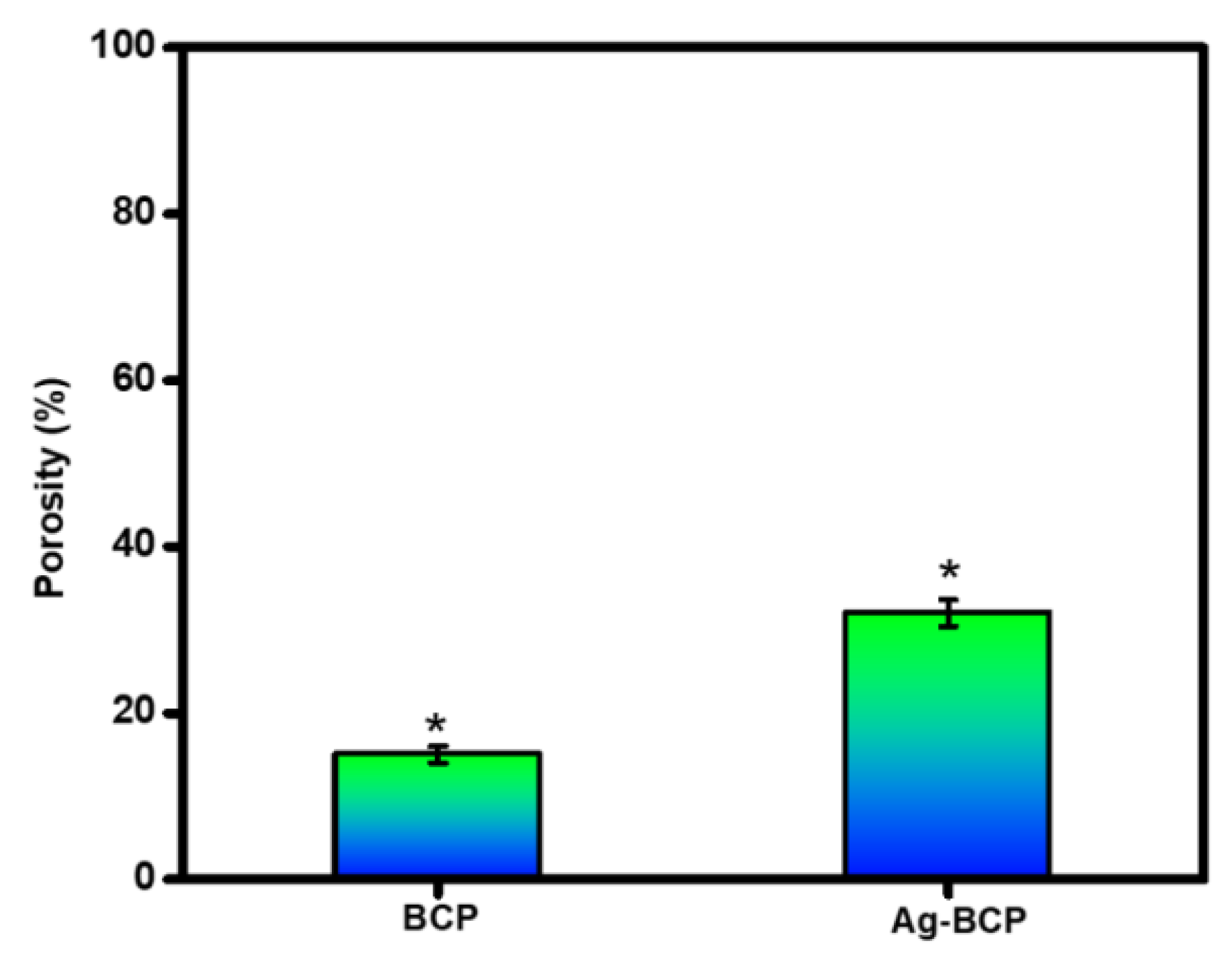
2.4. Measurement of Porosity
2.5. In-Vitro Bioactivity and Biodegradation Studies
2.6. Studies of Drug Loading and Release
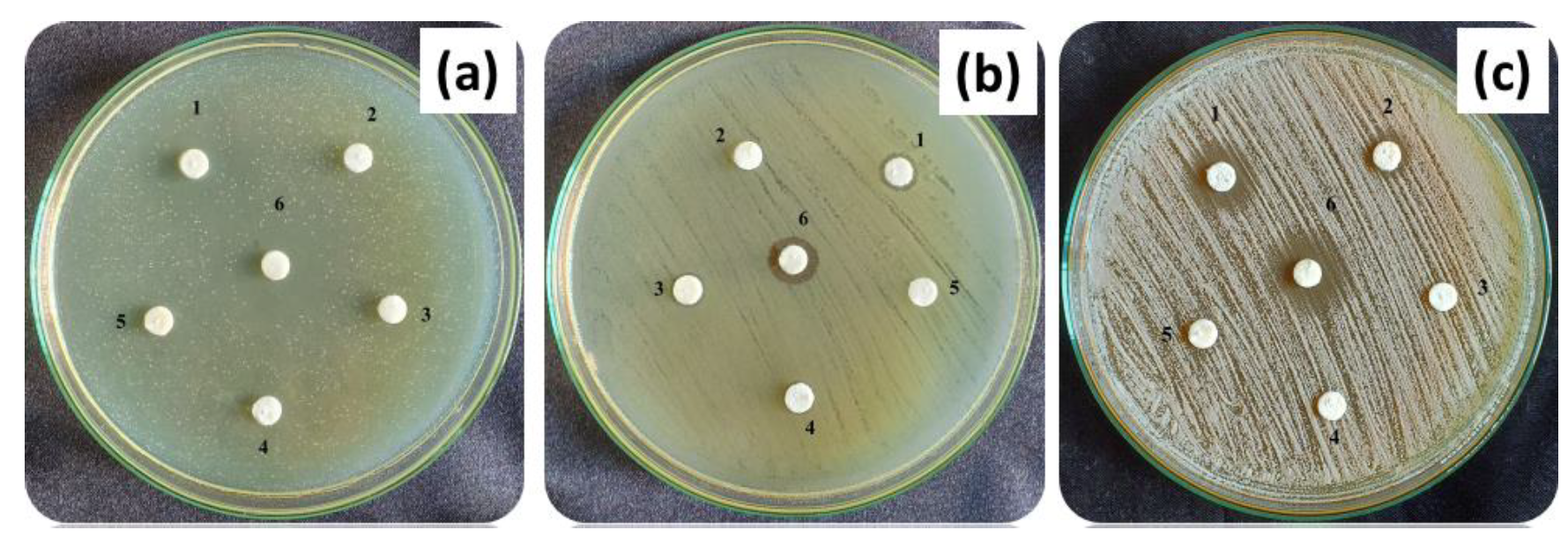
2.7. Antibacterial and Antifungal Activity
2.8. In Vitro Cell Viability Assay
2.9. Statistical Analysis
3. Results and Discussion
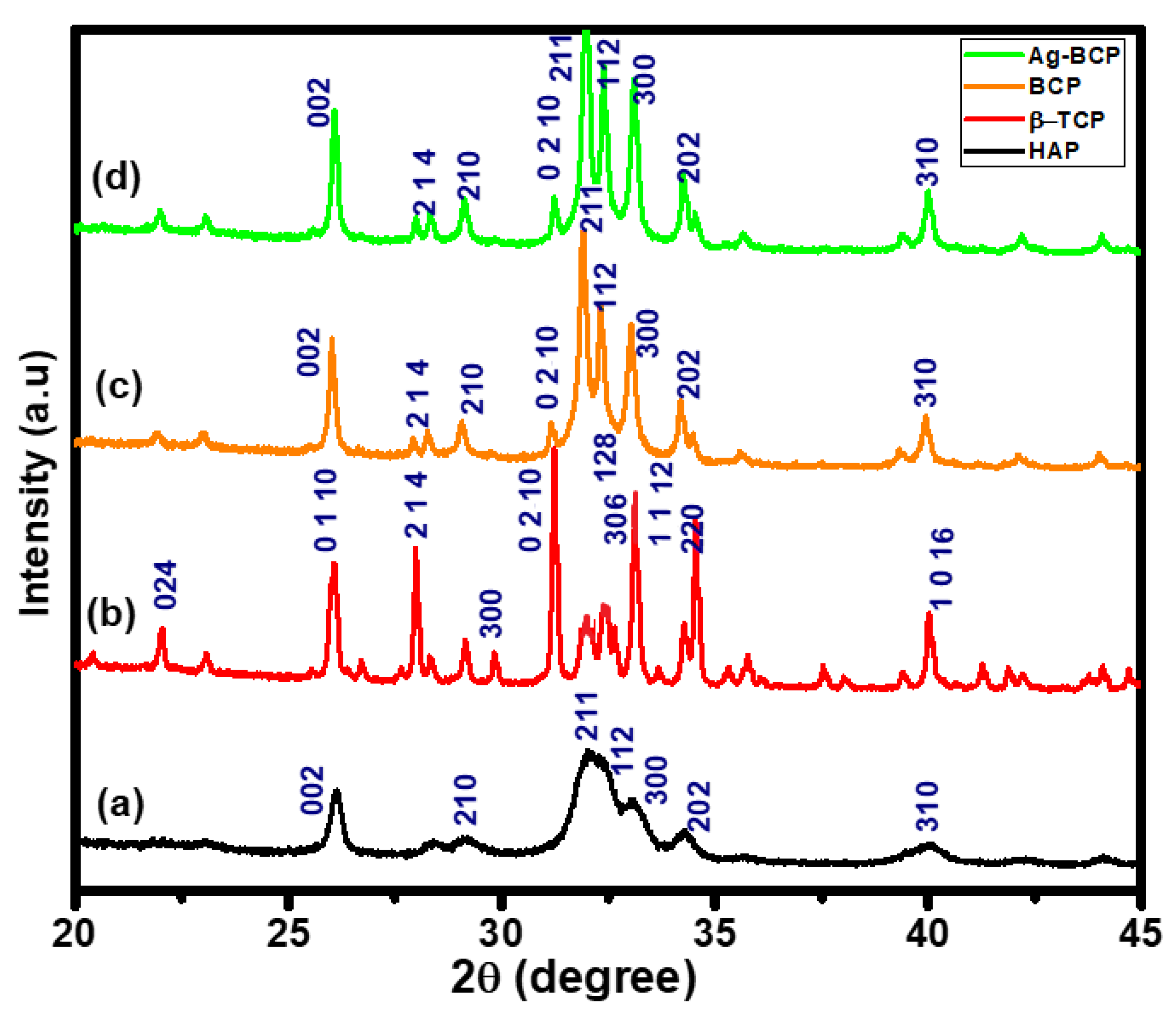
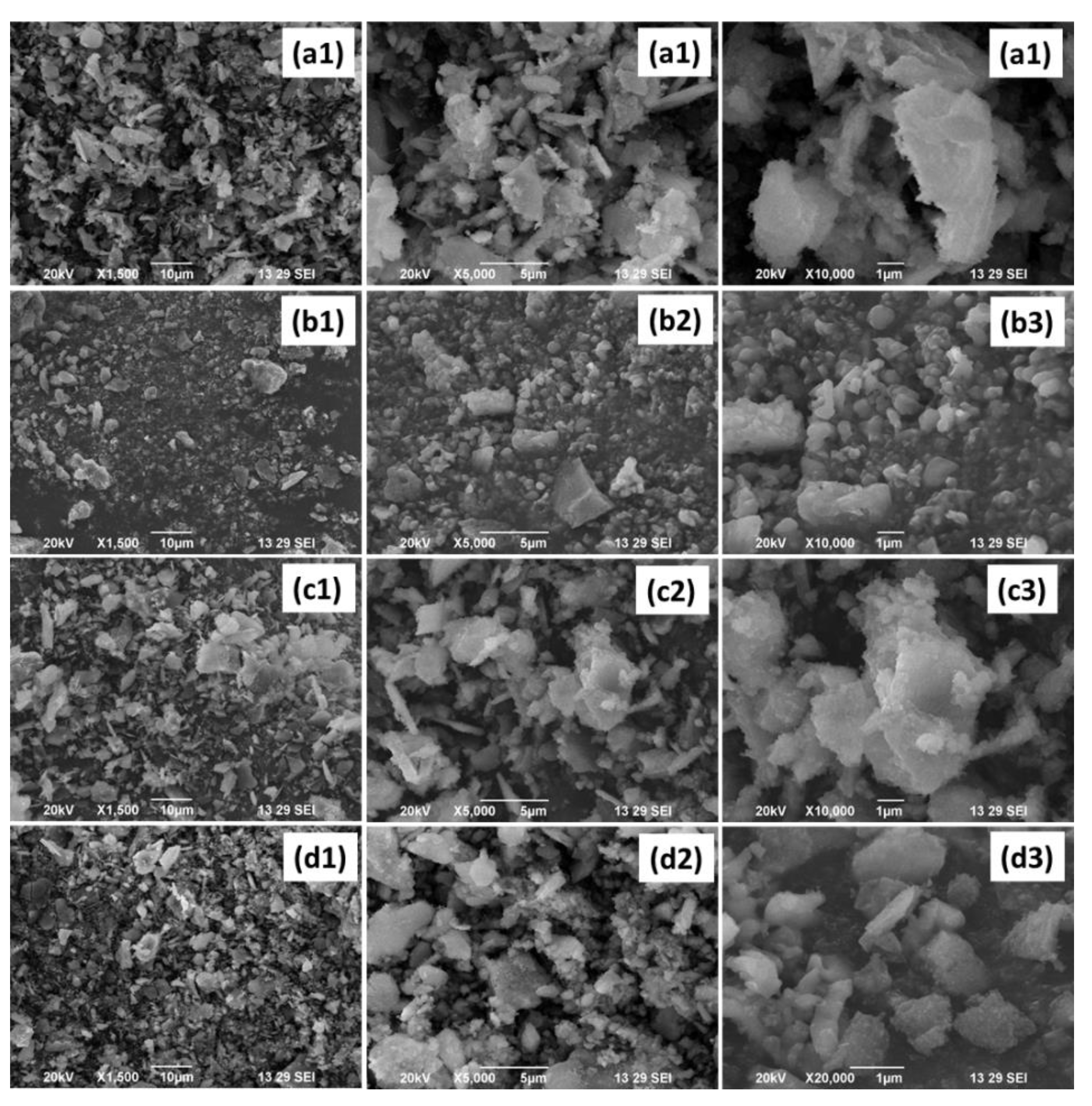
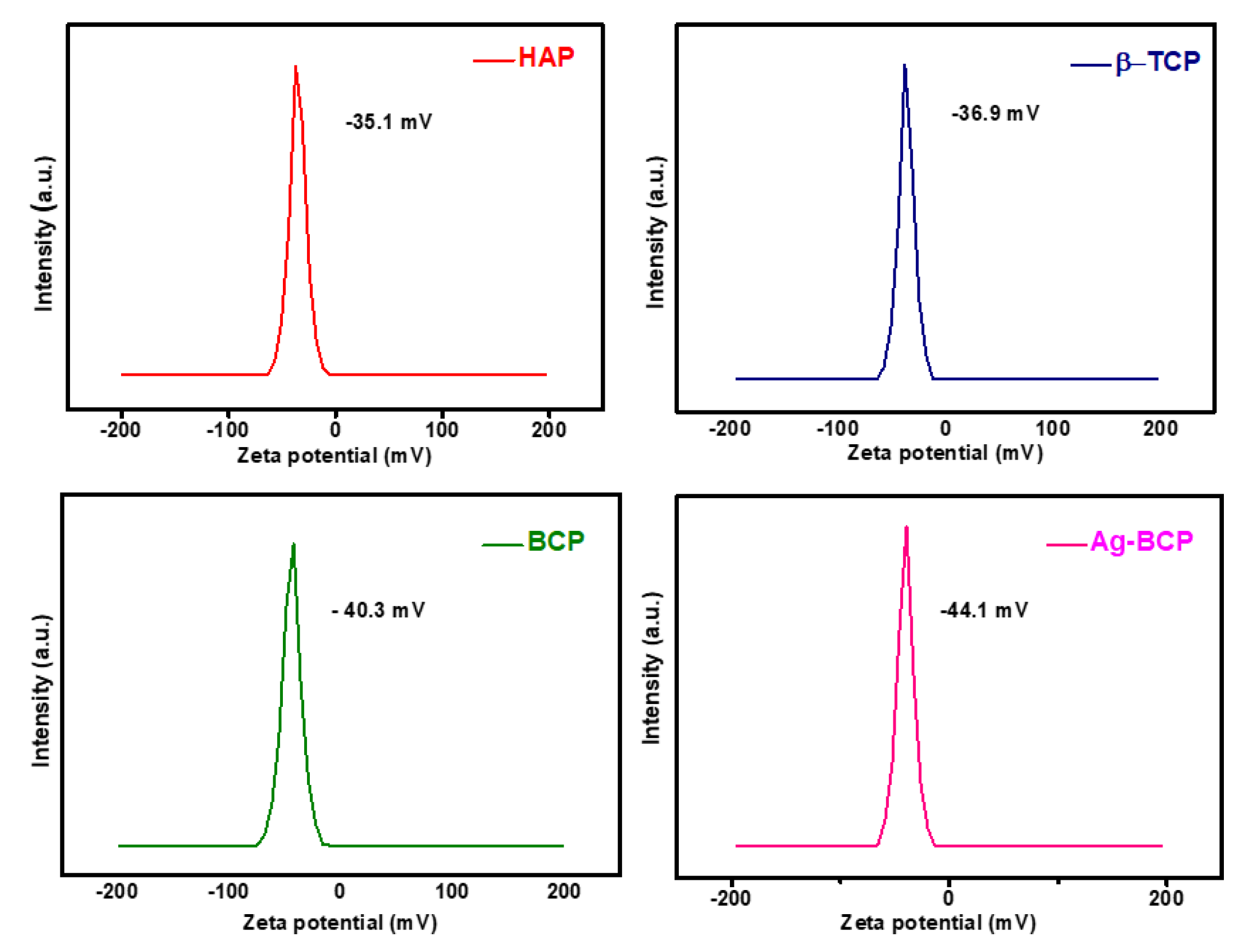
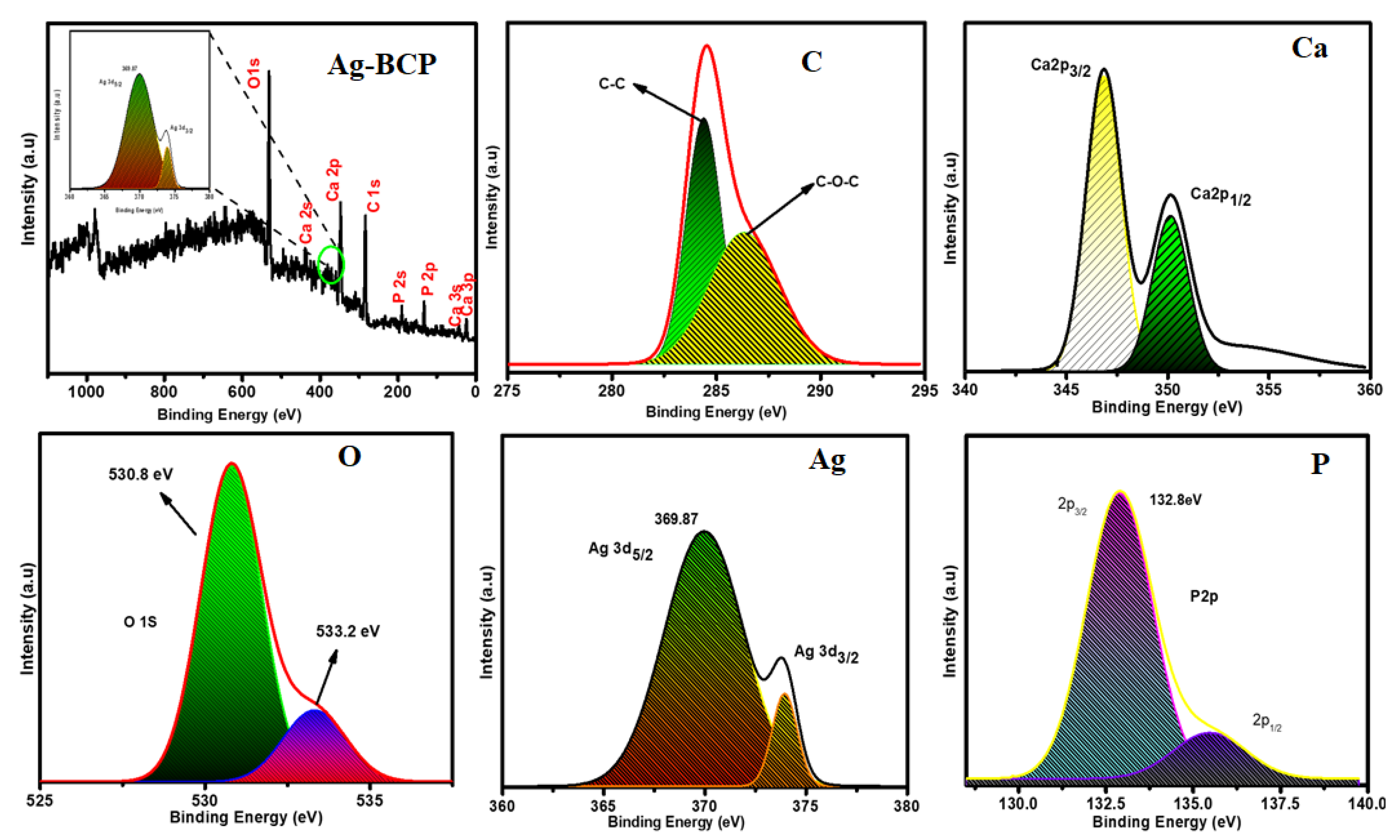
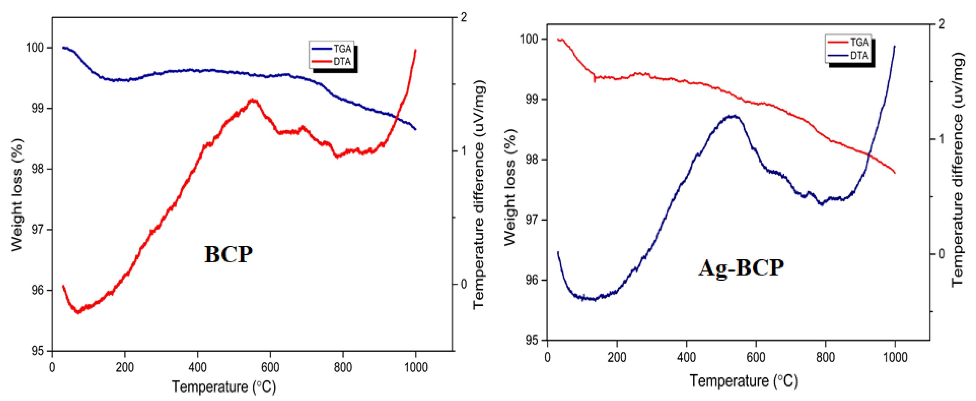
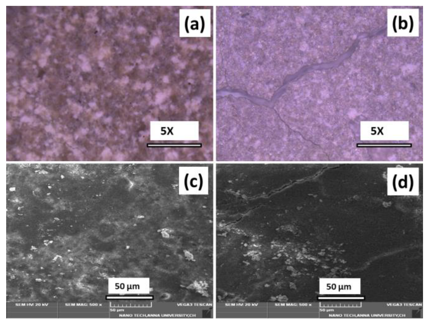
3.1. Physicochemical Analysis
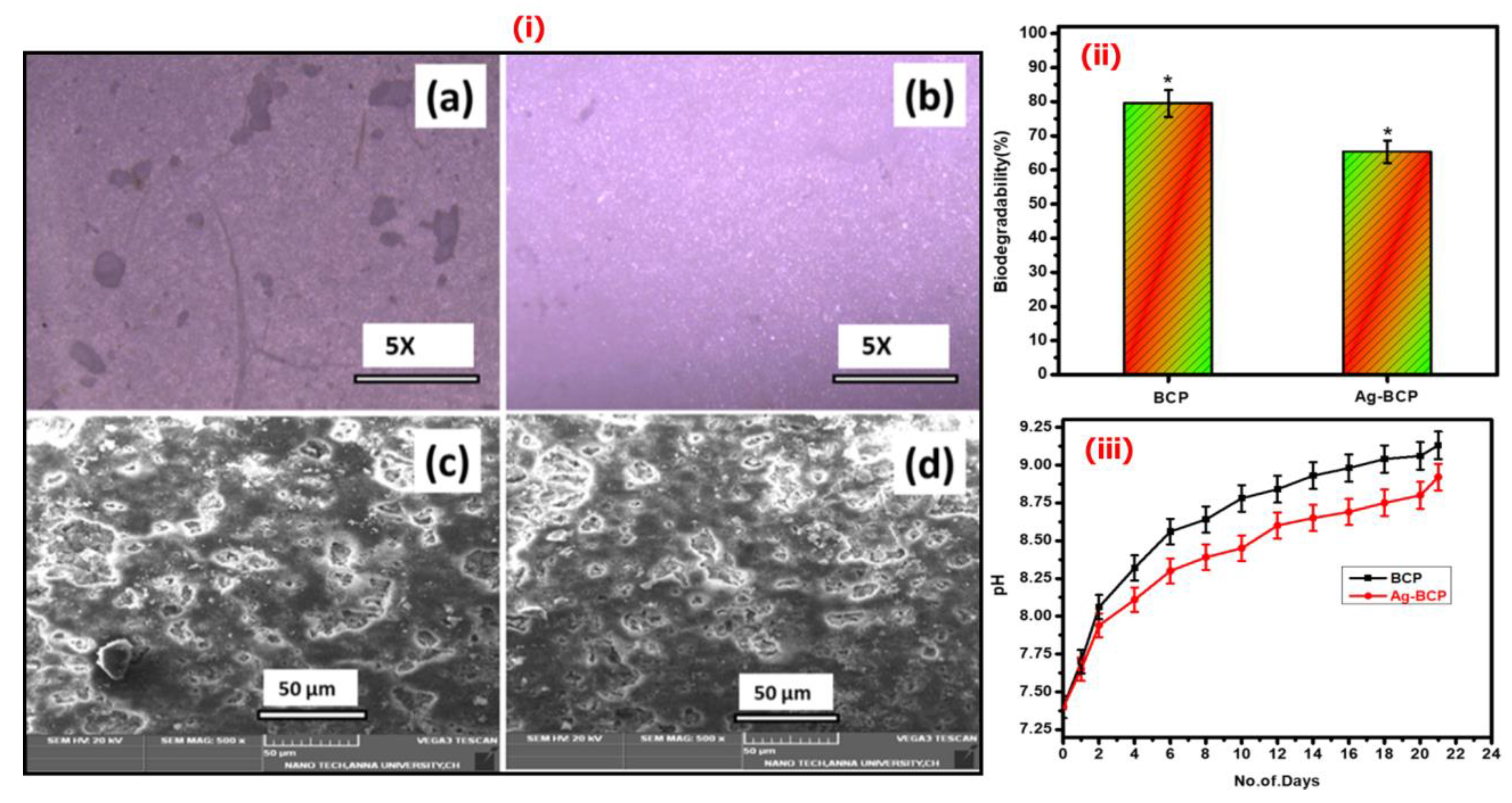
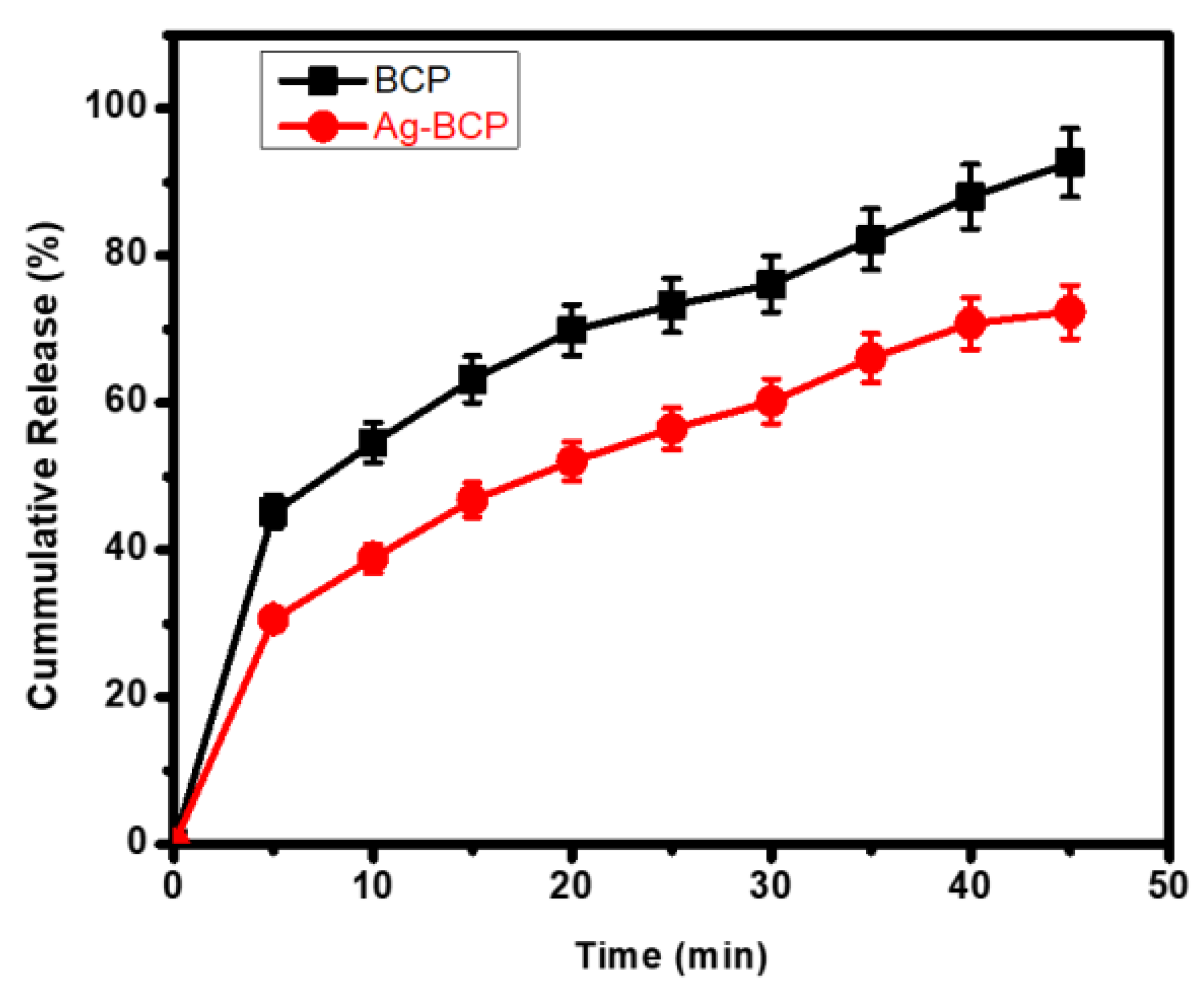
3.2. Studies of Bioactivity, Biodegradation, and Drug Release
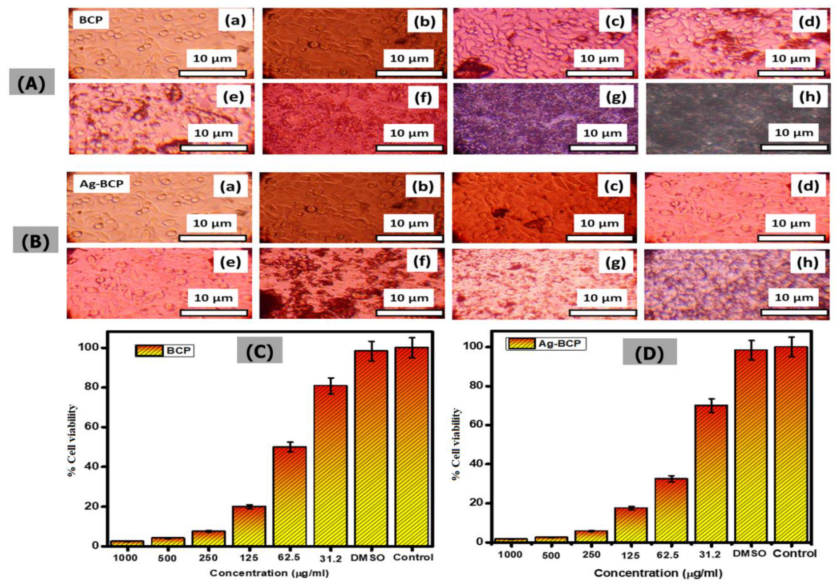
3.3. In Vitro Antimicrobial and Cytocompatibility Studies
4. Conclusions
Supplementary Materials
Author Contributions
Funding
Institutional Review Board Statement
Informed Consent Statement
Data Availability Statement
Acknowledgments
Conflicts of Interest
References
- Amini, A.R.; Laurencin, C.T.; Nukavarapu, S.P. Bone tissue engineering: Recent advances and challenges. Crit. Rev. Biomed. Eng. 2012, 40, 363. [Google Scholar] [CrossRef] [PubMed] [Green Version]
- Ghassemi, T.; Shahroodi, A.; Ebrahimzadeh, M.H.; Mousavian, A.; Movaffagh, J.; Moradi, A. Current Concepts in Scaffolding for Bone Tissue Engineering. Arch. Bone Jt. Surg. 2018, 6, 90–99. [Google Scholar] [PubMed]
- Zhang, D.; Wu, X.; Chen, J.; Lin, K. The development of collagen based composite scaffolds for bone regeneration. Bioact. Mater. 2018, 3, 129–138. [Google Scholar] [CrossRef] [PubMed]
- Balakrishnan, S.; Padmanabhan, V.P.; Kulandaivelu, R.; Nellaiappan, T.S.N.; Sagadevan, S.; Paiman, S.; Mohammad, F.; Al-Lohedan, H.A.; Obulapuram, P.K.; Oh, W.C. Influence of iron doping towards the physicochemical and biological characteristics of hydroxyapatite. Ceram. Int. 2021, 47, 5061–5070. [Google Scholar]
- O’brien, F.J. Biomaterials & scaffolds for tissue engineering. Mater. Today 2011, 14, 88–95. [Google Scholar]
- Liao, J.; Han, R.; Wu, Y.; Qian, Z. Review of a new bone tumor therapy strategy based on bifunctional biomaterials. Bone Res. 2021, 9, 1–3. [Google Scholar] [CrossRef]
- Canillas, M.; Pena, P.; Antonio, H.; Rodríguez, M.A. Calcium phosphates for biomedical applications. Boletín Soc. Española Cerámica Y Vidr. 2017, 56, 91–112. [Google Scholar] [CrossRef]
- Lobo, S.E.; Arinzeh, T.L. Biphasic calcium phosphate ceramics for bone regeneration and tissue engineering applications. Materials 2010, 3, 815–826. [Google Scholar] [CrossRef] [Green Version]
- Jeong, J.; Kim, J.H.; Shim, J.H.; Hwang, N.S.; Heo, C.Y. Bioactive calcium phosphate materials and applications in bone regeneration. Biomater. Res. 2019, 23, 1–11. [Google Scholar] [CrossRef] [Green Version]
- Chen, F.M.; Liu, X. Advancing biomaterials of human origin for tissue engineering. Prog. Polym. Sci. 2016, 53, 86–168. [Google Scholar]
- Eliaz, N.; Metoki, N. Calcium phosphate bioceramics: A review of their history, structure, properties, coating technologies and biomedical applications. Materials 2017, 10, 334. [Google Scholar] [CrossRef] [PubMed] [Green Version]
- Ebrahimi, M.; Botelho, M.G.; Dorozhkin, S.V. Biphasic calcium phosphates bioceramics (HA/TCP): Concept, physicochemical properties and the impact of standardization of study protocols in biomaterials research. Mater. Sci. Eng. C 2017, 71, 1293–1312. [Google Scholar] [CrossRef] [PubMed]
- de Oliveira Junior, J.M.; Montagner, P.G.; Carrijo, R.C.; Martinez, E.F. Physical characterization of biphasic bioceramic materials with different granulation sizes and their influence on bone repair and inflammation in rat calvaria. Sci. Rep. 2021, 11, 1–10. [Google Scholar] [CrossRef] [PubMed]
- Vallejos Baier, R.; Benjumeda Wijnhoven, I.; Irribarra del Valle, V.; Millán Giovanetti, C.; Vivanco, J.F. Microporosity clustering assessment in calcium phosphate bioceramic particles. Front. Bioeng. Biotechnol. 2019, 7, 281. [Google Scholar] [CrossRef] [PubMed] [Green Version]
- Khang, G.; Kim, S.H.; Kim, M.S.; Lee, H.B. Hybrid, composite, and complex biomaterials for scaffolds. Princ. Regen. Med. 2008, 1, 636–655. [Google Scholar]
- Zhang, F.; Chang, J.; Lu, J.; Lin, K.; Ning, C. Bioinspired structure of bioceramics for bone regeneration in load-bearing sites. Acta Biomater. 2007, 3, 896–904. [Google Scholar] [CrossRef]
- Levengood, S.K.; Polak, S.J.; Wheeler, M.B.; Maki, A.J.; Clark, S.G.; Jamison, R.D.; Johnson, A.J. Multiscale osteointegration as a new paradigm for the design of calcium phosphate scaffolds for bone regeneration. Biomaterials 2010, 31, 3552–3563. [Google Scholar] [CrossRef]
- Miranda, P.; Pajares, A.; Saiz, E.; Tomsia, A.P.; Guiberteau, F. Mechanical properties of calcium phosphate scaffolds fabricated by robocasting. J. Biomed. Mater. Res. Part A 2008, 85, 218–227. [Google Scholar] [CrossRef]
- Abou Neel, E.A.; Aljabo, A.; Strange, A.; Ibrahim, S.; Coathup, M.; Young, A.M.; Bozec, L.; Mudera, V. Demineralization–remineralization dynamics in teeth and bone. Key Eng. Mater. 2016, 11, 4743. [Google Scholar] [CrossRef]
- Ishikawa, K. Carbonate apatite bone replacement. Key Engineering Materials. 2014, 587, 17–20. [Google Scholar]
- Puttini, I.D.; Poli, P.P.; Maiorana, C.; Vasconcelos, I.R.; Schmidt, L.E.; Colombo, L.T.; Hadad, H.; Santos, G.M.; Carvalho, P.S.; Souza, F.Á. Evaluation of osteoconduction of biphasic calcium phosphate ceramic in the calvaria of rats: Microscopic and histometric analysis. J. Funct. Biomater. 2019, 10, 7. [Google Scholar] [CrossRef] [PubMed] [Green Version]
- Baghbani, F.; Moztarzadeh, F.; Nazari, A.G.; Kamran, A.R.; Tondnevis, F.; Nezafati, N.; Gholipourmalekabadi, M.; Mozafari, M. Biological response of biphasic hydroxyapatite/tricalcium phosphate scaffolds intended for low load-bearing orthopaedic applications. Adv. Compos. Lett. 2012, 21, 096369351202100102. [Google Scholar] [CrossRef] [Green Version]
- Burdușel, A.C.; Gherasim, O.; Grumezescu, A.M.; Mogoantă, L.; Ficai, A.; Andronescu, E. Biomedical applications of silver nanoparticles: An up-to-date overview. Nanomaterials 2018, 8, 681. [Google Scholar] [CrossRef] [PubMed] [Green Version]
- Dikshit, P.K.; Kumar, J.; Das, A.K.; Sadhu, S.; Sharma, S.; Singh, S.; Gupta, P.K.; Kim, B.S. Green synthesis of metallic nanoparticles: Applications and limitations. Catalysts 2021, 11, 902. [Google Scholar] [CrossRef]
- Dakal, T.C.; Kumar, A.; Majumdar, R.S.; Yadav, V. Mechanistic basis of antimicrobial actions of silver nanoparticles. Front. Microbiol. 2016, 7, 1831. [Google Scholar] [CrossRef]
- Nie, L.; Suo, J.; Zou, P.; Feng, S. Preparation and properties of biphasic calcium phosphate scaffolds multiply coated with HA/PLLA nanocomposites for bone tissue engineering applications. J. Nanomater. 2012, 2012, 213549 . [Google Scholar] [CrossRef] [Green Version]
- Xidaki, D.; Agrafioti, P.; Diomatari, D.; Kaminari, A.; Tsalavoutas-Psarras, E.; Alexiou, P.; Psycharis, V.; Tsilibary, E.C.; Silvestros, S.; Sagnou, M. Synthesis of hydroxyapatite, β-tricalcium phosphate and biphasic calcium phosphate particles to act as local delivery carriers of curcumin: Loading, release and in vitro studies. Materials 2018, 11, 595. [Google Scholar] [CrossRef] [Green Version]
- Kim, D.H.; Hwang, K.H.; Lee, J.D.; Park, H.C.; Yoon, S.Y. Long and short range order structural analysis of in-situ formed biphasic calcium phosphates. Biomater. Res. 2015, 19, 1–5. [Google Scholar] [CrossRef] [Green Version]
- Shameli, K.; Ahmad, M.B.; Jazayeri, S.D.; Sedaghat, S.; Shabanzadeh, P.; Jahangirian, H.; Mahdavi, M.; Abdollahi, Y. Synthesis and characterization of polyethylene glycol mediated silver nanoparticles by the green method. Int. J. Mol. Sci. 2012, 13, 6639–6650. [Google Scholar] [CrossRef] [Green Version]
- Ebrahimi, M.; Botelho, M. Biphasic calcium phosphates (BCP) of hydroxyapatite (HA) and tricalcium phosphate (TCP) as bone substitutes: Importance of physicochemical characterizations in biomaterials studies. Data Brief 2017, 10, 93–97. [Google Scholar] [CrossRef] [Green Version]
- Wu, F.; Lin, D.D.; Chang, J.H.; Fischbach, C.; Estroff, L.A.; Gourdon, D. Effect of the materials properties of hydroxyapatite nanoparticles on fibronectin deposition and conformation. Cryst. Growth Des. 2015, 15, 2452–2460. [Google Scholar] [CrossRef] [PubMed] [Green Version]
- van Dijk, L.A.; Duan, R.; Luo, X.; Barbieri, D.; Pelletier, M.; Christou, C.; Rosenberg, A.J.; Yuan, H.; Barrèrre-de Groot, F.; Walsh, W.R.; et al. Biphasic calcium phosphate with submicron surface topography in an Ovine model of instrumented posterolateral spinal fusion. JOR Spine 2018, 1, e1039. [Google Scholar] [CrossRef] [PubMed] [Green Version]
- Lee, Y.H.; Lee, J.W.; Yang, S.Y.; Lee, H.; Koh, Y.H.; Kim, H.E. Dual-scale porous biphasic calcium phosphate gyroid scaffolds using ceramic suspensions containing polymer microsphere porogen for digital light processing. Ceram. Int. 2021, 47, 11285–11293. [Google Scholar] [CrossRef]
- Kanchana, P.; Sekar, C. Effect of magnesium on the mechanical and bioactive properties of biphasic calcium phosphate. J. Miner. Mater. Charact. Eng. 2012, 11, 982. [Google Scholar] [CrossRef]
- Kumar, B.S.; Muthukumar, T.; Deepachitra, R.; Charumathy, R.K.; Hemalatha, T.; Sastry, T.P. In-vitro evaluation of biphasic calcium phosphate/casein incorporated with Myristica fragrans for bone tissue engineering. Ceram. Int. 2015, 41, 1725–1734. [Google Scholar] [CrossRef]
- Jariya, S.I.; Padmanabhan, V.P.; Kulandaivelu, R.; Prakash, N.; Mohammad, F.; Al-Lohedan, H.A.; Paiman, S.; Schirhagl, R.; Hossain, M.M.; Sagadevan, S. Drug delivery and antimicrobial studies of chitosan-alginate based hydroxyapatite bioscaffolds formed by the Casein micelle assisted synthesis. Mater. Chem. Phys. 2021, 272, 125019. [Google Scholar] [CrossRef]


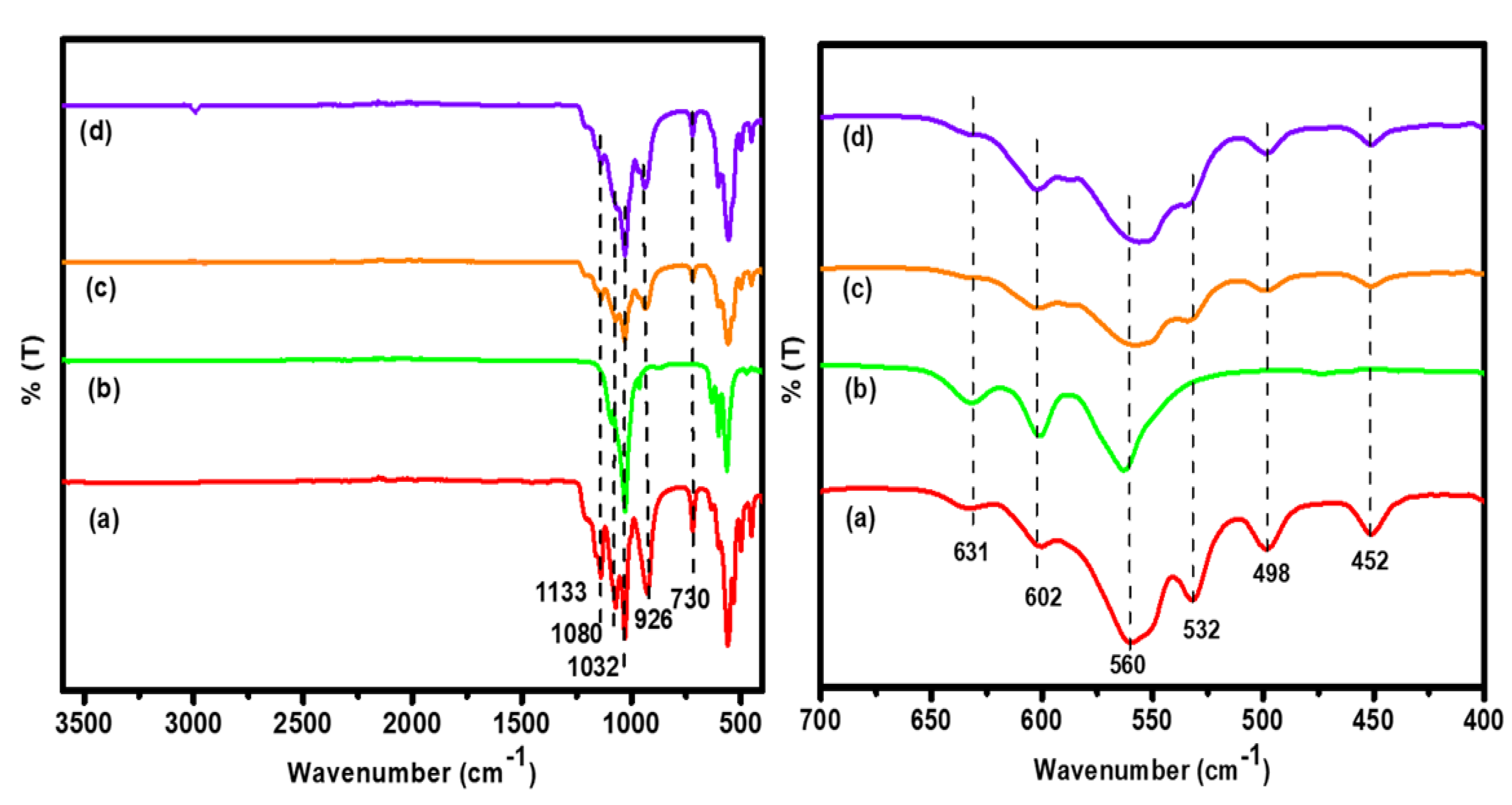
| Vibrational Frequency (cm−1) | Band Assignment | |||
|---|---|---|---|---|
| HAP | β-TCP | BCP | Ag-BCP | |
| 3572, 630 | -OH group | -OH group | -OH group | -OH group |
| 2920 | - | - | - | Glucose-assisted Ag NPs (C-H) stretching |
| 1032, 1098, 1133 | Asymmetric stretching vibrations of the P–O bonds | 1133 is absent | Asymmetric stretching of the P–O bonds | Asymmetric stretching of the P–O bonds |
| 1037 | - | PO43− ions found in β-TCP | PO43− ions found in β-TCP | PO43− ions found in β-TCP |
| 926 | Symmetric stretching (υ1) of P–O bond from PO43− group | - | Symmetric stretching (υ1) of P–O bond of PO43− group | Symmetric stretching (υ1) of P–O bond of PO43− group |
| 960 | - | Symmetric stretching (υ1) of P–O bond of PO43− group | Symmetric stretching (υ1) of P–O bond of PO43− group | Symmetric stretching (υ1) of P–O bond of PO43− group |
| 730 | Owing to H2O | - | - | Owing to H2O |
| 631 | Liberational OH group | Liberational OH group | Liberational OH group | Liberational OH group |
| 602, 560 | Phosphate bands (υ4) | Vibrational bands of PO43− | Phosphate bands (υ4) | Phosphate bands (υ4) |
| 498, 452 | Phosphate bands (υ2) | Phosphate bands (υ2) | Phosphate bands (υ2) | Phosphate bands (υ2) |
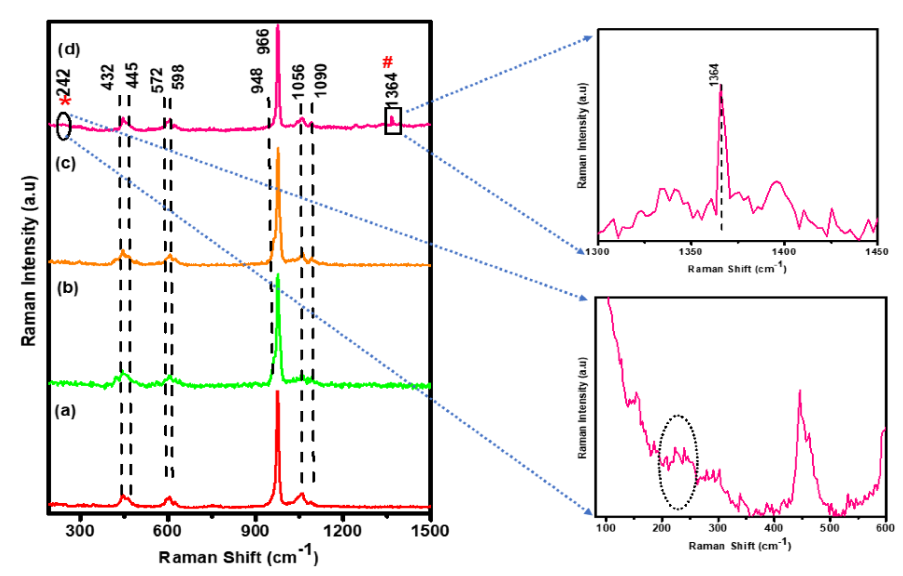
| Frequency of Vibration (cm−1) | Band Assignments | |||
|---|---|---|---|---|
| HAP | β-TCP | BCP | Ag-BCP | |
| 1364 | - | - | - | Gluconic acid |
| 1056 1090 | Asymmetric stretching vibrations (υ3) of PO4−3 | Asymmetric stretching vibrations (υ3) of PO4−3 | Asymmetric stretching vibrations (υ3) of PO4−3 | Asymmetric stretching vibrations (υ3) of PO4−3 |
| 966 | Symmetric stretching vibrations (υ1) of PO4−3 group | - | Symmetric stretching vibrations (υ1) of PO4−3 group | Symmetric stretching vibrations (υ1) of PO4−3 group |
| 964, 948 | - | Internal vibrations of β-TCP in BCP | Internal vibrations of β-TCP in BCP | Internal vibrations of β-TCP in BCP |
| 432, 445 | Symmetrical bending (υ2) | Symmetrical bending (υ2) | Symmetrical bending (υ2) | Symmetrical bending (υ2) |
| 572, 598 | Asymmetric bending (υ4) vibrations of PO4−3 | Asymmetric bending (υ4) vibrations of PO4−3 | Asymmetric bending (υ4) vibrations of PO4−3 | Asymmetric bending (υ4) vibrations of PO4−3 |
| Microorganism | Zone of Inhibition (mm) | |||||
|---|---|---|---|---|---|---|
| Ag-BCP | HAP | β-TCP | BCP | DMSO | Std (20 µL) | |
| E. coli | 8 ± 1.15 | - | - | - | - | 11 ± 1.75 |
| S. aureus | 10 ± 2.2 | - | - | - | - | 17 ± 1.2 |
| C. albicans | 10 ± 1.5 | - | - | - | - | 12 ± 1.0 |
Publisher’s Note: MDPI stays neutral with regard to jurisdictional claims in published maps and institutional affiliations. |
© 2022 by the authors. Licensee MDPI, Basel, Switzerland. This article is an open access article distributed under the terms and conditions of the Creative Commons Attribution (CC BY) license (https://creativecommons.org/licenses/by/4.0/).
Share and Cite
Padmanabhan, V.P.; Sivashanmugam, P.; Kulandaivelu, R.; Sagadevan, S.; Sridevi, B.; Govindasamy, R.; Thiruvengadam, M. Biosynthesised Silver Nanoparticles Loading onto Biphasic Calcium Phosphate for Antibacterial and Bone Tissue Engineering Applications. Antibiotics 2022, 11, 1780. https://doi.org/10.3390/antibiotics11121780
Padmanabhan VP, Sivashanmugam P, Kulandaivelu R, Sagadevan S, Sridevi B, Govindasamy R, Thiruvengadam M. Biosynthesised Silver Nanoparticles Loading onto Biphasic Calcium Phosphate for Antibacterial and Bone Tissue Engineering Applications. Antibiotics. 2022; 11(12):1780. https://doi.org/10.3390/antibiotics11121780
Chicago/Turabian StylePadmanabhan, Varun Prasath, Pugalmani Sivashanmugam, Ravichandran Kulandaivelu, Suresh Sagadevan, Balu Sridevi, Rajakumar Govindasamy, and Muthu Thiruvengadam. 2022. "Biosynthesised Silver Nanoparticles Loading onto Biphasic Calcium Phosphate for Antibacterial and Bone Tissue Engineering Applications" Antibiotics 11, no. 12: 1780. https://doi.org/10.3390/antibiotics11121780