Article

Characterization and Evaluation of Layered Bi2WO6 Nanosheets as a New Antibacterial Agent

by
Maria Karollyna do Nascimento Silva Leandro
1,2,
João Victor Barbosa Moura
3,
Paulo de Tarso Cavalcante Freire
4,
Maria Leticia Vega
5,
Cleânio da Luz Lima
5,
Ángel Alberto Hidalgo
5,
Ana Carolina Justino de Araújo
1,
Priscilla Ramos Freitas
1,
Cicera Laura Roque Paulo
1,
Amanda Karine de Sousa
1,2,
Janaina Esmeraldo Rocha
1,
Lívia Maria Garcia Leandro
2,
Rakel Olinda Macedo da Silva
2,
Natália Cruz-Martins
6,7,8,* and
Henrique Douglas Melo Coutinho
1,*
1
Department of Biological Chemistry, Regional University of Cariri, Crato 63105-000, Brazil
2
Department of Biomedicine, University Center Dr. Leão Sampaio, Juazeiro do Norte 63040-005, Brazil
3
Department of Physics, Science and Technology Center, Federal University of Maranhão, São Luís 65085-580, Brazil
4
Department of Physics, Campus do Pici, Federal University of Ceará, Fortaleza 60455-760, Brazil
5
Department of Physics, Campus Ministro Petrônio Portella, Federal University of Piauí, Teresina 64049-550, Brazil
6
Faculty of Medicine, University of Porto, Alameda Prof. Hernani Monteiro, 4200-319 Porto, Portugal
7
Institute for research and Innovation in Health (i3S), University of Porto, Rua Alfredo Allen, 4200-319 Porto, Portugal
8
Institute of Research and Advanced Training in Health Sciences and Technologies (CESPU), Rua Central de Gandra, 1317, 4585-116 Gandra, Portugal
*
Authors to whom correspondence should be addressed.
Antibiotics 2021, 10(9), 1068; https://doi.org/10.3390/antibiotics10091068
Submission received: 17 July 2021 / Revised: 22 August 2021 / Accepted: 31 August 2021 / Published: 3 September 2021
(This article belongs to the Special Issue The Global Need for New Antimicrobial and Antibiofilm Agents)

Abstract

:
Background: Pathogenic microorganisms are causing increasing cases of mortality and morbidity, along with alarming rates of ineffectiveness as a result of acquired antimicrobial resistance. Bi2WO6 showed good potential to be used as an antibacterial substance when exposed to visible light. This study demonstrates for the first time the dimension-dependent antibacterial activity of layered Bi2WO6 nanosheets. Materials and methods: The synthesized layered Bi2WO6 nanosheets were prepared by the hydrothermal method and characterized by powder X-ray diffraction (XRD), scanning electron microscopy (SEM), atomic force microscopy (AFM), and Raman and Fourier transform infrared spectroscopy (FTIR). Antibacterial and antibiotic-modulation activities were performed in triplicate by the microdilution method associated with visible light irradiation (LEDs). Results: Bi2WO6 nanosheets were effective against all types of bacteria tested, with MIC values of 256 μg/mL against Escherichia coli standard and resistant strains, and 256 μg/mL and 32 μg/mL against Staphylococcus aureus standard and resistant strains, respectively. Two-dimensional (2D) Bi2WO6 nanosheets showed antibacterial efficiency against both strains studied without the presence of light. Conclusions: Layered Bi2WO6 nanosheets revealed dimension-dependent antibacterial activity of the Bi2WO6 system.

1. Introduction

Microbial infections triggered by resistant pathogens have raised important public health problems in the world, being currently the focus of study by multiple researchers in an intent to provide safer, effective and less harmful antimicrobials [1]. Among the various fields under intense research, nanobiotechnology has been on high demand, with an increasing number of nanomaterials being highlighted in several scientific studies as a possible alternative to fight resistant microorganisms. Characterized by a very small size, but with a large surface area, such nanoformulations have revealed the capability of improving the delivery of drugs in specific tissues, ultimately boosting drugs’ effectiveness and a reduction in adverse effects, as both dose and time of action can be thoroughly controlled [2].
After the discovery of graphene, nanomaterials with a 2D dimension are increasingly being targeted by research seeking to elucidate physical properties in relation to their bulk precursors [3]. However, in the post-graphene era, numerous inorganic materials in the form of layers have been extensively investigated, such as transition metal dichalcogenides [4] and layered metal oxides [5], among others. Monolayered bismuth tungstate (Bi2WO6) nanosheets is a representative example; the Aurivillius oxide Bi2WO6 nanosheets has a sandwich substructure of [BiO]+—[WO4]2−—[BiO]+. Layered Bi2WO6 nanosheets were obtained by a cetyltrimethylammonium bromide-assisted bottom-up route [6].
Bismuth tungstate (Bi2WO6) has received huge attention as a visible light photocatalyst [7]. Bi2WO6 has shown interesting photochemical stability and reusability, being potentially useful for environmental treatment purposes, while also exhibiting photocatalytic degradation of Erichrome Black T (EBT) organic dye [8]. Furthermore, Bi2WO6 exhibited photocatalytic activity for degrading norfloxacin and enrofloxacin under visible light irradiation, meaning its feasible application along with fluoroquinolone antibiotics [9]. In a study, Aurivillius oxide Bi2WO6 was applied as an effective visible light-driven antibacterial photocatalyst for Escherichia coli’s inactivation, where neither visible light without the photocatalyst nor Bi2WO6 in the dark revealed bactericidal effects. Thus, the bactericidal effect in E. coli was certainly attributed to the photocatalytic reaction of Bi2WO6 under visible light irradiation [10].
In this sense, the present study aims to assess, for the first time, the antibacterial and antibiotic modulation effects of layered Bi2WO6 nanosheets against the standard and multidrug-resistant (MDR) Staphylococcus aureus and E. coli. Two-dimensional (2D) Bi2WO6 nanosheets showed antibacterial efficiency in both studied strains (Gram-negative and Gram-positive bacteria) without the presence of light (in the dark), showing a dimension-dependent antibacterial activity of the Bi2WO6 system and improving its properties in relation to the bulk material.

2. Results and Discussion

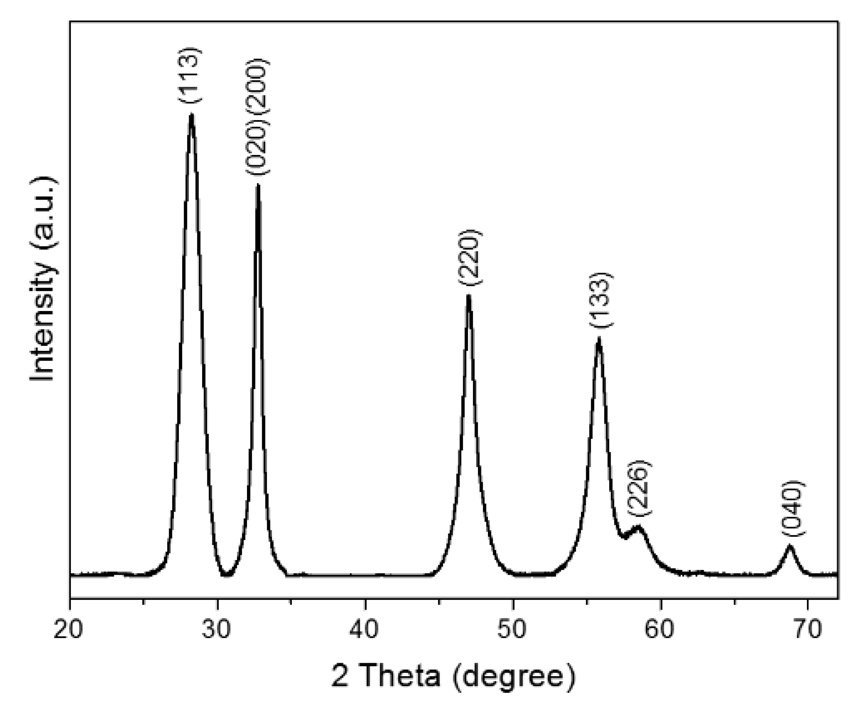
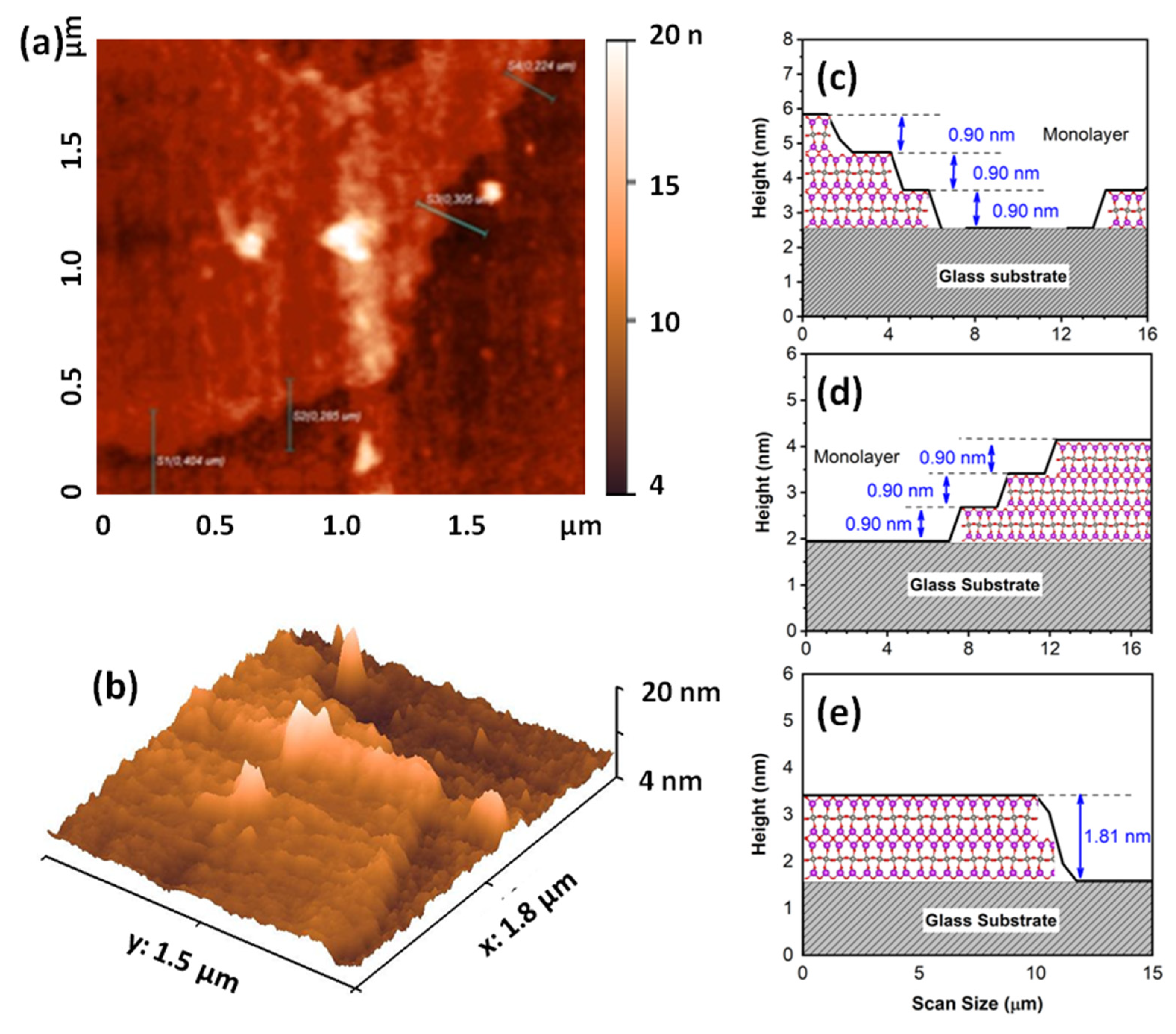
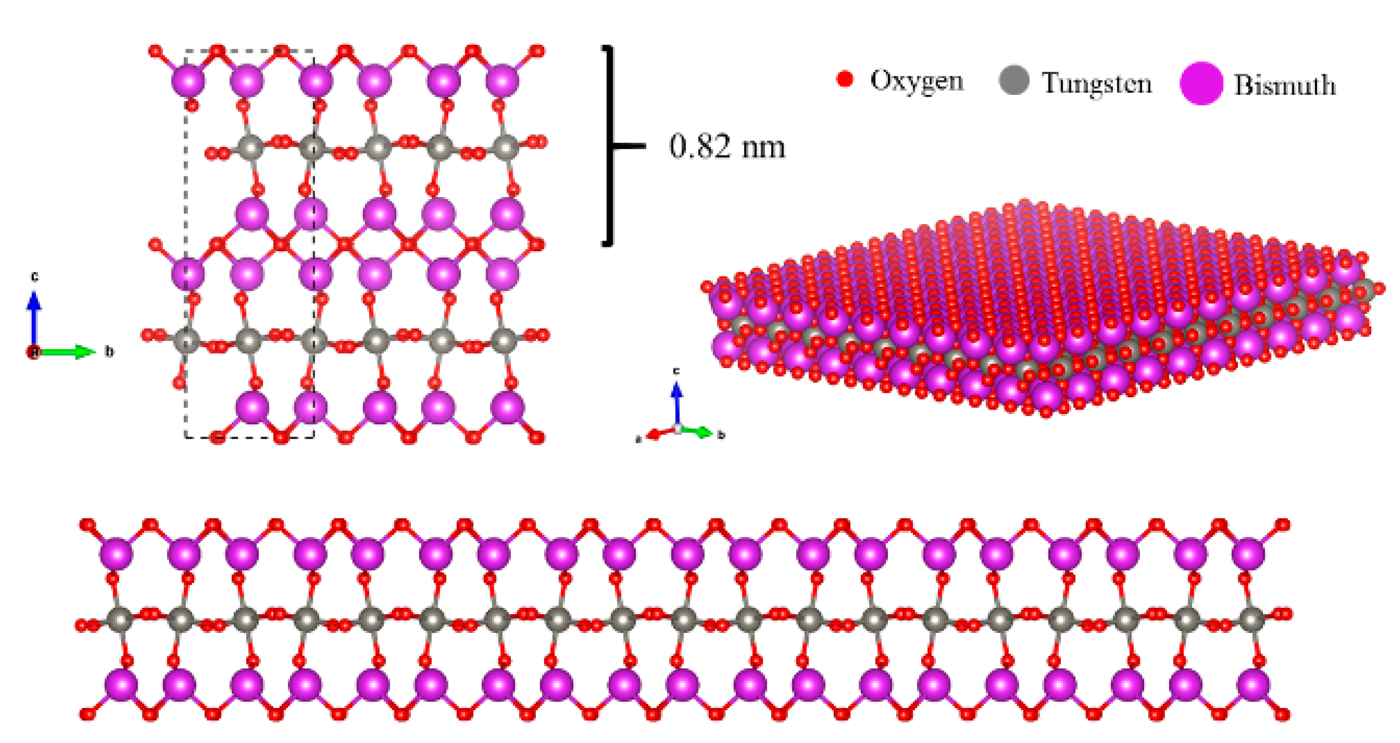
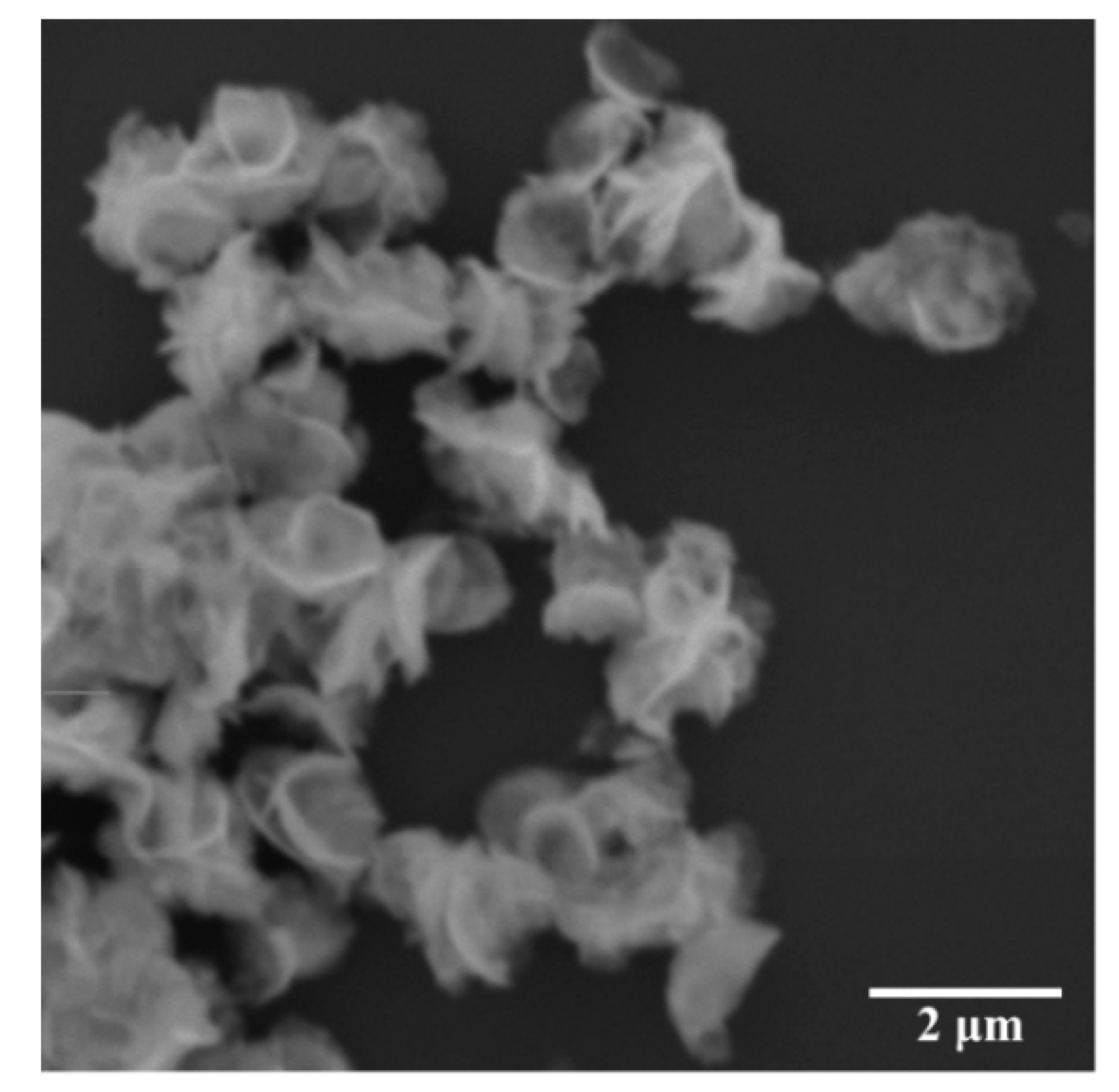
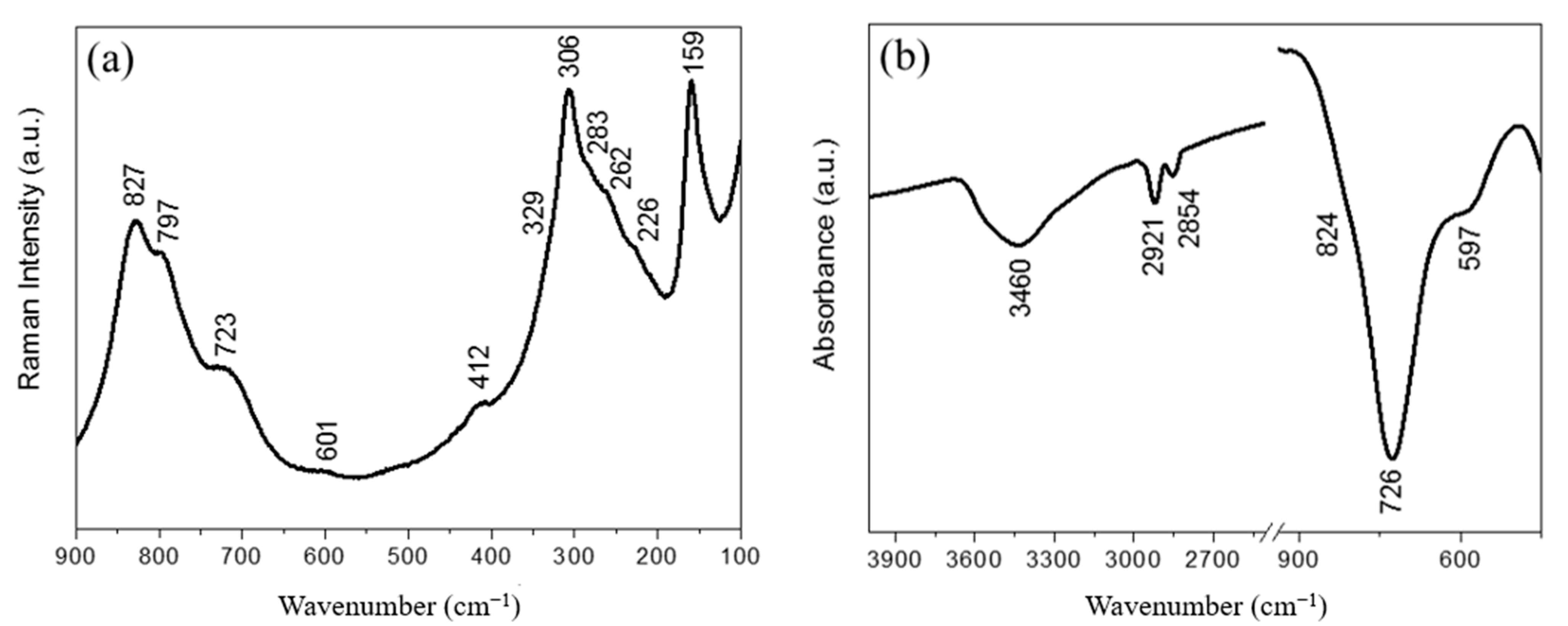
2.1. Characterization of Layered Bi2WO6 Nanosheets

Figure 1 shows the XRD powder pattern of Bi2WO6 obtained by a CTAB-assisted hydrothermal method. The crystalline nature of the samples was confirmed, and all diffraction peaks can be indexed to an orthorhombic Bi2WO6 phase with lattice parameters a = 5.457 Å, b = 5.436 Å and c = 16.427 Å (JCPDS Card N° 73-2020), without secondary phase. The intensity ratio of the (020)/(200) Bragg peak to the (113) peak was higher than the standard value. In addition, the full width at half maximum (FWHM) of the (200)/(020) Bragg peak was narrower than that of the (113) peak. This analysis indicates a higher grain size of the synthesized bismuth tungstate along the (100) and (010) directions compared to the (001) direction [11]. The AFM images (Figure 2a,b) and the corresponding height histograms (Figure 2c,d) of the layered Bi2WO6 nanosheets show that the monolayer has a thickness of about 0.9 nm, which is in agreement with that of Bi2WO6 monolayer along the (001) direction. In addition, the three-dimensional (3D) morphology in Figure 2b indicates that the layered Bi2WO6 nanosheets have a dense and flat morphology. Therefore, the AFM result agrees with the XRD results. Furthermore, this result is in perfect agreement with data obtained by Zhou et al. [6], who showed by high resolution transmission electron microscopy (HRTEM) and atomic force microscopy (AFM) that layered Bi2WO6 nanosheets obtained by CTAB-assisted hydrothermal synthesis exposes (001) facets. In addition, the authors showed that layers had a thickness of 0.8 nm, i.e., the Bi2WO6 monolayer along the (001) direction features 1/2 the size of the unit cell in the c-axis direction.
As stated above and looking at Figure 3, Aurivillius Bi2WO6 is a layered material built up of [Bi2O2] layers and corner-shared WO6 octahedral layers, whereas the Bi2WO6 monolayer has a sandwich substructure configuration of [BiO]+—[WO4]2−—[BiO]+ exposing the Bi atoms on the surface. The morphology of the two-dimensional Bi2WO6 nanosheets can be shown by the SEM images (Figure 4), which clearly reveal Bi2WO6 exhibiting thin two-dimensional structures.
The room temperature Raman and Fourier transform infrared (FTIR) spectra of the synthesized layered Bi2WO6 nanosheets are presented in Figure 5 and Table 1. The Raman spectrum of Bi2WO6 monolayers in the spectra range between 100 and 900 cm−1 presents broad Raman peaks, some even overlapping forming only shoulders, making them difficult to distinguish in the unpolarized spectrum (See Figure 5a). On one hand, the Raman spectrum exhibits significant changes as the crystallite size decreases, comparing the Raman spectrum of Bi2WO6 monolayers with the spectrum of Bi2WO6 bulk structures. On the other hand, the Raman spectrum obtained is in full agreement with what is shown in the literature for nano-sized materials [12]. Looking at the FTIR spectrum in Figure 5b, we see that the broad band at 3460 cm−1 corresponds to the stretching vibration of O–H bonds, indicating water molecules adsorbed on the surface of the sample. The 2921 cm−1 and 2854 cm−1 are associated with the C–H stretching vibrations of the methyl and methylene groups of CTAB used in hydrothermal synthesis [13]. The assignment of the Raman and FTIR modes that come from the W–O or Bi–O bonds are listed in Table 1. In summary, the FTIR and Raman spectra obtained are in full agreement with what is shown in the literature and in agreement with the XRD results [12,14,15].

2.2. Antibacterial Activity of Layered Bi2WO6 Nanosheets

Bi2WO6 nanosheets revealed to be effective against all kinds of bacteria tested, with a MIC of 256 μg/mL against E. coli standard and resistant strains, and of 256 μg/mL and 32 μg/mL against S. aureus standard and resistant strains, respectively (Table 2). Evaluating the antibacterial activity of Bi2WO6 nanosheets under LED irradiation with varying wavelengths (415, 620 and 590 nm), no photocatalytic reaction was observed, given that MIC values do not decrease under visible light irradiation. Thus, Bi2WO6 nanosheets exhibit excellent antibacterial inactivation on E. coli and S. aureus in the dark.
In the study by Li et al. [16], the Bi2WO6 crystals in hierarchical flower-like morphology did not reveal antibacterial activity against E. coli in the dark, as some effect was only observed when the material was irradiated with visible light. Likewise, Hen et al. [10] using Aurivillius oxide Bi2WO6 as an effective visible light-driven antibacterial photocatalyst for E. coli inactivation, did not report any bactericidal effect under visible light without the photocatalyst nor Bi2WO6 in the dark. It is, however, worth noting that the results obtained by Hen et al. [10] and Li et al. [16] attributed the bactericidal effect to the photocatalytic reaction of Bi2WO6 (bulk) under visible light irradiation. In the present study, it was demonstrated that the antibacterial activity of the layered Bi2WO6 nanosheets was intrinsic to the material (no need for the presence of visible light).
Regarding the antibacterial effect of Bi2WO6 nanosheets, an intrinsic clinical relevance was found with the standard stated by Houghton et al. [17]. Another interesting point to underline is that this material affects Gram-positive and Gram-negative bacteria equally, which highlights the great clinical and technological potential of this material. This is especially important in view of the significant increase in resistance mechanisms developed by E.coli and S. aureus, such as the alteration of cell membrane permeability, making it difficult for antibiotics to enter and change the target drug binding sites, respectively [18,19].
In addition, the crystalline structure of layered Bi2WO6 nanosheets has the ability to generate e–h+ pairs, while the sandwich substructure of [BiO]+—[WO4]2−—[BiO]+ monolayer Bi2WO6 simulates the heterojunction interface with space charge that promotes the separation of carriers generated in the interface [6]. As a result, such species interact with H2O, triggering the formation of OH, H+, and O2•−, which can act on the cells’ surface to degrade some components of bacterial cell membrane. Cell rupture can cause the bacteria to lose function and eventually lead to cell death [20]. Moreover, the high surface area/volume ratio of 2D Bi2WO6 nanosheets favors the interaction between the sample and the bacterial membrane, facilitating possible adsorption processes. Therefore, based on the results discussed above, it is possible to observe the possibility that the mechanism responsible for the antibacterial activity of Bi2WO6 monolayers is related to the two-dimensional property of the system.

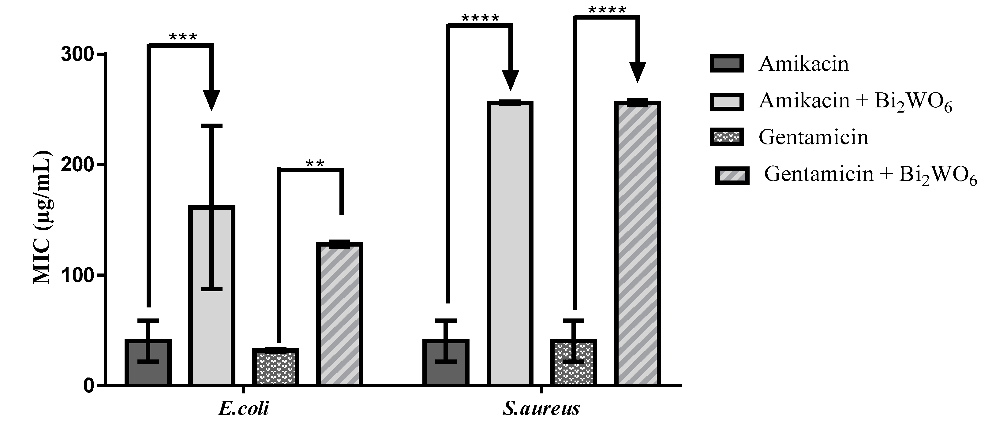
2.3. Modulation of Antibiotic Activity by Layered Bi2WO6 Nanosheets

Data obtained on the modulation of antibiotic activity demonstrated by Bi2WO6 nanosheets are shown in Figure 6. The compound significantly raised the MIC of both antibiotics (amikacin and gentamicin) against Gram-negative and Gram-positive strains, show no improvements in antibiotic activity, and the associations even resulted in antagonistic effects.
The association of aminoglycosides under visible light irradiation may represent a promising strategy in the treatment of skin infections caused by resistant bacteria. Therefore, in this study the antibiotic-modulating effects of LED light exposure associated or not with Bi2WO6 nanosheets was investigated (Figure 7). No potentiation of the action of aminoglycosides associated with Bi2WO6 nanosheets against Gram-positive and Gram-negative strains was stated, regardless of LED wavelength. Thus, the strong antagonistic effects observed when aminoglycosides were used in combination with Bi2WO6 nanosheets under visible light irradiation against Gram-positive and Gram-negative strains seem to be related to the high photocatalytic potential of bismuth tungstate in degrading organic compounds [8,9].
The emergence of antibiotic resistance by pathogenic bacteria occurs due to several factors and, among these, the inappropriate drugs’ disposal stands out. Specifically, wastewater from health facilities, mainly hospitals, stands out as an important source of emission of antibiotics in the environment and, thus, it is important to degrade these compounds before disposal. In this sense, current research is seeking new agents capable of acting as photocatalysts [21], and Bi2WO6 nanosheets showed a good potential in the treatment of effluents containing gentamicin and amikacin.

3. Materials and Methods

3.1. Synthesis

The synthesis of layered Bi2WO6 nanosheets was performed by a hydrothermal method, as described previously [6]. Sodium tungstate dihydrate [Na2WO4·2H2O] (≥99%, Sigma-Aldrich, St. Louis, MO, USA), bismuth nitrate pentahydrate [Bi(NO3)3·5H2O] (≥98.0%, Sigma-Aldrich, St. Louis, MO, USA) and hexadecyltrimethylammonium—CTAB [CH3(CH2)15N(Br)(CH3)3] were used as starting precursors. In a synthesis procedure, 1 mmol of Na2WO4·2H2O, 2 mmol of Bi(NO3)3·5H2O and 0.05 g of CTAB were dissolved in 80 mL of deionized water. This aqueous solution was stirred for 30 min at an average speed of 1500 rpm. The resulting solution was transferred to a 100 mL Teflon-lined stainless autoclave and maintained at 120 °C for 24 h. The white precipitates were repeatedly washed with deionized water and dried in an air oven at 60 °C for 10 h.

3.2. Structural Characterization

Structural characterization was performed by X-ray diffraction (XRD) using a Mini-Flex Rigaku diffractometer. Morphological analysis of layered Bi2WO6 nanosheets was carried out in a scanning electron microscope (SEM) model Vega3 Tescan. Atomic force microscopy images were recorded using NTMDT microscope. Fourier Transform Infrared (FT-IR) spectra were obtained using a Perkin Elmer Spectrum Two spectrophotometer. Raman measurements were performed by a Horiba LabRaman spectrometer.

3.3. Analysis of Antibacterial Activity and Antibiotic Resistance Modulation

The antibiotic-enhancing activity was assessed using the methodology of Coutinho et al. [22].

3.4. Experiments with LED Light Exposure

The Light Emitting Diodes-LED device (a light emitting diode; NEW Estética®, Fortaleza, Brazil) was used in the experimental protocols. The LEDs with a wavelength predetermined by the device used were blue (415 nm), red (620 nm) and yellow (590 nm). To assess the effect of LED light exposure on bacterial growth in modulating antibacterial activity, cultures and treatments were performed as described above. Plates were exposed to blue, red or yellow light for 20 min and then incubated at 37 °C for 24 h. Plates without exposure to LED light were used as experimental controls. Readings were performed as described above.

3.5. Statistical Analysis

Statistical analysis was performed using the GraphPad Prism 6.0 software, with an alpha set at 0.05. One-way analysis of variance (ANOVA) and Bonferroni’s post-hoc tests were used to address differences between groups. More details are shown in Supplementary File S1.

4. Conclusions

Data obtained in this study provide significant insights into the dimension-dependent antibacterial activity of layered Bi2WO6 nanosheets. The crystalline nature of the samples was confirmed, and all diffraction peaks were indexed to the orthorhombic Bi2WO6 phase. Two-dimensional (2D) Bi2WO6 nanosheets showed antibacterial action against the strains studied without the presence of light, and also revealed a possible catalytic effect of antibiotics. Further studies are needed toward a more in-depth understanding on this action.

Supplementary Materials

The following are available online at https://www.mdpi.com/article/10.3390/antibiotics10091068/s1, File S1: Synthesis, characterization and microbiological testing: extended methodology.

Author Contributions

Conceptualization, H.D.M.C., J.V.B.M., C.d.L.L. and P.d.T.C.F.; data curation, M.K.d.N.S.L. and J.V.B.M.; formal analysis, M.K.d.N.S.L. and J.V.B.M.; investigation, M.K.d.N.S.L., J.V.B.M., A.C.J.d.A., P.R.F., J.E.R., C.L.R.P., A.K.d.S., R.O.M.d.S. and L.M.G.L.; methodology, M.K.d.N.S.L., J.V.B.M., A.C.J.d.A., P.R.F., J.E.R., C.L.R.P., A.K.d.S., R.O.M.d.S. and L.M.G.L.; project administration, H.D.M.C., N.C.-M., J.V.B.M., M.K.d.N.S.L.; supervision, H.D.M.C.; writing—original draft, H.D.M.C.; J.V.B.M., M.K.d.N.S.L., M.L.V., N.C.-M. and Á.A.H.; writing—review and editing, H.D.M.C., N.C.-M., J.V.B.M., M.K.d.N.S.L. and Á.A.H. All authors have read and agreed to the published version of the manuscript.

Funding

This research received no external funding.

Data Availability Statement

Data presented in this study are available on request from the corresponding author.

Acknowledgments

The authors thank Conselho Nacional de Desenvolvimento Científico e Tecnológico—CNPq, Coordenação de Aperfeiçoamento de Pessoal de Nível Superior—CAPES, and Fundação Cearense de Apoio ao Desenvolvimento Científico e Tecnológico—FUNCAP (Proc. BP4-00172-00232.01.00/20 and Proc. PR2-0101-00006.01.00/15) for the financial support. The authors would also like to thank the educational institutions UFCA, URCA, and UNILEÃO for their support during the experiments.

Conflicts of Interest

The authors declare no conflict of interest.

References

  1. De Oliveira, D.R.; Tintino, S.R.; Morais-Braga, M.F.B.; Boligon, A.A.; Athayde, M.L.; Coutinho, H.D.M.; De Menezes, I.R.A.; Fachinetto, R. In Vitro Antimicrobial and Modulatory Activity of the Natural Products Silymarin and Silibinin. BioMed Res. Int. 2015, 2015, 292797. [Google Scholar] [CrossRef] [Green Version]
  2. Lombardo, D.; Kiselev, M.A.; Caccamo, M.T. Smart Nanoparticles for Drug Delivery Application: Development of Versatile Nanocarrier Platforms in Biotechnology and Nanomedicine. J. Nanomater. 2019, 2019, 3702518. [Google Scholar] [CrossRef]
  3. Momeni, K.; Ji, Y.; Wang, Y.; Paul, S.; Neshani, S.; Yilmaz, D.E.; Shin, Y.K.; Zhang, D.; Jiang, J.-W.; Park, H.; et al. Multiscale computational understanding and growth of 2D materials: A review. npj Comput. Mater. 2020, 6, 22. [Google Scholar] [CrossRef] [Green Version]
  4. Pan, B.; Zhang, K.; Ding, C.-C.; Wu, Z.; Fan, Q.; Luo, T.; Zhang, L.; Zou, C.; Huang, S. Universal Precise Growth of 2D Transition-Metal Dichalcogenides in Vertical Direction. ACS Appl. Mater. Interfaces 2020, 12, 35337–35344. [Google Scholar] [CrossRef]
  5. Puthirath Balan, A.; Radhakrishnan, S.; Woellner, C.F.; Sinha, S.K.; Deng, L.; Reyes, C.D.L.; Rao, B.M.; Paulose, M.; Neupane, R.; Apte, A.; et al. Exfoliation of a non-van der Waals material from iron ore hematite. Nat. Nanotechnol. 2018, 13, 602–609. [Google Scholar] [CrossRef]
  6. Zhou, Y.; Zhang, Y.; Lin, M.; Long, J.; Zhang, Z.; Lin, H.; Wu, J.C.-S.; Wang, X. Monolayered Bi2WO6 nanosheets mimicking heterojunction interface with open surfaces for photocatalysis. Nat. Commun. 2015, 6, 8340. [Google Scholar] [CrossRef] [PubMed] [Green Version]
  7. Tang, J.; Zou, Z.; Ye, J. Photocatalytic Decomposition of Organic Contaminants by Bi2WO6Under Visible Light Irradiation. Catal. Lett. 2004, 92, 53–56. [Google Scholar] [CrossRef]
  8. Lai, M.T.L.; Lai, C.W.; Lee, K.M.; Chook, S.W.; Yang, T.C.K.; Chong, S.; Juan, J.C. Facile one-pot solvothermal method to synthesize solar active Bi2WO6 for photocatalytic degradation of organic dye. J. Alloys Compd. 2019, 801, 502–510. [Google Scholar] [CrossRef]
  9. Huang, C.; Chen, L.; Li, H.; Mu, Y.; Yang, Z. Synthesis and application of Bi2WO6 for the photocatalytic degradation of two typical fluoroquinolones under visible light irradiation. RSC Adv. 2019, 9, 27768–27779. [Google Scholar] [CrossRef] [Green Version]
  10. Ren, J.; Wang, W.; Zhang, L.; Chang, J.; Hu, S. Photocatalytic inactivation of bacteria by photocatalyst Bi2WO6 under visible light. Catal. Commun. 2009, 10, 1940–1943. [Google Scholar] [CrossRef]
  11. Zhou, Y.; Lv, P.; Meng, X.; Tang, Y.; Huang, P.; Chen, X.; Shen, X.; Zeng, X. CTAB-Assisted Fabrication of Bi2WO6 Thin Nanoplates with High Adsorption and Enhanced Visible Light-Driven Photocatalytic Performance. Molecules 2017, 22, 859. [Google Scholar] [CrossRef] [PubMed] [Green Version]
  12. Ma̧czka, M.; Macalik, L.; Hermanowicz, K.; Kȩpiński, L.; Tomaszewski, P. Phonon properties of nanosized bismuth layered ferroelectric material-Bi2 WO6. J. Raman Spectrosc. 2009, 41, 1059–1066. [Google Scholar] [CrossRef]
  13. Su, G.; Yang, C.; Zhu, J.-J. Fabrication of Gold Nanorods with Tunable Longitudinal Surface Plasmon Resonance Peaks by Reductive Dopamine. Langmuir 2015, 31, 817–823. [Google Scholar] [CrossRef] [PubMed]
  14. Gupta, H.; Archana, H.C.; Luthra, V. Lattice dynamical investigations for Raman and infrared frequencies of Bi2WO6. J. Mol. Struct. 2011, 1005, 53–58. [Google Scholar] [CrossRef]
  15. Ma̧czka, M.; Macalik, L.; Kojima, S. Temperature-dependent Raman scattering study of cation-deficient Aurivillius phases: Bi2WO6and Bi2W2O9. J. Phys. Condens. Matter 2011, 23, 405902. [Google Scholar] [CrossRef]
  16. Li, Z.; Meng, X.; Zhang, Z. Hexagonal SnS nanoplates assembled onto hierarchical Bi2WO6 with enhanced photocatalytic activity in detoxification and disinfection. J. Colloid Interface Sci. 2019, 537, 345–357. [Google Scholar] [CrossRef]
  17. Houghton, P.; Howes, M.-J.; Lee, C.; Steventon, G. Uses and abuses of in vitro tests in ethnopharmacology: Visualizing an elephant. J. Ethnopharmacol. 2007, 110, 391–400. [Google Scholar] [CrossRef]
  18. Paitan, Y. Current Trends in Antimicrobial Resistance of Escherichia coli. Curr. Top. Microbiol. Immunol. 2018, 416, 181–211. [Google Scholar] [CrossRef]
  19. Alós, J.-I. Resistencia bacteriana a los antibióticos: Una crisis global. Enferm. Infecc. Microbiol. Clin. 2015, 33, 692–699. [Google Scholar] [CrossRef]
  20. Mardare, C.C.; Tanasic, D.; Rathner, A.; Muller, N.; Hassel, A.W. Growth inhibition of Escherichia coli by zinc molybdate with different crystalline structures. Phys. Status Solidi A 2016, 213, 1471–1478. [Google Scholar] [CrossRef]
  21. Cai, Q.; Hu, J. Decomposition of sulfamethoxazole and trimethoprim by continuous UVA/LED/TiO2 photocatalysis: Decomposition pathways, residual antibacterial activity and toxicity. J. Hazard. Mater. 2017, 323, 527–536. [Google Scholar] [CrossRef] [PubMed]
  22. Coutinho, H.D.; Costa, J.G.; Lima, E.O.; Falcão-Silva, V.S.; Siqueira-Júnior, J.P. Enhancement of the Antibiotic Activity against a Multiresistant Escherichia coli by Mentha arvensis L. and Chlorpromazine. Chemotherapy 2008, 54, 328–330. [Google Scholar] [CrossRef]
Figure 1. X-ray diffraction pattern of layered Bi2WO6 nanosheets obtained by CTAB-assisted hydrothermal synthesis.
Antibiotics 10 01068 g001
Figure 2. AFM images of the layered Bi2WO6 nanosheets: (a) Surface topography, (b) three-dimensional image and (ce) the corresponding height histograms.
Antibiotics 10 01068 g002
Figure 3. Bi2WO6 monolayer substructure configuration of [BiO]+—[WO4]2−—[BiO]+ exposing the Bi atoms on the surface.
Antibiotics 10 01068 g003
Figure 4. SEM image of layered Bi2WO6 nanosheets prepared by hydrothermal route showing two-dimensional structures.
Antibiotics 10 01068 g004
Figure 5. (a) Raman and (b) FTIR spectra of layered Bi2WO6 nanosheets at room temperature.
Antibiotics 10 01068 g005
Figure 6. MIC of amikacin and gentamicin alone, or in the presence of Bi2WO6 monolayers, against multi-drug resistant strains of S. aureus and E. coli. **** p < 0.0001, *** p < 0.001, ** p < 0.01.
Antibiotics 10 01068 g006
Figure 7. MIC of amikacin and gentamicin alone, or in the presence of Bi2WO6 monolayers and (a) blue, (b) yellow or (c) red LED lights, against MDR S. aureus and E. coli strains. **** p < 0.0001, ** p < 0.01, ns: value statistically non-significant.
Antibiotics 10 01068 g007
Table 1. Observed Raman and FTIR modes of layered Bi2WO6 nanosheets.
Observed Modes (cm−1)Assignment
RamanInfrared
827824Asymmetric stretching of WO6
797-Symmetric stretching of WO6
723726Asymmetric stretching of WO6
601597Bending of WO6
412-Bending of WO6
329-Bending of Bi-O bonds
306-Bending of WO6
283-Bending of WO6
262-Bending of WO6
226-Bending of WO6
159-Translational mode (Bi)
Table 2. MIC of layered Bi2WO6 nanosheets.
TreatmentE.C. ATCC 25922S.A. ATCC 25923E.C. 06S.A. 10
Bi2WO6 monolayers25625625632
Bi2WO6 + Blue Light341.325625632
Bi2WO6 + Yellow Light25625651264
Bi2WO6 + Red Light25625625632
S.A., Staphylococcus aureus; E.C., Escherichia coli. The MIC values are expressed in µg/mL.
Publisher’s Note: MDPI stays neutral with regard to jurisdictional claims in published maps and institutional affiliations.

