Article

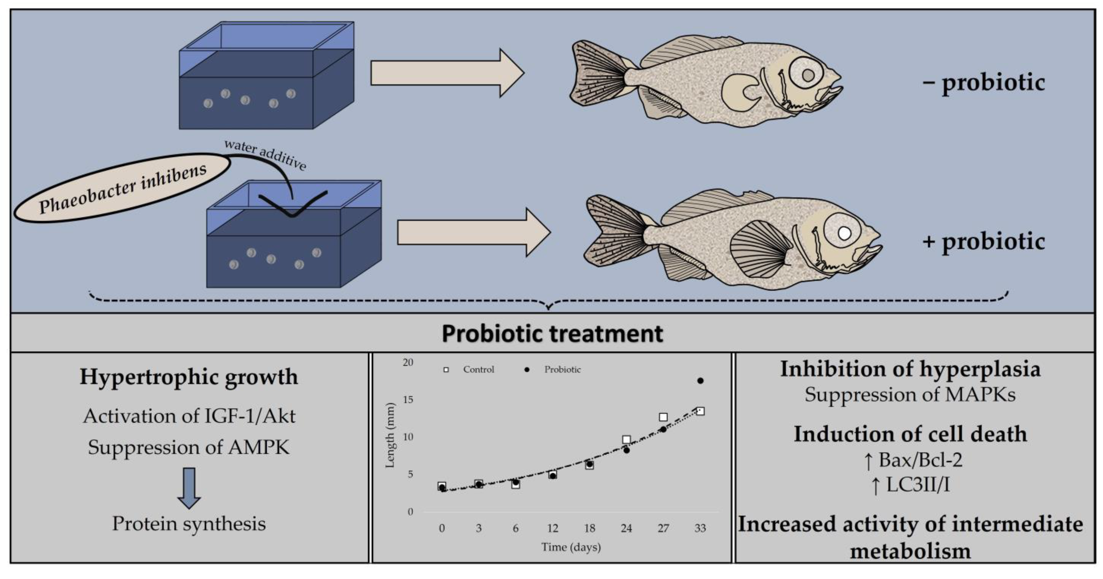
The Probiotic Phaeobacter inhibens Provokes Hypertrophic Growth via Activation of the IGF-1/Akt Pathway during the Process of Metamorphosis of Greater Amberjack (Seriola dumerili, Risso 1810)

by
Nikolas Panteli
1,*,
Konstantinos Feidantsis
1,2,
Maria Demertzioglou
1,
Vasiliki Paralika
3,
Stelios Karapanagiotis
4,
Constantinos C. Mylonas
5,
Konstantinos Ar. Kormas
6,7,
Eleni Mente
8,
Pavlos Makridis
3 and
Efthimia Antonopoulou
1,*
1
Department of Zoology, School of Biology, Faculty of Sciences, Aristotle University of Thessaloniki, 54124 Thessaloniki, Greece
2
Department of Fisheries & Aquaculture, University of Patras, 26504 Mesolonghi, Greece
3
Department of Biology, University of Patras, 26504 Rio Achaias, Greece
4
Galaxidi Marine Farm S.A., 33200 Galaxidi, Greece
5
Institute of Marine Biology, Biotechnology and Aquaculture, Hellenic Center for Marine Research, P.O. Box 2214, 71003 Heraklion, Greece
6
Department of Ichthyology and Aquatic Environment, School of Agricultural Sciences, University of Thessaly, 38446 Volos, Greece
7
Agricultural Development Institute, University Research and Innovation Centre “IASON”, Argonafton & Filellinon, 38221 Volos, Greece
8
Laboratory of Ichthyology-Culture and Pathology of Aquatic Animals, School of Veterinary Medicine, Aristotle University of Thessaloniki, 54124 Thessaloniki, Greece
*
Authors to whom correspondence should be addressed.
Animals 2023, 13(13), 2154; https://doi.org/10.3390/ani13132154
Submission received: 24 February 2023 / Revised: 4 June 2023 / Accepted: 27 June 2023 / Published: 29 June 2023
(This article belongs to the Special Issue Probiotics and Other Functional Feed Additives in Aquaculture)

Abstract

:

Simple Summary

Optimization of metamorphosis, a key stage in development where fish undergo several morphological and physiological changes, may assure the efficient mass production of greater amberjack (Seriola dumerili) juveniles. Application of probiotics as feed and/or water additive benefits nutrient utilization through modulation of the digestive enzymes. Therefore, following the addition of biofilters with the probiotic Phaeobacter inhibens in the rearing water throughout the early development of greater amberjack, crucial in metamorphosis, cellular pathways were investigated. The probiotic treatment increased the growth of greater amberjack, while hypertrophy was the main growth process in the metamorphosis. Additionally, the rearrangement of structures and tissues may have been facilitated through the observed induced cell death. The present findings are of great importance and may be applied in the aquaculture industry in order to enhance greater amberjack development and growth.

Abstract

Metamorphosis entails hormonally regulated morphological and physiological changes requiring high energy levels. Probiotics as feed supplements generate ameliorative effects on host nutrient digestion and absorption. Thereby, the aim of the present research was to investigate the impact of the probiotic Phaeobacter inhibens as a water additive on cellular signaling pathways in the metamorphosis of greater amberjack (Seriola dumerili). Activation of insulin-like growth factor type 1 receptor (IGF-1R), protein kinase B (Akt), mitogen-activated protein kinases (MAPKs) and AMP-activated protein kinase (AMPK), induction of heat shock proteins (Hsps), and programmed cell death were assessed through SDS-Page/immunoblot analysis, while energy metabolism was determined through enzymatic activities. According to the results, greater amberjack reared in P. inhibens-enriched water entered the metamorphic phase with greater body length, while protein synthesis was triggered to facilitate the hypertrophic growth as indicated by IGF-1/Akt activation and AMPK inhibition. Contrarily, MAPKs levels were reduced, whereas variations in Hsps response were evident in the probiotic treatment. Apoptosis and autophagy were mobilized potentially for the structural remodeling processes. Furthermore, the elevated enzymatic activities of intermediary metabolism highlighted the excess energy demands of metamorphosis. Collectively, the present findings demonstrate that P. inhibens may reinforce nutrient utilization, thus leading greater amberjack to an advanced growth and developmental state.

1. Introduction

Metamorphosis, a crucial stage in teleost post-embryonic development, encompasses the irreversible, abrupt, and nonsexual ontogenetic transition from larval to juvenile, a sexually immature form of the adult stage [1]. Teleosts undergo several changes in morphological, physiological, and behavioral traits that precede the juvenile transition, such as maturation of internal organs, changes in gill mitochondria-rich cells, and conformation of the adult pigmentation pattern [1,2,3]. Metamorphic remodeling is governed by thyroid hormones, while other endocrine factors, such as cortisol, prolactin, and the growth hormone (GH)/insulin-like growth factor (IGF) system, are also involved in the regulation of several cellular and developmental processes [4,5].
The GH/IGF system, pivotal to growth regulation [6], is highly linked to the metamorphic success of teleosts [7,8,9]. The effects of GH on somatic growth are mediated by IGF-1, a polypeptide hormone mainly produced in the liver that exerts a plethora of physiological and developmental actions on target tissues in an endocrine manner [9,10,11]. Consequent to its GH-stimulated secretion in circulation, IGF-1 acts on cells by binding to the IGF-1 receptor (IGF-1R) [10,12,13]. The latter triggers IGF-1R autophosphorylation, thus leading to the activation of two intracellular downstream signaling pathways, the phosphatidylinositol 3-kinase/protein kinase B (PI3K/Akt) and the mitogen-activated protein kinase/extracellular signal-regulated kinase (MAPK/ERK) [13,14]. The PI3K/Akt transduces stimulatory signals for cellular differentiation [15], hypertrophy [16], and protein synthesis [17], fundamental factors in animals’ development and growth. Cellular uptake of amino acids and glucose is also modulated via the PI3K/Akt signaling to supply the required substrates and energy for growth-related anabolic processes [3,18]. In contrast, the proliferative function of IGF-1 is mediated via the MAPK/ERK pathway [14], which also comprises a critical regulator of cell terminal differentiation [19]. Additionally, MAPK/ERK signaling is involved in the up-regulation of heat shock proteins (Hsp) expression [20,21], which are implicated in several developmental stages [22,23], including induction and completion of metamorphosis [24,25].
Hormonally regulated programmed cell death also constitutes an essential component of normal developmental progression, facilitating ongoing tissue remodeling and functional reorganization [26]. During metamorphosis, apoptotic cell death is triggered for the degeneration of larva transient structures and the sculpting of adult-specific organs [27,28]. Furthermore, apoptosis functions as a quality control mechanism, ensuring the elimination of defective and surplus cells, as well as compensation for mispatterning [28,29]. Apoptosis is determined by the interplay between pro-apoptotic and anti-apoptotic signals and is implemented via the activation of the caspases cascade, which orchestrates the cleavage of cellular substrates [28,30]. Nevertheless, deletion and renovation of cells and tissues may also be facilitated via autophagy, an intracellular catabolic system that promotes the degradation and recycling of cytoplasmic constituents in the lysosome, thereby preserving energetic homeostasis in cells undergoing developmental procedures [29,31,32].
The initiation of metamorphosis is coordinated in response to miscellaneous physiological and environmental cues, including endocrine signaling, and changes in temperature and food availability [4,33,34]. The metamorphosing period is accompanied by high requirements for specific nutrients, vital in both growth and physiological processes such as hormone synthesis [35,36]. The application of probiotics as an environmentally friendly alternative to antibiotics has emerged, encouraging the development of a sustainable aquaculture industry [37]. Probiotics, defined as adjuncts to beneficial microbes or microbial components in the host’s environment or feed [37,38], have been reported to efficiently inhibit pathogens’ adhesion and enhance development, growth, feed utilization, and digestibility in several fish species [39,40,41,42]. Species of the marine Roseobacter clade, including Phaeobacter inhibens, which has recently been characterized as a “safe-to-use” probiotic [43,44], are common members of the prokaryotic communities in aquaculture systems [44,45,46] and effectively colonize a wide range of inorganic and organic surfaces [47]. Due to the ability to produce several unique bioactives, including the bactericidal antibiotic tropodithietic acid (TDA) [48], P. inhibens antagonistic activity and suppresses the growth of potential opportunistic pathogens for the reared populations, such as Vibrio and Aeromonas [45,49,50] with no adverse effects on the host [44]. Thereby, the aim of the present study was to explore the effects of the probiotic bacterium P. inhibens as a water additive on cellular and metabolic signaling pathways during the metamorphosis of greater amberjack (Seriola dumerili), a potential candidate species for Mediterranean aquaculture, the production of which faces several major bottlenecks related to nutrition and reproduction [51,52].

2. Materials and Methods

2.1. Application of Biofilters with Phaeobacter inhibens to Greater Amberjack Larval Rearing Conditions

The probiotic bacterial strain Phaeobacter inhibens [53] was provided by the Leibniz Institute DSMZ-German Collection of Microorganisms and Cell Cultures GmbH in the form of a lyophilized preparation. The strain was reactivated in 5 mL of Marine Broth (MB, Difco Laboratories, Detroit, MI, USA) by incubation at 22 °C for 7 days in the dark and stagnant conditions, and thereafter inoculated in 2 L conical flasks of MB for 7 days. Commercial aquaculture biofilter supporting medium of porous ceramic cylinders (Premium Filter Material) made from natural quartzite was selected for the growth of P. inhibens [54]. The ceramic cylinders were sterilized at 121 °C for 20 min.
P. inhibens liquid cultures were harvested from the 2 L conical flasks by centrifugation at 12,000× g at 18 °C to remove the liquid culture medium. Afterward, filtered autoclaved seawater was added to the probiotic cells, and they were transferred and equally divided into the 5 L glass beakers with the ceramic cylinders. Additional filtered autoclaved seawater was added until the cylinders were completely covered with seawater added with bacteria. The beakers were covered with aluminum foil and incubated in the dark at 22 °C for 7 days. The ceramic cylinders were thereafter rinsed with sterile seawater and placed in three plastic 10 L containers, which were covered with a 350 μm net to prevent greater amberjack larvae from entering the biofilter. They were separately and carefully placed inside three larval tanks. Aeration with an air stone was added to these simple biofilters constructed in this way to oxygenate the cylinders and create a continuous water exchange between the plastic container and the larval tank. In this way, cells of P. inhibens were spread to the whole water volume in the rearing tanks.

2.2. Rearing of Greater Amberjack Larvae

Equal amounts of fertilized greater amberjack eggs were stocked in six 2800 L volume cylindroconical tanks in a flow-through system at the hatchery facilities of Galaxidi Marine Farm S.A., Greece. Borehole seawater was sand-filtered, aerated, and UV-treated prior to its use in the rearing system. The biofilters with the probiotic P. inhibens had been submerged in three tanks three days prior to the transfer of the amberjack eggs. The desired water temperature (>23 °C) was achieved using heat pumps. After hatching, the larval density was approximately 56 larvae/L. During the experimental period, the temperature and pH remained stable at 23–24 °C and 8, respectively. The dissolved oxygen saturation level was 85–95%. Water renewal was adjusted to 10% tank volume/h during egg incubation, was reduced to 5% tank volume/h on the day of hatching, and thereafter was increased gradually from 6 days post-hatching (dph) onwards up to 30% tank volume/h. Lights were turned on at 3 dph at 1000 lux intensity, decreased to 800 lux at 11 dph, 700 lux at 19 dph, and 500 lux at 23 dph. The live feed diet consisted of Rotifers (Brachionus sp.) from 3 to 22 dph, newly hatched Artemia nauplii from 12 to 33 dph, and enriched Artemia metanauplii from 18 dph up to the end of the trial. Formulated feed (Caviar Nature, BernAqua, Olen, Belgium) was used from 23 dph onwards.
Throughout the experimental trial, total length was used as the main morphometric index for the assessment of greater amberjack developmental progress [55,56]. On day 33, post-hatching during the process of metamorphosis and, subsequently, to the completion of notochord flexion, larvae from both experimental and control groups were sampled. From each of the 3 tanks, two biological replicates were collected (N = 2 + 2 + 2 = 6). Fish were captured, placed in 3 mL vials, and immediately frozen using liquid nitrogen. Samples were later transferred to −80 °C until further analyses.

2.3. SDS/PAGE and Immunoblot Analysis

The preparation of samples for SDS-PAGE, quantification of caspases and ubiquitinated proteins, and the immunoblot analysis are based on well-established protocols [57]. Specifically, frozen tissues were immediately homogenized in 3 mL g−1 of cold lysis buffer (20 mM β-glycerophosphate, 50 mM NaF, 2 mM EDTA, 20 mM Hepes, 0.2 mM Na3VO4, 10 mM benzamidine, pH 7, 200 μM leupeptin, 10 μΜ trans-epoxy succinyl-Lleucylamido-(4-guanidino)butane, 5 mM dithiotheitol, 300 μΜ phenyl methyl-sulfonyl fluoride (PMSF), 50 μg mL−1 pepstatin, 1% v/v Triton X-100), and extracted on ice for 30 min. Samples were centrifuged (10,000× g, 10 min, 4 °C), and the supernatant was boiled with 0.33 volumes of SDS/PAGE sample buffer (330 mM Tris-HCl, 13% v/v glycerol, 133 mM DTT, 10% w/v SDS, 0.2% w/v bromophenol blue). For the determination of LC3 II/I ratio and SQSTM1/p62 levels, samples were lysed in a buffer containing 150 mM NaCl, 20 mM Hepes, 5 mM DTT, 0.3 mM PMSF, 0.2 mM leupeptin, 0.01 mM E64 and 1% Triton X-100. Lysates were incubated on ice for 30 min and then centrifuged at 4 °C, for 5 min at 3000× g. Protein concentration was determined by using the BioRad protein assay (Bio-Rad Protein Assay Kit I, 5000001, BioRad, Hercules, CA, USA).
For the SDS-PAGE, equivalent amounts of proteins (50 μg) from samples of 5 individual batches of each developmental stage were separated either on 10% and 0.275% or 15% and 0.33% (w/v) acrylamide and bisacrylamide, followed by electrophoretic transfer onto membranes of nitrocellulose (0.45 μm, Schleicher and Schuell, Keene, NH, USA).
The resulting membranes were subjected to overnight incubation with: polyclonal rabbit anti-bcl2 (7973, Abcam, Cambridge, UK), polyclonal rabbit anti-bax (B-9) (7480, Santa Cruz Biotechnology, Dallas, TX, USA), monoclonal mouse anti-HSP70 (H5147, Sigma, St. Louis, MO, USA), monoclonal mouse anti-HSP90 (H1775, Sigma), anti-HSP60 (12165, Cell Signaling, Beverly, MA, USA), monoclonal mouse anti-phospho-SAPK-JNK (9255, Cell Signaling, Beverly, MA, USA), polyclonal rabbit anti-phospho-p38 MAP kinase (9211, Cell Signaling, Beverly, MA, USA), monoclonal rabbit anti-phospho p44/42 MAPK (4376, Cell Signaling, Beverly, MA, USA), polyclonal rabbit anti-SAPK-JNK (9252, Cell Signaling, Beverly, MA, USA), polyclonal rabbit anti-p44/42 MAPK (4695, Cell Signaling, Beverly, MA, USA), polyclonal rabbit anti-p38 MAPK (9212, Cell Signaling, Beverly, MA, USA), monoclonal rabbit anti-LC3B (3868, Cell Signaling, Beverly, MA, USA), polyclonal rabbit anti-p62/SQSTM1 (5114, Cell Signaling, Beverly, MA, USA), monoclonal rabbit anti-phospho AMPK (2535, Cell Signaling, Beverly, MA, USA), monoclonal rabbit anti-AMPK (5831, Cell Signaling, Beverly, MA, USA), anti-phospho-IGF-1R (3918, Cell Signaling, Beverly, MA, USA), anti-IGF-1R (9750, Cell Signaling, Beverly, MA, USA) and anti-phospho-Akt (9271, Cell Signaling, Beverly, MA, USA), anti-Akt (9272, Cell Signaling, Beverly, MA, USA). Quality transfer and protein loading Western blot were assured by Ponceau stain and actin (anti-β actin 3700, Cell Signaling, Beverly, MA, USA). Each of the examined proteins was normalized with its respective β-actin.
Concerning cleaved caspases and ubiquitination levels, protein samples were immunoblotted with a dot blot apparatus employment [58], and membranes were overnight incubated with monoclonal rabbit anti-cleaved caspase antibody (8698, Cell Signaling) and monoclonal mouse anti-ubiquitin conjugate (3936, Cell Signaling). Bands and blots were detected by enhanced chemiluminescence, while quantification was applied through laser-scanning densitometry (GelPro 4.0 Analyzer Software, GraphPad, San Diego, CA, USA).

2.4. Determination of Intermediate Metabolism Enzyme Activities

Activities of lactate dehydrogenase (L-LDH; E.C. 1.1.1.27.), citrate synthase (CS; E.C. 4.1.3.7.), and 3-hydroxyacyl CoA dehydrogenase (HOAD; 1.1.1.35) were estimated in samples according to well-established techniques [59,60,61,62]. For the analysis of L-LDH and HOAD activities, samples were homogenized in a buffer containing 150 mM imidazole, 1 mM EDTA, 5 mM dithiothreitol (DTT), and 1% Triton X-100, pH 7.4. For CS activity, tissue samples were homogenized in a buffer containing 20 mM HEPES, and 1 mM EDTA, with 1% Triton X-100, pH 7.4. To avoid loss of enzyme activity during sample preparation, procedures were performed on ice. Before analysis, homogenates were centrifuged at 13,000× g for 10 min at 4 °C. Maximum activity levels were determined spectrophotometrically at 18 °C. L-LDH and HOAD enzyme activities were measured following the oxidation of NADH at 340 nm (mM extinction coefficient = 6.22), and CS enzyme activities were determined based on the reaction of free coenzyme A with DTNB (5.5 V dithio-bis (2- nitrobenzoic acid) at 412 nm (mM extinction coefficient = 13.6). L-LDH was assayed in a medium containing 0.15 mM NADH, 1 mM KCN, and 50 mM imidazole, pH 7.4. The reaction was initiated by adding 1 mM pyruvate. CS was assayed in a medium containing 0.4 mM acetyl CoA, 0.25 mM DTNB and 75 mM Tris buffer, pH 8.0. The reaction was initiated by adding 0.5 mM oxaloacetate (OAA). 3-hydroxyacyl CoA dehydrogenase was assayed in a medium containing 0.15 NADH, 1 mM KCN, 1 mM EDTA, 50 mM Imidazole, pH 7.4. The reaction was initiated by the addition of 2.0 mM acetoacetate. Enzyme activities are expressed as micromoles of substrate min/mg protein. Protein concentration in supernatants was determined by using the BioRad protein assay (Bio-Rad Protein Assay Kit I, 5000001, BioRad, Hercules, CA, USA).

2.5. Microbiological Analysis of Rearing Water

In order to examine the effect of P. inhibens on bacterial load in the water, serial ten-fold dilutions of seawater from the rearing tanks from each sampling point were plated on 90 mm Petri dishes of Marine Agar (Difco Laboratories, Detroit, MI, USA) and thiosulfate-citrate-bile salts-sucrose agar (TCBS). The Petri dishes were thereafter incubated at room temperature (20 °C) for seven days in the dark, and the bacterial colonies were counted 1, 2, and 7 days after sampling.

2.6. Statistical Analysis
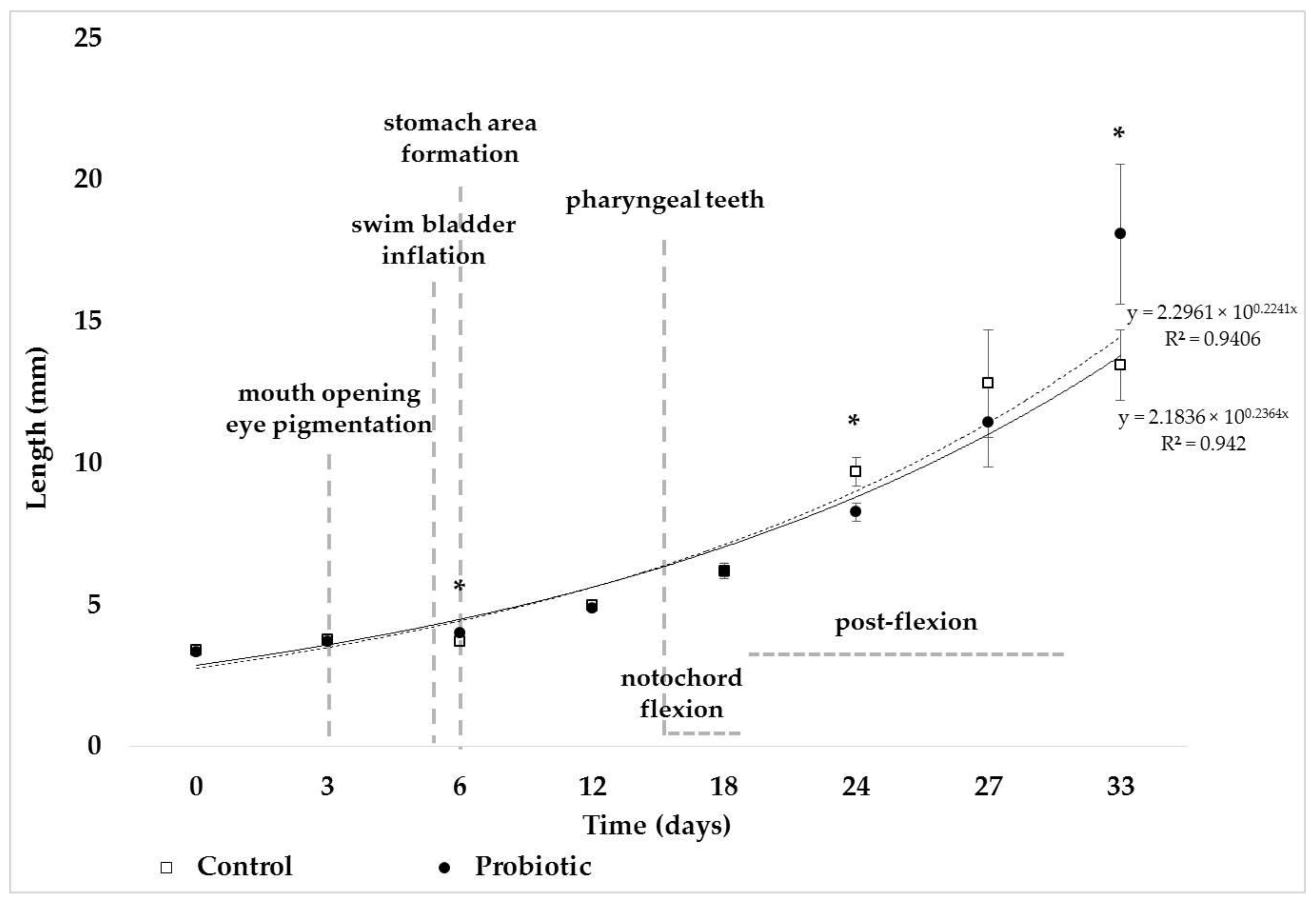
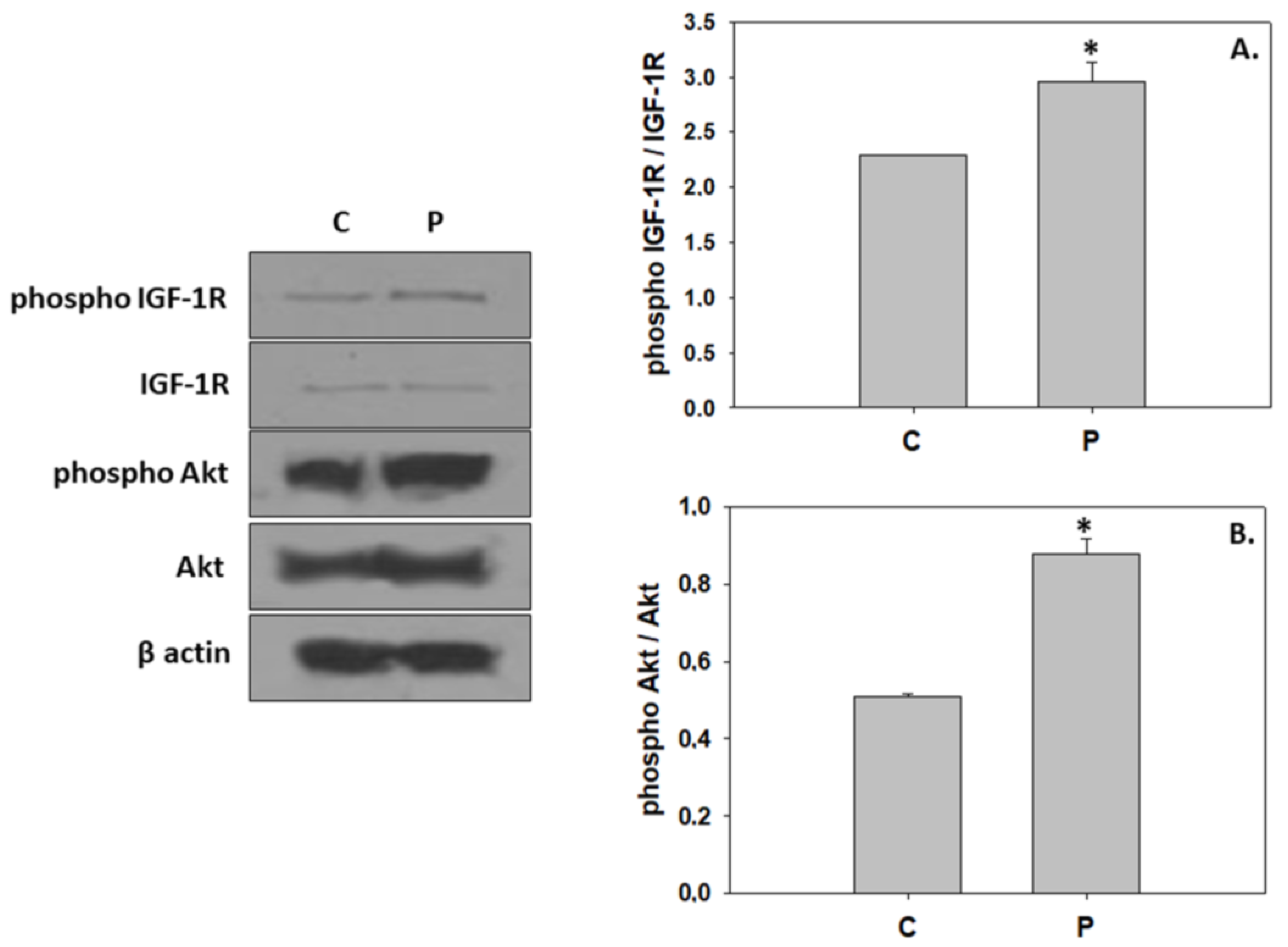
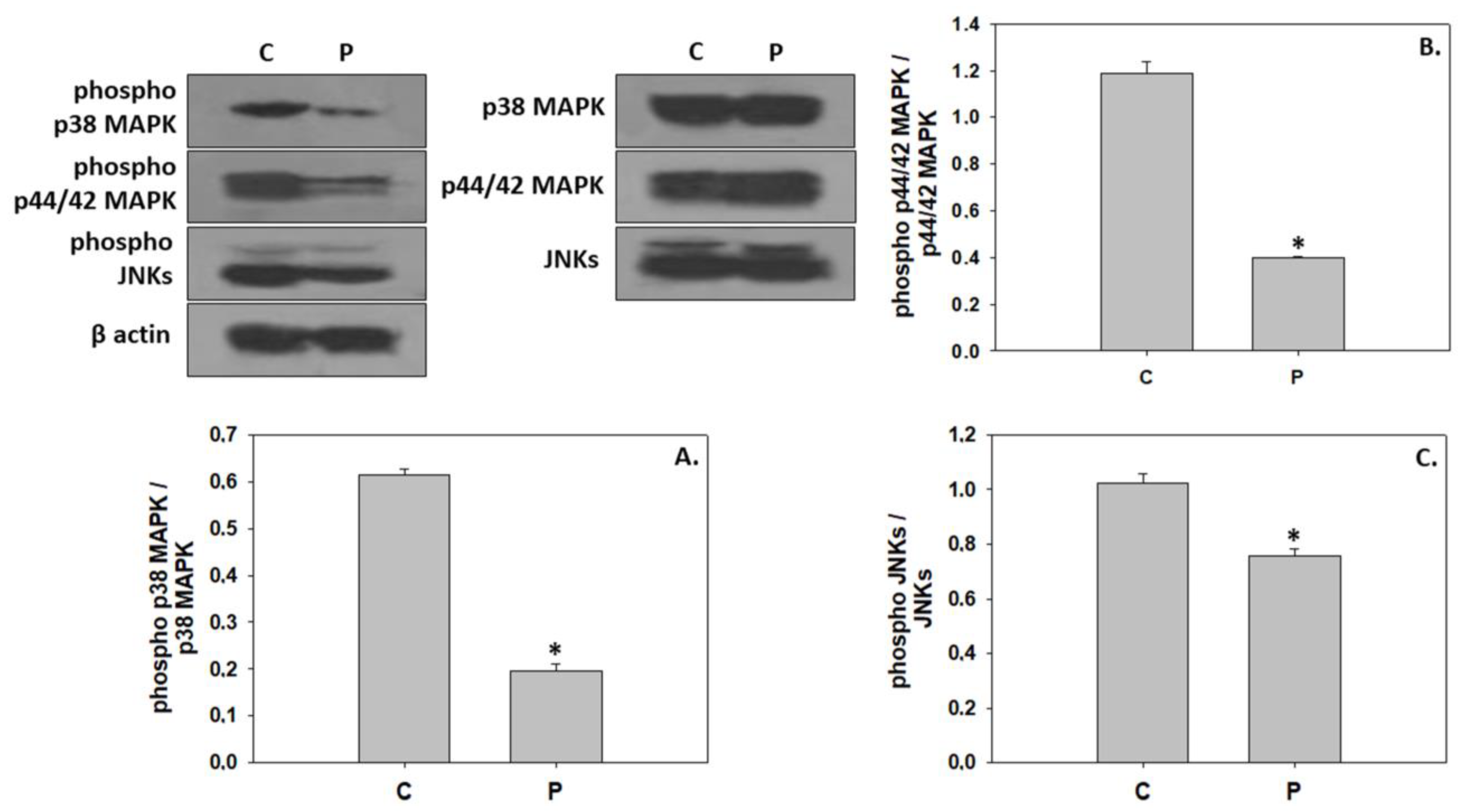
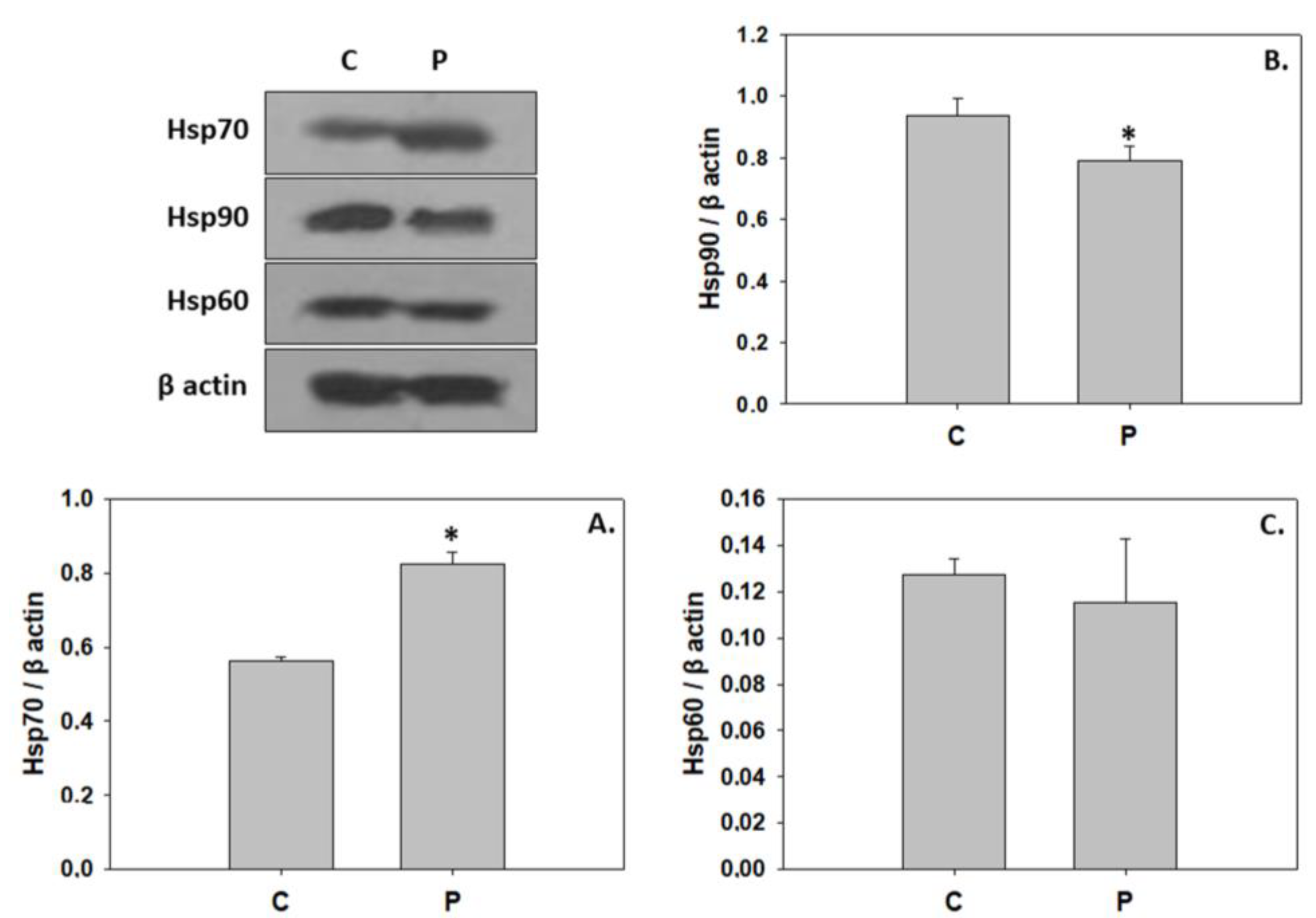
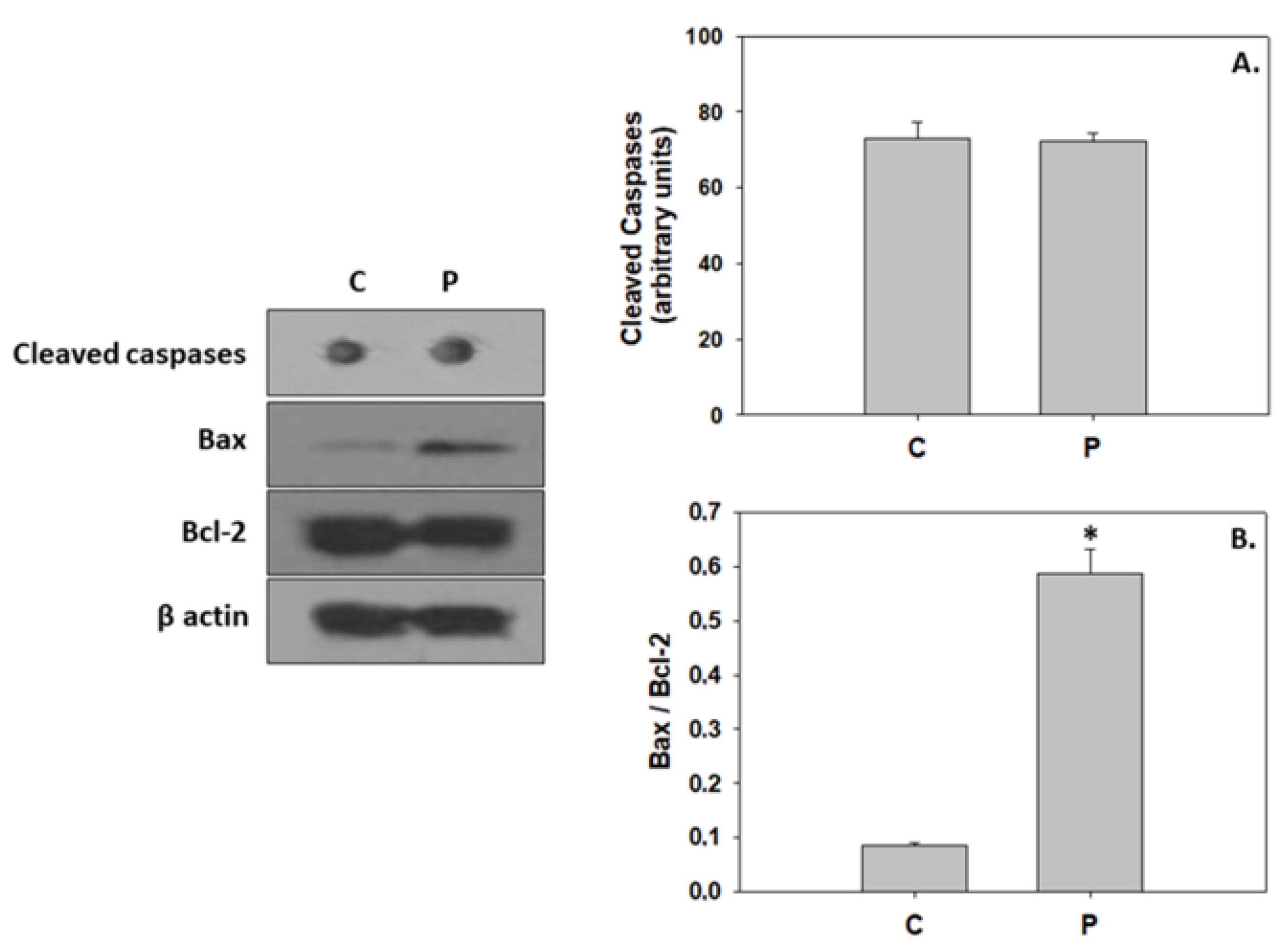
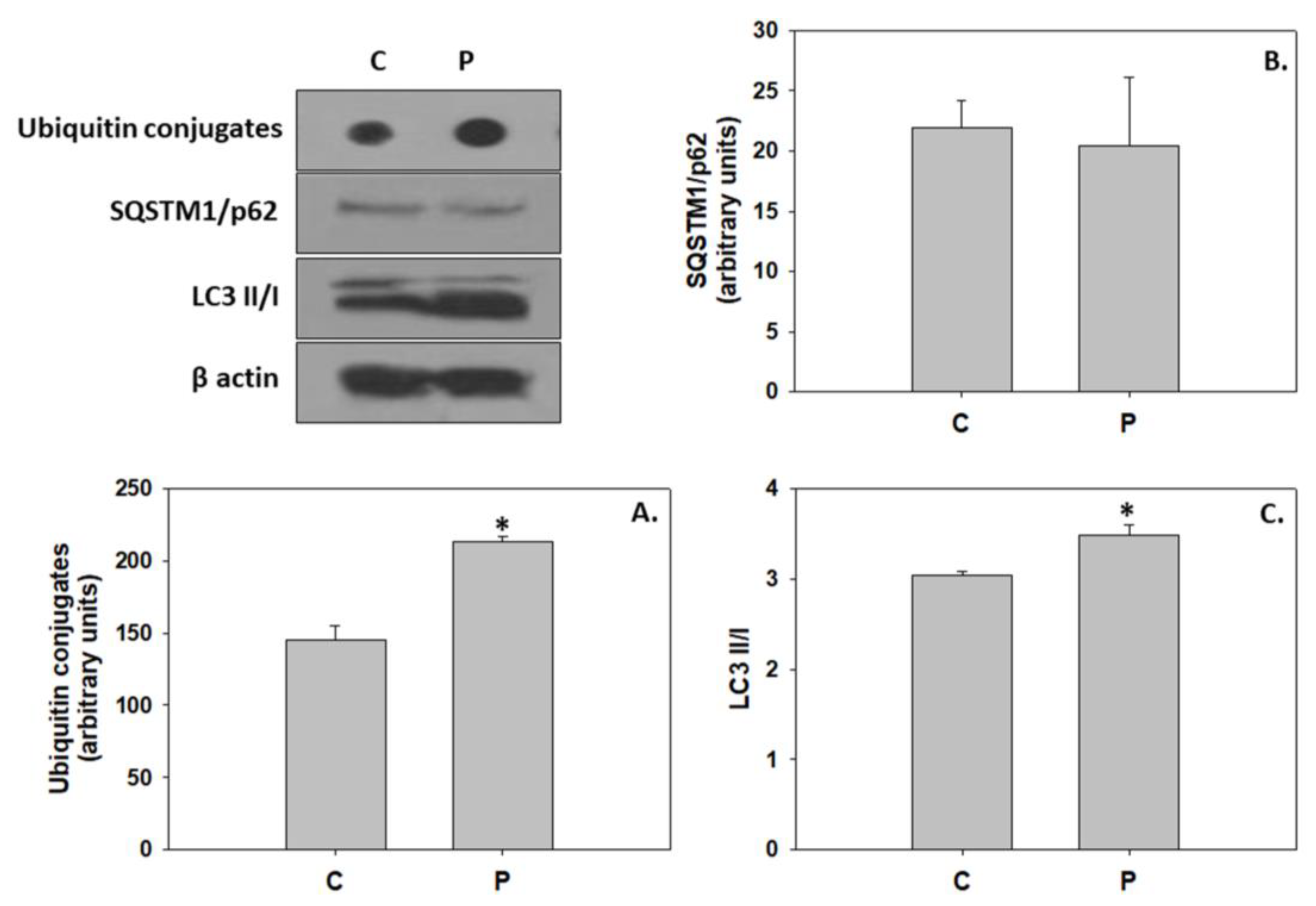
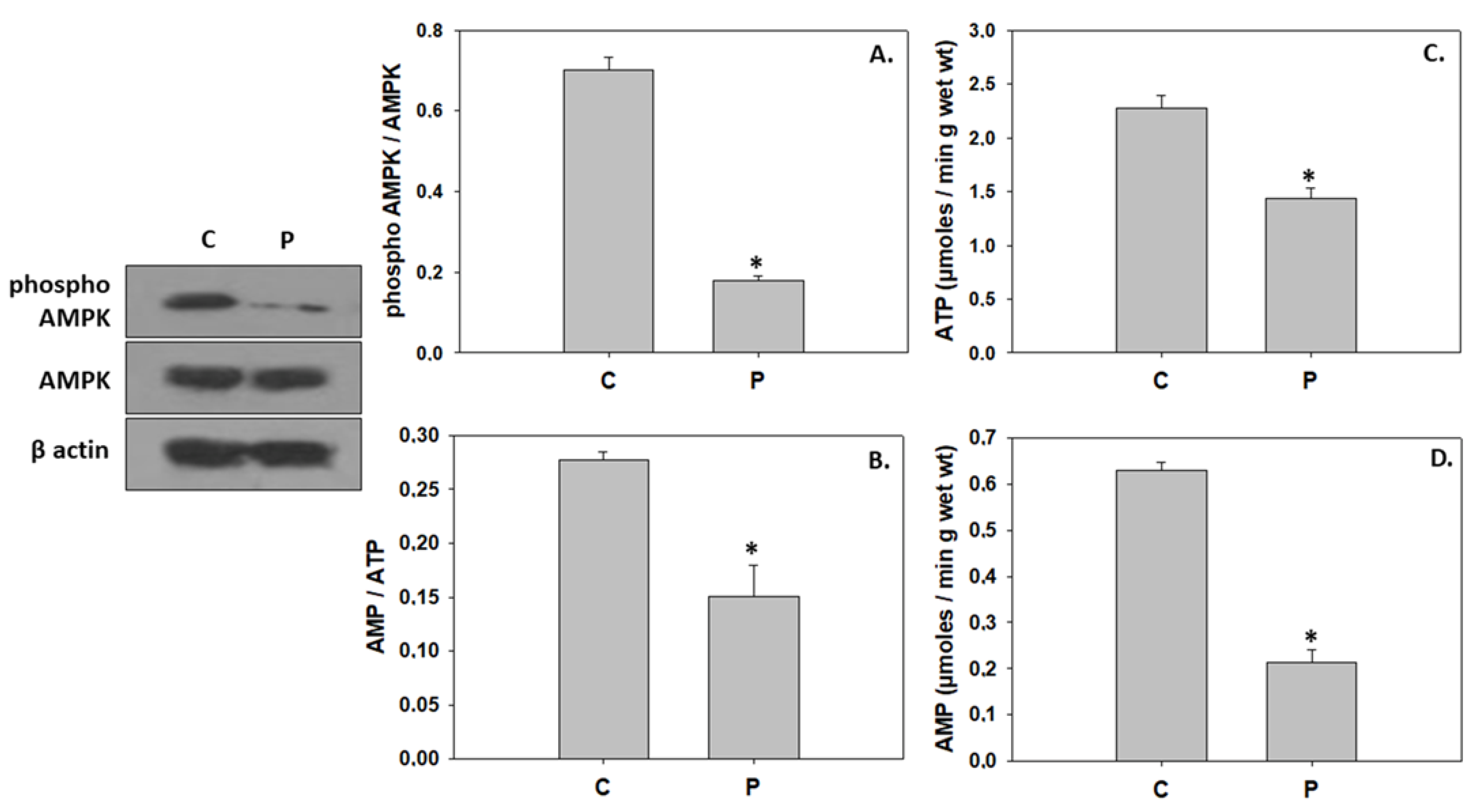
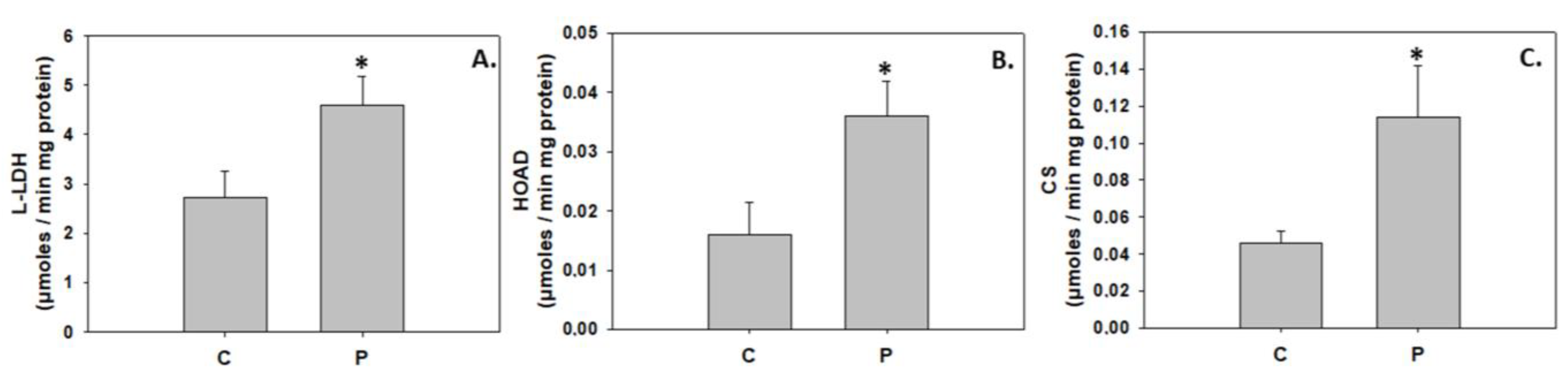
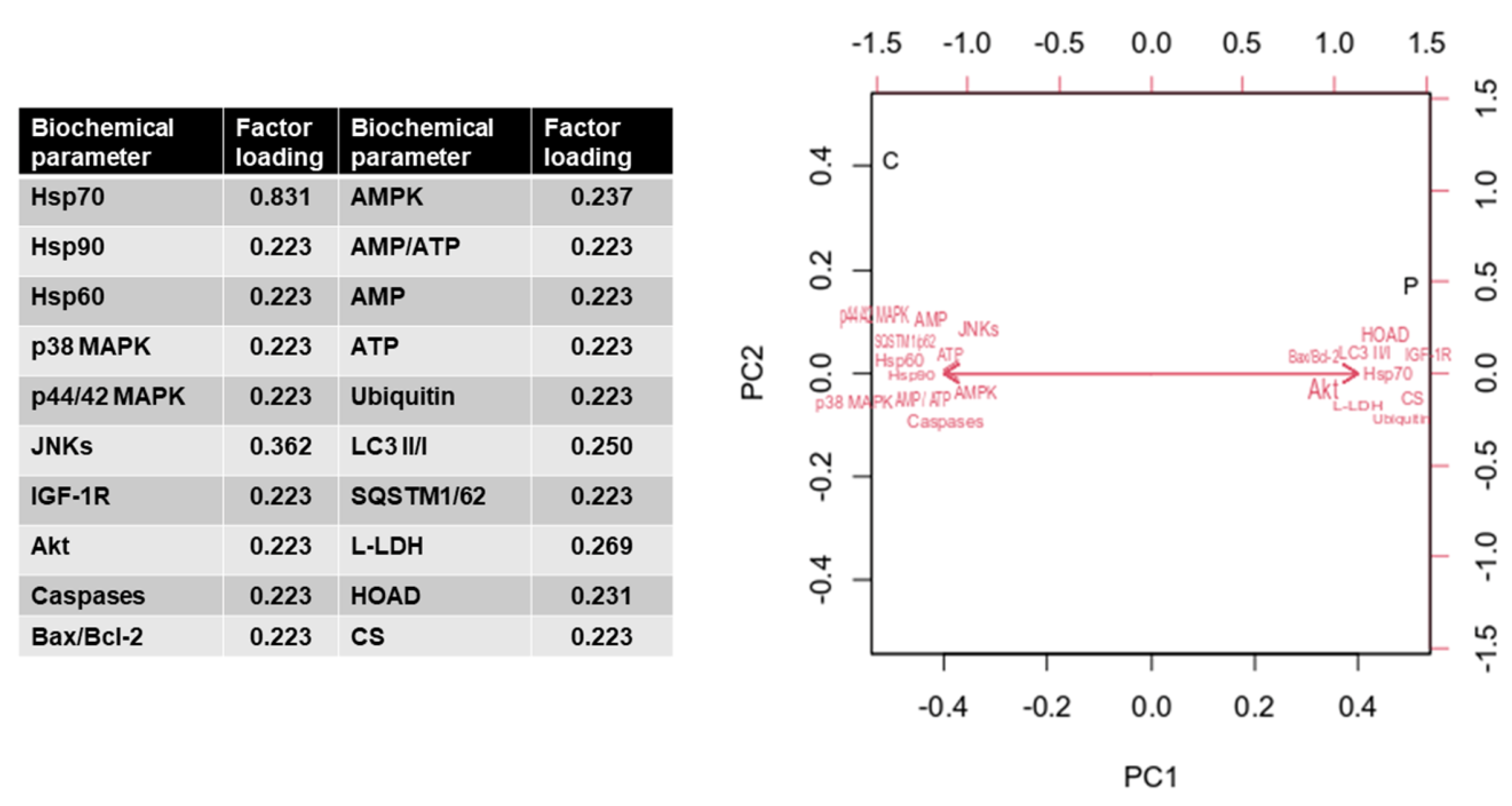
Protein expression data were tested for normality of distribution via the Shapiro–Wilk test, while homogeneity of variance was assessed using Levene’s Test for Equality of Variances. Changes in biochemical responses were tested for significance at the 5% level by using a one-way Analysis of variance (ANOVA) (GraphPad Instat 3.0). Values are presented as means ± S.D.
Principal components analysis (PCA) was conducted in the R package FactoMineR to assess patterns of possibly correlated variables. Moreover, simple linear correlation (Pearson’s test) analysis was employed for the estimation of significant correlations (at a 5% level) between the biochemical data obtained in the present study (GraphPad Prism 5.0).

3. Results

According to the measured total body length, the larval notochord flexion was completed by the end of the experimental trials. Changes in the total body length of greater amberjack from hatching day until metamorphosis (33 dph) are depicted in Figure 1. Growth in total body length was significantly higher (p < 0.05) in greater amberjack reared in the P. inhibens-enriched water at 6 dph and 33 dph. In contrast, lower total body length (p < 0.05) was evident in greater amberjack of the P. inhibens treatment at 24 dph compared to the control.
The probiotic P. inhibens as a water additive displayed similar effects on the phosphorylation ratios of IGF-1R and Akt, resulting in a significant activation (Figure 2). In contrast, probiotic-enriched water provoked a significant reduction in the phosphorylation ratio of all three MAPKs examined in the present study (Figure 3). The addition of the probiotic in the rearing water did not affect Hsp60 expression in 33 dph greater amberjack. However, probiotic addition significantly reduced Hsp90 levels, whereas Hsp70 levels were significantly increased (Figure 4).
The addition of the probiotic did not alter caspase levels compared to the control group. However, the probiotic significantly increased the Bax/Bcl-2 compared to the control (Figure 5). Regarding autophagic indicators, while the addition of the probiotic resulted in a statistically significant increase of ubiquitin conjugates levels and LC3 II/I ratio, no changes were observed in the SQSTM1/p62 levels (Figure 6). AMPK phosphorylation, the AMP/ATP ratio, and ATP and AMP levels were significantly reduced in the probiotic-enriched water compared to the control (Figure 7).
On the other hand, L-LDH, HOAD, and CS activity levels were significantly increased in the probiotic treatment (Figure 8).
According to the Principal Components Analysis (PCA), PC1 explained 31.62% of the variance (Figure 9). The biochemical variables that were positively correlated with PC1 scores were Hsp70, IGF-1R, Akt, Bax/Bcl-2, Ubiquitin, LC3 II/I, L-LDH, HOAD, and CS. In contrast, Hsp90, Hsp60, p38 MAPK, p44/42 MAPK, JNKs, caspases, AMPK, AMP/ATP, AMP, ATP, and SQSTM1/p62 were negatively correlated with PC1. Regarding PC2, which explained 27.75% of the variance, the scores that were positively correlated to this principal component were JNKs, IGF-1R, Akt, Bax/Bcl-2, AMP, ubiquitin, LC3 II/I, and L-LDH. In contrast, Hsp70, Hsp90, Hsp60, p38 MAPK, p44/42 MAPK, caspases, AMPK, AMP/ATP, ATP, SQSTM1/p62, HOAD, and CS were negatively correlated to PC2. The cumulative value of PC1 and PC2 was 66.09%.
The bacterial load in the rearing water, as determined by the total counts in colony-forming units (CFU) per mL, was not significantly modified among the P. inhibens-enriched tanks and the control tanks (p > 0.05) (Supplementary Material Figure S1). The number of presumptive Vibrios, as determined by CFU counts in TCBS agar, was similar in both treatments (p > 0.05), despite the higher numbers in the control tanks (Supplementary Material Figure S1).

4. Discussion

Growth in fish is highly reflected by the growth of skeletal muscle, the most abundant tissue and main contributor to total body mass [63]. In contrast to higher vertebrates, post-hatching muscle growth in teleost species is implemented by both hypertrophy (enlargement of existing muscle fibers) and hyperplasia (the genesis of new myofibers) [64]. During a teleost life span, the equilibrium in dominance among the two aforementioned processes is differentiated in a species-specific manner, where hypertrophy seems to constitute the main growth mechanism towards the later developmental stages of juvenile and adult, as has been previously demonstrated in Gilthead seabream, Sparus aurata, and white seabass, Atractoscion nobilis [63,65].

4.1. Induction of Hypertrophic Growth via Activation of IGF/Akt Pathway

According to the present results, administration of the probiotic P. inhibens in the rearing water promoted the activation of both IGF-1R and Akt, integral mediators of hypertrophic growth [16]. Nutritional regulation of the IGF-1/PI3K/Akt pathway has been previously reported in the skeletal muscle of fine flounder (Paralichthys adspersus) [66] and rainbow trout (Oncorhynchus mykiss) [67,68], where a dietary trial of fasting and refeeding led to PI3K/Akt inactivation and activation, respectively. The ameliorative effects of probiotics in host nutrition have been extensively documented in several teleost species. For instance, dietary supplementation of probiotics enhanced the activities of digestive enzymes, such as lipase, amylase, alkaline phosphatase, pepsin, trypsin, and chymotrypsin, and improved the growth performance in Caspian white fish (Rutilus frisii kutum) [69], California halibut (P. californicus) [70], zebrafish (Danio rerio) [71], European seabass (Dicentrarchus labrax) [72] (and gilthead sea bream [73]. In addition, probiotics may beneficially modulate gut microbiota composition and function, and ameliorate microbial balance, which may further improve the physiological processes in the digestive system (reviewed in [69,74]). Thus, the development of greater amberjack in rearing water supplemented with P. inhibens may have favored the digestion and absorption of nutrients such as proteins and, in consequence, altered cellular metabolic pathways.
Specifically, Akt activation observed herein, possibly due to digestion strengthening, may mediate the nutrient- and IGF-induced signaling for stimulation of protein synthesis via phosphorylation of the target of rapamycin (TOR), a downstream component of the PI3K/Akt pathway (reviewed in [75,76]). Induction of protein synthesis through the TOR pathway has been previously demonstrated in juvenile Jian carp (C. carpio var. Jian) in response to fish meal substitution with yeast hydrolysate [77]. In contrast, dietary supplementation of cottonseed meal protein hydrolysate at high levels repressed protein synthesis due to inhibition of the TOR signaling pathway via activation of AMPK [78]. The AMP-activated protein kinase is an energy-status sensor, which is activated responsively to changes in the intracellular AMP/ATP ratio and, more precisely, under a low cellular energetic regime in order to suppress energy-consuming processes such as protein synthesis and to stimulate catabolic pathways for ATP regeneration in the cell [76,79]. Thereby, the apparent low ratio of AMP/ATP and the inhibition of AMPK observed herein in greater amberjack reared in the probiotic-enriched water further confirms IGF/Akt/TOR-stimulated protein synthesis, which may have been recruited for the hypertrophic growth during metamorphosis. The present finding is consistent with previous studies that indicated the prevailing hypertrophy in postnatal growth, including the metamorphosing stage, in several teleosts [55,80,81]. However, in addition to the rapid growth of greater amberjack, the IGF/Akt-mediated hypertrophy may be implicated in the metamorphic remodeling and reorganization of miscellaneous larvae tissues and structures, such as the gastrointestinal tract as observed in other species [82], in order to functionally adapt to the juvenile form.

4.2. Inhibition of Downstream Components in the IGF/MAPK Signaling

In contrast to the IGF/Akt activation, P. inhibens as a water additive suppressed components of the MAPK signaling pathway during greater amberjack metamorphosis. Activation of the MAPK pathway via IGF-1 binding to the IGF-1R signals for cell proliferation and hyperplastic growth [15]. Proliferative events have previously been demonstrated to be crucial to the structural and functional development of tissues and organs during teleosts’ metamorphosis [83,84]. In regard to greater amberjack, Panteli et al. [85] indicated p38 MAPK, p44/42 MAPK, and JNK activation in the metamorphosing stage, thus indicating a potential involvement in organogenesis and muscle growth. Herein, P. inhibens may have acted as an inhibitor of the hyperplastic effect as observed by p38 MAPK, p44/42 MAPK, and JNK inactivation compared to the greater amberjack of the control group. In line with the present results, probiotic-supplemented diets increased the growth rate of Malaysian Mahseer (Tor tambroides) juveniles mainly through the prevalence of hypertrophic growth process in muscle fibers rather than hyperplasia [86].
The modification in MAPKs activation observed herein may be attributed to the evident differences in the greater amberjack length among the two groups. Specifically, according to the total length, the greater amberjack reared in the P. inhibens-enriched water may have initiated the stage of metamorphosis earlier in comparison to the control group. The relative contribution of hyperplasia in the growth procedure decreases with standard fish length, as reported by Zimmerman and Lowery [87]. Therefore, the apparent decrease in p38 MAPK, p44/42 MAPK, and JNK activation may indicate that P. inhibens favored a more rapid growth, potentially leading greater amberjack to advanced development, where the hyperplastic-facilitated procedures were mostly completed. Furthermore, hypertrophy and hyperplasia function in a cyclic phase pattern during teleosts’ early ontogeny [80,87], which reinforces the latter hypothesis. In this context, Weston et al. [88] suggested that p38 signaling is active early in myogenesis in order to prevent myocyte premature progression and is subsequently suppressed during the elongation, polarization, aggregation, and fusion of the differentiated cells. However, it is worth mentioning that fish species of large final body size, such as greater amberjack, continue to rely to an extent on hyperplasia for muscle growth in the adult phase [63].
Furthermore, consequent to the decrease in MAPKs phosphorylation, the expression level of Hsp90 was downregulated in the greater amberjack reared under the P. inhibens regime. As previously mentioned, MAPKs are regulators involved in Hsps induction [20,21]. According to previous studies, the metamorphosis of greater amberjack [85] and flatfish Senegalese sole (Solea senegalensis Kaup) [23] was accompanied by a decrease in Hsp90 expression levels. However, P. inhibens as a water additive induced Hsp70 expression in this instance, which was also evident in gilthead seabream following probiotics administration [89]. On the contrary, decreased Hsp70 levels were observed in teleost species under probiotic treatment [39,90,91]. Changes in Hsps induction due to probiotics vary according to miscellaneous factors such as fish species, development stage, bacterial strain and dose, supplementation mode, and rearing duration and conditions [90]. Molecular chaperones are involved in the regulation of signal-transduction pathways [23], and thus, the recruitment of such proteins is essential in several developmental stages, including metamorphosis for tissue resorption and rearrangement [92]. Upon entering the metamorphic phase, silver sea bream (Sparus sarba) exhibited induction of both Hsp70 and Hsp90, therefore indicating involvement in the larvae to juvenile transition processes [93]. However, Chung-Davidson et al. [94] demonstrated that Hsp90 inhibition facilitates the degeneration stage of the gall bladder during the liver metamorphosis of sea lamprey. Therefore, both up-regulation and down-regulation of Hsps due to the addition of P. inhibens may be deemed crucial in proper greater amberjack metamorphosis and accelerated growth, and hence, further investigations are required to clarify their distinct role in the organism’s development.

4.3. P. inhibens-Induced Developmental Cell Death

The commencement of metamorphosis in vertebrates is associated with the recruitment of extensive programmed cell death processes in order to facilitate the deletion of larval cells and tissues for the structural, morphological, and functional remodeling of the juveniles (reviewed in [26,30]). For instance, during the initial stage of the metamorphosis of the Japanese flounder (Paralichthys olivaceus), pro-apoptotic signals were evident in the frontal bone, and ethmoid and trabecular cartilage, which may have subsequently contributed to eye migration [95,96]. In addition, Gao et al. [97] demonstrated that the autophagic machinery facilitates eye migration in the aforementioned species through cell death in the orbital tissue. The addition of P. inhibens in the rearing water resulted in the induction of both apoptosis and autophagy in greater amberjack, as observed by the increased Bax/Bcl-2 and LC3 II/I ratios. The action of induced apoptosis was demonstrated in teleost cell lines following the addition of cytoplasmic extracts obtained from the probiotic Lactobacillus delbrüeckii subsp. lactis [98]. As indicated in the present results, probiotic treatment induced autophagy in zebrafish follicles, as observed by the increased autophagosomes and LC3-II protein levels, which may participate in ubiquitinated protein degradation [99]. Mobilization of apoptotic cell death observed herein may indicate the increased structural processes due to the potential accelerated growth. In specific, under the P. inhibens regime, greater amberjack seems to exhibit advanced developmental progress compared to the control.
However, probiotic-induced autophagy may subserve a dual role in greater amberjack metamorphosis. In addition to the aforementioned role in larval to juvenile reconstruction, autophagy may have also been employed to secure energy and nutrients required for the energetically demanding processes of metamorphosis and growth [29,31,100]. The latter is further supported by the increased activities of intermediary metabolism enzymes in the greater amberjack of the P. inhibens treatment despite the AMPK inhibition. In line with the above, probiotic-enhanced nutrient metabolism and growth performance was similarly suggested by Yi et al. [101], who demonstrated an up-regulation of genes related to carbohydrate metabolism and growth, including the IGF-1 gene, in zebrafish fed a diet supplemented with the potential probiotic Chromobacterium aquaticum. Skeletal muscle growth depends on protein anabolism [102], an energy-consuming procedure, and thus additional energy may be required by the greater amberjack reared under the P. inhibens regime, which also displayed higher body length.

5. Conclusions

Administration of probiotic Phaeobacter inhibens as water additive exerted a contrary indirect regulatory role on IGF-I/Akt and IGF-I/MAPK signaling in the metamorphosis of greater amberjack. Specifically, the potentially higher nutrient supply due to the P. inhibens-enhanced digestion triggered the activation of the IGF-I/Akt pathway, while downstream components of the IGF-I/MAPK, including p38 MAPK, p44/42 MAPK, and JNK exhibited inhibition (Figure 10). Moreover, inhibition of AMPK indicates that anabolic processes, such as protein synthesis, are active and facilitate hypertrophic growth. Induced programmed cell death may have been recruited in the probiotic treatment for the deletion and renovation of cells and tissues. According to the present results, it can be assumed that under the influence of the probiotic treatment, an apparent increase in the rate of development and muscle hyperplasia occurred earlier, thus leading to the subsequently enhanced hypertrophy observed. It should be emphasized that larval muscle hyperplasia and hypertrophy may affect the post-metamorphosis muscle cellularity, and the relative contributions of these two growth mechanisms exert long-term effects on muscle cellularity and growth of reared fish [103,104]. In conclusion, P. inhibens as a water additive seems to exert ameliorative effects in the development of reared greater amberjack. However, both developmental processes and responses to probiotics depend highly on species; thus, more species-oriented research is required to further explore the application of P. inhibens in the aquaculture industry, as well as the long-term change effects in hyperplasia and hypertrophy.

Supplementary Materials

The following supporting information can be downloaded at: https://www.mdpi.com/article/10.3390/ani13132154/s1, Figure S1: Total bacterial load and presumptive Vibrios in seawater from the rearing tanks as determined by counting of colony forming units (CFU) in Marine agar and in TCBS agar, respectively.

Author Contributions

Conceptualization, N.P., K.F., P.M. and E.A.; methodology, N.P., K.F., M.D., S.K., V.P. and E.A.; software, N.P., K.F., M.D. and E.A.; validation, N.P., K.F., M.D. and E.A.; formal analysis, N.P., K.F., M.D. and E.A; investigation, N.P., K.F., M.D., V.P., S.K., C.C.M., K.A.K., E.M., P.M. and E.A.; resources, P.M. and E.A.; data curation, N.P., K.F., M.D. and E.A.; writing—original draft preparation, N.P., K.F. and E.A.; writing—review and editing, N.P., K.F., M.D., V.P., S.K., C.C.M., K.A.K., E.M., P.M. and E.A.; visualization, N.P., K.F., M.D. and E.A.; supervision, E.M., P.M. and E.A.; project administration, E.A.; funding acquisition, C.C.M., E.M. and E.A. All authors have read and agreed to the published version of the manuscript.

Funding

This research was funded by European Union and Greek national funds through the National Strategic Reference Framework 2014–2020 (GR and EU), Special Actions Aquaculture-Industrial Materials-Open innovation in culture (project code: MIS 5045873), entitled “Improvement of broodstock management and fingerling production methods for greater amberjack (Seriola dumerili)” (acronym: Magiatiko).

Institutional Review Board Statement

The animal study protocol was performed in accordance with animal welfare guidelines of the Hellenic Aquaculture Producer Organization (HAPO) and approved and validated by the Hellenic Ministry of Rural Development and Food (1558/198651), alongside the company’s routine quality control samplings. All animal experiments complied with the ARRIVE guidelines and were carried out in accordance with the EU Directive 2010/63/EU for animal experiments.

Informed Consent Statement

Not applicable.

Data Availability Statement

The datasets generated and/or analyzed during the current study are not publicly available because, due to the nature of this research, accompanying data to the ones presented herein remain unpublished to date, but are available from the corresponding author on reasonable request.

Acknowledgments

The authors would like to thank Ioannis Papadakis for information sharing and fruitful discussions improving our manuscript.

Conflicts of Interest

The authors declare no conflict of interest. The funders had no role in the design of this study; in the collection, analyses, or interpretation of data; in the writing of the manuscript; or in the decision to publish the results.

References

  1. McMenamin, S.K.; Parichy, D.M. Chapter Five—Metamorphosis in Teleosts. In Current Topics in Developmental Biology; Shi, Y.-B., Ed.; Animal Metamorphosis; Academic Press: Cambridge, MA, USA, 2013; Volume 103, pp. 127–165. [Google Scholar]
  2. Schreiber, A.M.; Specker, J.L. Metamorphosis in the Summer Flounder, Paralichthys dentatus: Thyroidal Status Influences Gill Mitochondria-Rich Cells. Gen. Comp. Endocrinol. 2000, 117, 238–250. [Google Scholar] [CrossRef] [PubMed]
  3. Vélez, E.J.; Lutfi, E.; Azizi, S.; Perelló, M.; Salmerón, C.; Riera-Codina, M.; Ibarz, A.; Fernández-Borràs, J.; Blasco, J.; Capilla, E.; et al. Understanding Fish Muscle Growth Regulation to Optimize Aquaculture Production. Aquaculture 2017, 467, 28–40. [Google Scholar] [CrossRef]
  4. Campinho, M.A. Teleost Metamorphosis: The Role of Thyroid Hormone. Front. Endocrinol. 2019, 10, 383. [Google Scholar] [CrossRef] [PubMed]
  5. de Jesus, E.G.; Hirano, T.; Inui, Y. Flounder Metamorphosis: Its Regulation by Various Hormones. Fish Physiol. Biochem. 1993, 11, 323–328. [Google Scholar] [CrossRef]
  6. Fuentes, E.N.; Valdés, J.A.; Molina, A.; Björnsson, B.T. Regulation of skeletal muscle growth in fish by the growth hormone–insulin-like growth factor system. Gen. Comp. Endocrinol. 2013, 192, 136–148. [Google Scholar] [CrossRef]
  7. Hildahl, J.; Power, D.M.; Björnsson, B.T.; Einarsdóttir, I.E. Involvement of Growth Hormone-Insulin-like Growth Factor I System in Cranial Remodeling during Halibut Metamorphosis as Indicated by Tissue- and Stage-Specific Receptor Gene Expression and the Presence of Growth Hormone Receptor Protein. Cell Tissue Res. 2008, 332, 211–225. [Google Scholar] [CrossRef]
  8. Jia, Y. Roles of Insulin-like Growth Factors in Metamorphic Development of Turbot (Scophthalmus maximus). Gen. Comp. Endocrinol. 2018, 265, 61–63. [Google Scholar] [CrossRef]
  9. Zhang, J.; Shi, Z.; Cheng, Q.; Chen, X. Expression of Insulin-like Growth Factor I Receptors at MRNA and Protein Levels during Metamorphosis of Japanese Flounder (Paralichthys olivaceus). Gen. Comp. Endocrinol. 2011, 173, 78–85. [Google Scholar] [CrossRef]
  10. Moriyama, S.; Ayson, F.G.; Kawauchi, H. Growth Regulation by Insulin-like Growth Factor-I in Fish. Biosci. Biotech. Biochem. 2000, 64, 1553–1562. [Google Scholar] [CrossRef]
  11. Reinecke, M.; Björnsson, B.T.; Dickhoff, W.W.; McCormick, S.D.; Navarro, I.; Power, D.M.; Gutiérrez, J. Growth Hormone and Insulin-like Growth Factors in Fish: Where We Are and Where to Go. Gen. Comp. Endocrinol. 2005, 142, 20–24. [Google Scholar] [CrossRef]
  12. Duan, C. Nutritional and Developmental Regulation of Insulin-like Growth Factors in Fish. J. Nutr. 1998, 128, 306S–314S. [Google Scholar] [CrossRef] [PubMed] [Green Version]
  13. Dupont, J.; LeRoith, D. Insulin and Insulin-Like Growth Factor I Receptors: Similarities and Differences in Signal Transduction. Horm. Res. Paediatr. 2001, 55, 22–26. [Google Scholar] [CrossRef] [PubMed]
  14. Favelyukis, S.; Till, J.H.; Hubbard, S.R.; Miller, W.T. Structure and Autoregulation of the Insulin-like Growth Factor 1 Receptor Kinase. Nat. Struct. Mol. Biol. 2001, 8, 1058–1063. [Google Scholar] [CrossRef]
  15. Coolican, S.A.; Samuel, D.S.; Ewton, D.Z.; McWade, F.J.; Florini, J.R. The Mitogenic and Myogenic Actions of Insulin-like Growth Factors Utilize Distinct Signaling Pathways. J. Biol. Chem. 1997, 272, 6653–6662. [Google Scholar] [CrossRef] [PubMed] [Green Version]
  16. Bodine, S.C.; Stitt, T.N.; Gonzalez, M.; Kline, W.O.; Stover, G.L.; Bauerlein, R.; Zlotchenko, E.; Scrimgeour, A.; Lawrence, J.C.; Glass, D.J.; et al. Akt/MTOR Pathway Is a Crucial Regulator of Skeletal Muscle Hypertrophy and Can Prevent Muscle Atrophy in Vivo. Nat. Cell Biol. 2001, 3, 1014–1019. [Google Scholar] [CrossRef]
  17. Rommel, C.; Bodine, S.C.; Clarke, B.A.; Rossman, R.; Nunez, L.; Stitt, T.N.; Yancopoulos, G.D.; Glass, D.J. Mediation of IGF-1-Induced Skeletal Myotube Hypertrophy by PI(3)K/Akt/MTOR and PI(3)K/Akt/GSK3 Pathways. Nat. Cell Biol. 2001, 3, 1009–1013. [Google Scholar] [CrossRef]
  18. Codina, M.; García de la Serrana, D.; Sánchez-Gurmaches, J.; Montserrat, N.; Chistyakova, O.; Navarro, I.; Gutiérrez, J. Metabolic and Mitogenic Effects of IGF-II in Rainbow Trout (Oncorhynchus mykiss) Myocytes in Culture and the Role of IGF-II in the PI3K/Akt and MAPK Signalling Pathways. Gen. Comp. Endocrinol. 2008, 157, 116–124. [Google Scholar] [CrossRef]
  19. Li, J.; Johnson, S.E. ERK2 Is Required for Efficient Terminal Differentiation of Skeletal Myoblasts. Biochem. Biophys. Res. Commun. 2006, 345, 1425–1433. [Google Scholar] [CrossRef] [Green Version]
  20. Feidantsis, K.; Pörtner, H.O.; Markou, T.; Lazou, A.; Michaelidis, B. Involvement of P38 MAPK in the Induction of Hsp70 during Acute Thermal Stress in Red Blood Cells of the Gilthead Sea Bream, Sparus aurata. J. Exp. Zool. A Ecol. Genet. Physiol. 2012, 317, 303–310. [Google Scholar] [CrossRef]
  21. Sun, X.-C.; Xian, X.-H.; Li, W.-B.; Li, L.; Yan, C.-Z.; Li, Q.-J.; Zhang, M. Activation of P38 MAPK Participates in Brain Ischemic Tolerance Induced by Limb Ischemic Preconditioning by Up-Regulating HSP 70. Exp. Neurol. 2010, 224, 347–355. [Google Scholar] [CrossRef]
  22. Krone, P.H.; Sass, J.B. Hsp 90α and Hsp 90β Genes Are Present in the Zebrafish and Are Differentially Regulated in Developing Embryos. Biochem. Biophys. Res. Commun. 1994, 204, 746–752. [Google Scholar] [CrossRef] [PubMed]
  23. Manchado, M.; Salas-Leiton, E.; Infante, C.; Ponce, M.; Asensio, E.; Crespo, A.; Zuasti, E.; Cañavate, J.P. Molecular Characterization, Gene Expression and Transcriptional Regulation of Cytosolic HSP90 Genes in the Flatfish Senegalese Sole (Solea senegalensis Kaup). Gene 2008, 416, 77–84. [Google Scholar] [CrossRef] [PubMed]
  24. Kroiher, M.; Walther, M.; Berking, S. Heat Shock as Inducer of Metamorphosis in Marine Invertebrates. Roux’s Arch. Dev. Biol. 1992, 201, 169–172. [Google Scholar] [CrossRef] [PubMed]
  25. Ueda, N.; Degnan, S.M. Nitric Oxide Acts as a Positive Regulator to Induce Metamorphosis of the Ascidian Herdmania momus. PLoS ONE 2013, 8, e72797. [Google Scholar] [CrossRef] [PubMed] [Green Version]
  26. Kiess, W.; Gallaher, B. Hormonal Control of Programmed Cell Death/Apoptosis. Eur. J. Endocrinol. 1998, 138, 482–491. [Google Scholar] [CrossRef] [PubMed] [Green Version]
  27. Ishizuya-Oka, A.; Hasebe, T.; Shi, Y.-B. Apoptosis in Amphibian Organs during Metamorphosis. Apoptosis 2010, 15, 350–364. [Google Scholar] [CrossRef] [Green Version]
  28. Meier, P.; Finch, A.; Evan, G. Apoptosis in Development. Nature 2000, 407, 796–801. [Google Scholar] [CrossRef]
  29. Fuchs, Y.; Steller, H. Programmed Cell Death in Animal Development and Disease. Cell 2011, 147, 742–758. [Google Scholar] [CrossRef] [Green Version]
  30. Takle, H.; Andersen, Ø. Caspases and Apoptosis in Fish. J. Fish Biol. 2007, 71, 326–349. [Google Scholar] [CrossRef]
  31. Lee, E.; Koo, Y.; Ng, A.; Wei, Y.; Luby-Phelps, K.; Juraszek, A.; Xavier, R.J.; Cleaver, O.; Levine, B.; Amatruda, J.F. Autophagy Is Essential for Cardiac Morphogenesis during Vertebrate Development. Autophagy 2014, 10, 572–587. [Google Scholar] [CrossRef] [Green Version]
  32. Mizushima, N.; Levine, B. Autophagy in Mammalian Development and Differentiation. Nat. Cell Biol. 2010, 12, 823–830. [Google Scholar] [CrossRef]
  33. Fernández-Díaz, C.; Yýfera, M.; Cañavate, J.P.; Moyano, F.J.; Alarcón, F.J.; Díaz, M. Growth and Physiological Changes during Metamorphosis of Senegal Sole Reared in the Laboratory. J. Fish Biol. 2001, 58, 1086–1097. [Google Scholar] [CrossRef]
  34. Keefe, M.; Able, K.W. Patterns of Metamorphosis in Summer Flounder, Paralichthys dentatus. J. Fish Biol. 1993, 42, 713–728. [Google Scholar] [CrossRef]
  35. Hamre, K.; Moren, M.; Solbakken, J.; Opstad, I.; Pittman, K. The Impact of Nutrition on Metamorphosis in Atlantic Halibut (Hippoglossus hippoglossus L.). Aquaculture 2005, 250, 555–565. [Google Scholar] [CrossRef]
  36. Pinto, W.; Figueira, L.; Dinis, M.T.; Aragão, C. How Does Fish Metamorphosis Affect Aromatic Amino Acid Metabolism? Amino Acids 2009, 36, 177–183. [Google Scholar] [CrossRef]
  37. Gram, L.; Ringø, E. Chapter 17 Prospects of Fish Probiotics. In Biology of Growing Animals; Holzapfel, W.H., Naughton, P.J., Pierzynowski, S.G., Zabielski, R., Salek, E., Eds.; Microbial Ecology in Growing Animals; Elsevier: Amsterdam, The Netherlands, 2005; Volume 2, pp. 379–417. [Google Scholar]
  38. Merrifield, D.L.; Dimitroglou, A.; Foey, A.; Davies, S.J.; Baker, R.T.M.; Bøgwald, J.; Castex, M.; Ringø, E. The Current Status and Future Focus of Probiotic and Prebiotic Applications for Salmonids. Aquaculture 2010, 302, 1–18. [Google Scholar] [CrossRef]
  39. Avella, M.A.; Olivotto, I.; Silvi, S.; Place, A.R.; Carnevali, O. Effect of Dietary Probiotics on Clownfish: A Molecular Approach to Define How Lactic Acid Bacteria Modulate Development in a Marine Fish. Am. J. Physiol. Regul. Integr. Comp. Physiol. 2010, 298, R359–R371. [Google Scholar] [CrossRef] [Green Version]
  40. Dawood, M.A.O.; Koshio, S.; Ishikawa, M.; Yokoyama, S.; El Basuini, M.F.; Hossain, M.S.; Nhu, T.H.; Dossou, S.; Moss, A.S. Effects of Dietary Supplementation of Lactobacillus rhamnosus or/and Lactococcus lactis on the Growth, Gut Microbiota and Immune Responses of Red Sea Bream, Pagrus major. Fish Shellfish Immunol. 2016, 49, 275–285. [Google Scholar] [CrossRef]
  41. Gatesoupe, F.-J. Lactic Acid Bacteria Increase the Resistance of Turbot Larvae, Scophthalmus maximus, against Pathogenic Vibrio. Aquat. Living Resour. 1994, 7, 277–282. [Google Scholar] [CrossRef] [Green Version]
  42. Wu, Z.X.; Feng, X.; Xie, L.L.; Peng, X.Y.; Yuan, J.; Chen, X.X. Effect of Probiotic Bacillus subtilis Ch9 for Grass Carp, Ctenopharyngodon idella (Valenciennes, 1844), on Growth Performance, Digestive Enzyme Activities and Intestinal Microflora. J. Appl. Ichthyol. 2012, 28, 721–727. [Google Scholar] [CrossRef]
  43. Makridis, P.; Kokou, F.; Bournakas, C.; Papandroulakis, N.; Sarropoulou, E. Isolation of Phaeobacter sp. from Larvae of Atlantic Bonito (Sarda sarda) in a Mesocosmos Unit, and Its Use for the Rearing of European Seabass Larvae (Dicentrarchus labrax L.). Microorganisms 2021, 9, 128. [Google Scholar] [CrossRef] [PubMed]
  44. Sonnenschein, E.C.; Jimenez, G.; Castex, M.; Gram, L. The Roseobacter-group Bacterium Phaeobacter as a Safe Probiotic Solution for Aquaculture. Appl. Environ. Microbiol. 2021, 87, e02581-20. [Google Scholar] [CrossRef] [PubMed]
  45. Hjelm, M.; Riaza, A.; Formoso, F.; Melchiorsen, J.; Gram, L. Seasonal Incidence of Autochthonous Antagonistic Roseobacter spp. and Vibrionaceae Strains in a Turbot Larva (Scophthalmus maximus) Rearing System. Appl. Environ. Microbiol. 2004, 70, 7288–7294. [Google Scholar] [CrossRef] [PubMed] [Green Version]
  46. Prado, S.; Montes, J.; Romalde, J.L.; Barja, J.L. Inhibitory Activity of Phaeobacter Strains against Aquaculture Pathogenic Bacteria. Int. Microbiol. 2009, 12, 107–114. [Google Scholar] [CrossRef]
  47. Slightom, R.N.; Buchan, A. Surface Colonization by Marine Roseobacters: Integrating Genotype and Phenotype. Appl. Environ. Microbiol. 2009, 75, 6027–6037. [Google Scholar] [CrossRef] [PubMed] [Green Version]
  48. Brock, N.L.; Nikolay, A.; Dickschat, J.S. Biosynthesis of the Antibiotic Tropodithietic Acid by the Marine Bacterium Phaeobacter inhibens. Chem. Commun. 2014, 50, 5487–5489. [Google Scholar] [CrossRef] [PubMed]
  49. Porsby, C.H.; Nielsen, K.F.; Gram, L. Phaeobacter and Ruegeria Species of the Roseobacter Clade Colonize Separate Niches in a Danish Turbot (Scophthalmus maximus)-Rearing Farm and Antagonize Vibrio anguillarum under Different Growth Conditions. Appl. Environ. Microbiol. 2008, 74, 7356–7364. [Google Scholar] [CrossRef] [Green Version]
  50. Porsby, C.H.; Gram, L. Phaeobacter inhibens as Biocontrol Agent against Vibrio vulnificus in Oyster Models. Food Microbiol. 2016, 57, 63–70. [Google Scholar] [CrossRef]
  51. Papandroulakis, N.; Mylonas, C.C.; Maingot, E.; Divanach, P. First Results of Greater Amberjack (Seriola dumerili) Larval Rearing in Mesocosm. Aquaculture 2005, 250, 155–161. [Google Scholar] [CrossRef]
  52. Zupa, R.; Fauvel, C.; Mylonas, C.C.; Pousis, C.; Santamaria, N.; Papadaki, Μ.; Fakriadis, I.; Cicirelli, V.; Mangano, S.; Passantino, L.; et al. Rearing in Captivity Affects Spermatogenesis and Sperm Quality in Greater Amberjack, Seriola dumerili (Risso, 1810). J. Anim. Sci. 2017, 95, 4085–4100. [Google Scholar] [CrossRef] [Green Version]
  53. Martens, T.; Heidorn, T.; Pukall, R.; Simon, M.; Tindall, B.J.; Brinkhoff, T. Reclassification of Roseobacter Gallaeciensis Ruiz-Ponte et al. 1998 as Phaeobacter gallaeciensis gen. nov., comb. nov., Description of Phaeobacter inhibens sp. nov., Reclassification of Ruegeria algicola (Lafay et al. 1995) Uchino et al. 1999 as Marinovum algicola gen. nov., comb. nov., and Emended Descriptions of the Genera Roseobacter, Ruegeria and Leisingera. Int. J. Syst. Evol. Microbiol. 2006, 56, 1293–1304. [Google Scholar] [CrossRef] [PubMed] [Green Version]
  54. Prol-García, M.J.; Gómez, M.; Sánchez, L.; Pintado, J. Phaeobacter Grown in Biofilters: A New Strategy for the Control of Vibrionaceae in Aquaculture. Aquac. Res. 2014, 45, 1012–1025. [Google Scholar] [CrossRef]
  55. Koumoundouros, G.; Divanach, P.; Anezaki, L.; Kentouri, M. Temperature-Induced Ontogenetic Plasticity in Sea Bass (Dicentrarchus labrax). Mar. Biol. 2001, 139, 817–830. [Google Scholar] [CrossRef]
  56. Laggis, A.; Sfakianakis, D.G.; Divanach, P.; Kentouri, M. Ontogeny of the Body Skeleton in Seriola dumerili (Risso, 1810). Ital. J. Zool. 2010, 77, 303–315. [Google Scholar] [CrossRef]
  57. Feidantsis, K.; Georgoulis, I.; Giantsis, I.A.; Michaelidis, B. Treatment with ascorbic acid normalizes the aerobic capacity, antioxidant defence, and cell death pathways in thermally stressed Mytilus galloprovincialis. Comp. Biochem. Physiol. B 2021, 255, 110611. [Google Scholar] [CrossRef] [PubMed]
  58. Hofmann, G.E.; Somero, G.N. Interspecific Variation in Thermal Denaturation of Proteins in the Congeneric Mussels Mytilus trossulus and M. galloprovincialis: Evidence from the Heat-Shock Response and Protein Ubiquitination. Mar. Biol. 1996, 126, 65–75. [Google Scholar] [CrossRef]
  59. Driedzic, W.R.; Fonseca de Almeida-Val, V.M. Enzymes of Cardiac Energy Metabolism in Amazonian Teleosts and the Fresh-Water Stingray (Potamotrygon hystrix). J. Exp. Zool. 1996, 274, 327–333. [Google Scholar] [CrossRef]
  60. Moon, T.W.; Mommsen, T.P. Enzymes of Intermediary Metabolism in Tissue of the Little Skate, Raja erinacea. J. Exp. Zool. 1987, 244, 9–15. [Google Scholar] [CrossRef]
  61. Sidell, B.D.; Driedzic, W.R.; Stowe, D.B.; Johnston, I.A. Biochemical Correlations of Power Development and Metabolic Fuel Preferenda in Fish Hearts. Physiol. Zool. 1987, 60, 221–232. [Google Scholar] [CrossRef]
  62. Singer, T.D.; Ballantyne, J.S. Absence of Extrahepatic Lipid Oxidation in a Freshwater Elasmobranch, the Dwarf Stingray Potamotrygon magdalenae: Evidence from Enzyme Activities. J. Exp. Zool. 1989, 251, 355–360. [Google Scholar] [CrossRef]
  63. Zimmerman, A.M.; Lowery, M.S. Hyperplastic Development and Hypertrophic Growth of Muscle Fibers in the White Seabass (Atractoscion nobilis). J. Exp. Zool. 1999, 284, 299–308. [Google Scholar] [CrossRef]
  64. Higgins, P.J.; Thorpe, J.E. Hyperplasia and Hypertrophy in the Growth of Skeletal Muscle in Juvenile Atlantic Salmon, Salmo salar L. J. Fish Biol. 1990, 37, 505–519. [Google Scholar] [CrossRef]
  65. Rowlerson, A.; Mascarello, F.; Radaelli, G.; Veggetti, A. Differentiation and Growth of Muscle in the Fish Sparus aurata (L): II. Hyperplastic and Hypertrophic Growth of Lateral Muscle from Hatching to Adult. J. Muscle Res. Cell Motil. 1995, 16, 223–236. [Google Scholar] [CrossRef] [PubMed]
  66. Fuentes, E.N.; Björnsson, B.T.; Valdés, J.A.; Einarsdottir, I.E.; Lorca, B.; Alvarez, M.; Molina, A. IGF-I/PI3K/Akt and IGF-I/MAPK/ERK Pathways in Vivo in Skeletal Muscle Are Regulated by Nutrition and Contribute to Somatic Growth in the fine flounder. Am. J. Physiol. Regul. Integr. Comp. Physiol. 2011, 300, R1532–R1542. [Google Scholar] [CrossRef]
  67. Seiliez, I.; Panserat, S.; Skiba-Cassy, S.; Fricot, A.; Vachot, C.; Kaushik, S.; Tesseraud, S. Feeding Status Regulates the Polyubiquitination Step of the Ubiquitin-Proteasome-Dependent Proteolysis in Rainbow Trout (Oncorhynchus mykiss) Muscle. J. Nutr. 2008, 138, 487–491. [Google Scholar] [CrossRef] [Green Version]
  68. Seiliez, I.; Gabillard, J.-C.; Skiba-Cassy, S.; Garcia-Serrana, D.; Gutiérrez, J.; Kaushik, S.; Panserat, S.; Tesseraud, S. An in Vivo and in Vitro Assessment of TOR Signaling Cascade in Rainbow Trout (Oncorhynchus mykiss). Am. J. Physiol. Regul. Integr. Comp. Physiol. 2008, 295, R329–R335. [Google Scholar] [CrossRef] [Green Version]
  69. Mirghaed, A.T.; Yarahmadi, P.; Hosseinifar, S.H.; Tahmasebi, D.; Gheisvandi, N.; Ghaedi, A. The Effects Singular or Combined Administration of Fermentable Fiber and Probiotic on Mucosal Immune Parameters, Digestive Enzyme Activity, Gut Microbiota and Growth Performance of Caspian White Fish (Rutilus frisii kutum) Fingerlings. Fish Shellfish Immunol. 2018, 77, 194–199. [Google Scholar] [CrossRef]
  70. Zacarias-Soto, M.; Lazo, J.P.; Viana, M.T. Effect of Three Probiotics Administered through Live Feed on Digestive Enzyme Activity in California Halibut, Paralichthys californicus, Larvae. J. World Aquac. Soc. 2011, 42, 321–331. [Google Scholar] [CrossRef]
  71. Mohammadi Arani, M.; Salati, A.P.; Safari, O.; Keyvanshokooh, S. Dietary Supplementation Effects of Pediococcus acidilactici as Probiotic on Growth Performance, Digestive Enzyme Activities and Immunity Response in Zebrafish (Danio rerio). Aquac. Nutr. 2019, 25, 854–861. [Google Scholar] [CrossRef]
  72. Hamza, A.; Fdhila, K.; Zouiten, D.; Masmoudi, A.S. Virgibacillus proomii and Bacillus mojavensis as Probiotics in Sea Bass (Dicentrarchus labrax) Larvae: Effects on Growth Performance and Digestive Enzyme Activities. Fish Physiol. Biochem. 2016, 42, 495–507. [Google Scholar] [CrossRef]
  73. Suzer, C.; Çoban, D.; Kamaci, H.O.; Saka, Ş.; Firat, K.; Otgucuoğlu, Ö.; Küçüksari, H. Lactobacillus Spp. Bacteria as Probiotics in Gilthead Sea Bream (Sparus aurata, L.) Larvae: Effects on Growth Performance and Digestive Enzyme Activities. Aquaculture 2008, 280, 140–145. [Google Scholar] [CrossRef]
  74. Assan, D.; Kuebutornye, F.K.A.; Hlordzi, V.; Chen, H.; Mraz, J.; Mustapha, U.F.; Abarike, E.D. Effects of Probiotics on Digestive Enzymes of Fish (Finfish and Shellfish); Status and Prospects: A Mini Review. Comp. Biochem. Physiol. B Biochem. Mol. Biol. 2022, 257, 110653. [Google Scholar] [CrossRef] [PubMed]
  75. Ahmad, I.; Ahmed, I.; Fatma, S.; Peres, H. Role of Branched-Chain Amino Acids on Growth, Physiology and Metabolism of Different Fish Species: A Review. Aquac. Nutr. 2021, 27, 1270–1289. [Google Scholar] [CrossRef]
  76. Wullschleger, S.; Loewith, R.; Hall, M.N. TOR Signaling in Growth and Metabolism. Cell 2006, 124, 471–484. [Google Scholar] [CrossRef] [PubMed] [Green Version]
  77. Yuan, X.; Wang, C.; Liu, W.; Jiang, G.; Dai, Y. Evaluation of Yeast Hydrolysate as a Substitute to Dietary Fish Meal Of| Juvenile Jian Carp (Cyprinus carpio var. Jian): Protein Synthesis via TOR Pathway. Aquac. Nutr. 2021, 27, 1853–1860. [Google Scholar] [CrossRef]
  78. Yuan, X.-Y.; Liu, M.-Y.; Cheng, H.-H.; Huang, Y.-Y.; Dai, Y.-J.; Liu, W.-B.; Jiang, G.-Z. Replacing Fish Meal with Cottonseed Meal Protein Hydrolysate Affects Amino Acid Metabolism via AMPK/SIRT1 and TOR Signaling Pathway of Megalobrama amblycephala. Aquaculture 2019, 510, 225–233. [Google Scholar] [CrossRef]
  79. Magnoni, L.J.; Vraskou, Y.; Palstra, A.P.; Planas, J.V. AMP-Activated Protein Kinase Plays an Important Evolutionary Conserved Role in the Regulation of Glucose Metabolism in Fish Skeletal Muscle Cells. PLoS ONE 2012, 7, e31219. [Google Scholar] [CrossRef] [Green Version]
  80. Gwak, W.-S.; Tanaka, M. Changes in RNA, DNA and Protein Contents of Laboratory-Reared Japanese Flounder Paralichthys olivaceus during Metamorphosis and Settlement. Fish Sci. 2002, 68, 27–33. [Google Scholar] [CrossRef] [Green Version]
  81. Veggetti, A.; Mascarello, F.; Scapolo, P.A.; Rowlerson, A.; Carnevali, M.D.C. Muscle Growth and Myosin Isoform Transitions during Development of a Small Teleost Fish, Poecilia reticulata (Peters) (Atheriniformes, Poeciliidae): A Histochemical, Immunohistochemical, Ultrastructural and Morphometric Study. Anat. Embryol. 1993, 187, 353–361. [Google Scholar] [CrossRef]
  82. Pedersen, T.; Falk-Petersen, I.B. Morphological Changes during Metamorphosis in Cod (Gadus morhua L.), with Particular Reference to the Development of the Stomach and Pyloric Caeca. J. Fish Biol. 1992, 41, 449–461. [Google Scholar] [CrossRef]
  83. Sánchez-Amaya, M.I.; Ortiz-Delgado, J.B.; García-López, Á.; Cárdenas, S.; Sarasquete, C. Larval Ontogeny of Redbanded Seabream Pagrus auriga valenciennes, 1843 with Special Reference to the Digestive System. A Histological and Histochemical Approach. Aquaculture 2007, 263, 259–279. [Google Scholar] [CrossRef]
  84. Yúfera, M.; Ortiz-Delgado, J.B.; Hoffman, T.; Siguero, I.; Urup, B.; Sarasquete, C. Organogenesis of Digestive System, Visual System and Other Structures in Atlantic Bluefin Tuna (Thunnus thynnus) Larvae Reared with Copepods in Mesocosm System. Aquaculture 2014, 426–427, 126–137. [Google Scholar] [CrossRef]
  85. Panteli, N.; Demertzioglou, M.; Feidantsis, K.; Karapanagiotis, S.; Tsele, N.; Tsakoniti, K.; Gkagkavouzis, K.; Mylonas, C.C.; Kormas, K.A.; Mente, E.; et al. Advances in Understanding the Mitogenic, Metabolic, and Cell Death Signaling in Teleost Development: The Case of Greater Amberjack (Seriola dumerili, Risso 1810). Fish Physiol. Biochem. 2022, 48, 1665–1684. [Google Scholar] [CrossRef] [PubMed]
  86. Asaduzzaman, M.; Sofia, E.; Shakil, A.; Haque, N.F.; Khan, M.N.A.; Ikeda, D.; Kinoshita, S.; Abol-Munafi, A.B. Host Gut-Derived Probiotic Bacteria Promote Hypertrophic Muscle Progression and Upregulate Growth-Related Gene Expression of Slow-Growing Malaysian Mahseer Tor tambroides. Aquac. Rep. 2018, 9, 37–45. [Google Scholar] [CrossRef]
  87. Takii, K.; Seoka, M.; Takaoka, O.; Furuta, S.; Nakamura, M.; Kumai, H. Chemical Composition, RNA and DNA Contents, and Alkaline Phosphatase Activity with Growth of Striped Jack Larvae through Juveniles. Fish Sci. 1994, 60, 73–76. [Google Scholar] [CrossRef] [Green Version]
  88. Weston, A.D.; Sampaio, A.V.; Ridgeway, A.G.; Underhill, T.M. Inhibition of P38 MAPK Signaling Promotes Late Stages of Myogenesis. J. Cell Sci. 2003, 116, 2885–2893. [Google Scholar] [CrossRef] [Green Version]
  89. Rollo, A.; Sulpizio, R.; Nardi, M.; Silvi, S.; Orpianesi, C.; Caggiano, M.; Cresci, A.; Carnevali, O. Live Microbial Feed Supplement in Aquaculture for Improvement of Stress Tolerance. Fish Physiol. Biochem. 2006, 32, 167–177. [Google Scholar] [CrossRef]
  90. Makled, S.O.; Hamdan, A.M.; El-Sayed, A.-F.M.; Hafez, E.E. Evaluation of Marine Psychrophile, Psychrobacter namhaensis SO89, as a Probiotic in Nile Tilapia (Oreochromis niloticus) Diets. Fish Shellfish Immunol. 2017, 61, 194–200. [Google Scholar] [CrossRef]
  91. Mohapatra, S.; Chakraborty, T.; Prusty, A.K.; PaniPrasad, K.; Mohanta, K.N. Beneficial Effects of Dietary Probiotics Mixture on Hemato-Immunology and Cell Apoptosis of Labeo rohita Fingerlings Reared at Higher Water Temperatures. PLoS ONE 2014, 9, e100929. [Google Scholar] [CrossRef]
  92. Sanders, B.M. Stress Proteins in Aquatic Organisms: An Environmental Perspective. Crit. Rev. Toxicol. 1993, 23, 49–75. [Google Scholar] [CrossRef]
  93. Deane, E.E.; Woo, N.Y.S. Ontogeny of Thyroid Hormones, Cortisol, Hsp70 and Hsp90 during Silver Sea Bream Larval Development. Life Sci. 2003, 72, 805–818. [Google Scholar] [CrossRef] [PubMed]
  94. Chung-Davidson, Y.-W.; Yeh, C.-Y.; Bussy, U.; Li, K.; Davidson, P.J.; Nanlohy, K.G.; Brown, C.T.; Whyard, S.; Li, W. Hsp90 and Hepatobiliary Transformation during Sea Lamprey Metamorphosis. BMC Dev. Biol. 2015, 15, 47. [Google Scholar] [CrossRef] [PubMed] [Green Version]
  95. Baolong, B.; Guimei, Y.; Daming, R. Apoptosis in the metamorphosis of Japanese flounder Paralichthys olivaceus. Acta Zool. Sin. 2006, 52, 355–361. [Google Scholar]
  96. Sun, M.; Wei, F.; Li, H.; Xu, J.; Chen, X.; Gong, X.; Tian, Y.; Chen, S.; Bao, B. Distortion of Frontal Bones Results from Cell Apoptosis by the Mechanical Force from the Up-Migrating Eye during Metamorphosis in Paralichthys olivaceus. Mech. Dev. 2015, 136, 87–98. [Google Scholar] [CrossRef]
  97. Gao, L.; Huang, Y.; Sun, M.; Bao, B. The Role of Autophagy on Eye Migration during the Metamorphosis of Paralichthys olivaceus. Cells Dev. 2022, 169, 203751. [Google Scholar] [CrossRef]
  98. Salinas, I.; Meseguer, J.; Esteban, M.Á. Antiproliferative Effects and Apoptosis Induction by Probiotic Cytoplasmic Extracts in Fish Cell Lines. Vet. Microbiol. 2008, 126, 287–294. [Google Scholar] [CrossRef] [Green Version]
  99. Gioacchini, G.; Valle, L.D.; Benato, F.; Fimia, G.M.; Nardacci, R.; Ciccosanti, F.; Piacentini, M.; Borini, A.; Carnevali, O.; Gioacchini, G.; et al. Interplay between Autophagy and Apoptosis in the Development of Danio rerio Follicles and the Effects of a Probiotic. Reprod. Fertil. Dev. 2013, 25, 1115–1125. [Google Scholar] [CrossRef]
  100. Geffen, A.J.; van der Veer, H.W.; Nash, R.D.M. The Cost of Metamorphosis in Flatfishes. J. Sea Res. 2007, 58, 35–45. [Google Scholar] [CrossRef]
  101. Yi, C.-C.; Liu, C.-H.; Chuang, K.-P.; Chang, Y.-T.; Hu, S.-Y. A Potential Probiotic Chromobacterium aquaticum with Bacteriocin-like Activity Enhances the Expression of Indicator Genes Associated with Nutrient Metabolism, Growth Performance and Innate Immunity against Pathogen Infections in Zebrafish (Danio rerio). Fish Shellfish Immunol. 2019, 93, 124–134. [Google Scholar] [CrossRef]
  102. Thomson, D.M. The Role of AMPK in the Regulation of Skeletal Muscle Size, Hypertrophy, and Regeneration. Int. J. Mol. Sci. 2018, 19, 3125. [Google Scholar] [CrossRef] [Green Version]
  103. Johnston, I.A.; Cole, N.J.; Abercromby, M.; Vieira, V.L.A. Embryonic Temperature Modulates Muscle Growth Characteristics in Larval and Juvenile Herring. J. Exp. Biol. 1998, 201, 623–646. [Google Scholar] [CrossRef] [PubMed]
  104. López-Albors, O.; Abdel, I.; Periago, M.J.; Ayala, M.D.; Alcázar, A.G.; Graciá, C.M.; Nathanailides, C.; Vázquez, J.M. Temperature Influence on the White Muscle Growth Dynamics of the Sea Bass Dicentrarchus labrax, L. Flesh Quality Implications at Commercial Size. Aquaculture 2008, 277, 39–51. [Google Scholar] [CrossRef]
Figure 1. Temporal changes in total body length (mean values, N = 12) of greater amberjack (Seriola dumerili) from hatching day (D0) until metamorphosis (D33) in the absence and presence of Phaeobacter inhibens in the rearing water. Asterisks (*) depict significant differences (p < 0.05) between the treatments.
Animals 13 02154 g001
Figure 2. Levels of phospho IGF-1R/IGF-1R (A) and phospho Akt/Akt (B) (mean ± std) in the absence (C) and presence (P) of Phaeobacter inhibens in the rearing water of greater amberjack (Seriola dumerili). The quantitative histograms show the changes in the above-mentioned indicators under C and P treatment, normalized with β-actin. Representative blots are shown. N = 6 preparations from different animals. Significant differences (p < 0.05) are presented as *.
Animals 13 02154 g002
Figure 3. Levels of phospho p38 MAPK/p38 MAPK (A), phospho p44/42 MAPK/p44/42 MAPK (B), and phospho JNKs/JNKs (C) levels (mean ± std) in the absence (C) and presence (P) of Phaeobacter inhibens in the rearing water of greater amberjack (Seriola dumerili). The quantitative histograms show the changes in the above-mentioned indicators under C and P treatment, normalized with β-actin. Representative blots are shown. N = 6 preparations from different animals. Significant differences (p < 0.05) are presented as *.
Animals 13 02154 g003
Figure 4. Levels of Hsp70 (A), Hsp90 (B), and Hsp60 (C) induction (mean ± std) in the absence (C) and presence (P) of Phaeobacter inhibens in the rearing water of greater amberjack (Seriola dumerili). The quantitative histograms show the changes in the above-mentioned indicators under C and P treatment, normalized with β-actin. Representative blots are shown. N = 6 preparations from different animals. Significant differences (p < 0.05) are presented as *.
Animals 13 02154 g004
Figure 5. Cleaved caspases (A) and Bax/Bcl-2 ratio (B) levels (mean ± std) in the absence (C) and presence (P) of Phaeobacter inhibens in the rearing water of greater amberjack (Seriola dumerili). The quantitative histograms show the changes in the above-mentioned indicators under C and P treatment, normalized with β-actin. Representative dots and blots are shown. N = 6 preparations from different animals. Significant differences (p < 0.05) are presented as *.
Animals 13 02154 g005
Figure 6. Ubiquitin conjugates (A), SQSTM1/p62 (B), and LC3 II/I (C) levels (mean ± std) in the absence (C) and presence (P) of Phaeobacter inhibens in the rearing water of greater amberjack (Seriola dumerili). The quantitative histograms show the changes in the above-mentioned indicators under C and P treatment, normalized with β-actin. Representative dots and blots are shown. N = 6 preparations from different animals. Significant differences (p < 0.05) are presented as *.
Animals 13 02154 g006
Figure 7. Levels of phospho AMPK/AMPK (A), AMP/ATP (B), ATP (C), and AMP (D) (mean ± std) in the absence (C) and presence (P) of Phaeobacter inhibens in the rearing water of greater amberjack (Seriola dumerili). The quantitative histograms show the changes in the above-mentioned indicators under C and P treatment, normalized with β-actin. Representative blots are shown. N = 6 preparations from different animals. Significant differences (p < 0.05) are presented as *.
Animals 13 02154 g007
Figure 8. Levels of L-LDH (A), HOAD (B), and CS (C) activity (mean ± std) in the absence (C) and presence (P) of Phaeobacter inhibens in the rearing water of greater amberjack (Seriola dumerili). Representative blots are shown. N = 6 preparations from different animals. Significant differences (p < 0.05) are presented as *.
Animals 13 02154 g008
Figure 9. An analytical table of the contribution of biochemical parameters was studied according to factor loadings and variable correlations with each of the first two principal components (PCs) in the multivariate analysis. The PCA was generated from the complete biochemical and physiological dataset. Parameters with red vector arrows were included as predictors in constructing the PCA.
Animals 13 02154 g009
Figure 10. Summarized model of the modified biochemical indicators under the probiotic treatment during the metamorphosis of greater amberjack (Seriola dumerili).
Animals 13 02154 g010
Disclaimer/Publisher’s Note: The statements, opinions and data contained in all publications are solely those of the individual author(s) and contributor(s) and not of MDPI and/or the editor(s). MDPI and/or the editor(s) disclaim responsibility for any injury to people or property resulting from any ideas, methods, instructions or products referred to in the content.

