Article

A One-Year Retrospective Analysis of Viral and Parasitological Agents in Wildlife Animals Admitted to a First Aid Hospital

by
Maria Irene Pacini
1,*,
Maurizio Mazzei
1,
Micaela Sgorbini
1,
Rossella D’Alfonso
2 and
Roberto Amerigo Papini
1
1
Department of Veterinary Sciences, University of Pisa, Viale delle Piagge 2, 56124 Pisa, Italy
2
Department of Systems Medicine, University of Rome Tor Vergata, 00133 Rome, Italy
*
Author to whom correspondence should be addressed.
Animals 2023, 13(5), 931; https://doi.org/10.3390/ani13050931
Submission received: 2 February 2023 / Revised: 1 March 2023 / Accepted: 2 March 2023 / Published: 4 March 2023

Abstract

:

Simple Summary

In recent decades, wildlife populations in Italy have continued to expand, and some species are now present in large numbers with a wide geographical distribution. Viral and parasitic agents are an integral part of any wildlife population. The major changes in human land use, the tendency of some wild animals to get closer to urban areas in search of food, the increased interest in visiting protected natural areas, and the hunting of game as a food source increase the possibility of sharing natural areas between wild animals, livestock, pets, and humans. From an epidemiological point of view, these factors also increase the possibility of the exchange of pathogens between these groups. Therefore, wild animals can act as a source of infection for domestic animals and humans. This study represents a retrospective analysis including viral agents and parasites affecting a cohort of wild animals in Italy, providing a comprehensive overview of their health status. Overall, a large number of animals tested positive for at least one pathogen, and many were infected with more than two agents, showing a wide range of pathogens responsible for intra- and interspecific transmission in wild populations living in the study areas.

Abstract

This study aimed to provide information on the presence and frequency of viral and parasitic agents in wildlife presented to a Veterinary Teaching Hospital in 2020–2021. Serum and faecal samples were collected from 50 rescued animals (roe deer, fallow deer, foxes, badgers, pine martens, and porcupines) and examined by serological, molecular, and parasitological techniques. Transtracheal wash (TTW) was also collected post-mortem from roe deer. Overall, the results of the different techniques showed infections with the following viral and parasitic agents: Bovine Viral Diarrhea Virus, Small Ruminant Lentiviruses, Kobuvirus, Astrovirus, Canine Adenovirus 1, Bopivirus, gastrointestinal strongyles, Capillaria, Ancylostomatidae, Toxocara canis, Trichuris vulpis, Hymenolepis, Strongyloides, Eimeria, Isospora, Dictyocaulus, Angiostrongylus vasorum, Crenosoma, Dirofilaria immitis, Neospora caninum, Giardia duodenalis, and Cryptosporidium. Sequencing (Tpi locus) identified G. duodenalis sub-assemblages AI and BIV in one roe deer and one porcupine, respectively. Adult lungworms collected from the TTW were identified as Dictyocaulus capreolus (COX1 gene). This is the first molecular identification of G. duodenalis sub-assemblage AI and D. capreolus in roe deer in Italy. These results show a wide presence of pathogens in wild populations and provide an overview of environmental health surveillance.

1. Introduction

In recent decades the incidence of introduced and emerging infectious diseases in humans and animals has increased worldwide [1,2]. Wildlife plays an important role in the epidemiology of infectious and parasitic diseases, resulting in a source of various pathogens [1,3,4] with significant implications on human health, wild and domestic animal health, biodiversity, and economy, as it harbours most of the emerging and re-emerging pathogens and could play a significant role in their spread and persistence in ecosystems [1,5,6,7].
Numerous studies on wildlife have shown that wild animals act as hosts for some known and unknown pathogens that are transmissible to several other species, including domestic animals and humans. Therefore, in recent years, research on wildlife diseases has become an increasing international interest as a crucial aspect of wildlife conservation projects, such as reintroduction and translocation programmes, especially for species of high conservation value, and programs of disease surveillance for domestic animals and humans [1,8,9]. Unfortunately, the elusive and unique nature of these animals makes it difficult to sample and conduct health surveillance actions in these species and to understand their real role in the epidemiology and ecology of diseases [10].
Wildlife rescue centres can play an important sentinel role and represent an underutilised source of information on pathogens circulating in ecosystems, particularly at the wildlife–domestic animal interface [11,12,13]. Due to the increasing proximity of wild animals to urban areas, often in search of food, and the growing public awareness of biodiversity conservation, the number of wild animals admitted to rescue centres is steadily increasing. Each year, rescue centres recover hundreds of animals belonging to many different species, some of which were impossible to sample in the wild. In addition, wildlife rescue centres represent a system in which many injured, diseased, and stressed animals interact and where the risk of introducing, amplifying, and spreading pathogens is considerable. Similarly, the release of rescued animals into the wild habitats can lead to the spread of pathogens in the area, posing a health risk to the local population. It is, therefore, important to gather information on the pathogens that are or could be circulating among hospitalised animals.
Bovine viral diarrhoea virus (BVDV), family Flaviviridae, infects a variety of ungulate species and is associated with severe economic losses in livestock production worldwide. Several wild and domestic ruminant species cohabit during the grazing period, and BVDV circulation has been demonstrated in both domestic and wild ruminants and interspecific transmission has already been reported [14,15,16]. In Italy, a serosurvey was carried out in four large alpine ungulates in the High Valley of Susa, and antibodies were detected in chamois, wild boar, and red deer, but all roe deer were negative [15]. A similar result was obtained a few years later by Fernández-Sirera and colleagues.
Small Ruminant Lentivirus (SRLV), genus Lentivirus, family Retroviridae, are widespread throughout the world and have a detrimental economic impact on the small ruminant industry due to increased mortality and reduced animal performance [17,18].
Bopivirus is a novel genus of the family Picornaviridae recently discovered in faecal samples from wild and domestic ruminants. To date, genus Bopivirus contains the species Bopivirus A, detected in cattle, the novel Bopivirus B, detected in sheep and goats in Hungary; and the newest, tentatively designed Bopivirus C, detected for the first time in faeces of Australian fallow and red deer [19,20].
Astroviruses (AstV) are small, round, non-enveloped RNA viruses. First discovered in children with diarrhoea, they have been discovered in a variety of domestic and wild animals, both with and without gastroenteritis [21,22,23,24,25,26,27,28,29,30,31]. The wide host range and genetic diversity within the Astroviridae family have made classification difficult, and they are currently divided into two genera, Mamastrovirus (with 19 species) and Avastrovirus (with 3 species), infecting mammalian and avian hosts, respectively. However, numerous unclassified AstVs have yet to be accepted as species [32].
Kobuvirus is a genus belonging to the Picornaviridae family and consists of three species, Aichivirus A (formerly Aichi virus), Aichivirus B (formerly Bovine kobuvirus) and Aichivirus C (porcine kobuvirus). First recognised in humans with gastroenteritis, kobuviruses have recently been detected in a variety of animals in several countries with an ever-increasing number of hosts. In particular, they have been found in domestic and wild carnivores, ruminants, and suids [33,34,35,36,37,38,39,40,41,42].
Canine adenovirus 1 (CAdV-1), family Adenoviridae, genus Mastadenovirus, is the causative agent of infectious canine hepatitis in dogs and is characterised by high pathogenicity due to differential tissue tropism. In addition to the dog, CAdV-1 infection has been documented in several free-ranging wild mammalian carnivores, foxes, wolves, brown bears, striped skunks, and Eurasian otters, causing from subclinical to fatal diseases and sporadic epizootics [43,44,45,46,47].
As far as parasitic agents are concerned, coccidia are protozoa with a direct life cycle that infect the epithelial cells of the intestinal tract of their hosts and that usually have high host-specificity. The following Eimeria species are found in roe deer: Eimeria capreoli; Eimeria catubrina; Eimeria panda; Eimeria patavina; Eimeria ponderosa; Eimeria rotunda; and Eimeria superba [48]. Instead, Isospora canis, Isospora canivelocis, Isospora ohioensis, Isospora pavlodoratica, Isospora vulpina, and Isospora vulpis may occur in red foxes, though it must be considered that coccidia of canids is an exceptionally confused group of coccidia [49]. Eimeria melis and Isospora melis have been reported in badgers [50].
Gastrointestinal strongyles (GSI) are very common among wild ruminants and always occur in mixed infections with different nematode species whose eggs are morphologically indistinguishable from one another. Infections with helminths belonging to the genera Bunostomum, Chabertia, Haemonchus, Nematodirus, Oesophagostomum, Ostertagia, and Thychostrongylus have been reported in the cervids [51]. Hosts get infected by direct ingestion of third-stage larvae whilst grazing. Capillaria bovis and Capillaria bilobata have been reported in roe deer [52,53]. This intestinal nematode of ruminants has a direct life cycle and is considered to be of low pathogenic significance.
Dictyocaulus capreoli has previously been reported as a parasitic agent of bronchopulmonary infection in roe deer [54,55]. In particular, this lungworm is a specific parasite for C. capreolus, as the name suggests, and Alces alces (moose) [54]. The life cycle of the Dictyocaulus species is direct. Adult lungworms live in the bronchi and bronchioles of their hosts. Females are ovoviviparous. First-stage larvae hatch in the respiratory or intestinal tract and are expulsed with the faeces into the environment. Hosts get infected by ingesting L3 larvae whilst grazing.
Intestinal worms (Toxocara canis, Trichuris vulpis, Ancylostomatidae), lungworms (Capillaria spp.), and heartworms (Dirofilaria immitis, Angiostrongylus vasorum) can be found in red foxes. Their life cycle can be direct (T. vulpis, Capillaria spp.), indirect (D. immitis, A. vasorum), or both (T. canis, Ancylostomatidae), depending on the species. Within the family Ancylostomatidae, both Ancyclostoma caninum and Uncinaria stenocephala have been reported in V. vulpes [56].
Among Capillaria species, both Capillaria aerophila (syn. Eucoleus aerophilus) and Capillaria boehmi (syn. Eucoleus boehmi) are commonly found in the respiratory system of fox populations [57,58].
Overall, within the family Ancylostomatidae, four species belonging to the Uncinaria or Tetragomphius genera [59] and unidentified species belonging to the Ancylostoma genus [60] have been reported in badgers. Uncinaria criniformis is the species widely reported in European badger populations [59,61,62,63].
Lungworm species reported in Meles include Crenosoma melesi [63] and Crenosoma vulpis [64], as well as C. aerophile [60]. Crenosoma spp. has an indirect life cycle with terrestrial gastropods as intermediate hosts, while C. aerophila has a direct life cycle.
Strongyloides is a genus of small parasitic nematodes infecting the small intestine of mammals, characterised by an unusual life cycle involving one or more generations of free-living adult nematodes. Strongyloides sp. infections have been reported in European badgers [61,62]. Results of phylogenetic analysis in different carnivorous hosts suggested that badgers can be infected with a distinct species of Strongyloides [65].
The European badger may also be a definitive host for three species of heartworms. These include D. immitis as well as A. vasorum and Angiostrongylus daskalovi, which was considered a relatively rare species [61,62,66,67,68]. Angiostrongylus spp. has snails and slugs as intermediate hosts, while D. immitis is transmitted by mosquito bites. Reported prevalence values in badgers are 0.87% for D. immitis, 6.4 to 24% for A. vasorum, and 72% for A. daskalovi [61,62,66,67].
Neospora caninum, an apicomplexan protozoan, is among the main causes of abortion in cattle and has both horizontal and vertical transmission patterns. The existence of a sylvatic life cycle of N. caninum has been supported by some authors [69].
Giardia duodenalis is a flagellate protozoan with a direct orofecal life cycle. Molecular investigations have revealed that G. duodenalis is a complex species, including eight morphologically indistinguishable genot ypes or assemblages with different host ranges classified as A to H. Genotypes A and B infect humans but can also be found in animals with their respective sub-assemblages [70,71].
The Veterinary Teaching Hospital (VTH) of the Department of Veterinary Sciences of the University of Pisa has been providing a 24-h emergency service for wild mammals rescued in the Pisa area since 2010. The aim of the present study was to retrospectively investigate the presence and frequency of different virological and parasitic agents in a cohort of 50 injured wild mammals admitted to the VTH from September 2020 to August 2021. In addition, the study area was divided into three zones based on the different levels of urbanisation, population density, and land use and differences in the prevalence of viral pathogens concerning the rescue area were also investigated.

2. Materials and Methods
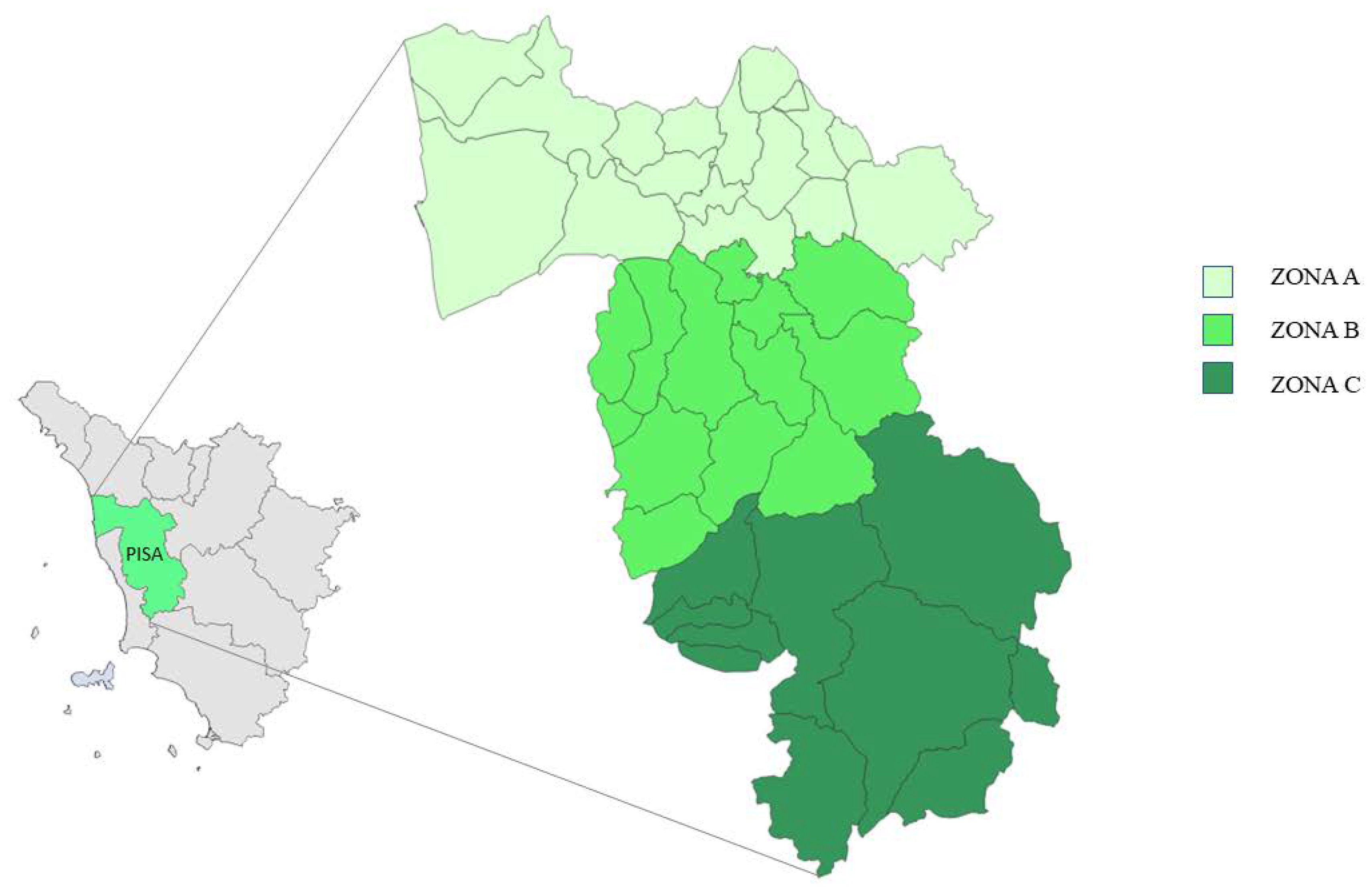
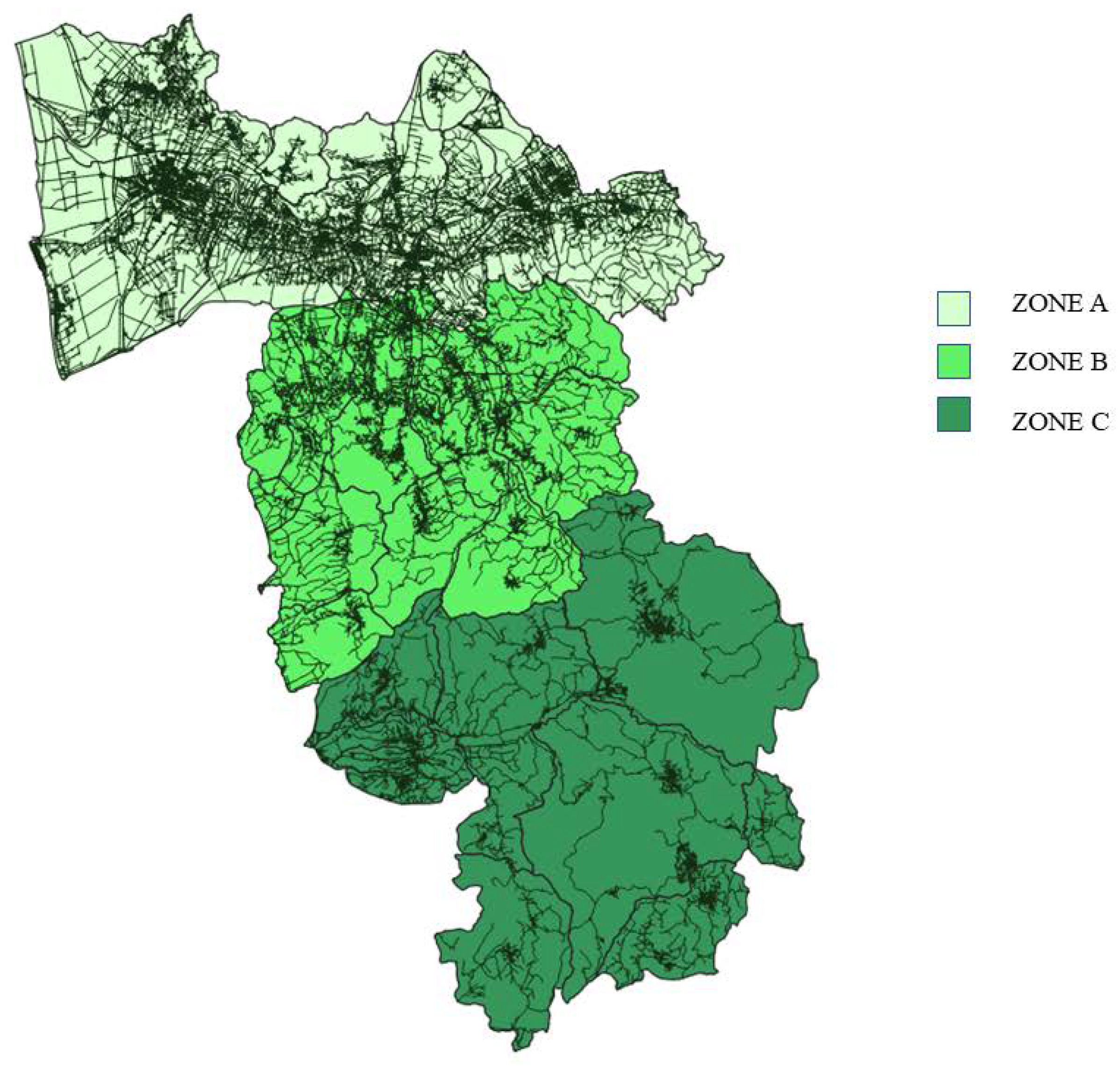
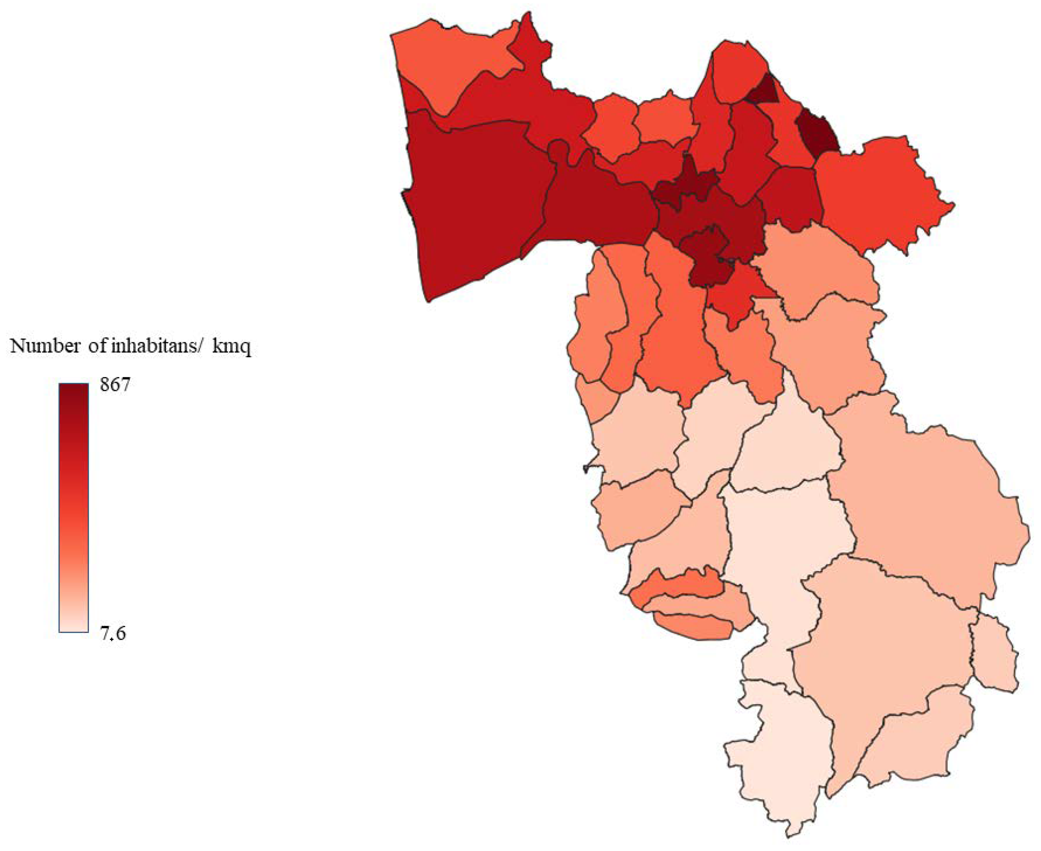
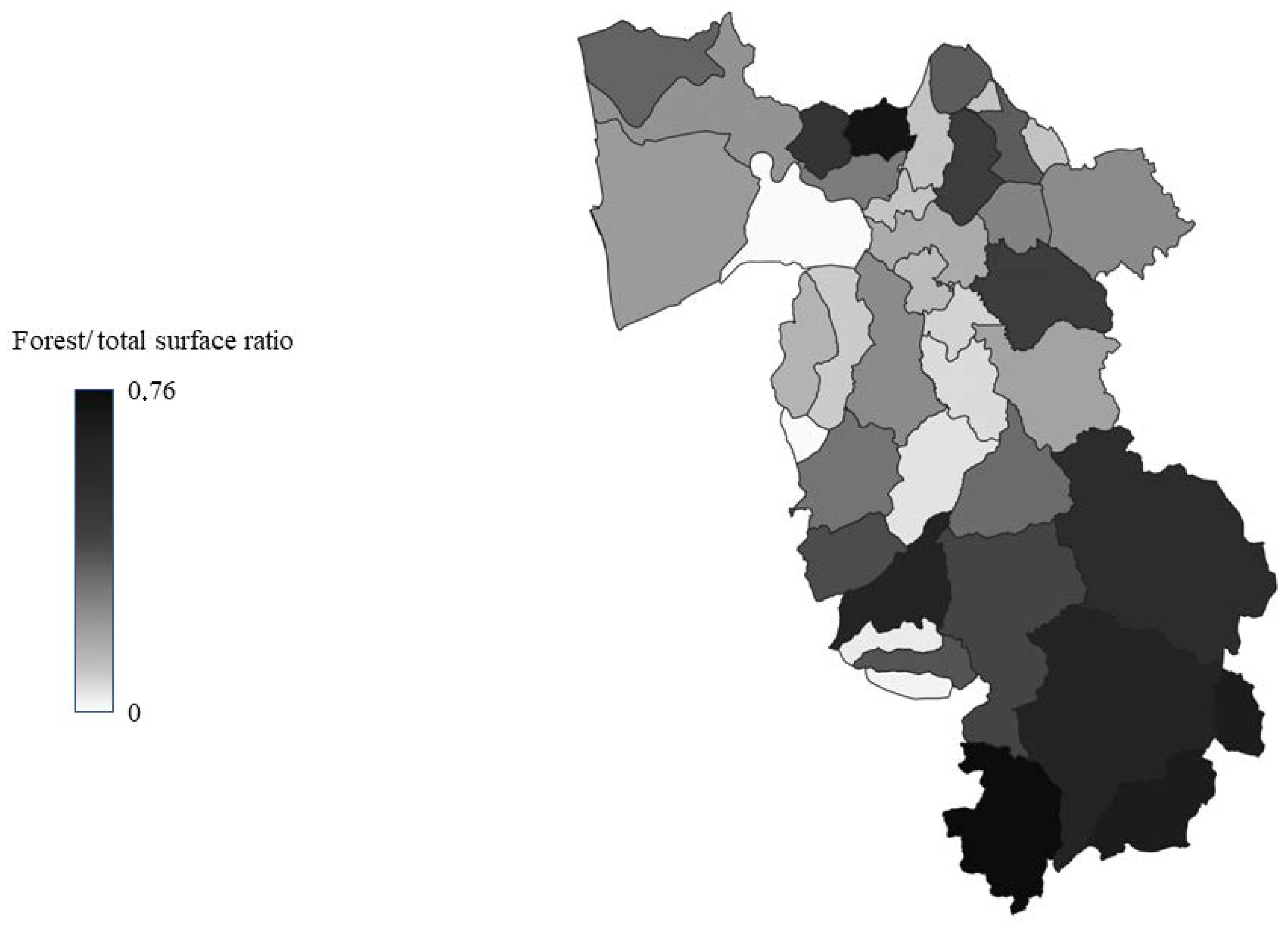
2.1. Study Area

Tuscany is the region of the Italian peninsula with the highest number of wild ungulates, with about 400,000 animals in constant increase, and it is estimated that 40% of roe deer, 45% of fallow deer, and 30% of wild boar of the entire country live there [72]. The province of Pisa, composed of 39 municipalities, covers an area of 2450 km2; 25% of the territory is made up of plains, with most of the inhabited centres, and the rest of the territory consists of hills and mountains suitable for the development of numerous and diverse wildlife populations [73]. Despite this, the data on land use shows that almost the entire territory of the province is used for industrial or agricultural purposes and that unused part is very limited [73]. Therefore, the uncontaminated and uninhabited areas are very limited, and wildlife habitats are fragmented and interrupted by an extensive network of roads and urban areas with many opportunities for contact and interaction between wildlife, domestic animals, and humans. Moreover, the territory of the province of Pisa is very diverse due to its considerable size, with the main urban settlements located in the northern area, where, in addition to the city of Pisa, some of the most important inhabited centres are located. For this study, the province was divided into three zones of similar size, characterised by different demographic densities, road networks, and land use (Figure 1, Figure 2, Figure 3 and Figure 4).
The zones have been named Zone A, B, and C (Figure 1). Data on zone extension, number of inhabitants, population density, and forest cover, expressed as its relationship with the total area surface, are shown in Table 1 [73,74].

2.2. Enrolled Animals

In this study, a cohort of 50 wild animals, injured and hospitalised during a one-year period (between September 2020 and August 2021), was retrospectively assessed for the presence and frequency of different virological and parasitic agents. The enrolled animals were older than 12 months (based on general conformation and teeth eruption), were of both sexes, and belonged to the following species: 23 roe deer (Capreolus capreolus); 4 fallow deer (Dama dama); 12 red foxes (Vulpes vulpes); 6 badgers (Meles meles); 4 porcupines (Hystrix cristata); and 1 pine marten (Martes martes). For each animal, information on the place and the cause of the rescue, as well as on age, sex, and species, was collected from the person who brought the animals (volunteers, citizens, local authorities) and during the first clinical examination and recorded in the hospital database.
Data on the species and sex of the enrolled cohort are presented in Table 2.
All the animals enrolled were hospitalised because they were injured. Overall, 34/50 (68%) of animals were injured in car accidents, 3/50 (6%) by predators, and 2/50 (4%) by fighting with conspecifics. In 11/50 (22%) animals, the cause for injuries could not be readily determined.

2.3. Sample Collection

Upon admission to the VTH, during the first clinical examination, a blood sample was usually collected from the jugular or cephalic vein of each animal under sedation or general anaesthesia to reduce the stress and handling time [75]. The blood sample was immediately used for routine laboratory analyses in order to perform a rapid diagnosis, while an aliquot was collected separately in a serum tube and centrifuged within 30 min after collection at 1000 rpm for 10 min. The serum sample obtained was stored at −20 °C to be used for future analyses.
A faecal sample was also usually collected from each animal after natural voiding; an aliquot of the faecal sample was immediately stored at +5 °C and examined within 12 h for routine parasitological examinations (passive flotation, Baermann technique, coproantigen detection) while an aliquot was stored at −20 °C pending further analyses.
Therefore, all the serological and molecular analyses on serum and faecal samples were performed after the period of hospitalisation and on stored frozen samples.

2.4. Viral Pathogens

A species-specific serological and molecular analysis panel was designed in agreement with previously reported data in the available literature on the most commonly detected viral agents in the enrolled species [28,33,76,77,78,79,80,81,82,83,84,85]. Molecular analyses were performed on faecal samples.

2.4.1. Serological Analysis

Roe deer (n = 23) and fallow deer (n = 4) sera were tested for the detection of antibodies against Bovine Viral Diarrhea Virus (BVDV) and Small Ruminant Lentivirus (SRLV), and all animals’ (n = 50) sera were tested for antibodies detection against Hepatitis E virus (HEV).
BVDV and SRLV antibodies were detected using the BVDV p80 Ab (IDEXX, Westbrook, ME, USA) and by Eradikit® SRLV Genotyping Kit (IN3 diagnostic, Grugliasco, Italy), respectively, after adaptation. The SRLV kit is suitable for the detection and genotyping of SRLV antibodies as it can discriminate whether a positive sample belongs to genotype A (Maedi Visna-like), B (CAEV-like), or E (Roccaverano strain).
HEV antibodies were tested in all enrolled animals using the HEV Ab version ULTRA kit (DIA.PRO, Sesto San Giovanni, Italy). Optical density was measured using a plate reader (Multiscan FC; Thermo Scientific, Waltham, MA, USA) at a wavelength of 450 nm. The procedures and interpretation analysis of the analyses were carried out according to the manufacturer’s instructions.

2.4.2. Molecular Analysis

Species-specific panels for molecular analysis are summarised in the Supplementary Material (Table S1). For species with little data in the literature about previous virological studies, such as porcupine and pine marten, large panels were developed that included viruses identified in related species.
Due to the multiple species tested in this study and the paucity of data on virus circulation in wildlife, broad-spectrum PCR protocols with consensus primers covering as many viral species as possible were used.
Molecular analyses were performed on DNA and RNA extracted from 200 mg of faecal samples using the AllPrep PowerFecal DNA/RNA Kit (Qiagen, Hilden, Germany) according to the manufacturer’s instructions. DNA and RNA were eluted in 30 µL of Buffer AE and RNase-free water, respectively, and stored at −80 °C until analysis. PCR for the detection of Canine bocavirus-2 (CboV-2), Feline/canine parvovirus (FPV/CPV), Torque teno virus 1 and 2 (TTV-1, TTV-2), and Canine adenovirus (CadV), and both rounds of nested PCR for CboV-1 and 3 were performed on DNA using the HotStarTaq® Plus Master Mix Kit (Qiagen, Germany). RT-PCR for the detection of Canine distemper virus (CDV), Bovine viral diarrhea virus (BVDV), Bopivirus (BoPV), and Kobuvirus (KoV), and first rounds of the nested RT-PCR for Coronavirus (CoV) and Astrovirus (AstV) were performed on RNA using the OneStep RT-PCR Kit (Qiagen, Germany). Secondary rounds were performed using the HotStarTaq® Plus Master Mix Kit (Qiagen, Germany). The protocol used for CadV detection is reported to discriminate between CadV 1 and CadV 2, resulting in distinct amplicons of 508 bp and 1030 bp, respectively [86]. Samples positive at molecular analysis were subjected to sequence analysis (BMR genomics, Padova, Italy) to confirm PCR results and obtain phylogenetic information. The primer sets used in the PCR and rt-PCR were included as Supplementary Data (Table S2) [20,77,85,86,87,88,89,90,91,92,93,94,95]. PCR was performed according to the manufacturer’s instruction.

2.5. Parasitological Investigations

2.5.1. Faecal Examinations

A passive flotation technique was carried out on the refrigerated aliquots to detect nematode eggs and coccidian oocysts, using a commercial saturated solution of sodium chloride (specific gravity = 1.20) as flotation fluid (Coprosol®, Candioli Farmaceutici, Beinasco, TO, Italy). The parasite burden was estimated by determining the number of eggs per gram (EPG) and oocysts per gram (OPG) of faeces by the modified McMaster method, with the lowest detection limit of 50 EPG/OPG per gram. In addition, faecal samples were examined via the Baermann technique to detect bronchopulmonary and cardiopulmonary nematode larvae. The number of larvae per gram (LPG) of faeces was estimated as above. The mean and range of OPG, EPG and LPG were calculated.

2.5.2. Commercial Assays

A panel of endoparasite infections was investigated by rapid in-clinic tests in a different range of species. In particular, a rapid diagnostic test to detect both Cryptosporidium and Giardia coproantigens (Fastest® Crypto-Giardia Strip, Megacor Diagnostik, Austria) was performed on all 47 faecal samples examined for parasitological infections. Moreover, sera from foxes (n = 10) and badgers (n = 5) were screened for the presence of antibodies against Leishmania infantum (Snap Leishmania, Idexx, USA), Angiostrongylus vasorum (Angio Detect Test, Idexx, Westbrook, ME, USA), and Dirofilaria immitis (Snap Heartworm Antigen Test, Idexx, Westbrook, ME, USA). Screening of seropositivity for Neospora caninum (Fastest® Neospora caninum test kit, Megacor Diagnostick, Gemeinde Hörbranz, Austria) was carried out on foxes, badgers, roe deer (n = 23), and fallow deer (n = 4). All the rapid in-clinic tests were performed according to their respective manufacturer’s instructions. Finally, serum samples from all animals were screened for Toxoplasma gondii infection by a commercial ELISA kit (ID Screen® Toxoplasmosis Indirect Multi-species, IDVET, Montpellier, France) according to the manufacturer’s protocol.

2.5.3. Transtracheal Wash Technique

In 4/13 (30.8%) roe deer that tested positive for lungworm larvae via the Baermann technique (see above), a transtracheal wash (TTW) was performed immediately after humane euthanasia due to severe injuries. Briefly, a polyurethane catheter (14G) (Terumo, Roma, Italy) was introduced 5 cm distal to the larynx at 45° to the skin between two tracheal rings identified by palpation. Once the catheter entered the tracheal lumen, the needle was retracted, and the stylet advanced. Then a catheter (1.3 mm × 50 cm) (Buster dog catheter, Kruuse, Denmark) was passed through the stylet, down the trachea, and towards the lungs, until resistance was found. A bolus of 30 Ml of sterile saline was injected using a 60 Ml syringe and the fluid was immediately aspirated to recover at least 10 Ml of fluid [96]. When the aspiration of fluid was complete, the catheter and stylet were withdrawn simultaneously. The TTW fluid was split into two 4 Ml EDTA tubes for parasitological examination. When lungworm specimens were detected in the amount of fluid recovered, they were stored for 24–48 h in physiological saline at 4 °C before the morphological examination and then stored in 70% alcohol pending molecular procedures for identification to species level.

2.5.4. Molecular Analysis for Giardia duodenalis Genotyping and Cryptosporidium Species Identification

Molecular investigation of G. duodenalis and Cryptosporidium was performed on faecal samples testing positive for coproantigens test (Fastest® Crypto-Giardia Strip, Megacor Diagnostik, Gemeinde Hörbranz, Austria). Genomic DNA extraction was performed by QIAamp DNA Stool Mini Kit (QIAGEN, Hilden, Germany) according to the manufacturer’s instructions. Giardia genotyping was determined through the amplification of tpi and β-giardin genes. In detail, a 530 bp fragment of the tpi gene was obtained via a two-step nested PCR protocol using AL3543/AL3546 primers for primary PCR and AL3544/AL3545 for secondary PCR [97]. A 384 bp fragment of the β-giardin gene was amplified with a forward primer G7, a forward inner primer G376, and a reverse primer G759 [98].
Cryptosporidium genotyping was determined through the amplification of 800–850 bp of the GP60 gene. In a two-step nested PCR protocol, AL3531/AL3535 primers were utilized for primary PCR and AL3532/AL3534 for the secondary PCR [99].
All amplicons obtained were purified using the mi-PCR Purification Kit (Metabion International AG, Planegg, Germania), and sequencing was performed in the laboratory of Bio-Fab Research (Rome, Italy). In order to check the quality of the sequences, the resulting chromatograms were manually analyzed using Finch TYV1.4 software (Geospiza, Inc, Seattle, WA, USA).
Consensus sequences were then compared with those available in the GenBank database by using the Standard Nucleotide BLAST search and aligned by Clustal Omega implemented in MEGA X with representative sequences of tpi, bg, and GP60 loci and used as references.

2.5.5. Molecular Identification of Adult Lungworms

Adult lungworms collected by TTW were identified to species level by PCR technique. Molecular analyses were performed by BMR Genomics (Padua, Italy, https://www.bmr-genomics.it/ accessed on 7 January 2022). Genomic DNA was extracted from the stored adult lungworms using the commercial PCRBIO Rapid Extract Lysis Kit (PCR Biosystems) following the manufacturer’s instructions. A fragment (about 700 base pairs in length) of the mitochondrial gene for cytochrome C oxidase subunit 1 (COX1) was used as a DNA barcoding system and amplified. The PCR amplification was carried out in a final mixture containing 12.5 μL of AmpliTaq Gold™ 360 Master Mix (Thermo Fisher), 2 μL of gDNA extracted, 1 μL (10 μM) of LCO1490 primer (5′-GGTCAACAAATCATAAAGATATTGG-3′), 1 μL (10 μM) of HCO2198 primer (5′-TAAACTTCAGGGTGACCAAAAAATCA-3′), and deionized water, reaching a total reaction volume of 25 μL. The PCR reactions were subjected to the following conditions in a thermal cycler (Mastercycler®, Eppendorf): 95 °C × 2 min, then 5 cycles (95 °C × 40 s, 45 °C for 90 s, and 72 °C for 90 s), followed by 35 cycles (95 °C for 40 s, 50 °C for 90 s, and 72 °C for 60 s), and finally, 72 °C for 10 min. The amplification products were visualized after electrophoresis on 1.5% agarose gel. PCR products were purified with ExoSAP-IT™ (Thermo Fisher Scientific, Massachusetts, MA, USA), sequenced, and aligned via BLAST analysis to detect their identity by retrieving similar sequences deposited in NCBI’s GenBank database [100,101].

2.6. Statistical Analysis

The prevalence of virological and parasitic agents was determined as the number of positive animals/numbers of examined animals X 100.
The Fisher test was used to determine statistical differences between rescue areas on positivity/negativity for Astrovirus, Kobuvirus, Bopivirus, and Adenovirus.

3. Results

3.1. Viral Pathogens

3.1.1. Serological Analysis

One out of 23 roe deer (4.3%) scored positive for antibodies against BVDV and one for SRLV, genotype A Maedi Visna-like, while all the fallow deer were serologically negative for both viruses. The positive roe deer was a male rescued in zone B. All the animals (50/50, 100%) scored negative for HEV antibodies.

3.1.2. Molecular Examination

All animals tested negative for CBoV, FPV, CPV, TTV, BVD, CDV, and CoV. Animal distribution in the three-rescue area and results of virological molecular analysis presented by viruses, species and rescue area are reported in Table 3 and Table 4.
Six out of 50 animals (12%) tested positive for KoV: 1/23 roe deer; 2/12 foxes; 2/4 porcupines; and 1/6 badgers, respectively. All positive roe deer, badger, foxes, and one porcupine were male; the other porcupine was female. All positive animals belonged to zone A; thus, no statistical comparison among zones was performed.
Eight out of 50 (16%) animals tested positive for AstV: 3/23 roe deer (1 male and 2 female) and 5/12 foxes (4 male and 1 female). Four out of 8 (50%) animals belonged to zone A (3 foxes and 1 roe deer), 3 (37.5%) animals belonged to zone B (2 foxes and 1 roe deer) and 1 (12.5%) roe deer belonged to zone C. No differences were found between zone A vs. B (p = 0.626), zone A vs. C (p = 0.536), or zone B vs. C (p = 0.779).
Foxes (n = 12), badgers (n = 6), and pine martens (n = 1) were tested for CAdV, and 5 out of 19 (26%) animals tested positive for CAdV type 1. Three out of 12 foxes and 2/6 badgers were positive; all the positive foxes were male, 2 from zone B and 1 from zona A. Both positive badgers were male, 1 from zone A and 1 from B. No animals tested for CAdV have been rescued in zone C. No statistical differences were found in CAdV type 1 prevalence between zones A vs. B (p = 0.161).
Roe deer (n = 23) and fallow deer (n = 4) were tested for BoPV, and 1 out of 27 (3%) tested positive for BoPV. This was a female fallow deer from zone A; thus, no statistical comparison among zones was performed.
Table S3 shows the accession numbers of the sequences entered in the GenBank database, along with the results of their analysis with Blast software. For each sequence in this study, the analysis identified a reference sequence and determined the per cent of nucleotide identity and E value obtained from their comparison.

3.2. Parasitological Investigations

Overall, parasitological analyses were performed in a total of 47/50 (94%) animals, including 10 foxes, 5 badgers, 4 porcupines, 23 roe deer, 4 fallow deer, and 1 marten, as faecal and serum samples from 2 foxes and 1 badger were not available. Results of faecal examinations by flotation and Baermann techniques are shown in Table 5.

3.2.1. Faecal Examinations

Roe deer. Overall, 19/23 (82.6%) roe deer were found to be coprologically positive by flotation and/or Baerman techniques. Ten out of 23, 8/23, and 1/23 roe deer showed two, one, or three parasitological infections, respectively. Eggs of gastrointestinal strongyles (mean = 7, range ≤ 50–100 EPG), Eimeria oocysts (mean = 250, range ≤ 50–>1000 OPG), and Capillaria spp. eggs (mean ≤50 EPG), were found in 14/23, 5/23, and 2/23 roe deer, respectively.
Lungworm larvae (mean = 23, range ≤ 50–150 LPG) consistent with first-stage larvae of Dictyocaulus sp. were detected in 13/23 (56.2%) roe deer.
Red foxes. All foxes (10/10, 100%) were found to be parasitised. Two, three, four, and even six parasitic co-infections were detected in 5/10, 2/10, 1/10, and 1/10 foxes, respectively. Therefore, only one fox out of 10 showed a single parasitic infection with coccidia oocysts. Parasite prevalence was as follows: Ancylostomatidae eggs (mean = 344, range ≤ 50–>1000 EPG) were found in 8/10 foxes; Toxocara canis eggs (mean ≤50 EPG) in 5/10; Capillaria spp. eggs (mean = 75, range ≤ 50–300 EPG) in 4/10; Trichuris vulpis eggs (mean ≤50 EPG) in 3/10; and coccidia oocysts (mean = 600, range = 200–>1000 OPG) in 2/10. In addition, the spurious passage of Hymemolepis eggs (<50 EPG) was detected in 1/10 foxes. Larvae of A. vasorum (mean = 275, range < 50–>1000 LPG) were detected in stool samples from 4/10 foxes.
Badgers. All badgers (5/5, 100%) harboured parasites. Infections with two and four parasitic agents or only a single infection were detected in 3/5, 1/5, and 1/5 of them, respectively. Ancylostomatidae eggs (mean = 80, range ≤ 50–300 EPG) and larvae of Crenosoma sp. (mean = 137, range ≤ 50–400 LPG) were found in stool samples from 5/5 and 4/5 badgers, respectively, while Strongyloides eggs (<50 EPG), Capillaria spp. eggs (<50 EPG), and coccidia oocysts (<50 OPG) were detected in 1/5 each.
All the fallow deer, all the porcupines, and the marten were found negative for both the flotation and Baermann techniques.

3.2.2. Commercial Assays

Results of the rapid in-clinic tests and the ELISA kit are summarized in Table 5. Briefly, eleven, six, and two samples from different host species showed positive results for N. caninum, G. duodenalis, and C. parvum, respectively. Six and four samples from foxes tested positive for A. vasorum and D. immitis, respectively, and one sample from badgers tested positive for D. immitis. No samples tested positive, either for L. infantum or T. gondii.

3.2.3. TTW Results

Four adult lungworms were detected in the TTW fluid from one of the four (25%) roe deer found positive for Dictyocaulus sp. larvae via the Baermann technique. The specimens measured on average about 5 cm in length and 0.4 mm in width at the mid-body and had an elongated oral opening, dorso-ventrally flattened. Based on the length of the body and the shape of the oral opening, the lungworms were presumptively identified as Dictyocaulus sp.

3.2.4. Molecular Identification of Giardia duodenalis and Cryptosporidium spp.

A total of six samples for G. duodenalis and two for Cryptosporidium spp., carried out positive by the coproantigens test, were tested by the two-step nested PCR protocols. By sequencing at the tpi locus, 1/6 (16.7%) sample from a roe deer and 1/6 (16.7%) from a porcupine were assigned to Assemblage A-AI and Assemblage B-BIV, respectively. Sequences obtained in this study were deposited in the GenBank database and are available under the accession numbers OP946512 and OP946513, respectively.

3.2.5. Molecular Identification of Adult Lungworms

The BLAST search results showed that COX1 of adult lungworms collected from a euthanized roe deer had 99–100% DNA homology with sequences of Dictyocaulus capreolus available in the GenBank database. The sequence obtained in this study was deposited in the GenBank database and is available under the accession number: OQ209836. To the best of our knowledge, this is the first molecular identification of D. capreolus in roe deer in Italy.

4. Discussion

Serological surveys in wildlife are a diagnostic tool to demonstrate the presence of antibodies against specific pathogens and to provide epidemiological data on pathogens; however, serological sampling in wild animals is not always feasible and can be very stressful for the animals themselves. In the present study, the hospitalisation of wild animals allowed blood sampling during clinical practice, and we decided to investigate by serological analysis a panel of pathogens for which direct virological diagnosis on faecal samples is not applicable.
The detection of BVDV antibodies indicates the presence and circulation of the virus in wild animals; moreover, the absence of vaccination plans for wildlife removes doubts about possible vaccine seropositivity. In our survey, one roe deer out of 21 was serologically positive. This result is in agreement with data from European studies, which reported the absence of seropositive animals (Spain and England) up to 12% of seroprevalence (Norway) [14,102,103,104,105]. In Italy, a serosurvey was carried out in four large alpine ungulates in the High Valley of Susa, and antibodies were detected in chamois, wild boar, and red deer, but all roe deer were negative [15]. A similar result was obtained a few years later when Fernández-Sirera and colleagues tested domestic ruminants, cattle, sheep, wild ruminants, chamois, and roe deer in the same research area for the presence of antibodies against BVD. High prevalence was found in both domestic ruminants, 25% in cattle, 17% in sheep, and 42 % in chamois, but none of the 213 roe deer tested positive [16]. The results of the Italian studies and the low seroprevalence found throughout Europe, even in areas with regular contact with domestic animals with high prevalence, suggest that roe deer play a marginal role in the epidemiology of the virus and that they are mostly an occasional guest and does not represent a major risk to domestic ruminants. The low prevalence of the virus among the European roe deer populations compared to other wild deer, especially the chamois, is explained by the different habits of the roe deer. The roe deer is mainly a solitary animal that prefers bushy feeding areas to open pastures where it would be easy to encounter domestic ruminants [15]. In addition, it is possible that this species has low susceptibility to common pestiviruses. Indeed, Fischer and colleagues have discovered a new pestivirus subtype in roe deer [106], and this may influence the serological results. However, as the presence of BVDV infection in roe deer is rarely detected, it would be advisable to conduct further surveys in the study area to increase the number of samples and obtain a more reliable estimate of prevalence.
In addition, 1 roe deer tested positive for SRLV, genotype A Maedi Visna-like. Although SRLV were originally thought to be species-specific pathogens, several lines of evidence showed that lentiviruses can efficiently cross the species barrier and adapt to new hosts [107,108]. Several studies reported the detection of SRLV antibodies in wild ruminants: Rocky Mountain goats; red deer; roe deer; and mouflon [109,110,111]. Therefore, it is possible that contact between wild and domestic small ruminants could result in cross-transmission of SRLV.
Regarding viral molecular analysis, positivities in foxes (CAdV-1, KoV, AstV), roe deer (KoV, AstV,) fallow deer (BoPV), badgers (CAdV-1, KoV), and porcupine (KoV) have been detected.
A recent investigation in Italy revealed the presence of bopiviruses in sheep, roe deer, chamois, and Alpine ibex, with sequences clustering with bopivirus strains previously detected in goats and sheep from Hungarian farms and in red deer with sequences closely related to Bopivirus C sequences identified in fallow and red deer in Australia [112]. In our study, one fallow deer sample resulted positive for Bopivirus, whereas all samples from roe deer were negative. According to BLAST analysis, the sequence shared 94–92% nucleotide (nt) identity with deer Bopivirus strain (Bopivirus C), 88–85% with bovine Bopivirus strain (Bopivirus A), and 72–70% with ovine–goat Bopivirus strain (Bopivirus B). The sample, therefore, showed the highest nucleotide identity to Bopivirus C previously identified in Australian fallow deer and Italian red deer. Although numerous viruses of the Picornavidae family are responsible for diseases with relevant clinical forms in humans and animals, the pathogenicity of Bopivirus in deer remains undetermined. The population In our study, as in previous studies, was apparently healthy or not affected by symptoms attributable to an infectious disease.
Astroviruses were detected in foxes and roe deer representing, to our knowledge, the first report of astrovirus in these two species in Italy. Astroviruses have already been identified in foxes in the Netherlands and Australia using metagenomic techniques. In the first study, the sequences were mainly identified as two novel astroviruses called Fox Astrovirus F5 and F4, in the second, sequences clustered with feline astrovirus with high nt identity. Little research has been reported on AstVs in deer, but astroviruses have been identified in European roe deer in Denmark in 2010 [113] from animals with gastrointestinal disease, and in Slovenia from healthy hunted animals [114]. The following sequence analysis identified a new species of Mamastrovirus, the Capreolus capreolus Astrovirus (CcAstV strain 1 and 2), related to other bovine, porcine, yak, porcupine, and dromedary AstVs strains, possibly forming a group of currently unassigned sequences that are distantly related to mAstV genogroups I and II. Our sequences, both from foxes and roe deer, were obtained using broad-range primers amplifying a portion of RdRp without phylogenetic information; however, BLAST analysis of the amplicons confirms that this virus clustered with other mammalian-associated viruses within the mamastrovirus genera. In general, AstV infection is associated with mild to severe gastrointestinal tract disease, typically with diarrhoea and vomiting [115], but has also been associated with other diseases, such as shaking syndrome in mink, neurological disease in cattle and sheep, and encephalitis in humans [116,117,118,119]; infection may be asymptomatic [115]. There is no information on the symptoms in foxes, although it is likely that CcAstV can cause gastroenteritis in roe deer. In our study, the sampled animals did not have gastrointestinal disease or other disorders attributed to astroviruses.
In Italy, kobuviruses have been detected in dogs, foxes, wolves, roe deer, goats, swine, cats, and cattle [33,38,39,40,76,120]. In this study, kobuviruses were detected in two foxes, one roe deer, one badger, and two porcupines, and none of the positive animals showed symptoms suggestive of enteritis. In foxes, Canine kobuvirus (CaKoV) has been detected in free-ranging foxes in Italy with a prevalence of 14.7%. To date, the known host range of CaKoV includes several species of Canidae (domestic dog, red fox, wolf, golden jackal, and side-striped jackal), and the virus has been detected in both diarrhoeic and asymptomatic animals, so the pathogenic potential of kobuviruses in carnivores remains to be elucidated. Our sequence of RdRp partial region from fox shared the highest identity with the CaKoV strains from Italian foxes with an nt sequence identity of 89–94% showing E-value ranging from 2e−30 to 9e−39. CaKoV is often found in co-infections as a potential secondary infection after primary infections by mainly viral agents responsible for immunosuppression, such as canine distemper virus or parvovirus [121,122,123]. In the studies of Di Martino and colleagues, both a fox and some dogs were co-infected by Kobuvirus and cCoV [40].
In our study, all foxes were negative to molecular analysis for CPV, CDV, and cCoV, but two foxes positive for kobuvirus were also PCR-positive for astrovirus, and one also for adenovirus; both were responsible for clinical gastroenteritis. Coinfection with CaKoV and cAstV and with CaKoV and cAdV has been described in diarrhoeic dogs in China and Korea, respectively [124,125]. In roe deer, kobuvirus RNA was found in 6.6% of rectal swabs collected from healthy roe deer in northern Italy between 2012 and 2014; most of these strains showed high homology with bovine kobuvirus, and others with caprine kobuvirus [76]. In our study, kobuvirus has been detected in 1/23 roe deer with a prevalence in line with previous studies. BLAST analysis of the partial 3D gene sequence showed 75% and 71% of nucleotide identity, respectively, with bovine and caprine kobuvirus detected in Italian roe deer, while the highest identities were shared with Italian fox kobuvirus. This result contrasts with what resulted in the previous study and it cannot be excluded that the roe deer may have ingested food contaminated with faecal contents of other species. However, it is important to consider that this is the second study to report Kobuvirus in wild ruminants and that very little is still known about which strains circulate among the populations of these animals and about the possibility of transmission of strains between different species. Kobuvirus has only been identified in one member of Mustelidae, a domestic ferret in the Netherlands, using a metagenomic approach, and the almost complete kobuvirus genome clustered with bovine and ovine kobuvirus (Aichivirus B) possibly indicates a past cross species transmission events [126]. No data is currently present about the circulation of kobuvirus in free-ranging mustelids that share the ecosystem with other wild animals susceptible to infection. In the present study, kobuvirus has been identified in 1/6 badger representing its first detection in this species. The badger resulted also positive for adenovirus. Our sequence clustered with sequences of CaKoV detected in wild and domestic carnivores with an nt sequence identity of 85–87% and an e value of 3 e−80–1 e−65. Finally, in our study, kobuvirus was also identified in two porcupines representing its first detection in this species. To date, kobuvirus has been detected in other rodents, such as rabbits, grey squirrels, and some species of mice (Peromyscus crinitus, Apodemus agrarius, Rattus norvegicus, Rattus losea, and Rattus argentiventer, Mus musculus, Rattus losea, Rattus tanezumi, and Rattus norvegicus) [30,36,127,128,129]. The BLAST analysis of our sequence showed a high homology both with both CaKoV (90–95% of nt identity, e value of 9 e−98–2 e−42), and murine kobuvirus (85–90% of nt identity, e value of 6 e−81–1 e−64), both belonging to the same genetic cluster within Aichivirus A, and more in-depth phylogenetic studies would be needed to better classify the virus. Interestingly, a high percentage of nt identity resulted between all our sequences (87–95%). This evidence, together with the common origin of all positive animals from zone A, suggests the possibility that a similar kobuvirus could infect different animal species sharing the same habitat.
Moreover, three foxes and two badgers tested positive for cAdV-1. In foxes, cAdV was first identified in 1925 as the causative agent of severe neurologic disease, the epizootic fox encephalitis [130]. Subsequently, evidence of cAdV circulation in foxes has been reported in several geographical areas, with serological prevalence in the European population ranging from 3% in Germany to 64.4% in the UK [131,132]. In Italy, and more specifically in the Pisan area, the virological prevalence reported in previous studies is around 28% [132], which is in line with the value of 25% obtained in our study. All the positive animals were traumatised, and none showed clinical signs attributable to infectious diseases. Our data, together with the high prevalence values reported worldwide [44,45,132,133,134] and the identification of the virus in healthy animals reported in previous studies [132,134], suggest that red foxes may play a role in maintaining cAdV-1 in the territory and may be a source of viral spread in wild ecosystems. In addition, although cAdV-1 can be controlled in domestic dogs by vaccination, cases of infectious canine hepatitis in domestic dogs have been diagnosed in Italy in recent years [135], and the presence of infected foxes may also be a source of infection for domestic dogs, especially foxes living in peri-urban areas [135]. Adenovirus has previously been detected in Mustelidae, in a young striped skunk with hepatitis, in a seriously diseased Eurasian otter in the Seoul Grand Park Zoo, and in several pine martens and Eurasian otters in the UK, with prevalences of 33% and 88%, respectively [80,136,137]. However, our evidence is the first detection of adenovirus in badgers.
Both fox and badger viral sequences clustered with Canine adenovirus 1 with a high percentage of nt identity (94–98%). The presence of cAdV-1 in badgers was expected as specific adenoviruses have also been identified to date in otters and pine martens (marten adenovirus and lutrine adenovirus) and it is already known that mustelids are susceptible to CAdV-1 infection [80]. Moreover, the home ranges of mustelids and other predators such as red foxes (Vulpes vulpes) may overlap and there is potential for cross-infection with pathogens through indirect contact with urine, faeces, and other infected fomites.
Moving on to discuss the parasitological findings, although coccidiosis was presumably harmless for the animals examined in this study, coccidia may pose a major threat to some species of wildlife; for instance, juvenile coccidiosis may be associated with impaired growth and increased mortality in badger cubs [50]. In this study, oocysts of Eimeria were detected in fecal samples from roe deer, red foxes, and badgers, but identification to species level could not be reached.
Normally GSI are of little clinical significance in wild ruminants, but if combined, they can cause profuse and watery diarrhea, anaemia of varying degrees, edema under the lower jaw extending along the abdomen, progressive weight loss, rough hair coat, anorexia, hypoproteinemia, reduced growth, and even death in severe cases [138].
Since a much higher prevalence of C. bovis (26.6%) than C. bilobata (6%) has been detected in C. capreolus [52,53], it is likely that C. bovis was the Capillaria species involved in roe deer of this study.
Adults of D. capreoli were detected in the transtracheal wash fluid from a roe deer, as shown by the results of the molecular analysis. D. capreoli has been reported to cause anorexia, poor haircoat quality, delayed haircoat change, diarrhea, small apical atelectatic areas, peri-bronchial inflammation, and total bronchopneumopatia in roe deer [55].
In agreement with previous studies carried out in Italy [56,57], combining results of coprological analyses and commercial assays, intestinal worms, lungworms, and heartworms were found in red foxes. It has been reported that the infection with intestinal helminths, especially T. canis, and the number of helminth species per individual may negatively affect the host body condition in red foxes [139,140]. All parasites of foxes are shared with dogs [141], and some species can be zoonotic agents [142]. In particular, foxes may be considered an important source of infections with cardiopulmonary nematodes, which may be responsible for overt clinical disease in dogs [143,144].
The eggs of A. caninum and U stenocephala cannot be reliably distinguished microscopically. Likewise, eggs of C. aerophila and C bohemi are microscopically indistinguishable from one species to another because all Capillaridae eggs are morphologically characterized by a similar barrel shape with polar plugs [134]. Therefore, it was not possible to determine whether the foxes examined in this study were infected with A. caninum, U. stenocephala, or both and with C. aerophila, C. boehmi, or both. In addition, the spurious expulsion of Capillaria hepatica (syn. Calodium hepaticum) eggs, after preying or scavenging on infected hosts, cannot be excluded. Indeed, foxes are known to scavenge on carcasses and eat a wide range of rodents, including mice and rats, which commonly show a very high prevalence of infection with C. hepatica [145]. In this study, the spurious expulsion of typical eggs of Hymenolepis spp. was detected. Hymenolepis is a genus of zoonotic cestodes that commonly infect rodents, mostly mice and rats, with usually a high prevalence [146]. This suggests that at least some of the foxes examined were feeding on rodents at the time of sampling and that foxes may act as carriers for the dispersal of the eggs of Hymenolepis species into the environment. Conversely, though Capillaria plica may be highly prevalent in foxes in central Italy [147], shedding of C. plica eggs in faeces was excluded because adult parasites of C. plica live in the bladder of definitive hosts, and eggs are expulsed with urine.
Within the family, Ancylostomatidae U. criniformis is the species widely reported in European badger populations [59,61,62,63]. Therefore, it may be suggested that U. criniformis was the species infecting badgers examined in this study.
Lungworm infections in M. meles can be identified based on the typical morphology of L1 larvae (Crenosoma spp.) and eggs (C. aerophila). However, the limited information available on the morphological differences between L1 larvae of C. melesi and C. vulpis did not allow differentiation from one species to another. Therefore, it is likely that C. aerophila was the Capillaria species involved in the badgers of this study, but it was not possible to determine whether C. melesi, C. vulpis, or both were infecting the badgers examined. Similarly, to foxes, the shedding of spurious C. hepatica eggs, merely passing through the intestinal tract, cannot be completely excluded. Indeed, C. aerophila and C. hepatica eggs are hardly distinguishable by microscopy, while badgers are known to be scavenger animals and to eat small mammals, including mice and rats (often heavily infected by C. hepatica, as aforementioned).
Oval, thin-shelled eggs with a fully developed coiled larva inside (typical of rhabditiform nematode parasites) were found in a badger faecal sample and were identified as eggs of Strongyloides sp. To our knowledge, the identification of Strongyloides species infecting the European badger is still unknown since Strongyloides specimens recovered in M. meles have not been identified to the species level [61,62]. Strongyloides specimens isolated from the Japanese badger (Meles anakuma) are closely related to Strongyloides procyonis on a phylogenetic tree and are morphologically similar to S. procyonis and Strongyloides martis [65]. However, the results of molecular phylogenetic analyses using mitochondrial genome sequencing as a marker suggested that they belonged to a species genetically separate from S. procyonis [65]
Concerning Leishmania and Toxoplasma, it cannot be excluded that the negative results found in this study may be due to limitations (uncertain sensitivity) attributable to the screening tests. A rapid immunochromatographic test (marketed for dogs) has been previously used to determine the seroprevalence of anti-Leishmania antibodies in wild canids, such as red foxes and coyotes [148]. Likewise, a commercial indirect multi-species ELISA (marketed for ruminants, swine, dogs, and cats) has been previously used to determine the seroprevalence of anti-Toxoplasma antibodies in free-ranging and captive large African carnivores [149]. Therefore, at present, it can be assumed that these two commercial assays may be suitable for wildlife species, but they have not yet been validated for the diagnosis of Leishmania and Toxoplasma infections in the species examined in this study.
Neospora caninum-antibodies were detected in 3/4 fallow deer, 7/23 roe deer, and 1/5 badgers. These findings agree with those of other studies where the reported seroprevalence values for Neospora were 11% in fallow deer [150] and 28% in roe deer [151], while 10.9% of badgers were found positive for Neospora DNA in brain, liver, or neck muscles [152]. Red foxes are considered natural intermediate hosts for N. caninum, with reported prevalences of 10.7% by the PCR [153] to 69.8% by the agglutination test [154]. However, no foxes tested positive for Neospora in this survey. The possible existence of a sylvatic life cycle of N. caninum has been postulated [69]. Therefore, the present results suggest that fallow deer, roe deer, and badgers may contribute to the sylvatic life cycle of N. caninum, if any, in some areas of central Italy. Further investigations are needed to elucidate the role they may play in maintaining the circulation of the parasite in nature.
For the detection of A. vasorum infection in red foxes and badgers, the Angio Detect test was used in combination with the typical recovery of cardiopulmonary larvae by the Baermann technique. Angio Detect is commercialized as an in-clinic assay to detect A. vasorum circulating antigen in dogs but it has also been successfully used in other host species, such as red foxes and badgers [155,156]. Likewise, commercially available antigen detection tests, commercialized for use in dogs, have been successfully used to identify D. immitis infection in other host species, such as wolves and red foxes, coyotes, and island foxes [157,158,159]. In this study, the use of the Angio Detect rapid test enhanced the global sensitivity of the diagnostic procedure in foxes as it allowed the detection of two additional positive subjects concerning the traditional Baermann apparatus. It is likely that these two A. vasorum-antigen positive/Baermann negative foxes were in the prepatent period that characterises the early phase of any parasitic infection.
The European badger may be a definitive host for the cardiopulmonary nematodes D. immitis, A. vasorum, and A. daskalovi [61,62,66,67,68]. In this study, D. immitis circulating antigen was detected in 1/5 badgers by the in-clinic test, but no one was found positive either for circulating antigen or larvae of Angiostrongylus spp.
In this study, the molecular analysis of G. duodenalis was successfully amplified at the tpi locus in one roe deer and in one porcupine. Among isolates of G. duodenalis from C. capreolus, assemblage A was previously identified in the Netherlands and Poland, while assemblage B was detected in Poland [160,161,162]. Identification to sub-assemblage level was not carried out in those investigations. G. duodenalis sub-assemblage AII was reported in roe deer in Spain [163]. In the present study, sub-assemblage AI was identified. To the best of our knowledge, this is the first report of G. Duodenalis sub-assemblage AI in C. capreolus in Italy. This sub-assemblage has previously been isolated in other wild species in the same geographical area [164]. Likewise, the finding of G. duodenalis sub-assemblage BIV in a porcupine is consistent with data previously reported for the same host species in the same geographical area [165]. G. duodenalis sub-assemblages AI and BIV are mainly found in animals and, to a lesser extent, in humans. Therefore, they are considered potentially zoonotic [70].

5. Conclusions

The results highlight the widespread presence of pathogens in the population studied, confirming the role of wildlife in the epidemiology of several viruses and parasites. Although the impact on wild species’ health is not yet known for all the pathogens identified, they can pose a threat to wildlife and can potentially have important effects on the population dynamics of their hosts, especially when they are experiencing the cumulative effects of other pathogens and stressors.
Moreover, some of the parasitic agents found can be transmitted to domestic animals, and some of them are zoonotic. Therefore, they can affect the health status of livestock or pets and can have public health implications, especially in a highly urbanised area such as the study area.
The study highlights the key role that structures admitting wild animals can play as focal points for collecting information on the health of wildlife populations and monitoring the health of the environments in which they live.

Supplementary Materials

The following supporting information can be downloaded at: https://www.mdpi.com/article/10.3390/ani13050931/s1, Table S1: Summary of the specie-specific panels for virological molecular investigations; Table S2: The primer sets used in the study; Table S3: Accession number of the sequences entered on the GenBank database and results of their analysis with the Blast software.

Author Contributions

Conceptualization, M.I.P., M.M., M.S. and R.A.P.; methodology, M.I.P., M.M., M.S. and R.A.P.; software, M.I.P. and M.S.; formal analysis, M.I.P., M.M., M.S., R.D. and R.A.P.; investigation, M.I.P., R.D. and R.A.P.; resources, M.M., M.S., R.D. and R.A.P.; data curation, M.I.P. and R.A.P.; writing—original draft preparation, M.I.P., M.M., M.S., R.D. and R.A.P.; writing—review and editing, M.I.P., M.M., M.S., R.D. and R.A.P.; supervision, M.S.; project administration, M.M., M.S. and R.A.P.; funding acquisition, M.M, M.S. and R.A.P. All authors have read and agreed to the published version of the manuscript.

Funding

This research was funded by Fondi di Ateneo (Univesity of Pisa) and Maurizio Mazzei. Micaela Sgorbini, and Roberto Amerigo Papini.

Institutional Review Board Statement

Our study was carried out on injured animals admitted to the Veterinary Teaching Hospital (VTH). Since July 2010, the Fauna Defense Service of Tuscany has entrusted the VTH to provide a 24-h emergency service for wild animals found injured within the province of Pisa, and the ethical approval of clinical procedures was implicit in this agreement.

Informed Consent Statement

Not applicable.

Data Availability Statement

Not applicable.

Conflicts of Interest

The authors declare no conflict of interest.

References

  1. Daszak, P.; Cunningham, A.A.; Hyatt, A.D. Emerging Infectious Diseases of Wildlife—Threats to Biodiversity and Human Health. Science 2000, 287, 443–449. [Google Scholar] [CrossRef]
  2. Dobson, A.; Foufopoulos, J. Emerging Infectious Pathogens of Wildlife. Philos. Trans. R. Soc. B Biol. Sci. 2001, 356, 1001–1012. [Google Scholar] [CrossRef] [PubMed]
  3. Cunningham, A.A. A Walk on the Wild Side—Emerging Wildlife Diseases. BMJ 2005, 331, 1214–1215. [Google Scholar] [CrossRef] [Green Version]
  4. Cleaveland, S.; Haydon, D.T.; Taylor, L. Overviews of Pathogen Emergence: Which Pathogens Emerge, When and Why? Curr. Top. Microbiol. Immunol. 2007, 315, 85–111. [Google Scholar]
  5. Artois, M.; Delahay, R.; Guberti, V.; Cheeseman, C. Control of Infectious Diseases of Wildlife in Europe. Vet. J. 2001, 162, 141–152. [Google Scholar] [CrossRef]
  6. Morner, T.; Obendorf, D.L.; Artois, M.; Woodford, M.H. Diseases of Wildlife Occur in Many Different Forms in a Wide Range of Animal Species and Populations. Rev. Sci. Tech. 2002, 21, 67–76. [Google Scholar] [CrossRef]
  7. Artois, M.; Bengis, R.; Delahay, R.J.; Duchêne, M.J.; Duff, J.P.; Ferroglio, E.; Gortazar, C.; Hutchings, M.R.; Kock, R.A.; Leighton, F.A.; et al. Wildlife Disease Surveillance and Monitoring. In Management of Disease in Wild Mammals; Springer: Berlin/Heidelberg, Germany, 2009; pp. 187–213. [Google Scholar] [CrossRef]
  8. Bengis, R.G.; Leighton, F.A.; Fischer, J.R.; Artois, M.; Mörner, T.; Tate, C.M. The Role of Wildlife in Emerging and Re-Emerging Zoonoses. OIE Rev. Sci. Tech. 2004, 23, 497–511. [Google Scholar] [CrossRef] [Green Version]
  9. Ryser-Degiorgis, M.P. Wildlife Health Investigations: Needs, Challenges and Recommendations. BMC Vet. Res. 2013, 9, 223. [Google Scholar] [CrossRef] [Green Version]
  10. Wobeser, G.A. Disease in Wild Animals: Investigation and Management; Springer: Berlin/Heidelberg, Germany, 2007; pp. 1–393. [Google Scholar] [CrossRef]
  11. Stitt, T.; Mountifield, J.; Stephen, C. Opportunities and Obstacles to Collecting Wildlife Disease Data for Public Health Purposes: Results of a Pilot Study on Vancouver Island, British Columbia. Can. Vet. J. 2007, 48, 83–90. [Google Scholar] [CrossRef] [PubMed]
  12. Sleeman, J.M. Use of Wildlife Rehabilitation Centers as Monitors of Ecosystem Health. In Zoo and Wild Animal; Elsevier: Amsterdam, The Netherlands, 2008; pp. 97–104. [Google Scholar] [CrossRef]
  13. Trocini, S.; Pacioni, C.; Warren, K.; Butcher, J.; Robertson, I. Wildlife Disease Passive Surveillance: The Potential Role of Wildlife Rehabilitation Centres. In Proceedings of the 6th National Wildlife Rehabilitation Conference, Canberra, Australia, 9–13 August 2008. [Google Scholar]
  14. Jiménez-Ruiz, S.; Vicente, J.; García-Bocanegra, I.; Cabezón, Ó.; Arnal, M.C.; Balseiro, A.; Ruiz-Fons, F.; Gómez-Guillamón, F.; Lázaro, S.; Escribano, F.; et al. Distribution of Pestivirus Exposure in Wild Ruminants in Spain. Transbound. Emerg. Dis. 2020, 68, 1577–1585. [Google Scholar] [CrossRef]
  15. Olde Riekerink, R.G.M.; Dominici, A.; Barkema, H.W.; De Smit, A.J. Seroprevalence of Pestivirus in Four Species of Alpine Wild Ungulates in the High Valley of Susa, Italy. Vet. Microbiol. 2005, 108, 297–303. [Google Scholar] [CrossRef] [PubMed]
  16. Fernández-Sirera, L.; Cabezón, O.; Dematteis, A.; Rossi, L.; Meneguz, P.G.; Gennero, M.S.; Allepuz, A.; Rosell, R.; Lavín, S.; Marco, I. Survey of Pestivirus Infection in Wild and Domestic Ungulates from South-Western Italian Alps. Eur. J. Wildl. Res. 2012, 58, 425–431. [Google Scholar] [CrossRef]
  17. Kaba, J.; Strzałkowska, N.; Jóźwik, A.; Krzyzewski, J.; Bagnicka, E. Twelve-Year Cohort Study on the Influence of Caprine Arthritis-Encephalitis Virus Infection on Milk Yield and Composition. J. Dairy Sci. 2012, 95, 1617–1622. [Google Scholar] [CrossRef] [PubMed] [Green Version]
  18. Martínez-Navalón, B.; Peris, C.; Gómez, E.A.; Peris, B.; Roche, M.L.; Caballero, C.; Goyena, E.; Berriatua, E. Quantitative Estimation of the Impact of Caprine Arthritis Encephalitis Virus Infection on Milk Production by Dairy Goats. Vet. J. 2013, 197, 311–317. [Google Scholar] [CrossRef] [PubMed]
  19. Huaman, J.L.; Pacioni, C.; Sarker, S.; Doyle, M.; Forsyth, D.M.; Pople, A.; Carvalho, T.G.; Helbig, K.J. Novel Picornavirus Detected in Wild Deer: Identification, Genomic Characterisation, and Prevalence in Australia. Viruses 2021, 13, 2412. [Google Scholar] [CrossRef] [PubMed]
  20. László, Z.; Pankovics, P.; Reuter, G.; Cságola, A.; Bálint, Á.; Albert, M.; Boros, Á. Multiple Types of Novel Enteric Bopiviruses (Picornaviridae) with the Possibility of Interspecies Transmission Identified from Cloven-Hoofed Domestic Livestock (Ovine, Caprine and Bovine) in Hungary. Viruses 2021, 13, 66. [Google Scholar] [CrossRef]
  21. Snodgrass, D.R.; Gray, E.W. Detection and Transmission of 30 Nm Virus Particles (Astroviruses) in Faeces of Lambs with Diarrhoea. Arch. Virol. 1977, 55, 287–291. [Google Scholar] [CrossRef]
  22. Woode, G.N.; Bridger, J.C. Isolation of Small Viruses Resembling Astroviruses and Caliciviruses from Acute Enteritis of Calves. J. Med. Microbiol. 1978, 11, 441–452. [Google Scholar] [CrossRef] [Green Version]
  23. Campbell, S.J.; Ashley, W.; Gil-Fernandez, M.; Newsome, T.M.; Di Giallonardo, F.; Ortiz-Baez, A.S.; Mahar, J.E.; Towerton, A.L.; Gillings, M.; Holmes, E.C.; et al. Red Fox Viromes in Urban and Rural Landscapes. Virus Evol. 2020, 6, veaa065. [Google Scholar] [CrossRef]
  24. Wang, L.; Shen, H.; Zheng, Y.; Schumacher, L.; Li, G. Astrovirus in White-Tailed Deer, United States, 2018. Emerg. Infect. Dis. 2020, 26, 374. [Google Scholar] [CrossRef]
  25. Hoshino, Y.; Zimmer, J.F.; Moise, N.S.; Scott, F.W. Detection of Astroviruses in Feces of a Cat with Diarrhea. Arch. Virol. 1981, 70, 373–376. [Google Scholar] [CrossRef]
  26. Kurtz, J.B.; Lee, T.W. Astroviruses: Human and Animal. Ciba Found. Symp. 1987, 128, 92–107. [Google Scholar] [CrossRef] [PubMed]
  27. Toffan, A.; Jonassen, C.M.; De Battisti, C.; Schiavon, E.; Kofstad, T.; Capua, I.; Cattoli, G. Genetic Characterization of a New Astrovirus Detected in Dogs Suffering from Diarrhoea. Vet. Microbiol. 2009, 139, 147–152. [Google Scholar] [CrossRef] [PubMed] [Green Version]
  28. Walter, J.E.; Mitchell, D.K. Astrovirus infection in children. Curr. Opin. Infect. Dis. 2003, 16, 247–253. [Google Scholar] [CrossRef] [PubMed]
  29. . Reuter, G.; Nemes, C.; Boros, Á.; Kapusinszky, B.; Delwart, E.; Pankovics, P. Astrovirus in Wild Boars (Sus scrofa) in Hungary. Arch. Virol. 2012, 157, 1143–1147. [Google Scholar] [CrossRef]
  30. Pankovics, P.; Boros, Á.; Kiss, T.; Delwart, E.; Reuter, G. Detection of a Mammalian-like Astrovirus in Bird, European Roller (Coracias garrulus). Infect. Genet. Evol. 2015, 34, 114–121. [Google Scholar] [CrossRef]
  31. Kowalczyk, M.; Jakubczak, A. Mink Astrovirus Diseases—Cause, Symptoms, Diagnosis. Wiadomości Zootech. 2016, 54, 96–101. [Google Scholar]
  32. Guix, S.; Bosch, A.; Pintó, R.M. Astrovirus Taxonomy. In Astrovirus Research: Essential Ideas, Everyday Impacts, Future Directions; Springer: Berlin/Heidelberg, Germany, 2013; pp. 97–118. ISBN 978-1-4614-4735-1. [Google Scholar] [CrossRef]
  33. Di Martino, B.; Di Profio, F.; Melegari, I.; Robetto, S.; Di Felice, E.; Orusa, R.; Marsilio, F. Molecular Evidence of Kobuviruses in Free-Ranging Red Foxes (Vulpes vulpes). Arch. Virol. 2014, 159, 1803–1806. [Google Scholar] [CrossRef]
  34. Reuter, G.; Egyed, L. Bovine Kobuvirus in Europe. Emerg. Infect. Dis. 2009, 15, 822. [Google Scholar] [CrossRef]
  35. Reuter, G.; Boros, Á.; Pankovics, P.; Egyed, L. Kobuvirus in Domestic Sheep, Hungary. Emerg. Infect. Dis. 2010, 16, 869. [Google Scholar] [CrossRef]
  36. Reuter, G.; Boros, Á.; Pankovics, P. Kobuviruses—A Comprehensive Review. Rev. Med. Virol. 2011, 21, 32–41. [Google Scholar] [CrossRef] [PubMed]
  37. Reuter, G.; Nemes, C.; Boros, Á.; Kapusinszky, B.; Delwart, E.; Pankovics, P. Porcine Kobuvirus in Wild Boars (Sus scrofa). Arch. Virol. 2013, 158, 281–282. [Google Scholar] [CrossRef] [PubMed]
  38. Melegari, I.; Di Profio, F.; Sarchese, V.; Martella, V.; Marsilio, F.; Di Martino, B. First Molecular Evidence of Kobuviruses in Goats in Italy. Arch. Virol. 2016, 161, 3245–3248. [Google Scholar] [CrossRef]
  39. Melegari, I.; Sarchese, V.; Di Profio, F.; Robetto, S.; Carella, E.; Bermudez Sanchez, S.; Orusa, R.; Martella, V.; Marsilio, F.; Di Martino, B. First Molecular Identification of Kobuviruses in Wolves (Canis lupus) in Italy. Arch. Virol. 2018, 163, 509–513. [Google Scholar] [CrossRef]
  40. Di Martino, B.; Di Felice, E.; Ceci, C.; Di Profio, F.; Marsilio, F. Canine Kobuviruses in Diarrhoeic Dogs in Italy. Vet. Microbiol. 2013, 166, 246–249. [Google Scholar] [CrossRef]
  41. Carmona-Vicente, N.; Buesa, J.; Brown, P.A.; Merga, J.Y.; Darby, A.C.; Stavisky, J.; Sadler, L.; Gaskell, R.M.; Dawson, S.; Radford, A.D. Phylogeny and Prevalence of Kobuviruses in Dogs and Cats in the UK. Vet. Microbiol. 2013, 164, 246–252. [Google Scholar] [CrossRef] [PubMed] [Green Version]
  42. Olarte-Castillo, X.A.; Heeger, F.; Mazzoni, C.J.; Greenwood, A.D.; Fyumagwa, R.; Moehlman, P.D.; Hofer, H.; East, M.L. Molecular Characterization of Canine Kobuvirus in Wild Carnivores and the Domestic Dog in Africa. Virology 2015, 477, 89–97. [Google Scholar] [CrossRef] [Green Version]
  43. Woods, L.W. Adenoviral Diseases. In Infectious Diseases of Wild Mammals; Wiley: Hoboken, NJ, USA, 2008; pp. 202–213. [Google Scholar] [CrossRef]
  44. Åkerstedt, J.; Lillehaug, A.; Larsen, I.L.; Eide, N.E.; Arnemo, J.M.; Handeland, K. Serosurvey for Canine Distemper Virus, Canine Adenovirus, Leptospira Interrogans, and Toxoplasma Gondii in Free-Ranging Canids in Scandinavia and Svalbard. J. Wildl. Dis. 2010, 46, 474–480. [Google Scholar] [CrossRef] [Green Version]
  45. Thompson, H.; Philbey, A.W.; Lewis, J.C.M.; Stocker, L.R.; Laurenson, M.K. Infectious Canine Hepatitis in Red Foxes (Vulpes vulpes) in the United Kingdom. Vet. Rec. 2010, 166, 111–114. [Google Scholar] [CrossRef]
  46. Millán, J.; López-Bao, J.V.; Garcıá, E.J.; Oleaga, Á.; Llaneza, L.; Palacios, V.; De La Torre, A.; Rodríguez, A.; Dubovi, E.J.; Esperón, F. Patterns of Exposure of Iberian Wolves (Canis lupus) to Canine Viruses in Human-Dominated Landscapes. Ecohealth 2016, 13, 123–134. [Google Scholar] [CrossRef]
  47. Knowles, S.; Bodenstein, B.L.; Hamon, T.; Saxton, M.W.; Hall, J.S. Infectious Canine Hepatitis in a Brown Bear (Ursus Arctos Horribilis) from Alaska, USA. J. Wildl. Dis. 2018, 54, 642–645. [Google Scholar] [CrossRef]
  48. Pyziel, A. Studies on Eimerians (Apicomplexa: Eimeriidae) of Wild Ruminants. Ann. Parasitol. 2012, 58, 105–106. [Google Scholar] [PubMed]
  49. Duszynski, D.W.; Couch, L.; Upton, S.J. Coccidia (Eimeriidae) of Canidae and Felidae. Available online: http://eimeria.unl.edu/carniv1.html (accessed on 2 January 2023).
  50. Newman, C.; Macdonald, D.W.; Anwar, M.A. Coccidiosis in the European Badger, Meles Meles in Wytham Woods: Infection and Consequences for Growth and Survival. Parasitology 2001, 123, 133–142. [Google Scholar] [CrossRef]
  51. Burlinski, P.; Janiszewski, P.; Anna, K.; Gonkowski, S. Parasitofauna in the Gastrointestinal Tract of the Cervids (Cervidae) in Northern Poland. Acta Vet. 2011, 61, 269–282. [Google Scholar] [CrossRef]
  52. Borgsteede, F.H.M.; Jansen, J.; van Nispen tot Pannerden, H.P.M.; van der Burg, W.P.J.; Noorman, N.; Poutsma, J.; Kotter, J.F. A Study of the Endoparasitic Helminth Fauna of Roe Deer (Capreolus capreolus L.) in the Netherlands. Z. Jagdwiss. 1990, 36, 104–109. [Google Scholar] [CrossRef]
  53. Bolukbas, C.S.; Gurler, A.T.; Beyhan, Y.E.; Acici, M.; Umur, S. Helminths of Roe Deer (Capreolus capreolus) in the Middle Black Sea Region of Turkey. Parasitol. Int. 2012, 61, 729–730. [Google Scholar] [CrossRef]
  54. Gibbons, L.; Höglund, J. Dictyocaulus Capreolus n. sp. (Nematoda: Trichostrongyloidea) from Roe Deer, Capreolus Capreolus and Moose, Alces Alces in Sweden. J. Helminthol. 2002, 76, 119–124. [Google Scholar] [CrossRef] [PubMed]
  55. Jurankova, J.; Jirsova, D.; Pafco, B.; Forejtek, P. The Molecular and Morphometric Identification of Dictyocaulus Capreolus in Clinically Affected Roe Deer (Capreolus capreolus L.). Vet. Med. 2019, 64, 386–391. [Google Scholar] [CrossRef]
  56. Fiocchi, A.; Gustinelli, A.; Gelmini, L.; Rugna, G.; Renzi, M.; Fontana, M.C.; Poglayen, G. Helminth Parasites of the Red Fox Vulpes vulpes (L., 1758) and the Wolf Canis lupus italicus Altobello, 1921 in Emilia-Romagna, Italy. Ital. J. Zool. 2016, 83, 503–513. [Google Scholar] [CrossRef] [Green Version]
  57. Magi, M.; Guardone, L.; Prati, M.C.; Mignone, W.; Macchioni, F. Extraintestinal Nematodes of the Red Fox Vulpes Vulpes in North-West Italy. J. Helminthol. 2015, 89, 506–511. [Google Scholar] [CrossRef]
  58. Veronesi, F.; Morganti, G.; di Cesare, A.; Lepri, E.; Cassini, R.; Zanet, S.; Deni, D.; Chiari, M.; Ferroglio, E. Eucoleus Boehmi Infection in Red Fox (Vulpes vulpes) from Italy. Vet. Parasitol. 2014, 206, 232–239. [Google Scholar] [CrossRef]
  59. Seguel, M.; Gottdenker, N. The Diversity and Impact of Hookworm Infections in Wildlife. Int. J. Parasitol. Parasites Wildl. 2017, 6, 177–194. [Google Scholar] [CrossRef]
  60. András, T.; László, S.; Attila, T.A.; Tamás, T.P.; Miklós, H. Data on the Parasitological State of the Eurasian Badger (Meles meles) in Hungary. Magy. Állatorvosok Lapja 2012, 134, 106–110. [Google Scholar]
  61. Torres, J.; Miquel, J.; Motjé, M. Helminth Parasites of the Eurasian Badger (Meles meles L.) in Spain: A Biogeographic Approach. Parasitol. Res. 2001, 87, 259–263. [Google Scholar] [CrossRef] [PubMed]
  62. Millán, J.; Sevilla, I.; Gerrikagoitia, X.; García-Pérez, A.L.; Barral, M. Helminth Parasites of the Eurasian Badger (Meles meles L.) in the Basque Country (Spain). Eur. J. Wildl. Res. 2004, 50, 37–40. [Google Scholar] [CrossRef]
  63. Byrne, R.L.; Fogarty, U.; Mooney, A.; Harris, E.; Good, M.; Marples, N.M.; Holland, C.V. The Helminth Parasite Community of European Badgers (Meles meles) in Ireland. J. Helminthol. 2019, 94, e37. [Google Scholar] [CrossRef]
  64. Latrofa, M.S.; Lia, R.P.; Giannelli, A.; Colella, V.; Santoro, M.; D’Alessio, N.; Campbell, B.E.; Parisi, A.; Dantas-Torres, F.; Mutafchiev, Y.; et al. Crenosoma Vulpis in Wild and Domestic Carnivores from Italy: A Morphological and Molecular Study. Parasitol. Res. 2015, 114, 3611–3617. [Google Scholar] [CrossRef]
  65. Ko, P.P.; Suzuki, K.; Canales-Ramos, M.; Aung, M.P.P.T.H.H.; Htike, W.W.; Yoshida, A.; Montes, M.; Morishita, K.; Gotuzzo, E.; Maruyama, H.; et al. Phylogenetic Relationships of Strongyloides Species in Carnivore Hosts. Parasitol. Int. 2020, 78, 102151. [Google Scholar] [CrossRef]
  66. Ionică, A.M.; Deak, G.; Boncea, R.; Gherman, C.M.; Mihalca, A.D. The European Badger as a New Host for Dirofilaria Immitis and an Update on the Distribution of the Heartworm in Wild Carnivores from Romania. Pathogens 2022, 11, 420. [Google Scholar] [CrossRef] [PubMed]
  67. Nagy, E.; Benedek, I.; Zsolnai, A.; Halász, T.; Csivincsik, Á.; Ács, V.; Nagy, G.; Tari, T. Habitat Characteristics as Potential Drivers of the Angiostrongylus Daskalovi Infection in European Badger (Meles meles) Populations. Pathogens 2021, 10, 715. [Google Scholar] [CrossRef]
  68. Gherman, C.M.; Deak, G.; Matei, I.A.; IonicǍ, A.M.; D’Amico, G.; Taulescu, M.; Barbu-Tudoran, L.; Sarmaşi, A.; Mihalca, A.D.; Cozma, V. A Rare Cardiopulmonary Parasite of the European Badger, Meles meles: First Description of the Larvae, Ultrastructure, Pathological Changes and Molecular Identification of Angiostrongylus Daskalovi Janchev & Genov 1988. Parasites Vectors 2016, 9, 1–10. [Google Scholar] [CrossRef] [Green Version]
  69. Almería, S. Neospora Caninum and Wildlife. ISRN Parasitol. 2013, 2013. [Google Scholar] [CrossRef] [Green Version]
  70. Ryan, U.; Cacciò, S.M. Zoonotic Potential of Giardia. Int. J. Parasitol. 2013, 43, 943–956. [Google Scholar] [CrossRef]
  71. Cai, W.; Ryan, U.; Xiao, L.; Feng, Y. Zoonotic Giardiasis: An Update. Parasitol. Res. 2021, 120, 4199–4218. [Google Scholar] [CrossRef] [PubMed]
  72. Regione Toscana. Risultati di Applicazione Della Legge Regionale n. 10 del 9 Febbraio 2016 “Legge Obiettivo per la Gestione Degli Ungulati in Toscana”. 2018. Available online: https://www.consiglio.regione.toscana.it/upload/COCCOINA/documenti/nota%20informativa%2040_pub.pdf (accessed on 10 September 2022).
  73. Piano Locale di Sviluppo Rurale (PLSR) Della Provincia di Pisa. 2008. Available online: https://www.regione.toscana.it/documents/10180/11243391/pisa/9b71a602-921f-44ab-8f1c-5454527ffed4 (accessed on 10 September 2022).
  74. Statistiche Demografiche. Provincia Di PISA, Densità Abitativa, Popolazione, Età Media, Famiglie, Stranieri. Available online: https://ugeo.urbistat.com/AdminStat/it/it/demografia/dati-sintesi/pisa/50/3 (accessed on 5 December 2022).
  75. Pacini, M.I.; Bonelli, F.; Briganti, A.; Citi, S.; Papini, R.A.; Sgorbini, M. Wildlife Ungulate Rescue and Emergency Services in the Pisa Area (Tuscany, Italy): Evaluation of a 9-Years Period (2010–2018). Front. Vet. Sci. 2020, 7, 626. [Google Scholar] [CrossRef] [PubMed]
  76. Di Martino, B.; Di Profio, F.; Melegari, I.; Di Felice, E.; Robetto, S.; Guidetti, C.; Orusa, R.; Martella, V.; Marsilio, F. Molecular Detection of Kobuviruses in European Roe Deer (Capreolus capreolus) in Italy. Arch. Virol. 2015, 160, 2083–2086. [Google Scholar] [CrossRef]
  77. Conceição-Neto, N.; Godinho, R.; Álvares, F.; Yinda, C.K.; Deboutte, W.; Zeller, M.; Laenen, L.; Heylen, E.; Roque, S.; Petrucci-Fonseca, F.; et al. Viral Gut Metagenomics of Sympatric Wild and Domestic Canids, and Monitoring of Viruses: Insights from an Endangered Wolf Population. Ecol. Evol. 2017, 7, 4135–4146. [Google Scholar] [CrossRef] [PubMed] [Green Version]
  78. Ricci, S.; Bartolini, S.; Morandi, F.; Cuteri, V.; Preziuso, S. Genotyping of Pestivirus A (Bovine Viral Diarrhea Virus 1) Detected in Faeces and in Other Specimens of Domestic and Wild Ruminants at the Wildlife-Livestock Interface. Vet. Microbiol. 2019, 235, 180–187. [Google Scholar] [CrossRef]
  79. van den Brand, J.M.A.; van Leeuwen, M.; Schapendonk, C.M.; Simon, J.H.; Haagmans, B.L.; Osterhaus, A.D.M.E.; Smits, S.L. Metagenomic Analysis of the Viral Flora of Pine Marten and European Badger Feces. J. Virol. 2012, 86, 2360–2365. [Google Scholar] [CrossRef] [Green Version]
  80. Walker, D.; Gregory, W.F.; Turnbull, D.; Rocchi, M.; Meredith, A.L.; Philbey, A.W.; Sharp, C.P. Novel Adenoviruses Detected in British Mustelids, Including a Unique Aviadenovirus in the Tissues of Pine Martens (Martes martes). J. Med. Microbiol. 2017, 66, 1177. [Google Scholar] [CrossRef] [Green Version]
  81. Di Sabatino, D.; Di Francesco, G.; Zaccaria, G.; Malatesta, D.; Brugnola, L.; Marcacci, M.; Portanti, O.; De Massis, F.; Savini, G.; Teodori, L.; et al. Lethal Distemper in Badgers (Meles meles) Following Epidemic in Dogs and Wolves. Infect. Genet. Evol. 2016, 46, 130–137. [Google Scholar] [CrossRef]
  82. Bodewes, R.; Ruiz-Gonzalez, A.; Schapendonk, C.M.E.; Van Den Brand, J.M.A.; Osterhaus, A.D.M.E.; Smits, S.L. Viral Metagenomic Analysis of Feces of Wild Small Carnivores. Virol. J. 2014, 11, 89. [Google Scholar] [CrossRef] [Green Version]
  83. Balboni, A.; Verin, R.; Morandi, F.; Poli, A.; Prosperi, S.; Battilani, M. Molecular Epidemiology of Canine Adenovirus Type 1 and Type 2 in Free-Ranging Red Foxes (Vulpes vulpes) in Italy. Vet. Microbiol. 2013, 162, 551–557. [Google Scholar] [CrossRef] [PubMed]
  84. Martella, V.; Pratelli, A.; Cirone, F.; Zizzo, N.; Decaro, N.; Tinelli, A.; Foti, M.; Buonavoglia, C. Detection and Genetic Characterization of Canine Distemper Virus (CDV) from Free-Ranging Red Foxes in Italy. Mol. Cell. Probes 2002, 16, 77–83. [Google Scholar] [CrossRef]
  85. Ndiana, L.A.; Lanave, G.; Desario, C.; Berjaoui, S.; Alfano, F.; Puglia, I.; Fusco, G.; Colaianni, M.L.; Vincifori, G.; Camarda, A.; et al. Circulation of Diverse Protoparvoviruses in Wild Carnivores, Italy. Transbound. Emerg. Dis. 2021, 68, 2489–2502. [Google Scholar] [CrossRef]
  86. Hu, R.L.; Huang, G.; Qiu, W.; Zhong, Z.H.; Xia, X.Z.; Yin, Z. Detection and Differentiation of CAV-1 and CAV-2 by Polymerase Chain Reaction. Vet. Res. Commun. 2001, 25, 77–84. [Google Scholar] [CrossRef] [PubMed]
  87. Chu, D.K.W.; Leung, C.Y.H.; Gilbert, M.; Joyner, P.H.; Ng, E.M.; Tse, T.M.; Guan, Y.; Peiris, J.S.M.; Poon, L.L.M. Avian Coronavirus in Wild Aquatic Birds. J. Virol. 2011, 85, 12815–12820. [Google Scholar] [CrossRef] [PubMed] [Green Version]
  88. Chu, D.K.W.; Poon, L.L.M.; Guan, Y.; Peiris, J.S.M. Novel Astroviruses in Insectivorous Bats. J. Virol. 2008, 82, 9107. [Google Scholar] [CrossRef] [Green Version]
  89. Schatzberg, S.J.; Haley, N.J.; Barr, S.C.; Parrish, C.; Steingold, S.; Summers, B.A.; Delahunta, A.; Kornegay, J.N.; Sharp, N.J.H. Polymerase Chain Reaction (PCR) Amplification of Parvoviral DNA from the Brains of Dogs and Cats with Cerebellar Hypoplasia. J. Vet. Intern. Med. 2003, 17, 538–544. [Google Scholar] [CrossRef]
  90. Kapoor, A.; Mehta, N.; Dubovi, E.J.; Simmonds, P.; Govindasamy, L.; Medina, J.L.; Street, C.; Shields, S.; Ian Lipkin, W. Characterization of Novel Canine Bocaviruses and Their Association with Respiratory Disease. J. Gen. Virol. 2012, 93, 341. [Google Scholar] [CrossRef]
  91. Lau, S.K.P.; Woo, P.C.Y.; Yeung, H.C.; Teng, J.L.L.; Wu, Y.; Bai, R.; Fan, R.Y.Y.; Chan, K.H.; Yuen, K.Y. Identification and Characterization of Bocaviruses in Cats and Dogs Reveals a Novel Feline Bocavirus and a Novel Genetic Group of Canine Bocavirus. J. Gen. Virol. 2012, 93, 1573–1582. [Google Scholar] [CrossRef]
  92. Li, L.; Pesavento, P.A.; Leutenegger, C.M.; Estrada, M.; Coffey, L.L.; Naccache, S.N.; Samayoa, E.; Chiu, C.; Qiu, J.; Wang, C.; et al. A Novel Bocavirus in Canine Liver. Virol. J. 2013, 10, 54. [Google Scholar] [CrossRef] [Green Version]
  93. Segalés, J.; Martínez-Guinó, L.; Cortey, M.; Navarro, N.; Huerta, E.; Sibila, M.; Pujols, J.; Kekarainen, T. Retrospective Study on Swine Torque Teno Virus Genogroups 1 and 2 Infection from 1985 to 2005 in Spain. Vet. Microbiol. 2009, 134, 199–207. [Google Scholar] [CrossRef] [PubMed] [Green Version]
  94. Vilček, S.; Herring, A.J.; Herring, J.A.; Nettleton, P.F.; Lowings, J.P.; Paton, D.J. Pestiviruses Isolated from Pigs, Cattle and Sheep Can Be Allocated into at Least Three Genogroups Using Polymerase Chain Reaction and Restriction Endonuclease Analysis. Arch. Virol. 1994, 136, 309–323. [Google Scholar] [CrossRef]
  95. Reuter, G.; Boldizsár, Á.; Pankovics, P. Complete Nucleotide and Amino Acid Sequences and Genetic Organization of Porcine Kobuvirus, a Member of a New Species in the Genus Kobuvirus, Family Picornaviridae. Arch. Virol. 2009, 154, 101–108. [Google Scholar] [CrossRef]
  96. Sheehan, M.; Markey, B.; Cassidy, J.; Ball, H.; Duane, M.; Doherty, M.L. New Transtracheal Bronchoalveolar Lavage Technique for the Diagnosis of Respiratory Disease in Sheep. Vet. Rec. 2005, 157, 309–313. [Google Scholar] [CrossRef]
  97. Sulaiman, I.M.; Fayer, R.; Bern, C.; Gilman, R.H.; Trout, J.M.; Schantz, P.M.; Das, P.; Lal, A.A.; Xiao, L. Triosephosphate Isomerase Gene Characterization and Potential Zoonotic Transmission of Giardia Duodenalis. Emerg. Infect. Dis. 2003, 9, 1444–1452. [Google Scholar] [CrossRef] [PubMed]
  98. Cacciò, S.M.; De Giacomo, M.; Pozio, E. Sequence Analysis of the β-Giardin Gene and Development of a Polymerase Chain Reaction-Restriction Fragment Length Polymorphism Assay to Genotype Giardia Duodenalis Cysts from Human Faecal Samples. Int. J. Parasitol. 2002, 32, 1023–1030. [Google Scholar] [CrossRef] [PubMed]
  99. Alves, M.; Xiao, L.; Sulaiman, I.; Lal, A.A.; Matos, O.; Antunes, F. Subgenotype Analysis of Cryptosporidium Isolates from Humans, Cattle, and Zoo Ruminants in Portugal. J. Clin. Microbiol. 2003, 41, 2744–2747. [Google Scholar] [CrossRef] [Green Version]
  100. Zhang, Z.; Schwartz, S.; Wagner, L.; Miller, W. A Greedy Algorithm for Aligning DNA Sequences. J. Comput. Biol. 2000, 7, 203–214. [Google Scholar] [CrossRef]
  101. Morgulis, A.; Coulouris, G.; Raytselis, Y.; Madden, T.L.; Agarwala, R.; Schäffer, A.A. Database Indexing for Production MegaBLAST Searches. Bioinformatics 2008, 24, 1757–1764. [Google Scholar] [CrossRef] [Green Version]
  102. Nielsen, S.S.; Roensholt, L.; Bitsch, V. Bovine Virus Diarrhea Virus in Free-Living Deer from Denmark. J. Wildl. Dis. 2000, 36, 584–587. [Google Scholar] [CrossRef] [PubMed]
  103. Lillehaug, A.; Vikøren, T.; Larsen, I.-L.; Kerstedt, J.A.; Tharaldsen, J.; Handeland, K. Antibodies to ruminant alpha-herpesviruses and pestiviruses in norwegian cervids. J. Wildl. Dis. 2003, 39, 779–786. [Google Scholar] [CrossRef] [Green Version]
  104. Böhm, M.; White, P.C.L.; Chambers, J.; Smith, L.; Hutchings, M.R. Wild Deer as a Source of Infection for Livestock and Humans in the UK. Vet. J. 2007, 174, 260–276. [Google Scholar] [CrossRef]
  105. Tavernier, P.; Sys, S.U.; De Clercq, K.; De Leeuw, I.; Caij, A.B.; De Baere, M.; De Regge, N.; Fretin, D.; Roupie, V.; Govaerts, M.; et al. Serologic Screening for 13 Infectious Agents in Roe Deer (Capreolus capreolus) in Flanders. Infect. Ecol. Epidemiol. 2015, 5, 29862. [Google Scholar] [CrossRef]
  106. Fischer, S.; Weiland, E.; Frolich, K. Characterization of a Bovine Viral Virus from. J. Wildl. Dis. 1998, 34, 47–55. [Google Scholar] [CrossRef] [PubMed] [Green Version]
  107. Franklin, S.P.; Troyer, J.L.; Terwee, J.A.; Lyren, L.M.; Boyce, W.M.; Riley, S.P.D.; Roelke, M.E.; Crooks, K.R.; VandeWoude, S. Frequent Transmission of Immunodeficiency Viruses among Bobcats and Pumas. J. Virol. 2007, 81, 10961–10969. [Google Scholar] [CrossRef] [PubMed] [Green Version]
  108. da Cruz, J.C.M.; Singh, D.K.; Lamara, A.; Chebloune, Y. Small Ruminant Lentiviruses (SRLVs) Break the Species Barrier to Acquire New Host Range. Viruses 2013, 5, 1867–1884. [Google Scholar] [CrossRef] [PubMed] [Green Version]
  109. Patton, K.M.; Bildfell, R.J.; Anderson, M.L.; Cebra, C.K.; Valentine, B.A. Fatal Caprine Arthritis Encephalitis Virus-like Infection in 4 Rocky Mountain Goats (Oreamnos americanus). J. Vet. Diagn. Investig. 2012, 24, 392–396. [Google Scholar] [CrossRef] [PubMed] [Green Version]
  110. Olech, M.; Osiński, Z.; Kuźmak, J. Seroprevalence of Small Ruminant Lentivirus (SRLV) Infection in Wild Cervids in Poland. Prev. Vet. Med. 2020, 176, 104905. [Google Scholar] [CrossRef]
  111. Kuhar, U.; Vengušt, D.Ž.; Vengušt, G. Serological Survey of Small Ruminant Lentivirus Infections in Free-Ranging Mouflon and Chamois in Slovenia. Animals 2022, 12, 1032. [Google Scholar] [CrossRef] [PubMed]
  112. Palombieri, A.; Fruci, P.; Di Profio, F.; Sarchese, V.; Robetto, S.; Martella, V.; Di Martino, B. Detection and Characterization of Bopiviruses in Domestic and Wild Ruminants. Transbound. Emerg. Dis. 2022, 69, 3972–3978. [Google Scholar] [CrossRef]
  113. Smits, S.L.; Van Leeuwen, M.; Kuiken, T.; Hammer, A.S.; Simon, J.H.; Osterhaus, A.D.M.E. Identification and Characterization of Deer Astroviruses. J. Gen. Virol. 2010, 91, 2719–2722. [Google Scholar] [CrossRef]
  114. Jamnikar-Ciglenecki, U.; Civnik, V.; Kirbis, A.; Kuhar, U. A Molecular Survey, Whole Genome Sequencing and Phylogenetic Analysis of Astroviruses from Roe Deer. BMC Vet. Res. 2020, 16, 68. [Google Scholar] [CrossRef] [PubMed] [Green Version]
  115. Moser, L.A.; Schultz-Cherry, S. Pathogenesis of Astrovirus Infection. Viral Immunol. 2005, 18, 4–10. [Google Scholar] [CrossRef]
  116. Blomström, A.L.; Widén, F.; Hammer, A.S.; Belák, S.; Berg, M. Detection of a Novel Astrovirus in Brain Tissue of Mink Suffering from Shaking Mink Syndrome by Use of Viral Metagenomics. J. Clin. Microbiol. 2010, 48, 4392–4396. [Google Scholar] [CrossRef] [Green Version]
  117. Li, L.; Diab, S.; McGraw, S.; Barr, B.; Traslavina, R.; Higgins, R.; Talbot, T.; Blanchard, P.; Rimoldi, G.; Fahsbender, E.; et al. Divergent Astrovirus Associated with Neurologic Disease in Cattle. Emerg. Infect. Dis. 2013, 19, 1385–1392. [Google Scholar] [CrossRef]
  118. Naccache, S.N.; Peggs, K.S.; Mattes, F.M.; Phadke, R.; Garson, J.A.; Grant, P.; Samayoa, E.; Federman, S.; Miller, S.; Lunn, M.P.; et al. Diagnosis of Neuroinvasive Astrovirus Infection in an Immunocompromised Adult with Encephalitis by Unbiased Next-Generation Sequencing. Clin. Infect. Dis. 2015, 60, 919–923. [Google Scholar] [CrossRef] [Green Version]
  119. Pfaff, F.; Schlottau, K.; Scholes, S.; Courtenay, A.; Hoffmann, B.; Höper, D.; Beer, M. A Novel Astrovirus Associated with Encephalitis and Ganglionitis in Domestic Sheep. Transbound. Emerg. Dis. 2017, 64, 677–682. [Google Scholar] [CrossRef]
  120. Di Bartolo, I.; Angeloni, G.; Tofani, S.; Monini, M.; Ruggeri, F.M. Infection of Farmed Pigs with Porcine Kobuviruses in Italy. Arch. Virol. 2015, 160, 1533–1536. [Google Scholar] [CrossRef] [PubMed]
  121. von Messling, V.; Springfeld, C.; Devaux, P.; Cattaneo, R. A Ferret Model of Canine Distemper Virus Virulence and Immunosuppression. J. Virol. 2003, 77, 12579–12591. [Google Scholar] [CrossRef] [Green Version]
  122. Miyabe, F.M.; Ribeiro, J.; Alfieri, A.F.; Alfieri, A.A. Detection of Canine Kobuvirus RNA in Diarrheic Fecal Samples of Dogs with Parvoviruses. Braz. J. Microbiol. 2019, 50, 871–874. [Google Scholar] [CrossRef] [Green Version]
  123. Kaiser, F.K.; van Dyck, L.; Jo, W.K.; Schreiner, T.; Pfankuche, V.M.; Wohlsein, P.; Baumann, I.; Peters, M.; Baumgärtner, W.; Osterhaus, A.D.M.E.; et al. Detection of Systemic Canine Kobuvirus Infection in Peripheral Tissues and the Central Nervous System of a Fox Infected with Canine Distemper Virus. Microorganisms 2021, 9, 2521. [Google Scholar] [CrossRef] [PubMed]
  124. Oem, J.K.; Choi, J.W.; Lee, M.H.; Lee, K.K.; Choi, K.S. Canine Kobuvirus Infections in Korean Dogs. Arch. Virol. 2014, 159, 2751–2755. [Google Scholar] [CrossRef]
  125. Wang, Y.; Li, Y.; Cui, Y.; Jiang, S.; Liu, H.; Wang, J.; Li, Y. Duplex SYBR Green I-Based Real-Time PCR Assay for the Rapid Detection of Canine Kobuvirus and Canine Astrovirus. J. Virol. Methods 2021, 290, 114066. [Google Scholar] [CrossRef] [PubMed]
  126. Smits, S.L.; Raj, V.S.; Oduber, M.D.; Schapendonk, C.M.E.; Bodewes, R.; Provacia, L.; Stittelaar, K.J.; Osterhaus, A.D.M.E.; Haagmans, B.L. Metagenomic Analysis of the Ferret Fecal Viral Flora. PLoS ONE 2013, 8, e71595. [Google Scholar] [CrossRef] [Green Version]
  127. Phan, T.G.; Kapusinszky, B.; Wang, C.; Rose, R.K.; Lipton, H.L.; Delwart, E.L. The Fecal Viral Flora of Wild Rodents. PLOS Pathog. 2011, 7, e1002218. [Google Scholar] [CrossRef] [PubMed] [Green Version]
  128. Dastjerdi, A.; Benfield, C.; Everest, D.; Stidworthy, M.F.; Zell, R. Novel Enteric Viruses in Fatal Enteritis of Grey Squirrels. J. Gen. Virol. 2020, 101, 746–750. [Google Scholar] [CrossRef]
  129. Zhang, M.; You, F.; Wu, F.; He, H.; Li, Q.; Chen, Q. Epidemiology and Genetic Characteristics of Murine Kobuvirus from Faecal Samples of Rattus Losea, Rattus Tanezumi and Rattus Norvegicus in Southern China. J. Gen. Virol. 2021, 102, 1646. [Google Scholar] [CrossRef]
  130. Green, R.G.; Ziegler, N.R.; Green, B.B.; Dewey, E.T. Epizootic Fox Encephalitis. I. General Description. Am. J. Epidemiol. 1930, 12, 109–129. [Google Scholar] [CrossRef]
  131. Walker, D.; Abbondati, E.; Cox, A.L.; Mitchell, G.B.B.; Pizzi, R.; Sharp, C.P.; Philbey, A.W. Infectious Canine Hepatitis in Red Foxes (Vulpes vulpes) in Wildlife Rescue Centres in the UK. Vet. Rec. 2016, 178, 421. [Google Scholar] [CrossRef]
  132. Verin, R.; Forzan, M.; Schulze, C.; Rocchigiani, G.; Balboni, A.; Poli, A.; Mazzei, M. Multicentric Molecular and Pathologic Study On Canine Adenovirus Type 1 in Red Foxes (Vulpes vulpes) in Three European Countries. J. Wildl. Dis. 2019, 55, 935–939. [Google Scholar] [CrossRef]
  133. Robinson, A.J.; Crerar, S.K.; Sharma, W.; Müller, W.J.; Bradley, M.P.; Jackson, A.; Anderson, N.; Browning, G.; Carmel, B.; Condron, R.; et al. Prevalence of Serum Antibodies to Canine Adenovirus and Canine Herpesvirus in the European Red Fox (Vulpes vulpes) in Australia. Aust. Vet. J. 2005, 83, 356–361. [Google Scholar] [CrossRef] [PubMed]
  134. Walker, D.; Fee, S.A.; Hartley, G.; Learmount, J.; O’Hagan, M.J.H.; Meredith, A.L.; Bronsvoort, B.M.D.C.; Porphyre, T.; Sharp, C.P.; Philbey, A.W. Serological and Molecular Epidemiology of Canine Adenovirus Type 1 in Red Foxes (Vulpes vulpes) in the United Kingdom. Sci. Rep. 2016, 6, 36051. [Google Scholar] [CrossRef] [PubMed] [Green Version]
  135. Decaro, N.; Campolo, M.; Elia, G.; Buonavoglia, D.; Colaianni, M.L.; Lorusso, A.; Mari, V.; Buonavoglia, C. Infectious Canine Hepatitis: An “Old” Disease Reemerging in Italy. Res. Vet. Sci. 2007, 83, 269–273. [Google Scholar] [CrossRef] [PubMed]
  136. Park, N.Y.; Lee, M.C.; Kurkure, N.V.; Cho, H.S. Canine Adenovirus Type 1 Infection of a Eurasian River Otter (Lutra lutra). Vet. Pathol. 2007, 44, 536–539. [Google Scholar] [CrossRef] [PubMed]
  137. Kozak, R.A.; Ackford, J.G.; Slaine, P.; Li, A.; Carman, S.; Campbell, D.; Welch, M.K.; Kropinski, A.M.; Nagy, É. Characterization of a Novel Adenovirus Isolated from a Skunk. Virology 2015, 485, 16–24. [Google Scholar] [CrossRef] [Green Version]
  138. ADM Technical Bulletin—Internal Parasites of Deer; ADM Monograms; ADM: Chicago, IL, USA, 2005.
  139. Vervaeke, M.; Dorny, P.; De Bruyn, L.; Vercammen, F.; Jordaens, K.; Van Den Berge, K.; Verhagen, R. A Survey of Intestinal Helminths of Red Foxes (Vulpes vulpes) in Northern Belgium. Acta Parasitol. 2005, 50, 221–227. [Google Scholar]
  140. Eira, C.; Vingada, J.; Torres, J.; Miquel, J. The Helminth Community of the Red Fox, Vulpes vulpes, In Dunas de Mira (Portugal) and Its Effect on Host Condition. Wildl. Biol. Pract. 2006, 2, 26–36. [Google Scholar] [CrossRef]
  141. Hernández, F.A.; Manqui, J.; Mejías, C.; Acosta-Jamett, G. Domestic Dogs and Wild Foxes Interactions in a Wildlife-Domestic Interface of North-Central Chile: Implications for Multi-Host Pathogen Transmission. Front. Vet. Sci. 2021, 8, 631788. [Google Scholar] [CrossRef]
  142. Karamon, J.; Dabrowska, J.; Kochanowski, M.; Samorek-Pieróg, M.; Sroka, J.; Różycki, M.; Bilska-Zajac, E.; Zdybel, J.; Cencek, T. Prevalence of Intestinal Helminths of Red Foxes (Vulpes vulpes) in Central Europe (Poland): A Significant Zoonotic Threat. Parasites Vectors 2018, 11, 436. [Google Scholar] [CrossRef] [PubMed]
  143. Traversa, D.; Torbidone, A.; Malatesta, D.; Guglielmini, C. Occurrence of Fatal Canine Angiostrongylus Vasorum Infection in Italy. Vet. Parasitol. 2008, 152, 162–166. [Google Scholar] [CrossRef]
  144. Morchón, R.; Carretón, E.; González-Miguel, J.; Mellado-Hernández, I. Heartworm Disease (Dirofilaria immitis) and Their Vectors in Europe—New Distribution Trends. Front. Physiol. 2012, 3, 196. [Google Scholar] [CrossRef] [PubMed] [Green Version]
  145. Fuehrer, H.P. An Overview of the Host Spectrum and Distribution of Calodium Hepaticum (Syn. Capillaria Hepatica): Part 1—Muroidea. Parasitol. Res. 2014, 113, 619–640. [Google Scholar] [CrossRef] [Green Version]
  146. Khan, W.; Nisa, N.N.; Pervez, S.; Ahmed, S.; Ahmed, M.S.; Alfarraj, S.; Ali, A.; Tahreem, S. Occurrence of Hymenolepis diminuta: A Potential Helminth of Zoonotic Importance in Murid Rodents. Braz. J. Biol. 2022, 82, e242089. [Google Scholar] [CrossRef] [PubMed]
  147. Eleni, C.; Mariacher, A.; Grifoni, G.; Cardini, E.; Tonon, S.; Lombardo, A.; Barone, A.; Fichi, G. Pathology of Urinary Bladder in Pearsonema spp. Infected Wildlife from Central Italy. Pathogens 2021, 10, 474. [Google Scholar] [CrossRef]
  148. Rosypal, A.C.; Alexander, A.; Byrd, D.; Weaver, M.; Stewart, R.; Gerhold, R.; Houston, A.; Van Why, K.; Dubey, J.P. Survey of Antibodies to Leishmania spp. in Wild Canids from Pennsylvania and Tennessee. J. Zoo Wildl. Med. 2013, 44, 1131–1133. [Google Scholar] [CrossRef]
  149. Ferreira, S.C.M.; Torelli, F.; Klein, S.; Fyumagwa, R.; Karesh, W.B.; Hofer, H.; Seeber, F.; East, M.L. Evidence of High Exposure to Toxoplasma Gondii in Free-Ranging and Captive African Carnivores. Int. J. Parasitol. Parasites Wildl. 2019, 8, 111–117. [Google Scholar] [CrossRef]
  150. De La Torre, J.R.; Bautista-Piña, C.; Alfonso Ortega, S.J.; Cantu-Covarruvias, A.; Genoveva Alvarez-Ojeda, M.; Romero-Salas, D.; Henke, S.E.; Hilton, C.D.; Hewitt, D.G.; De Young, R.W.; et al. Neospora Caninum in Axis Deer (Axis Axis) and Fallow Deer (Dama Dama) in Northern Mexico. J. Wildl. Dis. 2017, 53, 186–187. [Google Scholar] [CrossRef]
  151. Rocchigiani, G.; Nardoni, S.; D’Ascenzi, C.; Nicoloso, S.; Picciolli, F.; Amerigo Papini, R.; Mancianti, F. Seroprevalence of Toxoplasma Gondii and Neospora Caninum in Red Deer from Central Italy. Ann. Agric. Environ. Med. 2016, 23, 699–701. [Google Scholar] [CrossRef] [PubMed] [Green Version]
  152. Bartley, P.M.; Wright, S.E.; Zimmer, I.A.; Roy, S.; Kitchener, A.C.; Meredith, A.; Innes, E.A.; Katzer, F. Detection of Neospora Caninum in Wild Carnivorans in Great Britain. Vet. Parasitol. 2013, 192, 279–283. [Google Scholar] [CrossRef] [PubMed]
  153. Almería, S.; Ferrer, D.; Pabón, M.; Castellà, J.; Mañas, S. Red Foxes (Vulpes vulpes) Are a Natural Intermediate Host of Neospora Caninum. Vet. Parasitol. 2002, 107, 287–294. [Google Scholar] [CrossRef]
  154. Marco, I.; Ferroglio, E.; López-Olvera, J.R.; Montané, J.; Lavín, S. High Seroprevalence of Neospora Caninum in the Red Fox (Vulpes vulpes) in the Pyrenees (NE Spain). Vet. Parasitol. 2008, 152, 321–324. [Google Scholar] [CrossRef] [PubMed]
  155. Hamnes, I.S.; Enemark, H.L.; Henriksen, K.; Madslien, K.; Er, C. The Surveillance Programme for Angiostrongylus Vasorum in Red Foxes (Vulpes vulpes) in Norway 2016; Norwegian Veterinary Institute: Oslo, Norway, 2020.
  156. Deak, G.; Gherman, C.M.; Ionică, A.M.; Daskalaki, A.A.; Matei, I.A.; D’Amico, G.; Domşa, C.; Pantchev, N.; Mihalca, A.D.; Cozma, V. Use of a Commercial Serologic Test for Angiostrongylus Vasorum for the Detection of A. chabaudi in Wildcats and A. daskalovi in Badgers. Vet. Parasitol. 2017, 233, 107–110. [Google Scholar] [CrossRef] [PubMed]
  157. Gomes-de-Sá, S.; Santos-Silva, S.; Moreira, A.d.S.; Barradas, P.F.; Amorim, I.; Cardoso, L.; Mesquita, J.R. Dirofilaria Immitis Antigenemia and Microfilaremia in Iberian Wolves and Red Foxes from Portugal. Parasites Vectors 2022, 15, 119. [Google Scholar] [CrossRef]
  158. Sobotyk, C.; Nguyen, N.; Negrón, V.; Varner, A.; Saleh, M.N.; Hilton, C.; Tomeček, J.M.; Esteve-Gasent, M.D.; Verocai, G.G. Detection of Dirofilaria Immitis via Integrated Serological and Molecular Analyses in Coyotes from Texas, United States. Int. J. Parasitol. Parasites Wildl. 2022, 18, 20–24. [Google Scholar] [CrossRef]
  159. Roemer, G.W.; Coonan, T.J.; Garcelon, D.K.; Starbird, C.H.; McCall, J.W. Spatial and Temporal Variation in the Seroprevalence of Canine Heartworm Antigen in the Island Fox. J. Wildl. Dis. 2000, 36, 723–728. [Google Scholar] [CrossRef] [Green Version]
  160. van der Giessen, J.W.B.; de Vries, A.; Roos, M.; Wielinga, P.; Kortbeek, L.M.; Mank, T.G. Genotyping of Giardia in Dutch Patients and Animals: A Phylogenetic Analysis of Human and Animal Isolates. Int. J. Parasitol. 2006, 36, 849–858. [Google Scholar] [CrossRef] [Green Version]
  161. Solarczyk, P.; Majewska, A.C.; Moskwa, B.; Cabaj, W.; Dabert, M.; Nowosad, P. Multilocus Genotyping of Giardia Duodenalis Isolates from Red Deer (Cervus elaphus) and Roe Deer (Capreolus capreolus) from Poland. Folia Parasitol. 2012, 59, 237–240. [Google Scholar] [CrossRef] [Green Version]
  162. Stojecki, K.; Sroka, J.; Cacció, S.M.; Cencek, T.; Dutkiewicz, J.; Kusyk, P. Prevalence and Molecular Typing of Giardia Duodenalis in Wildlife from Eastern Poland. Folia Parasitol. 2015, 62, 42. [Google Scholar] [CrossRef] [Green Version]
  163. García-Presedo, I.; Pedraza-Díaz, S.; González-Warleta, M.; Mezo, M.; Gómez-Bautista, M.; Ortega-Mora, L.M.; Castro-Hermida, J.A. The First Report of Cryptosporidium bovis, C. ryanae and Giardia Duodenalis Sub-Assemblage A-II in Roe Deer (Capreolus capreolus) in Spain. Vet. Parasitol. 2013, 197, 658–664. [Google Scholar] [CrossRef] [PubMed]
  164. Procesi, I.G.; Di Filippo, M.M.; De Liberato, C.; Lombardo, A.; Brocherel, G.; Perrucci, S.; Di Cave, D.; Berrilli, F. Giardia Duodenalis in Wildlife: Exploring Genotype Diversity in Italy and across Europe. Pathogens 2022, 11, 105. [Google Scholar] [CrossRef] [PubMed]
  165. Coppola, F.; Maestrini, M.; Berrilli, F.; Guadano Procesi, I.; Felicioli, A.; Perrucci, S. First Report of Giardia Duodenalis Infection in the Crested Porcupine (Hystrix cristata L., 1758). Int. J. Parasitol. Parasites Wildl. 2020, 11, 108–113. [Google Scholar] [CrossRef] [PubMed]
Figure 1. Map of the province of Pisa divided into the 3 zones.
Animals 13 00931 g001
Figure 2. Development of the road network in the province of Pisa.
Animals 13 00931 g002
Figure 3. Municipalities in the province of Pisa classified according to population density.
Animals 13 00931 g003
Figure 4. Municipalities in the province of Pisa classified according to the forest/total surface ratio.
Animals 13 00931 g004
Table 1. Data relating to area extension, number of inhabitants, population density, and forest extension (expressed as its relationship with the total area surface) for each zone.
Surface (kmq)PopulationPopulation DensityForest/Total Surface Ratio
ZONE A824.4328,8733980.3
ZONE B588.363,3131070.2
ZONE C939.625,763270.5
Table 2. Number of enrolled animals presented by species and sex.
SpeciesSexTotal
MalesFemales
Roe deer17/23 (74%)6/23 (26%)23
Fallow deer3/4 (75%)1/4 (25%)4
Foxes9/12 (75%)3/12 (25%)12
Badgers4/6 (67%)2/6 (33%)6
Pine martens-1/1 (100%)1
Porcupines2/4 (50%)2/4 (50%)4
Total35/50 (70%)15/50 (30%)50
Table 3. Animal distribution in the three-rescue zones.
SpeciesZoneTotal
Zone AZone BZone C
Roe deer9/23 (39%)11/23 (48%)3/23 (13%)23
Fallow deer4/4 (100%)--4
Foxes9/12 (75%)3/12 (25%)-12
Badgers5/6 (83%)1/6 (17%)-6
Pine martens-1/1 (100%)-1
Porcupines3/4 (75%)-1/4 (25%)4
Total30/50 (60%)16/50 (32%)4/50 (8%)50
Table 4. Results of virological molecular analyses presented by viruses, species and rescue zones. A, B, and C refer to rescue zones.
CAdV 1KoVAstVBoPV
Roe deer--130
ABCABC
1/90/110/31/91/111/3
Fallow deer--001
ABC
1/40/00/0
Foxes325--
ABCABCABC
1/92/30/02/90/30/03/92/30/0
Badger210--
ABCABC
1/51/10/01/50/10/0
Pine
Marten
000--
Porcupine--20--
ABC
1/30/00/1
Total5681
ABCABCABCABC
2/143/50/06/300/160/44/303/161/41/130/110/3
Table 5. Parasite infections found by faecal flotation, the Baermann technique, and different commercial assays in the cohort of 47 wildlife animals examined. * GIS = Gastrointestinal strongyles; ^ ND = Not Determined.
Host SpeciesFaecal
Flotation
Baermann TechniqueCommercial Assays
GiardiaCryptosporidiumToxoplasmaNeosporaLeishmaniaDirofilariaAngiostrongylus
Roe deer
(n = 23)
* GIS (10)
Eimeria (5)
Capillaria (2)
Dictyocaulus (13)1007NDNDND
Fallow deer
(n = 4)
NegativeNegative0003NDNDND
Red foxes
(n = 10)
Ancylostomatidae (8)
T canis (5)
Capillaria (4)
T vulpis (3)
Coccidia (2)
Hymenolepis (1)
Angiostrongylus (4)1100046
Badgers
(n = 5)
Ancylostomatidae (5)
Capillaria (1)
Coccidia (1)
Strongyloides (1)
Crenosoma (4)2001010
Porcupines
(n = 4)
NegativeNegative110ND ^NDNDND
Marten
(n = 1)
NegativeNegative100NDNDNDND
Disclaimer/Publisher’s Note: The statements, opinions and data contained in all publications are solely those of the individual author(s) and contributor(s) and not of MDPI and/or the editor(s). MDPI and/or the editor(s) disclaim responsibility for any injury to people or property resulting from any ideas, methods, instructions or products referred to in the content.

