

Haematological Alterations Associated with Selected Vector-Borne Infections and Exposure in Dogs from Pereira, Risaralda, Colombia
 , , , , and
  add
Show full author list
add
Show full author list
Abstract
:Simple Summary
Abstract
1. Introduction
2. Materials and Methods
2.1. Study Area
2.2. Animals
2.3. Collection of Blood Samples
2.4. Samples Analysis
2.5. Statistical Analysis
3. Results
4. Discussion
5. Conclusions
Author Contributions
Funding
Institutional Review Board Statement
Informed Consent Statement
Data Availability Statement
Acknowledgments
Conflicts of Interest
References
- Bonilla-Aldana, D.K.; Pomares-Cantillo, L.H.; Beltran-Sanchez, C.A.; Bettin-Martinez, A.C.; Campo-Urbina, M.L.; Rodriguez-Morales, A.J.; Perez-Doria, A. Molecular detection of Anaplasma spp. in domestics dogs from urban areas of Soledad, Atlantico, Colombia. Infez. Med. 2020, 28, 373–383. [Google Scholar] [PubMed]
- Buelvas, F.; Alvis, N.; Buelvas, I.; Miranda, J.; Mattar, S. A high prevalence of antibodies against Bartonella and Babesia microti has been found in villages and urban populations in Cordoba, Colombia. Rev. Salud Publica 2008, 10, 168–177. [Google Scholar] [CrossRef] [Green Version]
- Tobón-Castaño, A.; Gonzalez, J.; Echaide, I.; Pabón, A.; Piñeros, J.J.G.; Blair, S. Babesiosis prevalence in malaria-endemic regions of Colombia. J. Vector Borne Dis. 2018, 55, 222. [Google Scholar] [CrossRef] [PubMed]
- Godbole, R.; Gaur, A.; Nayar, P.; Kiruthiga, K.G.; D’Costa, P.; Manchanda, R.; Khilari, A.; Shanmugam, D.; Muglikar, K.D.; Kundu, K. Case Report: A Fatal Case of Babesiosis in a Splenectomized Male Patient from Western India. Am. J. Trop. Med. Hyg. 2022, 106, 1421–1425. [Google Scholar] [CrossRef] [PubMed]
- Maksimović, Z.; Dervišević, M.; Zahirović, A.; Rifatbegović, M. Seroprevalence of Anaplasma spp. and Ehrlichia spp. and molecular detection of Anaplasma phagocytophilum and Anaplasma platys in stray dogs in Bosnia and Herzegovina. Ticks Tick-borne Dis. 2021, 13, 101875. [Google Scholar] [CrossRef] [PubMed]
- Miranda, E.A.; Han, S.-W.; Rim, J.-M.; Cho, Y.-K.; Choi, K.-S.; Chae, J.-S. Serological evidence of Anaplasma spp., Borrelia burgdorferi and Ehrlichia canis in dogs from the Republic of Korea by rapid diagnostic test kits. J. Vet. Sci. 2022, 23, e20. [Google Scholar] [CrossRef] [PubMed]
- Sarker, B.R.; Mitpasa, T.; Macotpet, A.; Bupata, P.A.; Sangmaneedet, S.; Taweenan, W. First report on molecular prevalence and identification of Anaplasma platys in dogs in Khon Kaen, Thailand. Vet. World 2021, 14, 2613–2619. [Google Scholar] [CrossRef] [PubMed]
- Kidd, L.; Hamilton, H.; Stine, L.; Qurollo, B.; Breitschwerdt, E.B. Vector-borne disease and its relationship to hematologic abnormalities and microalbuminuria in retired racing and show-bred greyhounds. J. Vet. Intern. Med. 2022, 36, 1287–1294. [Google Scholar] [CrossRef]
- Cevidanes, A.; Di Cataldo, S.; Muñoz-San Martín, C.; Latrofa, M.S.; Hernández, C.; Cattan, P.E.; Otranto, D.; Millán, J. Co-infection patterns of vector-borne zoonotic pathogens in owned free-ranging dogs in central Chile. Vet. Res. Commun. 2022. [Google Scholar] [CrossRef]
- Pantchev, N.; Pluta, S.; Huisinga, E.; Nather, S.; Scheufelen, M.; Vrhovec, M.G.; Schweinitz, A.; Hampel, H.; Straubinger, R. Tick-borne Diseases (Borreliosis, Anaplasmosis, Babesiosis) in German and Austrian Dogs: Status quo and Review of Distribution, Transmission, Clinical Findings, Diagnostics and Prophylaxis. Parasitol. Res. 2015, 114, 19–54. [Google Scholar] [CrossRef]
- Thongsahuan, S.; Chethanond, U.; Wasiksiri, S.; Saechan, V.; Thongtako, W.; Musikacharoen, T. Hematological profile of blood parasitic infected dogs in Southern Thailand. Vet. World 2020, 13, 2388–2394. [Google Scholar] [CrossRef] [PubMed]
- Qurollo, B.A.; Buch, J.; Chandrashekar, R.; Beall, M.J.; Breitschwerdt, E.B.; Yancey, C.B.; Caudill, A.H.; Comyn, A. Clinicopathological findings in 41 dogs (2008–2018) naturally infected with Ehrlichia ewingii. J. Vet. Intern. Med. 2018, 33, 618–629. [Google Scholar] [CrossRef] [PubMed] [Green Version]
- Gianopoulos, A.; Mylonakis, M.E.; Theodorou, K.; Christopher, M.M. Quantitative and qualitative leukocyte abnormalities in dogs with experimental and naturally occurring acute canine monocytic ehrlichiosis. Vet. Clin. Pathol. 2016, 45, 281–290. [Google Scholar] [CrossRef] [PubMed]
- Domínguez, G. Prevalencia e Identificación de Hemoparásitos (Ehrlichia Canis, Babesia Canis y Anaplasma Phagocytophilum) en Perros de la Ciudad de Cuenca; Universidad de Cuenca: Cuenca, Ecuador, 2011. [Google Scholar]
- Greene, C.E. Feline Enteric Viral Infections. In Infectious Diseases of the Dog and Cat, 4th ed.; Saunders, Ed.; Elsevier: Amsterdam, The Netherlands, 2012; pp. 80–91. [Google Scholar]
- Maegraith, B.; Gilles, H.; Devakul, K. Pathological Processes in Babesia cam’s Infections. Z. Trop. Parasitol. 1957, 8, 485–514. [Google Scholar]
- Boozer, A.L.; Macintire, D.K. Canine babesiosis. Vet. Clin. N. Am. Small Anim. Pract. 2003, 33, 885–904. [Google Scholar] [CrossRef] [PubMed]
- Rikihisa, Y. Mechanisms of Obligatory Intracellular Infection with Anaplasma phagocytophilum. Clin. Microbiol. Rev. 2011, 24, 469–489. [Google Scholar] [CrossRef] [PubMed] [Green Version]
- Waner, T.; Mahan, S.; Kelly, P.; Harrus, S. Rickettsiales. Pathogenesis of Bacterial Infections in Animals; Wiley-Blackwell: New York, NY, USA, 2010; pp. 589–621. [Google Scholar] [CrossRef]
- Erp, E.; Fahrney, D. Exit of Anaplasma marginale from bovine red blood cells. Am. J. Vet. Res. 1975, 36. [Google Scholar]
- Rodríguez-Morales, A.J.; Orrego-Acevedo, C.A.; Zambrano-Muñoz, Y.; García-Folleco, F.J.; Herrera-Giraldo, A.C.; Lozada-Riascos, C.O. Mapping malaria in municipalities of the Coffee Triangle region of Colombia using Geographic Information Systems (GIS). J. Infect. Public Heal. 2015, 8, 603–611. [Google Scholar] [CrossRef] [Green Version]
- Petruccelli, A.; Ferrara, G.; Iovane, G.; Schettini, R.; Ciarcia, R.; Caputo, V.; Pompameo, M.; Pagnini, U.; Montagnaro, S. Seroprevalence of Ehrlichia spp., Anaplasma spp., Borrelia burgdorferi sensu lato, and Dirofilaria immitis in Stray Dogs, from 2016 to 2019, in Southern Italy. Animals 2020, 11, 9. [Google Scholar] [CrossRef]
- Chandrashekar, R.; Mainville, C.A.; Beall, M.J.; O’Connor, T.; Eberts, M.D.; Alleman, A.R.; Gaunt, S.D.; Breitschwerdt, E.B. Performance of a commercially available in-clinic ELISA for the detection of antibodies against Anaplasma phagocytophilum, Ehrlichia canis, and Borrelia burgdorferi and Dirofilaria immitis antigen in dogs. Am. J. Vet. Res. 2010, 71, 1443–1450. [Google Scholar] [CrossRef]
- Stillman, B.A.; Monn, M.; Liu, J.; Thatcher, B.; Foster, P.; Andrews, B.; Little, S.; Eberts, M.; Breitschwerdt, E.B.; Beall, M.J.; et al. Performance of a commercially available in-clinic ELISA for detection of antibodies against Anaplasma phagocytophilum, Anaplasma platys, Borrelia burgdorferi, Ehrlichia canis, and Ehrlichia ewingii and Dirofilaria immitis antigen in dogs. J. Am. Vet. Med Assoc. 2014, 245, 80–86. [Google Scholar] [CrossRef] [PubMed]
- Bonilla-Aldana, D.K.; Gutiérrez-Grajales, E.J.; Martínez-Arboleda, J.P.; Reina-Mora, M.A.; Trejos-Mendoza, A.E.; Pérez-Vargas, S.; Valencia-Mejía, L.; Marín-Arboleda, L.F.; Osorio-Navia, D.; Chacón-Peña, M.; et al. Seroprevalence canine survey for selected vector-borne pathogens and its relationship with poverty in metropolitan Pereira, Colombia, 2020. Parasite Epidemiology Control 2022, 17, e00249. [Google Scholar] [CrossRef] [PubMed]
- Moreira, M.A.; Luvizotto, M.C.; Garcia, J.F.; Corbett, C.E.; Laurenti, M.D. Comparison of parasitological, immunological and molecular methods for the diagnosis of leishmaniasis in dogs with different clinical signs. Vet. Parasitol 2007, 145, 245–252. [Google Scholar] [CrossRef] [PubMed]
- Blevins, S.M.; Greenfield, R.A.; Bronze, M.S. Blood smear analysis in babesiosis, ehrlichiosis, relapsing fever, malaria, and Chagas disease. Clevel. Clin. J. Med. 2008, 75, 521–530. [Google Scholar] [CrossRef] [PubMed] [Green Version]
- Knoll, J.S.; Rowell, S.L. Clinical hematology. In-clinic analysis, quality control, reference values, and system selection. Vet. Clin. N. Am Small Anim. Pract. 1996, 26, 981–1002. [Google Scholar] [CrossRef] [PubMed]
- Bouzouraa, T.; René-Martellet, M.; Chêne, J.; Attipa, C.; Lebert, I.; ChalVet-Monfray, K.; Cadoré, J.L.; Halos, L.; Chabanne, L. Clinical and laboratory features of canine Anaplasma platys infection in 32 naturally infected dogs in the Mediterranean basin. Ticks Tick-borne Dis. 2016, 7, 1256–1264. [Google Scholar] [CrossRef] [PubMed]
- Kohn, B.; Galke, D.; Beelitz, P.; Pfister, K. Clinical features of canine granulocytic anaplasmosis in 18 naturally infected dogs. J. Vet. Intern. Med. 2008, 22, 1289–1295. [Google Scholar] [CrossRef] [PubMed]
- Mylonakis, M.E.; Koutinas, A.F.; Breitschwerdt, E.B.; Hegarty, B.C.; Billinis, C.D.; Leontides, L.S.; Kontos, V.S. Chronic canine ehrlichiosis (Ehrlichia canis): A retrospective study of 19 natural cases. J. Am. Anim. Hosp. Assoc. 2004, 40, 174–184. [Google Scholar] [CrossRef]
- Chochlios, T.A.; Angelidou, E.; Kritsepi-Konstantinou, M.; Koutinas, C.K.; Mylonakis, M.E. Seroprevalence and risk factors associated with Ehrlichia canis in a hospital canine population. Vet. Clin. Pathol. 2019, 48, 305–309. [Google Scholar] [CrossRef]
- Kottadamane, M.R.; Dhaliwal, P.S.; Das Singla, L.; Bansal, B.K.; Uppal, S.K. Clinical and hematobiochemical response in canine monocytic ehrlichiosis seropositive dogs of Punjab. Vet. World 2017, 10, 255–261. [Google Scholar] [CrossRef]
- Parashar, R.; Sudan, V.; Jaiswal, A.K.; Srivastava, A.; Shanker, D. Evaluation of clinical, biochemical and haematological markers in natural infection of canine monocytic ehrlichiosis. J. Parasit. Dis. 2015, 40, 1351–1354. [Google Scholar] [CrossRef] [PubMed]
- Tozon, N.; Petrovec, M.; Avsic-Zupanc, T. Clinical and Laboratory Features of the First Detected Cases ofA. phagocytophilaInfections in Dogs from Slovenia. Ann. New York Acad. Sci. 2003, 990, 424–428. [Google Scholar] [CrossRef] [PubMed]
- Palacios, M.; Arteaga, R.; Calvo, G. High-Dose Filgrastim Treatment of Nonregenerative Pancytopenia Associated With Chronic Canine Ehrlichiosis. Top. Companion Anim. Med. 2017, 32, 28–30. [Google Scholar] [CrossRef] [PubMed]
- Bulla, C.; Takahira, R.K.; Araújo, J.P., Jr.; AparecidaTrinca, L.; Lopes, R.S.; Wiedmeyer, C.E. The relationship between the degree of thrombocytopenia and infection with Ehrlichia canis in an endemic area. Vet. Res. 2004, 35, 141–146. [Google Scholar] [CrossRef] [Green Version]
- Kaewmongkol, G.; Lukkana, N.; Yangtara, S.; Kaewmongkol, S.; Thengchaisri, N.; Sirinarumitr, T.; Jittapalapong, S.; Fenwick, S.G. Association of Ehrlichia canis, Hemotropic Mycoplasma spp. and Anaplasma platys and severe anemia in dogs in Thailand. Vet. Microbiol. 2017, 201, 195–200. [Google Scholar] [CrossRef]
- Rautenbach, Y.; Schoeman, J.; Goddard, A. Prevalence of canine Babesia and Ehrlichia coinfection and the predictive value of haematology. Onderstepoort J. Vet. Res. 2018, 85, e1–e5. [Google Scholar] [CrossRef] [Green Version]
- Kettner, F.; Reyers, F.; Miller, D. Thrombocytopaenia in canine babesiosis and its clinical usefulness. J. South Afr. Vet. Assoc. 2003, 74, 63–68. [Google Scholar] [CrossRef] [Green Version]
- Kakoma, I.; Sainz, A.; Tesouro, M.; Amusategui, I.; Kim, C.H.; Biggerstaff, J.; McPeak, J.; Levy, M.G. Standardization of the diagnostic criteria for canine ehrlichiosis. Towards a universal case definition. Ann. N. Y. Acad. Sci. 2006, 916, 396–403. [Google Scholar] [CrossRef]
- Harrus, S.; Waner, T.; Bark, H.; Jongejan, F.; Cornelissen, A.W. Recent advances in determining the pathogenesis of canine monocytic ehrlichiosis. J. Clin. Microbiol. 1999, 37, 2745–2749. [Google Scholar] [CrossRef] [Green Version]
- Rungsipipat, A.; Oda, M.; Kumpoosiri, N.; Wangnaitham, S.; Poosoonthontham, R.; Komkaew, W.; Suksawat, F.; Ryoji, Y. Clinicopathological study of experimentally induced canine monocytic ehrlichiosis. Comp. Clin. Pathol. 2008, 18, 13–22. [Google Scholar] [CrossRef]
- Smith, R.D.; Ristic, M.; Huxsoll, D.L.; Baylor, R.A. Platelet kinetics in canine ehrlichiosis: Evidence for increased platelet destruction as the cause of thrombocytopenia. Infect. Immun. 1975, 11, 1216–1221. [Google Scholar] [CrossRef] [PubMed]


| Total (n = 100) | Infected Dogs (n = 85) | Non-Infected Dogs (n = 15) | ||||||||
|---|---|---|---|---|---|---|---|---|---|---|
| Haematological Variables | Median Values | IQR | Median Values | IQR | Median Values | IQR | p-Value * | |||
| Hematocrit (%) (RV ≥ 38) | 39.65 | 34.70 | 45.35 | 39.5 | 34.3 | 45.9 | 40.70 | 38.70 | 42.75 | 0.602 |
| Hemoglobin (g/dL) (RV ≥ 14) | 29.60 | 29.20 | 30.33 | 29.6 | 29.2 | 30.2 | 30.40 | 29.40 | 30.75 | 0.074 |
| Red blood cells (×106/μL) (RV ≥ 5.5) | 7.18 | 6.33 | 8.09 | 7.11 | 6 | 8 | 7.42 | 6.83 | 7.58 | 0.938 |
| Mean corpuscular volume (MCV) (RV ≥ 62) | 55.60 | 53.60 | 58.33 | 56 | 53 | 59 | 56.40 | 54.90 | 57.65 | 0.409 |
| % with low MCV | 96 | 96 | 93 | 0.568 | ||||||
| Mean corpuscular haemoglobin (MCH) (RV ≥ 22) | 16.65 | 15.80 | 17.40 | 17 | 16 | 17 | 16.90 | 16.60 | 17.65 | 0.309 |
| % with low MCH | 98 | 98 | 100 | 0.548 | ||||||
| Mean corpuscular haemoglobin concentration (MCHC) (RV ≥ 32) | 29.60 | 29.20 | 30.33 | 30 | 29 | 30 | 30.40 | 29.40 | 30.75 | 0.075 |
| % with low MCHC | 97 | 96 | 100 | 0.460 | ||||||
| % with anaemia | 100 | 100 | 100 | N/A | ||||||
| White blood cells (×109/L) (RV 6000–17,000) | 12,550 | 8900 | 16,250 | 12,000 | 8200 | 16,000 | 13,500 | 11,850 | 19,050 | 0.087 |
| % with leukopenia | 11 | 13 | 0 | 0.140 | ||||||
| % with leukocytosis | 23 | 20 | 40 | 0.090 | ||||||
| Neutrophil count (×109/L) | 6903 | 4986 | 9517 | 6591 | 4806 | 9112 | 8804 | 6677 | 13,555 | 0.013 |
| % of neutrophils (RV 60–70) | 61 | 50 | 68 | 59 | 49 | 68 | 66 | 58.5 | 70.5 | 0.044 |
| % with neutropenia | 47 | 51 | 27 | 0.087 | ||||||
| % with neutrophilia | 14 | 12 | 27 | 0.125 | ||||||
| % of lymphocytes (RV 12–30) | 20 | 14 | 30 | 20 | 14 | 32 | 18 | 12 | 22.5 | 0.161 |
| % with lymphopenia | 16 | 14 | 27 | 0.222 | ||||||
| % with lymphocytosis | 23 | 27 | 0 | 0.022 | ||||||
| % of monocytes (RV 3–10) | 15 | 9 | 19.25 | 15 | 9 | 20 | 11 | 9.5 | 18.5 | 0.667 |
| % with monocytopenia | 2 | 1 | 7 | 0.161 | ||||||
| % with monocytosis | 61 | 62 | 53 | 0.509 | ||||||
| % of eosinophils (RV ≤ 10) | 3 | 1 | 7 | 3 | 1 | 7 | 3 | 1 | 7 | 0.911 |
| % with eosinophilia | 10 | 8 | 20 | 0.161 | ||||||
| % of basophils | 0 | 0 | 0 | 0 | 0 | 0 | 0 | 0 | 0 | N/A |
| % of reticulocytes | 0 | 0 | 0 | 0 | 0 | 0 | 0 | 0 | 0 | N/A |
| Ortochromatic normocites | 2 | 1 | 2 | 3 | 1 | 2 | 0 | 0 | 0 | N/A |
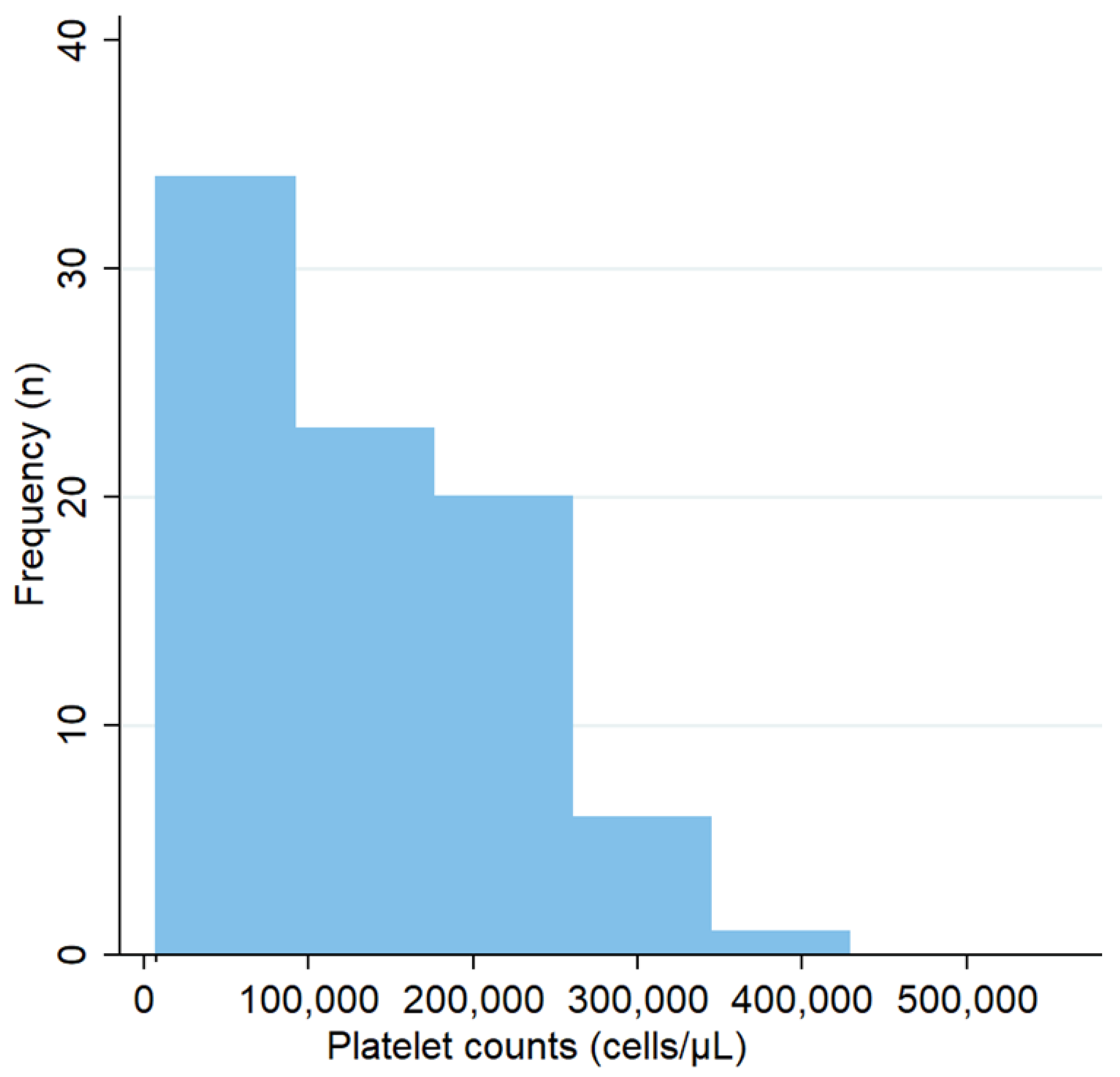
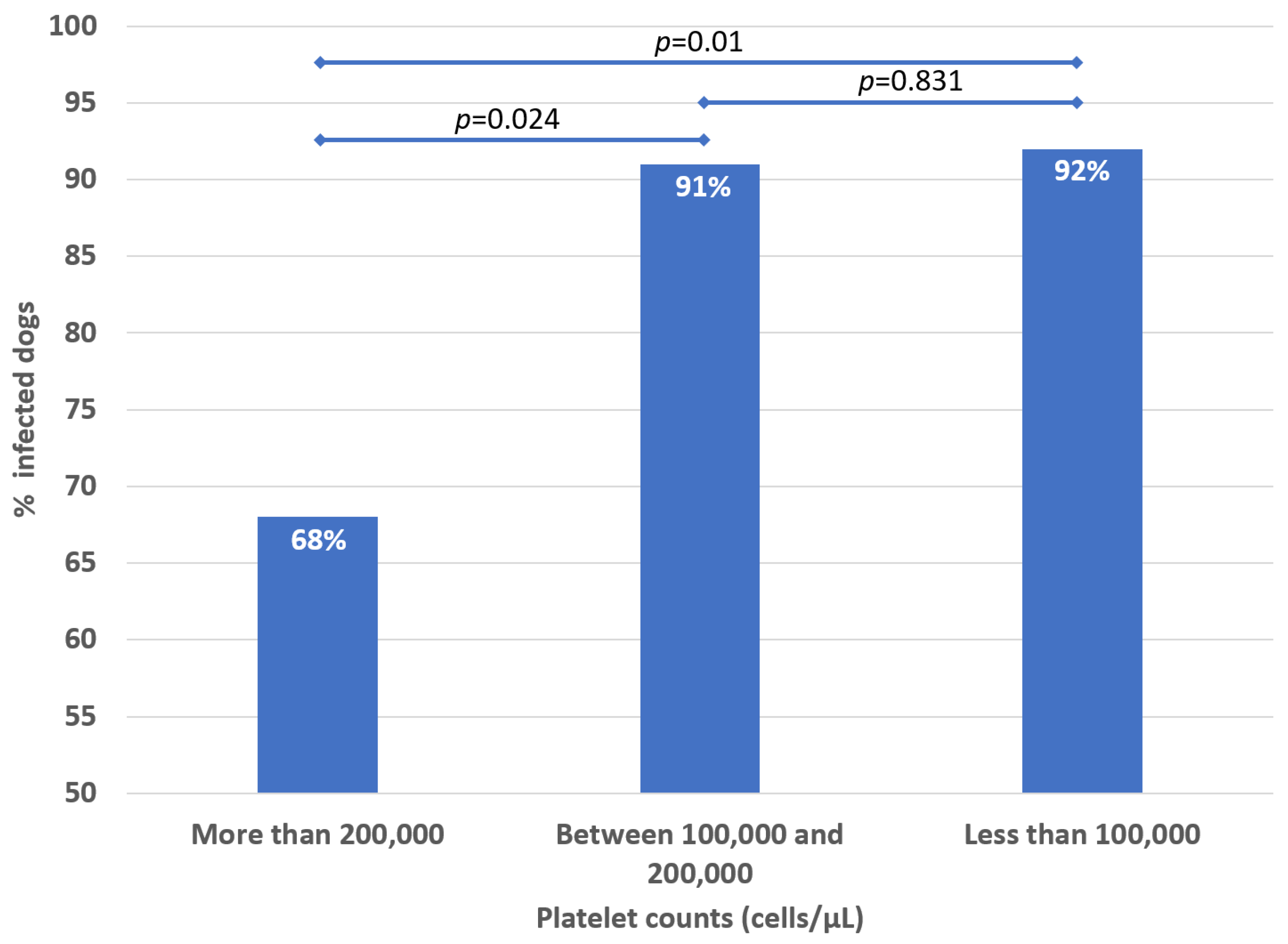
| Platelet counts (cells/μL) (RV ≥ 200,000) ** | 143,000 | 62,750 | 214,250 | 126,000 | 55,000 | 191,000 | 221,000 | 180,000 | 303,500 | 0.003 |
| % with thrombocytopenia | 70 | 75 | 40 | 0.006 | ||||||
| % with severe thrombocytopenia (<35,000 platelets/μL) | 13 | 14 | 7 | 0.429 | ||||||
| % with macroplatelets | 13 | 19 | 13 | 0.470 | ||||||
| % with anisocytosis | 11 | 16 | 0 | 0.336 | ||||||
| % with hypochromia | 10 | 18 | 7 | 0.758 | ||||||
| % with polychromacy | 8 | 9 | 7 | 0.913 | ||||||
| % with poikilocytosis | 4 | 4 | 7 | 0.568 | ||||||
| % with toxic neutrophils | 4 | 7 | 0 | 0.692 | ||||||
| % with reactive lymphocytes | 2 | 1 | 7 | 0.161 | ||||||
| % with agglutination | 1 | 2 | 0 | 0.673 | ||||||
| % with spherocytes | 0 | 0 | 0 | N/A | ||||||
| % with bicytopenia | 59 | 62 | 40 | 0.105 | ||||||
| % with pancytopenia | 11 | 13 | 0 | 0.140 | ||||||
| Total proteins (g/dL) (RV ≥ 5.5) | 10.50 | 9.88 | 11.28 | 10.5 | 10 | 12 | 9.50 | 8.25 | 10.25 | 0.003 |
| % with hypoproteinemia | 1 | 1 | 0 | 0.673 | ||||||
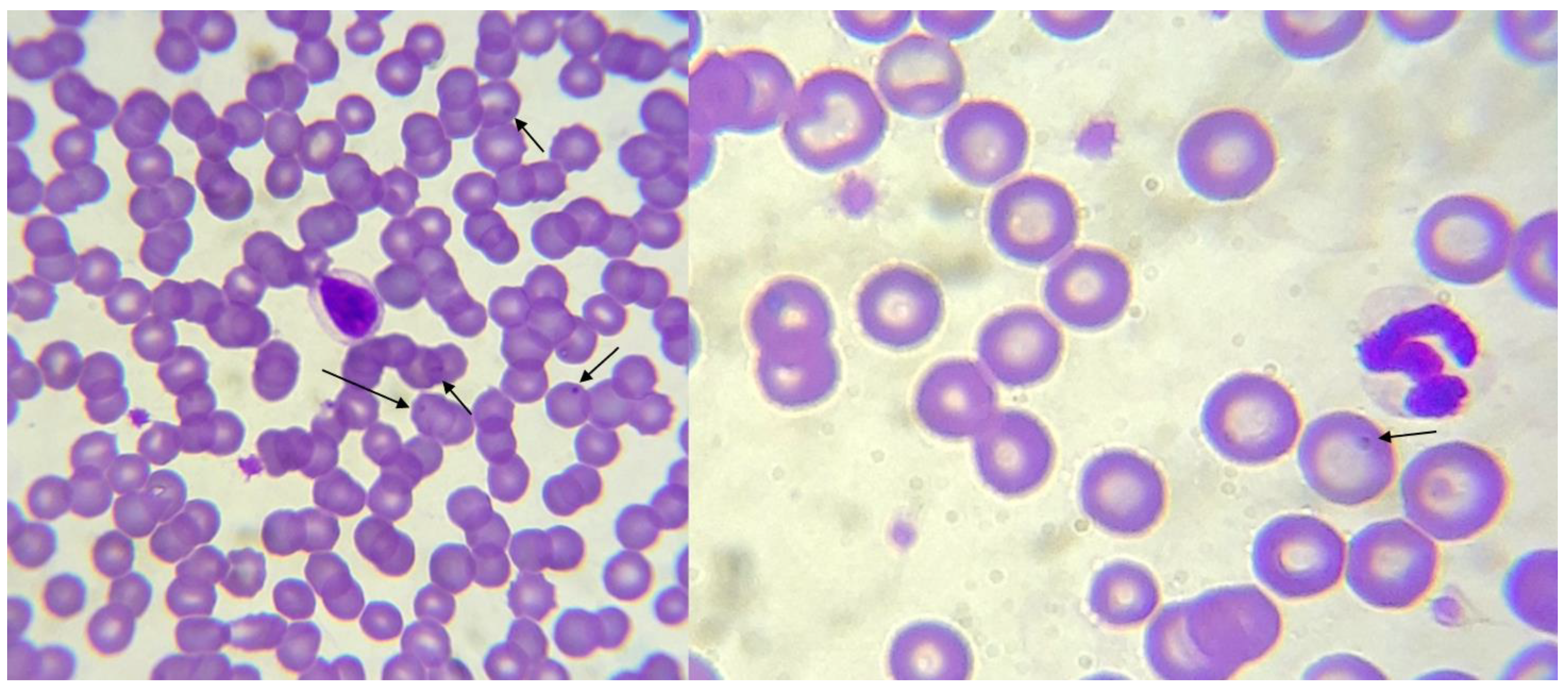
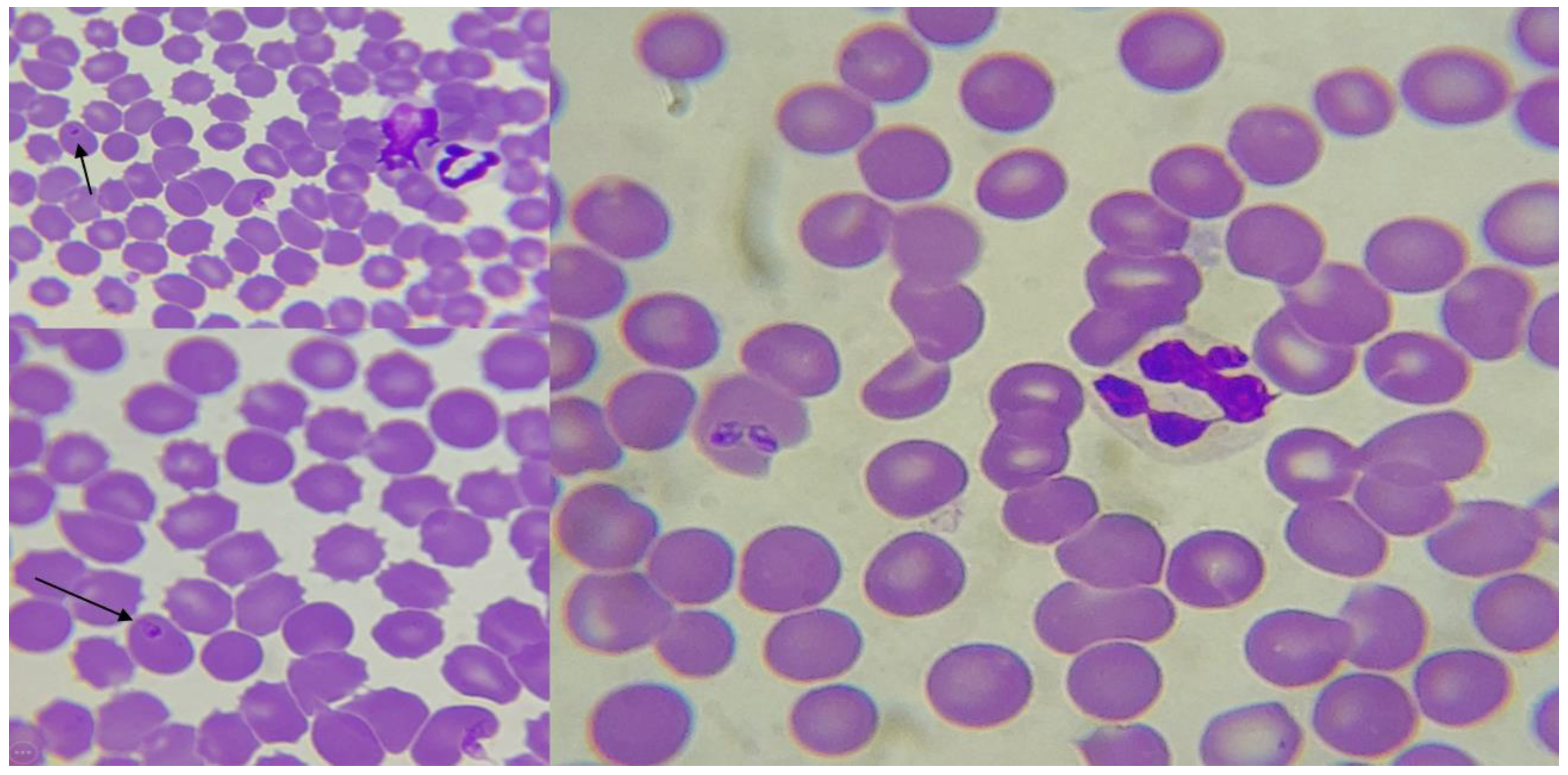
| % with organisms in the blood smear | 5 | 6 | 0 | N/A | ||||||
| Suggestive of Anaplasma marginale, marginal at erythrocytes | 2 | 2 | 0 | N/A | ||||||
| Suggestive of Babesia | 2 | 2 | 0 | N/A | ||||||
| Suggestive of Anaplasma, pyriform inclusions, Howell–Jolly bodies | 1 | 1 | 0 | N/A | ||||||
| Suggestive of Theileria | 1 | 1 | 0 | N/A | ||||||
| Infected Dogs | p (Male vs. Female) | Non-Infected Dogs | p (Male vs. Female) | p (Infected vs. Non-Infected by Sex) | ||||||
|---|---|---|---|---|---|---|---|---|---|---|
| Sex | Median Value | IQR | Median Values | IQR | ||||||
| Platelet counts (cells/μL) | Male | 136,500 | 48,250 | 197,750 | 0.705 | 281,000 | 236,000 | 320,000 | 0.270 | 0.003 |
| Female | 105,000 | 64,500 | 178,000 | 206,000 | 106,750 | 270,500 | 0.104 | |||
| % | p (Male vs. Female) | % | p (Male vs. Female) | p (Infected vs. Non-Infected by Sex) | ||||||
| n | Yes | No | n | Yes | No | |||||
| Thrombocytopenia | Male | 42 | 74 | 26 | 0.754 | 5 | 20 | 80 | 0.264 | 0.015 |
| Female | 43 | 77 | 23 | 10 | 50 | 50 | 0.091 | |||
Publisher’s Note: MDPI stays neutral with regard to jurisdictional claims in published maps and institutional affiliations. |
© 2022 by the authors. Licensee MDPI, Basel, Switzerland. This article is an open access article distributed under the terms and conditions of the Creative Commons Attribution (CC BY) license (https://creativecommons.org/licenses/by/4.0/).
Share and Cite
Bonilla-Aldana, D.K.; Gutiérrez-Grajales, E.J.; Osorio-Navia, D.; Chacón-Peña, M.; Trejos-Mendoza, A.E.; Pérez-Vargas, S.; Valencia-Mejía, L.; Marín-Arboleda, L.F.; Martínez-Hidalgo, J.P.; Reina-Mora, M.A.; et al. Haematological Alterations Associated with Selected Vector-Borne Infections and Exposure in Dogs from Pereira, Risaralda, Colombia. Animals 2022, 12, 3460. https://doi.org/10.3390/ani12243460
Bonilla-Aldana DK, Gutiérrez-Grajales EJ, Osorio-Navia D, Chacón-Peña M, Trejos-Mendoza AE, Pérez-Vargas S, Valencia-Mejía L, Marín-Arboleda LF, Martínez-Hidalgo JP, Reina-Mora MA, et al. Haematological Alterations Associated with Selected Vector-Borne Infections and Exposure in Dogs from Pereira, Risaralda, Colombia. Animals. 2022; 12(24):3460. https://doi.org/10.3390/ani12243460
Chicago/Turabian StyleBonilla-Aldana, D. Katterine, Erwin J. Gutiérrez-Grajales, Daniela Osorio-Navia, Mariana Chacón-Peña, Adrián E. Trejos-Mendoza, Soffia Pérez-Vargas, Lorenzo Valencia-Mejía, Luisa F. Marín-Arboleda, J. Paola Martínez-Hidalgo, María Angelica Reina-Mora, and et al. 2022. "Haematological Alterations Associated with Selected Vector-Borne Infections and Exposure in Dogs from Pereira, Risaralda, Colombia" Animals 12, no. 24: 3460. https://doi.org/10.3390/ani12243460