Article

Modulatory Impacts of Multi-Strain Probiotics on Rabbits’ Growth, Nutrient Transporters, Tight Junctions and Immune System to Fight against Listeria monocytogenes Infection

1
Department of Microbiology, Faculty of Veterinary Medicine, Zagazig University, Zagazig 44511, Egypt
2
Department of Nutrition and Clinical Nutrition, Faculty of Veterinary Medicine, Zagazig 44511, Egypt
3
Department of Poultry Diseases, Reference Laboratory for Quality Control of Poultry Production (RLQP), Animal Health Research Institute (AHRI), Zagazig Branch, Agriculture Research Center (ARC), Zagazig 44516, Egypt
4
Department of Veterinary Public Health, Faculty of Veterinary Medicine, Zagazig University, Zagazig 44511, Egypt
5
Veterinary Educational Hospital, Faculty of Veterinary Medicine, Zagazig University, Zagazig 44511, Egypt
6
Department of Biotechnology, Reference Laboratory for Quality Control of Poultry Production (RLQP), Animal Health Research Institute (AHRI), Sharkia Branch, Agriculture Research Center (ARC), Zagazig 44511, Egypt
7
Department of Bacteriology, Animal Health Research Institute (AHRI), Ismailia Branch, Agriculture Research Center (ARC), Ismailia 41522, Egypt
8
Department of Microbiology, Faculty of Veterinary Medicine, Matrouh University, Matrouh 51511, Egypt
9
Department of Clinical Laboratory Sciences, Turabah University College, Taif University, P.O. Box 11099, Taif 21944, Saudi Arabia
10
Department of Avian and Rabbit Medicine, Faculty of Veterinary Medicine, Zagazig University, Zagazig 44519, Egypt
*
Author to whom correspondence should be addressed.
Animals 2022, 12(16), 2082; https://doi.org/10.3390/ani12162082
Submission received: 7 July 2022 / Revised: 1 August 2022 / Accepted: 8 August 2022 / Published: 15 August 2022

Abstract

:

Simple Summary

Weaning is a crucial period associated with great stress and susceptibility to infection, implying adverse impacts on farmed rabbits’ production. Recently, probiotics have been provided as direct microbial feed supplements, which are considered the ideal antibiotic substitutes during pathogenic infections with an emphasis on promoting rabbits’ growth and modulating their immune functions. Therefore, our experiment was carried out to explore the efficacy of multi-strain probiotics (MSP) on rabbits’ growth, molecular aspects, such as nutrients transporters, cytokines, and intestinal integrity, and effectiveness against Listeria monocytogenes (L. monocytogenes) infection. Altogether, our findings proposed the beneficial consequences of MSP on rabbits’ growth, gut health, and immunity. After post-experimental infection of rabbits with L. monocytogenes, administration of MSP during the whole rearing period greatly reduced the detrimental impact of infection and consequently renovated efficient rabbits’ production.

Abstract

Multi-strain probiotics (MSP) are considered innovative antibiotics’ substitutes supporting superior gut health and immunity of farmed rabbits. The promising roles of MSP on performance, intestinal immunity, integrity and transporters, and resistance against Listeria monocytogenes (L. monocytogenes) were evaluated. In the feeding trial, 220 rabbits were fed a control diet or diet supplemented with three MSP graded levels. At 60 days of age, rabbits were experimentally infected with L. monocytogenes and the positive control, enrofloxacin, prophylactic MSP (MSPP), and prophylactic and therapeutic MSP (MSPTT) groups were included. During the growing period, MSP at the level of 1 × 10⁸ CFU/kg diet (MSPIII) promoted the rabbits’ growth, upregulated the nutrient transporters and tight-junction-related genes, and modified cytokines expression. Supplementing MSPTT for L. monocytogenes experimentally-infected rabbits restored the impaired growth and intestinal barriers, reduced clinical signs of severity and mortalities, and attenuated the excessive inflammatory reactions. Notably, enrofloxacin decreased L. monocytogenes and beneficial microbial loads; unlike MSPTT, which decreased pathogenic bacterial loads and sustained the beneficial ones. Histopathological changes were greatly reduced in MSPTT, confirming its promising role in restricting L. monocytogenes translocation to different organs. Therefore, our results suggest the use of MSPTT as an alternative to antibiotics, thereby conferring protection for rabbits against L. monocytogenes infection.

1. Introduction

Rabbit breeding is becoming important due to the rapid growth rate, superior productive ability, and easily digestible and healthy meat of rabbits [1]. Weaning is the most stressful period associated with significant financial losses in rabbit husbandry as it can increase their susceptibility to numerous infections [2]. To overcome this period, strengthening the intestinal barrier and modifying the immune systems of rabbits are urgently needed, given that the intestinal barrier is the main site for the dynamic contact of enteric pathogens with the host [3]. It is well recognized that intestinal immune system development is triggered by commensal bacteria that inhibit the colonization of harmful bacteria and provide a protective barrier to resist the invasion of exogenous pathogens, thus preventing intestinal inflammation and ensuring intestinal integrity [4,5]. Any imbalance of microflora can lead to a change of pH and pathogen multiplication with harmful consequences for the animal’s health [6,7]. Moreover, several enteric pathogens can escape from commensal-mediated colonization resistance [8,9].
Listeria monocytogenes (L. monocytogenes) is one of the most devious pathogens associated with listeriosis, and is uniquely difficult to monitor [10]. Previous investigations showed that farmed rabbits are predisposed to L. monocytogenes, and their meat is a potential source of listerial foodborne pathogens [11,12]. The hardiness of L. monocytogenes against eradication is due to its capacity to adapt and persist in a variety of environments, its intracellular site, and the weak intracellular diffusion of some antibiotics, which makes it a serious ongoing concern in animal production [13,14]. Additionally, the virulence of L. monocytogenes arises from its ability for invasion, adhesion, and translocation across the intestinal barrier throughout the gastrointestinal phase of infection [15]. Therefore, restricting L. monocytogenes at the gastrointestinal stage of infection is the best way to restrict pathogen spread to the deeper tissues and avoid consequent lethality. Despite the fact that antibiotics are the treatment of choice for listeriosis, they can disrupt the gut microbiota and impair host defense immunity [16]. Besides, the progress of drug tolerant bacteria and antibiotic residues in the meat were serious drawbacks raised from the excessive use of antibiotics [17,18,19,20]. This in turn has facilitated the development of new antibiotics alternatives [21,22], such as probiotics, as a control strategy against listeria infection. Probiotics are living commensal micro-organisms that struggle with pathogens at the sites of adhesion, enhance nutrient utilization, enhance the epithelial immune response, augment microbial balance, restore epithelial barrier function, and, therefore, prevent infection and subsequent pathological lesions [23,24,25,26,27,28,29]. Probiotics belonging to either Lactobacillus or Bacillus species are commonly used in rabbits’ feed [24]. However, knowing what physiological changes the multi-strain probiotics (MSP) stimulate in healthy rabbits and their further efficacy under L. monocytogenes infection over long-term use is very limited. Moreover, the precise MSP mechanism of actions that assess their immune modulation and promising roles in preventing L. monocytogenes induced intestinal barrier dysfunction need further investigation. Keeping the above-mentioned facts in view, the present work was undertaken to investigate, for the first time, the effects of MSP—in comparison with enrofloxacin—on the growth performance and expression of nutrient transporters, cytokines, and tight-junction-protein-related genes and their roles in fighting L. monocytogenes experimental infection in rabbits.

2. Materials and Methods

2.1. Probiotics Preparation

Three probiotic bacterial strains, including Lactobacillus acidophilus NBIMCC 8242, Bacillus subtilis DSM 17,299 and Enterococcus faecium NBIMCC 8270, were mixed at equal ratios (1:1:1) before being added into the formulated feed. The probiotic strains were grown separately in de Man, Rogosa, and Sharpe broth (Oxoid, Cambridge, UK) at 37 °C for 48 h. After incubation, the cells were centrifuged and then spray-dried before being added to the rabbit feed.

2.2. Rabbits and Experimental Protocol

A total of 220 weaned New Zealand white male rabbits, aged approximately 30 days and having an 802.8 g average body weight, were obtained from commercial rabbit farms. To conduct the experiment, feeding, experimental infection, and clinical trials were formed. A four-week feeding trial was carried out to examine the effect of supplementing the basal diet with three graded levels of the three probiotic bacterial strains at equal ratios and to choose the appropriate and effective dose of the mixed probiotic strains to be used as a prophylactic and/or a novel therapeutic tool for L. monocytogenes experimental infection in the clinical trial.

2.2.1. Feeding Trial

For this trial, 220 rabbits were randomly allocated into four equal experimental groups; each had 55 rabbits arranged in 5 replicates (11 rabbits each). The rabbits were offered the basal diet (control or untreated group) or fed experimental diets supplemented with a mixture of equal doses of Lactobacillus acidophilus, Bacillus subtilis, and Enterococcus faecium at graded levels—1 × 10⁶ (MSPI), 1 × 10⁷ (MSPII), and 1 × 10⁸ (MSPIII) colony forming units (CFU)/kg diet, respectively. All rabbits were kept in cages, fed pelleted diets, and provided free access to water and feed throughout the experimental rearing period. The diets were formulated in accordance with the nutrient recommendations for rabbits [30]. The ingredients and composition of the basal experimental diet are listed in Table 1.

2.2.2. Experimental Infection and Clinical Trials

These trials were conducted on two experimental groups comprised of the control (rabbits fed the basal diet) and MSP (rabbits fed the experimental diets supplemented with the most effective dose of the mixed probiotic strains) groups. All rabbits were checked to be free from detectable L. monocytogenes. The experimental infection trial was conducted according to the protocol of an earlier study [31] using the field L. monocytogenes strain previously recovered from clinically-diseased rabbits. The used strain was revitalized in Listeria enrichment broth (Oxoid, Cambridge, UK) at 37 °C for 18 h under microaerophilic conditions. Subsequently, 0.1 mL of the enrichment broth was inoculated for 24 h at 37 °C onto a Listeria-selective agar base supplemented with Listeria-selective supplement (Oxoid, Cambridge, UK). The typical colonies of L. monocytogenes were confirmed by Gram’s stain and biochemical tests including lactose, sucrose and xylose fermentation, catalase, oxidase and urease according to FDA bacteriological analytical manual. The strain was further confirmed via umbrella shaped motility, hemolysis onto sheep blood agar (Oxoid, Cambridge, UK), CAMP test and molecular identification methods. The inoculum suspension was subsequently prepared to obtain a concentration of 107 CFU/mL [32]. At 60 days of age, each rabbit in the two previous groups were orally administered with 1 mL of the prepared L. monocytogenes inoculum. Establishment of L. monocytogenes infection was verified by observing the characteristic clinical symptoms and post-mortem lesions of the sacrificed rabbits as well as bacteriological re-isolation and identification of the used strain.
In the clinical trial, the above-mentioned experimental groups (n = 50/group) were further subdivided into 2 subgroups (n = 25/subgroup and each subgroup contained 5 replicates). Regarding the MSP group, the rabbits in the first subgroup received the MSP prophylactically for 30 days during the previous feeding trial (from 30 to 60 days of age) without further MSP supplementation after L. monocytogenes experimental infection (MSPP); meanwhile, those in the second subgroup were offered the same MSP for an additional 30 days after L. monocytogenes experimental infection (from 30 to 90 days of age, MSPTT). Concerning the control group, the rabbits in the first subgroup were kept infected only and did not receive any treatment, but those in the second subgroup were treated with commercial enrofloxacin hydrochloride (ENR) at a dose of 40 mg/kg for 7 consecutive days, as was recommended by the producer [32]. The treatment program with ENR antibiotic was applied when the rabbits presented the clinical manifestations associated with listeriosis to support successful antibiotic treatment of L. monocytogenes infection.

2.3. Growth Performance Traits and Clinical Examination

The growth performance parameters of rabbits, in each replicate, were evaluated at the end of both the growing and finishing periods (60 and 90 days of age, respectively). The feed intake (FI) and body weight gain (BWG) were verified and then the feed conversion ratio (FCR) was estimated as following: amount of consumed feed (g)/BWG (g) [33,34]. Additionally, clinical signs and post-mortem (PM) findings were recorded. Moreover, the mortality rates of rabbits were calculated during the course of the experimental period.

2.4. Samples Collection

At 60 (before experimental infection) and 90 (4 weeks after L. monocytogenes experimental infection) days of age, 5 experimental rabbits were randomly selected and sacrificed by cervical dislocation. The slaughtering and dissection of rabbits were performed according to the recommendations of the World Rabbit Science Association [35]. The cecal and ileal contents were collected for further quantitative analysis of intestinal microbiota. The collected cecal and ileal contents were stored at −80 °C until analyzed with commercial DNA extraction kits. Moreover, slaughtered rabbits were skinned, eviscerated, and tissues from the jejunum and cecum were then collected and stored in RNAlater® (Sigma Aldrich, St. Louis, MO, USA) for subsequent mRNA expression assays. At 7 and 14 days post-experimental infection, L. monocytogenes counts were enumerated in the cecal contents. Finally, samples of liver, brain, and spleen tissues were harvested and fixed in 10% neutral buffered formalin for histopathological examination at 14 days post-infection according to [36,37].

2.5. Investigated Parameters

2.5.1. Quantitative DNA-Based Analysis of Intestinal Bacterial Populations

Total DNA was extracted from the cecal and ileal contents using QIAamp Fast DNA Stool Mini (Qiagen, Hilden, Germany). The extracted DNA quality and concentration were assessed by a Thermo Scientific NanoDrop 2000 spectrophotometer (Thermo Fisher Scientific Inc., Waltham, MA, USA). Ultimately, the purified DNA samples were stored at −80 °C for posterior quantitative PCR analysis. Real-time PCR (RT-PCR) assays were performed to calculate the populations of some intestinal microbial species, including total bacteria, and Lactobacillus, Bifidobacterium, Enterobacteriaceae, and L. monocytogenes strains using a Stratagene MX3005P quantitative PCR machine. The sequences of the primers targeting the specific bacterial genes are shown in Table 2. The PCR amplification assays were carried out, in triplicate, in a reaction volume of 25 μL containing 12.5 μL of SYBR Green PCR Master Mix (Qiagen, Hilden, Germany), 1 μL of of each primer (10 mM), 2 μL of target genomic DNA, and 8.5 μL of sterile PCR-grade water. Standard curves were prepared with ten-fold serial dilutions of genomic DNA isolated from pure bacterial cultures. The standard calibration curves were then generated by plotting the threshold cycle (Ct) values versus the bacterial DNA copy numbers. The bacterial concentrations in each DNA sample were measured using the generated standard curves in terms of log10 CFU/gram of the cecal and ileal contents.

2.5.2. Expression Profiling by Reverse Transcription Quantitative Real-Time PCR (qRT-PCR) Assays

The extracted total RNA from frozen jejunum and cecum tissues was done via QIAamp RNeasy Mini kit (Qiagen, Hilden, Germany) in compliance with the manufacturer’s instructions. RNA concentration and purity were assessed through a Nano Drop 2000 spectrophotometer (Thermo Scientific Inc., Waltham, MA, USA). The expression analysis was determined by one-step qRT-PCR assays using 2× QuantiTect SYBR Green RT-PCR Kit (Qiagen, Hilden, Germany). The mRNA expression levels of genes encoding nutrient transporters including sodium–glucose co-transporter-1 (SGLT-1), glucose transporter-2 (GLUT-2), cationic amino acid transporter-1 (CAT-1), and fatty acid-binding protein-2 (FABP-2); tight junction proteins (TJP) comprising claudins-1 (CLDN-1), junctional adhesion molecule-2 (JAM-2), occludin, and mucin-2 (MUC-2); and cytokines including interleukin-6 (IL-6), IL-8, IL-1β, IL-10, transforming growth factor-beta-1 (TGF-β1), and tumor necrosis factor-alpha (TNF-α) were determined at 60 and 90 days of age. Moreover, the mRNA expression level of gene encoding interferon gamma (IFNγ) was detected at 90 days of age only. All qRT-PCR reactions were run, in triplicate, on a Stratagene Mx3005P real-time thermal cycler (Agilent Technologies, Inc., Santa Clara, CA, USA). The transcript levels of the genes of interest were normalized to the expression of the housekeeping gene, glyceraldehyde 3-phosphate dehydrogenase (GAPDH), as a calibrator. All the investigated target genes and the sequences of their appropriate primers are characterized in Table 2. After the qRT-PCR reactions were completed, the specificity of the PCR amplifications and the purity of qPCR products were verified by post-PCR melting curves analyses. The relative changes in gene expression levels were analyzed using the 2−ΔΔCt method [38].
Table 2. Primer sequences of target genes analyzed in quantitative real-time PCR assays.
Encoding GenePrimer Sequence (5′-3′)Accession No./Reference
Nutrient transporters
SGLT-1F:GATTTCCCGTATGATTACCGAG
R:AAGAGGGAGACAACCACAACG
NM_001101692.1
GLUT-2F-CAGAGGCACTGTCCACCACC
R:TGTCTCCAAGCCACCCACC
NM_001277382.1
CAT-1F:CCAGTCTATTAGGTTCCATGTTCC
R: CGATTATTGGCGTTTTGGTC
XM_002721425.3
FABP-2F: GTGGGGTTTTCCCTTTTGC
R: CGCACTTTGGCCTTCACC
XM_002722970
Tight junction proteins
MUC-2F:TATACCGCAAGCAGCCAGGT
R:GCAAGCAGGACACAGACCAG
L41544.1
JAM-2F:ATATCGCAGGTGTCCTGGAA
R: GAGCATAGCACACGCCAAG
XM_017346699
CLDN-1F: GGAGCAAAAGATGCGGATGG
R: AATTGACAGGGGTCAAAGGGT
NM_001089316.1
occludinF: GCAAGAGGCCGTATCCAGAG
R: AGTCCGTCTCGTAGTGGTCT
XM_008262320.1
Cytokines
IL-6F:GCCAACCCTACAACAAGA
R:AGAGCCACAACGACTGAC
NC_013678
IL-8F:CTCTCTTGGCAACCTTCCTG
R:TTGCACAGTGAGGTCCACTC
KT216053.1
IL-10F:AAAAGCTAAAAGCCCCAGGA
R:CGGGAGCTGAGGTATCAGAG
NM001082045.1
IL-1βF:TTCCGGATGTATCTCGAGCA
R:GTGGATCGTGGTCGTCTTCA
NC_013670
TNF-αF:CTGCACTTCAGGGTGATCG
R:CTACGTGGGCTAGAGGCTTG
XM_008262537.2
IFNγF:TTCTTCAGCCTCACTCTCTCC
R:TGTTGTCACTCTCCTCTTTCC
NM_001081991.1
TGF-β1F: CAGTGGAAAGACCCCACATCTC
R: GACGCAGGCAGCAATTATCC
NM_001082660
House keeping
GAPDHF:TGTTTGTGATGGGCGTGAA
R:CCTCCACAATGCCGAAGT
NC_013676.1
Listeria monocytogenes
16S rRNAF: CCTTTGACCACTCTGGAGACAGAGC
R: AAGGAGGTGATCCAACCGCACCTTC
[39]
Total bacteria
16S rRNA F: CGGCAACGAGCGCAACCC
R: CCATTGTAGCACGTGTGTAGCC
[40]
Genus Lactobacillus
16S rRNA F: AGCAGTAGGGAATCTTCCA
R: CACCGCTACACATGGAG
[40]
Genus Bifidobacterium
16S rRNA F: TCGCGTCYGGTGTGAAAG
R: CCACATCCAGCRTCCAC
[40]
Enterobacteriaceae
16S rRNA F: CATTGACGTTACCCGCAGAAGAAGC
R: CTCTACGAGACTCAAGCTTGC
[40]
SGLT-1: sodium–glucose co-transporter-1; GLUT-2: glucose transporter-2; CAT-1: cationic amino acid transporter-1; FABP-2: fatty acid-binding protein-2; MUC-2: mucin-2; JAM-2: junctional adhesion molecule-2; CLDN-1: claudins-1; IL: interleukin; TNF-α: tumor necrosis factor-alpha; IFNγ: interferon gamma; TGF-β1: transforming growth factor-beta-1; GAPDH: glyceraldehyde 3-phosphate dehydrogenase and 16S rRNA: 16S ribosomal RNA.

2.5.3. Histomorphological Examination

The fixed liver, brain, and spleen tissues were cut to size, washed with fresh water, dehydrated in ascending grades of absolute ethanol, transparentized in xylene, and ultimately impregnated in paraffin wax. Thin sections (5 μm in thickness) of paraffin-embedded tissues were stained with eosin and hematoxylin and examined under the light microscope equipped with a computerized digital camera [37,41,42]. Stained slides were analyzed, and the lesions were accordingly detected and documented.

2.6. Statistical Analysis

The results were analyzed by the GLM procedure of SPSS and a subsequent Tukey’s post-hoc test was used to estimate the statistically significant differences among the treatment groups. Homogeneity and normality among our groups were evaluated through Levene’s and Shapiro–Wilk’s tests, correspondingly. The achieved data were conveyed as standard error of mean (SEM). For all tests, the significance was determined at p < 0.05.

3. Results

3.1. Growth Performance and Clinical Observations

The results regarding the growth performance attributes of rabbits are shown in Table 3 and Table 4. At the end of the growing period, the BWG of rabbits were significantly (p < 0.05) increased with increasing levels of MSP. Moreover, supplementation with MSP at a level of 1 × 10⁸ CFU/kg displayed the most significant (p < 0.05) improvement in rabbits’ FCR (Table 3). At the end of finishing period (90 d), infecting rabbits with L. monocytogenes significantly (p < 0.05) decreased BWG and impaired FCR. Interestingly, this impaired growth rate and FCR were improved in the probiotics- or enrofloxacin-treated groups. Meanwhile, rabbits receiving MSP (prophylactic and therapeutic) had the highest significant (p < 0.05) BWG and improved FCR. The experimentally-infected rabbits showed various clinical signs in the form of depression, anorexia and diarrhea, and generalized septicemia in PM examination of freshly dead rabbits. Notably, MSP over the whole rearing period (MSPTT) reduced the severity of both clinical signs and PM lesions. Moreover, the mortality percentages were significantly (p < 0.05) reduced, especially after administration of MSP during the whole rearing period when compared with the positive control group (24 vs. 6%) (Table 4).

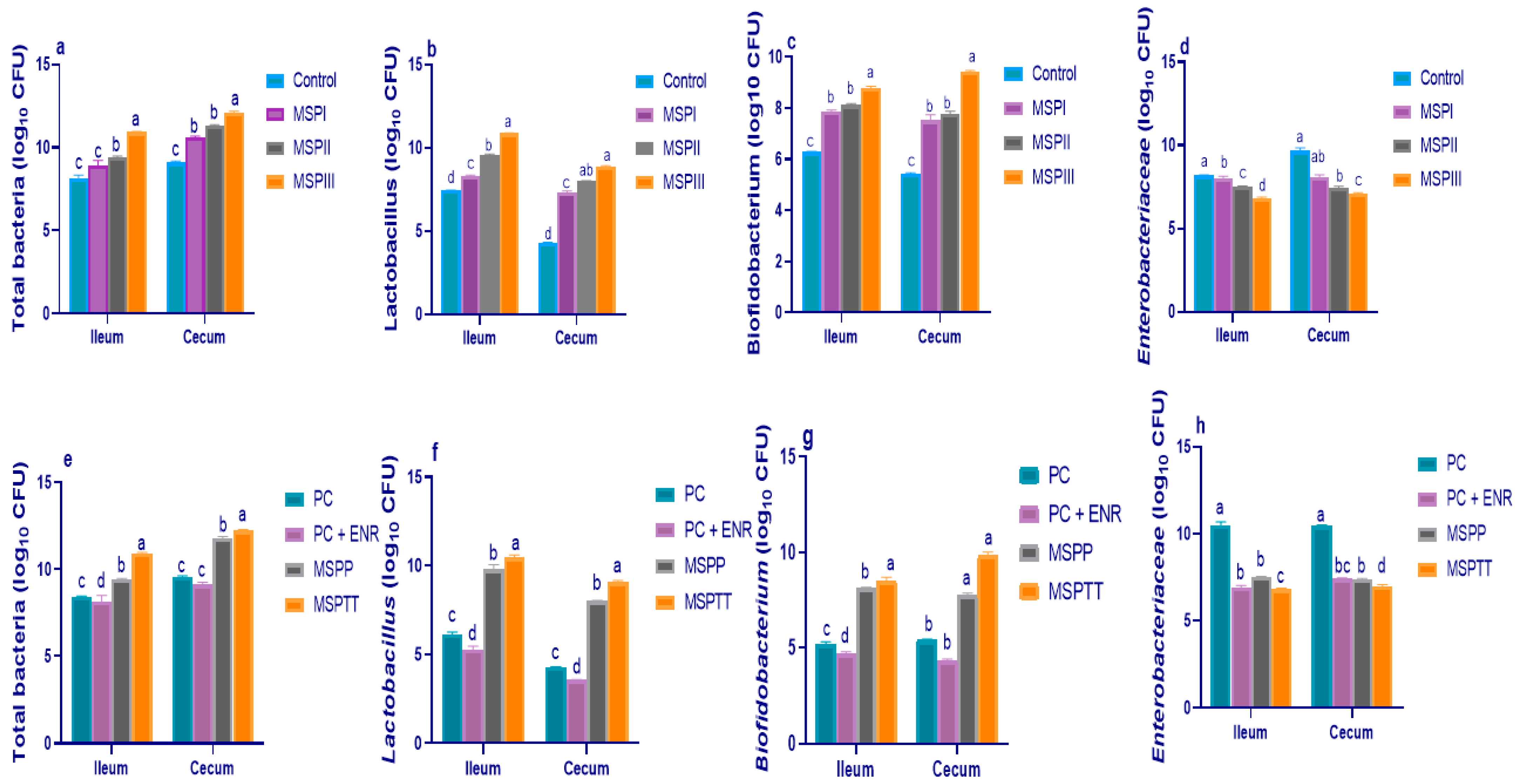
3.2. Microbial Populations of Intestinal Contents

At 60 days of age (before L. monocytogenes experimental infection), MSP supplementation at various levels resulted in a differential increase in the abundance of the total aerobic bacterial (Figure 1a), Lactobacillus (Figure 1b), and Bifidobacterium (Figure 1c) populations and decreased Enterobacteriaceae counts (Figure 1d) compared to the control group. Furthermore, compared with the control group, supplementing the rabbit diet with MSP at 1 × 10⁸ CFU/kg remarkably elevated (p < 0.05) the abundance of Lactobacillus and Bifidobacterium counts by 3.45 and 2.51 CFU/g of the ileal contents and 4.65 and 4 CFU/g of the cecal contents, respectively. In contrast, Enterobacteriaceae counts were notably decreased when increasing the levels of MSP in rabbits’ diets. At 90 days of age, rabbits experimentally infected with L. monocytogenes exhibited the highest (p < 0.05) colonization of harmful Enterobacteriaceae and the lowest (p < 0.05) populations of beneficial ones (i.e., Lactobacillus and Bifidobacterium species) in the ileum and cecum. Notably, enrofloxacin-treated rabbits exhibited the lowest (p < 0.05) ileal and cecal total bacteria (Figure 1e), and Lactobacillus (Figure 1f) and Bifidobacterium (Figure 1g) populations. Meanwhile, the cecum and ileum of experimentally-infected rabbits that received MSP had lower Enterobacteriaceae (Figure 1h) counts and higher Lactobacillus and Bifidobacterium populations, with prominent beneficial effects in rabbit groups that received MSP during the whole rearing period (prophylactic and therapeutic, MSPTT).

3.3. Gene Expression Analysis

The results of a gene expression analysis of nutrient-transporter-related genes analyzed by RT-PCR are illustrated in Figure 2. Before L. monocytogenes experimental infection (60 d of age), the data indicated that GLUT-2 gene expression levels were significantly (p < 0.05) increased when increasing the MSP levels (Figure 2a). Additionally, the mRNA expression levels of SGLT-1 (Figure 2b) and FABP-2 (Figure 2c) genes reached their peaks in rabbits supplemented with MSP at the level of 1 × 10⁸ CFU/kg (1.26- and 1.28-fold, respectively). Moreover, the transcriptional levels of CAT-1 genes (Figure 2d) were increased post-MSP supplementation, unlike the control group—with no significant differences detected among different MSP levels. At 90 d of age, expression analysis displayed significant (p < 0.05) upregulation of GLUT-2 genes (Figure 2e) in rabbits fed MSP-supplemented diets over the whole rearing period (MSPTT), followed by those fed MSP as a prophylactic, unlike the experimentally-infected (PC) group. Additionally, rabbits that received MSP during the whole rearing period exhibited the highest (p < 0.05) upregulation in SGLT-1 (Figure 2f), FABP-2 (Figure 2g), and CAT-1 (Figure 2h) gene expression (1.24-, 1.28-, and 1.27-fold, respectively).
The results of a gene expression analysis of tight-junction-protein-related genes are illustrated in Figure 3 and Figure 4. Before experimental infection (60 d of age), the expression levels of MUC-2 (Figure 3a) and JAM-2 (Figure 3b) were prominently upregulated (p < 0.05) in groups fed MSP at a level of 1 × 10⁸ CFU/kg when compared with the control group (1.73- and 1.44-fold, respectively). Moreover, CLDN-1 (Figure 3c) and occludin (Figure 3d) transcriptional levels were increased (p < 0.05) post-supplementation with MSP in a dose-dependent manner. At 90 d of age, the maximum expression levels of MUC-2 (Figure 4a) and JAM-2 (Figure 4b) were observed in rabbits fed MSP for either 30 or 60 days; meanwhile, their expressions were significantly (p < 0.05) impaired in rabbits treated with enrofloxacin. Of note, the expression results showed significant (p < 0.05) upregulation of CLDN-1(Figure 4c) and occludin (Figure 4d) genes in rabbits supplemented with MSP during the whole rearing period (MSPTT), followed by those fed MSP as a prophylactic (MSPP)—in comparison with the experimentally-infected (PC) group.
The results of a gene expression analysis of cytokine-related genes are illustrated in Figure 5 and Figure 6. Before experimental infection with L. monocytogenes (60 d of age), the relative expression levels of IL-8 (Figure 5a) and IL-6 (Figure 5b) genes were significantly (p < 0.05) decreased with increasing the levels of dietary MSP, while TNF-α (Figure 5c) and IL-1β (Figure 5d) expression levels were significantly (p < 0.05) decreased in rabbits fed MSP at levels of 1 × 10⁷ and 1 × 10⁸ CFU/kg unlike the control group. Moreover, IL-10 (Figure 5e) and TGF-β1 (Figure 5f) gene expression levels were significantly (p < 0.05) upregulated, especially with higher MSP supplementation levels. At 90 d of age, other remarkable findings emerged from the data analyses were the prominant decreases in the expression levels of the IL-8 (Figure 6a), IL-6 (Figure 6b), TNF-α (Figure 6c), and IL-1β (Figure 6d) genes in rabbits fed MSP over the whole rearing period (MSPTT) and those treated with enrofloxacin. Additionally, the enrofloxacin- and prophylactic and therapeutic MSP (MSPTT) treated groups, followed by prophylactic MSP (MSPP), displayed higher (p < 0.05) IL-10 (Figure 6e) and TGF-β1 (Figure 6f) transcriptional levels compared with the experimentally-infected group. Moreover, the most prominent (p < 0.05) upregulation of the IFNγ gene (Figure 6g) was detected in rabbits fed MSP during the whole rearing period (MSPTT) and those treated with enrofloxacin (1.45- and 1.55-fold, respectively).

3.4. Quantification of L. monocytogenes

At 7 days post-experimental infection, significantly (p < 0.05) lower log10 copies of L. monocytogenes populations were found in the cecal contents of rabbits treated with enrofloxacin, followed by those fed MSP during the whole rearing period (MSPTT; 2.75 and 2.92 log10 CFU/g, respectively) (Figure 7). Another remarkable observation that emerged from the data analyses was that L. monocytogenes counts reached their lowest levels in rabbits either treated with enrofloxacin or those that were supplemented with MSP during the whole rearing period (MSPTT) at 14 days post-infection (Figure 7).

3.5. Histopathological Alterations

At 14-days post-infection, the liver of L. monocytogenes experimentally-infected rabbits (positive control group) showed congestion of the hepatic blood vessels and sinusoids. Moreover, thrombus was formed within blood vessels and neutrophilic infiltrations were seen within sinusoids. The brain showed suppurative meningitis, which is characterized by an increase in the thickness of the meninges due to meningeal congestion and leukocytic infiltrations, mainly neutrophils. The spleen showed shrinkage of some lymphoid follicles and necrotic changes of some lymphoid elements besides dilated splenic sinusoids (Figure 8a). The histopathological findings of rabbits experimentally infected with L. monocytogenes and treated with enrofloxacin are shown in Figure 8b. The liver showed a preserved lobular pattern, cord arrangement, central veins, and portal triads structures with the presence of thrombus in the blood vessels and neutrophilic infiltrations within sinusoids in the hepatic parenchyma. The brain tissue showed abundant cellular and karyorrhectic debris of inflammatory cells within periventricular tissue with ventricular congestion, edema, and necrosis of some choroid plexus epithelium. The examined sections of the spleen showed depleted lymphocytes within some white pulp. Additionally, the histopathological outcomes of the liver, brain, and spleen of rabbits that received a diet supplemented with MSP as a prophylactic and experimentally infected with L. monocytogenes (MSPP) are shown in Figure 8c. The examined liver section tissues showed normal hepatic cords and blood vessels with prominent kupffur cells. Most of the cerebral parenchyma had normal structures. However, the periventricular inflammatory cells were seen. The spleen revealed mild to moderate proliferation of the white pulp lymphoid population. The red pulp showed congestion of splenic blood vessels and it was infiltrated by a large number of mature and immature lymphocytes and proliferated macrophages. The histopathological alterations of rabbits fed MSP over the whole rearing period (MSPTT) and experimentally infected with L. monocytogenes are shown in Figure 8d. The liver showed normal hepatic parenchyma. The brain showed normal cerebral tissue with the presence of round cells infiltration within the meninges. Normal splenic histomorphology with a preserved white pulp lymphoid arrangement (germinal centers, central arteriole, marginal, and mantle zone) and preserved red pulp, including sinusoids, reticular fibers network, and the supported lymphoid cells beside normal megakaryocytes, supported the stroma and capsule. The red pulp showed moderate infiltration of mature and immature lymphocytes, as well as other inflammatory cells.

4. Discussion

Listeria monocytogenes is an opportunistic foodborne pathogen that can infect a wide range of farm animals including rabbits resulting in life-threatening listeriosis [10,12]. The challenges associated with L. monocytogenes ubiquity renders it a dangerous ongoing concern in animal production and food safety because of growth depression and great economic losses [10]. Indiscriminate therapeutic application of antibiotics for treating L. monocytogenes disrupts the host’s normal microbiotic balance and leads to the development of antimicrobial-resistant micro-organisms. Thus, an awareness of excessive antibiotics use can encourage the development of alternative safe ways, particularly, on the prophylaxis level.
Among these safe alternatives, we claimed that MSP could block the pathogen attachment or invasion of epithelial cells and consequently improve the rabbits’ growth performance and confer protection against L. monocytogenes infection. Especially in rabbits, mechanisms of how MSP modulate their tight junctions and immunity and counteract listeriosis are not fully understood until now. In the current study, use of MSP at a level of 1 × 10⁸ CFU/kg diet during the growing period contributed to the highest growth performance parameters and good health conditions of rabbits, suggesting the growth-promoting role of selected MSP. In accordance, supplementing rabbits’ diets with B. subtilis improved growth performance, immune organs’ indices, intestinal homeostasis, innate immune response, and disease resistance [43]. Moreover, [44,45,46] found an enhancement in growth performance after using various probiotic compositions. The previous authors attributed the improved growth performance to an increase in the digestive enzymes’ activities coupled with other changes, such as the maintenance of gastrointestinal tract (GIT) beneficial microbiota and a decrease in the production of ammonia. Besides, Bacillus species could produce extracellular enzymes and some necessary nutrients and provide essential growth factors to boost animal growth [47]. Similarly, greater BWG and lower FCR were observed in weaning rabbits fed with dietary L. acidophilus alone or a combination of B. subtilis and L. acidophilus [48]. These findings could result from greater nitrogen retention and nutrient digestibility in the L. acidophilus- and B. subtilis-supplemented rabbits [48,49]. Interestingly, the suppressed growth performance parameters induced after L. monocytogenes experimental infection were compensated in groups supplemented with MSP, suggesting their potential role in attenuating L. monocytogenes infection in rabbits. In the same line, L. monocytogenes infection impaired the growth performance parameters of rabbits; however, L. acidophilus administration recovered the final BWG of rabbits [50]. In this context, the growth-promoting activity of MSP may be increased from competing with pathogenic microbes in the gut and augmenting the immune system, thereby leading to higher resistance to dangerous infectious agents [51,52]. Additionally, the probiotic function mechanisms were heterogeneous, complex, and specific to each probiotic strain. They comprise pathogens’ competitive exclusion [53], ability to colonize the intestine [54], improvement in intestinal barrier functions by upregulating the TJP, and mucin expression, along with immune system regulation [55].
The digestion end products of dietary ingredients in the small intestine are principally absorbed through nutrient transporters that play a crucial role in nutrient absorption and feed utilization [40,56]. The absorption of monosaccharides in the intestine is crucial for the homeostasis of energy. SGLT-1 and GLUT-2 engage in the intestinal absorption of monosaccharides and their concentrations, and, consequently, control monosaccharide uptake in the small intestine. SGLT-1 is specified in the apical membrane and it mediates glucose uptake from the intestinal lumen through the brush-border membrane into the intestinal enterocytes [57,58], while GLUT-2 is the primary fructose and glucose transporter in the basolateral membrane [59,60,61]. FABP-2 is implicated in the trafficking of intracellular free fatty acids and eventually enhances intestinal nutrient absorption [62]. Moreover, CAT-1 can transport cationic amino acids (e.g., arginine, histidine, and lysine) from enterocytes to the vascular supply or vice versa. In this respect, probiotic strains have been previously shown to improve nutrient absorption [63]; however, the mechanism by which they can affect nutrient transporters at the molecular level needs further investigation. Herein, the long-term administration of MSP upregulated SGLT-1, GLUT-2, CAT1, and FABP-2 genes associated with nutrient transportation even after L. monocytogenes experimental infection. Similarly, the relative gene expression of glucose and protein transporters showed a significant (p < 0.05) upregulation after feeding on multi-strain probiotics in broiler chickens [64].
The intestinal microbiota could greatly affect the host’s gut health via modulating intestinal pH, transporter gene expression, and mucosal immunity [65]. Probiotics can modify the composition of microbial species in the host gut by sustaining the balance and suppressing the growth of pathogenic bacteria [66]. The data of the present study revealed that supplementation of MSP at the dose of 1 × 10⁸ CFU/kg diet shifted the ileal and cecal microbiome composition of growing rabbits at 60 days of age towards the beneficial bacteria when compared to the control group. In contrast, the abundance of Enterobacteriaceae opportunistic pathogens was decreased after MSP supplementation, especially at higher doses. In accordance, dietary probiotics increased the numbers of beneficial bacterial organisms in the rabbits GIT than the pathogenic ones [67]. Moreover, feeding on probiotics increased the populations of Lactobacillus and Bifidobacterium species in the gut of rabbits [40,68]. The proposed probiotics mechanism of the intestinal microbiota modulation can result from reducing the oxygen or intestinal pH in the digestive tract [69]. Moreover, they could improve the intestinal tract habitat to antagonize the proliferation of pathogenic bacteria and therefore optimize the intestinal flora structure [70]. Additionally, an increase of the cecal lactobacilli counts in the rabbits supplemented with L. acidophilus led to higher concentrations of cecal acetic acids and total volatile fatty acids and decreased intestinal coliform colonization [48,71]. Moreover, rabbits fed B. subtilis/L. acidophilus-supplemented diets showed greater intestinal lactobacilli counts than those fed diets supplemented with B. subtilis alone [48]. This could be explained by the increased activities of lactobacilli after co-culturing with Bacillus species by the stimulating effect of the latter one on the biosynthesis of lactobacillus strains [72]. On the other hand, treatment with antibiotics affects not only the target bacterial microorganisms, but also the intestinal microbial communities. It has been shown that antibiotics exhibited long-lasting alterations in the intestinal microbiota, which is associated with disease occurrence [73]. Notably, after infection with L. monocytogenes and treatment with enrofloxacin, all counted bacterial populations were decreased, while early and continuous MSP supplementation after infection enhanced beneficial bacterial communities and decreased the colonization of harmful ones in the cecum and ileum, which are in agreement with previous studies [74,75]. Herein, reducing gut microbial damage after antibiotics treatment can be attributed to their wide antibacterial action that does not distinguish between types of bacteria [76,77]. Meanwhile, the beneficial effects of MSP on gut microbiota could be likely mediated through competitive exclusion, which mainly results from lowering luminal pH, thereby causing direct inhibition of enteric pathogens and the secretion of bactericidal proteins [78].
Cytokines are known to have a key regulatory impact in monitoring the intestinal inflammatory response. Probiotic bacteria exert immunostimulatory and immunoregulatory activities, and thus they can be extensively applied in the treatment of numerous diseases [79,80]. The beneficial effects of probiotics on the inflammatory process have been documented [54,81] and they are mainly due to their modulatory functions through the induction of various kinds of cytokines in gut-associated lymphoid tissues [80]. This was evidenced by the reduction of pro-inflammatory cytokine expression and boosting the anti-inflammatory ones. Similarly, Lactobacillus species triggered the immune system by boosting the expression of anti-inflammatory cytokine genes, IFN-γ and IL-10 [82,83], or by preventing the expression of the pro-inflammatory cytokine genes, TNF-α, IL-6, and IL-8 [84,85]. Additionally, the probiotics immunoregulatory effect can be related to IL-10 production, which blunts the excessive inflammatory response [86]. On the other hand, the invasion of bacterial pathogens into intestinal epithelial cells triggers the gastrointestinal immune cells to produce cytokines, which stimulate immune responses against pathogens [87]. In this context, L. monocytogenes could upregulate TNF-α and IL-6 genes, thereby leading to an increase in the permeability of the intestinal epithelium [88]. Stimulation of pro-inflammatory cytokines has been well studied in rabbits in response to L. monocytogenes [11]. IL-1β is a key pro-inflammatory cytokine that stimulates its own expression and the expression of other pro-inflammatory cytokines and chemokines, which in turn recruit inflammatory responses and trigger the induction of antimicrobial cells [54]. As anticipated in our study, elevated expression levels of pro-inflammatory cytokines (TNF-α, IL-6, IL-8 and IL-1β) genes were noted in rabbits fed a basal diet and experimentally infected with L. monocytogenes compared with those experimentally infected and treated with either enrofloxacin or probiotics. Similarly, probiotics could blunt intestinal inflammation [89] through TNF-α inhibitory metabolites production and nuclear factor-kB signaling inhibition in the enterocytes [90]. Regarding IFNγ, its initial production is a crucial step for producing an immune response and regulating L. monocytogenes infection [91]. Moreover, IL-10 has a predominantly antagonistic impact on inflammation, besides its crucial role in depressing the inflammatory and immune responses [92]. In the current study, oral administration of MSP induced the production of IFNγ, which promoted effective L. monocytogenes clearance and upregulated IL-10 and TGFβ genes that suppress excessive inflammation and maintain intestinal immune homeostasis. The robust inflammatory response in the L. monocytogenes experimentally-infected group was effectively counteracted by MSP administration over the whole rearing period (prophylactic and therapeutic), thereby indicating their strong anti-inflammatory properties. In previous studies, higher transcript levels of IFN-γ genes were detected after B. subtilis pre-treatment, even after Citrobacter rodentium infection [93,94]. In accordance, Lactobacillus species triggered pathogen clearance and suppressed intestinal inflammation via stimulating the anti-inflammatory cytokines (IL-10 and TGFβ) production [95,96]. On the other hand, supplementation with antibiotic growth promoters seemed to relatively downregulate the expression of inflammatory cytokine genes in the intestine after infection with live pathogens [97]. In a separate study, broilers fed a bacitracin-supplemented diet and received lipopolysaccharide (LPS) had reduced intestinal levels of inflammatory cytokines compared with bacitracin-unsupplemented and LPS-received controls [98]. Moreover, pathogen-infected chickens fed an antibiotics-supplemented diet had decreased IL-2, IL-8, L-1β, and IL-6 transcript levels [97,99].
A key function of the intestinal epithelium is to form a natural barrier, which hinders pathogens and toxic constituents from entering the mucosa and coming into contact with the immune system, thereby ensuring intestinal homeostasis [100,101]. Intestinal tight junctions and their related proteins, including CLDN, zonula occludens (ZO) and occludin, are the main factors that stimulate tight junction barrier formation [102,103]. Previous reports have described their differing effects on intestinal permeability [102,104]. It has been demonstrated that intestinal microbiota efficiently contributed to intestinal immune system development, epithelial barrier augmentation, and pathogen colonization restriction [8]. Nevertheless, the intestinal microbiota can be modified by many causes and diseases involving dietary changes, stress, antibiotics, and infection [105], leading to an imbalance in intestinal homeostasis. During weaning, the decrease in the gene expression of TJP; ZO-1, CLDN-1, and occludin, could lead to barrier integrity impairment. In this context, probiotics have a diverse mechanism of action to enhance the function of the intestinal barrier and sustain homeostasis, and thus it may counteract the weaning stress. Herein, higher expression levels of ZO-1, CLDN-1, occludin, MUC-2, and JAM-2 in the intestine of rabbits described that MSP, especially at higher doses, enhanced TJPs. In accordance, probiotics could enhance gut barrier function by increasing the expression of TJP genes [106,107,108]. Moreover, some Lactobacillus species, such as L. plantarum, abated barrier disruption via upregulation of TJPs [109,110]. In this study, relative TJP transcripts were significantly reduced after L. monocytogenes infection. However, after supplementing MSP during the whole rearing period (prophylactic and therapeutic), these reductions were abrogated. This could be attributed to the beneficial roles of MSP in fortifying the intestinal epithelial resistance to pathogens by sustaining TJP abundance. A previous study utilizing different probiotic strains described parallel in-vivo and in-vitro results [111]. It has been asserted that lactic acid bacteria (LAB) can restore the intestinal epithelial barrier damage generated by pathogenic infection [109]. A recent report found that lactobacilli ameliorated the intestinal barrier damage induced by Salmonella species [112]. Moreover, [113] described that lactobacilli probiotics prevented L. monocytogenes-induced intestinal permeability through the preservation of the cell junctional architecture of CLDN-1, occludin, and E-cadherin. Additionally, L. plantarum had protective effects on the intestinal barrier by rearranging TJPs (ZO-1, CLDN-1, and occludin) disturbed by Escherichia coli (E. coli) and accordingly ameliorated the barrier function [113,114]. Another in-vitro study described that L. rhamnosus and L. fermentum significantly improved E. coli-disturbed TJP (Occludin, ZO-1, cingulin-1, and CLDN-1) [115]. Notably, maintaining tight junction integrity in rabbits that received MSP during the rearing period was in line with our results of downregulating the pro-inflammatory cytokine genes (IL-6, IL-8, and TNFa). Aside from their important roles in immunity, cytokines were also demonstrated to affect tight junction integrity as pro-inflammatory cytokines could induce tight junction disruption. Commensal bacteria such as Lactobacillus and Bifidobacterium species in intestinal microflora could improve intestinal barrier functions and regulate cytokines secretion [84]. Similarly, L. plantarum pre-treatment downregulated the pro-inflammatory cytokine genes (IL-6, IL-8, and TNF-α) and alleviated the reduction in TJP (CLDN-1, occludin, and ZO-1) caused by E. coli [54]. Furthermore, the intestinal barrier is reinforced by a glycosylated mucin-rich layer secreted by goblet cells. In view of this, probiotics have been demonstrated to strengthen the integrity of intestinal barriers by increasing the number of goblet cells that reinforce the mucus layer [116]. Previous in-vitro studies described that many Lactobacillus species were proved to increase the expression of intestinal mucin [117,118,119]. Moreover, lactobacilli probiotics triggered MUC secretion and thus fortified the mucus barrier and limited L. monocytogenes-induced MUC-2 loss and apoptotic responses [113]. Although antibiotics can combat pathogenic agents, their application exhibited several adverse effects, such as intestinal barrier dysfunction [40]. This is supported by our results, which evidence that treatment with enrofloxacin decreased the expression of TJP genes. Similarly, [120] verified that antibiotics reduced the expression of TJP genes and increased intestinal paracellular permeability, suggesting their negative impact on intestinal tight junction barriers. Herein, the impairment of intestinal barrier functions is associated with dysbiosis of intestinal microbiota and the decrease in the expression of ZO-1, CLDN-1, and occludin genes post-treatment with enrofloxacin.
Regarding the infection with L. monocytogenes, it enters the host via the intestines and then infects the spleen, liver, and immune cells, where they proliferate until attacked by the host’s immune system [121]. As an important weapon to struggle against infectious diseases, antibiotics have been employed to cure bacterial infections for several years. Even though antibiotics bring considerable advantages for the hosts, they produce a variety of grievous adverse concerns. Among them, infections with multidrug-resistant pathogens might be the most dangerous one [122]. From this view, the potential effects of probiotic bacteria have been intended as a rational attempt for counteracting the intestinal pathogens [47,123]. Regarding farmed rabbits, there is limited data concerning the prevention or control of L. monocytogenes infection by probiotics. Our results proved that MSP administration during the rearing period greatly reduced the intestinal counts of L. monocytogenes. In accordance, [113] described that the lactobacilli probiotics were able to co-aggregate with L. monocytogenes and inhabit the membrane expressed epithelial Hsp60 receptor sites on epithelial cells to competitively eliminate it. There are several mechanisms underlying the effect of probiotic strains on the inhibition of gut translocation of bacteria to other organs [124]. It is proposed that probiotics may compete with pathogens for adhesion sites, prevent pathogen-induced disruption of epithelial integrity, and modulate the immune system, thereby conferring resistance to L. monocytogenes [23]. Besides, LAB also produced microbicidal substances that have a great impact on the gastric and intestinal pathogens and other microbes that compete for cell surfaces and mucin binding sites. Current in-vitro findings revealed that L. acidophilus, L. plantarum, and E. faecium, or their secondary metabolites, prevented the formation of L. monocytogenes biofilm [92,125,126]. Additionally, the reduced consequences of the probiotic on L. monocytogenes loads were endorsed by recent results in rabbits [11]. After infection with L. monocytogenes, significant histological changes were detected in rabbits’ livers, brains, and spleens with a picture of septicemia (positive control group). Similar findings were previously detected in the organs of rabbits infected with L. monocytogenes [127]. Administration of MSP during the whole rearing period attenuated the translocation of L. monocytogenes to other organs, as evidenced by restoring the normal histopathological architecture of rabbits’ livers, brains, and spleens. Similarly, significant improvements were found in the histological pictures of rabbits` tissues after administration of Lactobacillus acidophilus [128]. This could be attributed to the effective roles of MSP, which was proven in our results, on enhancing the immunity of rabbits against infection with intestinal pathogens and strengthening the intestinal barriers with a consequent restriction of pathogen spread to different organs.

5. Conclusions

Taken together, our findings suggested the favorable outcomes of multi-strain probiotics during the whole rearing period on rabbits’ growth, immune response, and intestinal barriers, as evidenced by modulating the expression of cytokines and tight-junction-protein-related genes. Additionally, administration of MSP for rabbits infected with L. monocytogenes attenuated the severity of clinical signs, PM lesions, and pathogen localization or translocation by decreasing its load in the cecum of rabbits, thereby downregulating the pro-inflammatory cytokines and subsiding the excessive inflammatory response. Hence, our results recommend the application of multi-strain probiotics, as an alternative to antibiotics, to offer protection for rabbits against L. monocytogenes infection.

Author Contributions

Conceptualization, M.I.A.E.-H., D.I., R.I.H., E.K., E.M.A.-A., H.H.N., H.B., M.H.E., A.S.A.E.O., G.A.I., T.A.I. and N.F.S.A.; methodology, M.I.A.E.-H., D.I., R.I.H., E.K., E.M.A.-A., H.H.N., H.B., M.H.E., T.A.I., A.S.A.E.O., G.A.I. and N.F.S.A.; software, M.I.A.E.-H., D.I., R.I.H., E.K., E.M.A.-A., H.H.N., H.B., M.H.E., T.A.I. and N.F.S.A.; validation, M.I.A.E.-H., D.I., R.I.H., E.K., E.M.A.-A., H.H.N., H.B., A.S.A.E.O., G.A.I., M.H.E., T.A.I. and N.F.S.A.; formal analysis, M.I.A.E.-H., D.I., R.I.H., E.K., E.M.A.-A., H.H.N., H.B., M.H.E., T.A.I. and N.F.S.A.; investigation, M.I.A.E.-H., D.I., R.I.H., E.K., E.M.A.-A., A.S.A.E.O., G.A.I., H.H.N., H.B., M.H.E., T.A.I. and N.F.S.A.; resources, M.I.A.E.-H., D.I., R.I.H., E.K., E.M.A.-A., H.H.N., H.B., M.H.E., T.A.I. and N.F.S.A.; data curation, M.I.A.E.-H., D.I., R.I.H., E.K., E.M.A.-A., H.H.N., H.B., M.H.E., T.A.I. and N.F.S.A.; writing—original draft preparation, M.I.A.E.-H., D.I., R.I.H., E.K., E.M.A.-A., H.H.N., H.B., M.H.E., T.A.I. and N.F.S.A.; writing—review and editing, M.I.A.E.-H., D.I., R.I.H., E.K., E.M.A.-A., H.H.N., H.B., M.H.E., T.A.I. and N.F.S.A.; visualization, M.I.A.E.-H., D.I., R.I.H., E.K., E.M.A.-A., H.H.N., H.B., M.H.E., T.A.I., A.S.A.E.O., G.A.I. and N.F.S.A.; supervision, M.I.A.E.-H., D.I., R.I.H., E.K., E.M.A.-A., H.H.N., H.B., M.H.E., T.A.I. and N.F.S.A.; project administration, M.I.A.E.-H., D.I., R.I.H., E.K., E.M.A.-A., H.H.N., H.B., M.H.E., T.A.I. and N.F.S.A.; funding acquisition, M.I.A.E.-H., D.I., R.I.H., E.K., E.M.A.-A., H.H.N., H.B., M.H.E., A.S.A.E.O., G.A.I., T.A.I. and N.F.S.A. All authors have read and agreed to the published version of the manuscript.

Funding

This research was supported by the Taif University Researchers Supporting Project number (TURSP-2020/134), Taif University, Taif, Saudi Arabia.

Institutional Review Board Statement

All research protocols and experimental design were carried out in accordance with regulations and approved guidelines of the Institutional Ethics Committee on Animal Care and Use established by Faculty of Veterinary Medicine, Zagazig University (Approval No. ZU-IACUC/F/137/2021). This article does not contain any studies with human participants performed by any of the authors.

Informed Consent Statement

Not applicable.

Data Availability Statement

The data presented in this study are available on request from the corresponding author.

Acknowledgments

The authors would like to acknowledge and thank the Taif University Researchers Supporting Project number (TURSP-2020/134), Taif University, Taif, Saudi Arabia. Furthermore, we want to thank the Faculty of Veterinary Medicine, Zagazig University, Egypt.

Conflicts of Interest

The authors declare no conflict of interest.

References

  1. Cullere, M.; Dalle Zotte, A. Rabbit meat production and consumption: State of knowledge and future perspectives. Meat Sci. 2018, 143, 137–146. [Google Scholar] [CrossRef]
  2. Simonová, M.P.; Lauková, A.; Žitňan, R.; Chrastinová, Ľ. Effect of rabbit-origin enterocin-producing probiotic strain Enterococcus faecium CCM7420 application on growth performance and gut morphometry in rabbits. Czech J. Anim. Sci. 2015, 60, 509–512. [Google Scholar] [CrossRef]
  3. Pastorelli, L.; De Salvo, C.; Mercado, J.R.; Vecchi, M.; Pizarro, T.T. Central role of the gut epithelial barrier in the pathogenesis of chronic intestinal inflammation: Lessons learned from animal models and human genetics. Front. Immunol. 2013, 4, 280. [Google Scholar] [CrossRef] [PubMed]
  4. Becattini, S.; Littmann, E.R.; Carter, R.A.; Kim, S.G.; Morjaria, S.M.; Ling, L.; Gyaltshen, Y.; Fontana, E.; Taur, Y.; Leiner, I.M. Commensal microbes provide first line defense against Listeria monocytogenes infection. J. Exp. Med. 2017, 214, 1973–1989. [Google Scholar] [CrossRef] [PubMed]
  5. Farahat, M.; Ibrahim, D.; Kishawy, A.; Abdallah, H.; Hernandez-Santana, A.; Attia, G. Effect of cereal type and plant extract addition on the growth performance, intestinal morphology, caecal microflora, and gut barriers gene expression of broiler chickens. Animal 2020, 15, 100056. [Google Scholar] [CrossRef] [PubMed]
  6. Michelland, R.J.; Combes, S.; Monteils, V.; Cauquil, L.; Gidenne, T.; Fortun-Lamothe, L. Molecular analysis of the bacterial community in digestive tract of rabbit. Anaerobe 2010, 16, 61–65. [Google Scholar] [CrossRef] [PubMed]
  7. Ibrahim, D.; Ismail, T.A.; Khalifa, E.; El-Kader, A.; Shaimaa, A.; Mohamed, D.I.; Mohamed, D.T.; Shahin, S.E.; El-Hamid, A.; Marwa, I. Supplementing Garlic Nanohydrogel Optimized Growth, Gastrointestinal Integrity and Economics and Ameliorated Necrotic Enteritis in Broiler Chickens Using a Clostridium perfringens Challenge Model. Animals 2021, 11, 2027. [Google Scholar] [CrossRef]
  8. Kamada, N.; Chen, G.Y.; Inohara, N.; Núñez, G. Control of pathogens and pathobionts by the gut microbiota. Nat. Immunol. 2013, 14, 685–690. [Google Scholar] [CrossRef]
  9. Awad, N.F.; Abd El-Hamid, M.I.; Hashem, Y.; Erfan, A.; Abdelrahman, B.; Mahmoud, H.I. Impact of single and mixed infections with Escherichia coli and Mycoplasma gallisepticum on Newcastle disease virus vaccine performance in broiler chickens: An in vivo perspective. J. Appl. Microbiol. 2019, 127, 396–405. [Google Scholar] [CrossRef] [PubMed]
  10. Johansson, J.; Freitag, N.E. Regulation of Listeria monocytogenes virulence. Microbiol. Spectr. 2019, 7, 1–20. [Google Scholar] [CrossRef]
  11. Zhao, H.; Zhang, F.; Chai, J.; Wang, J. Effect of lactic acid bacteria on Listeria monocytogenes infection and innate immunity in rabbits. Czech J. Anim. Sci. 2020, 65, 23–30. [Google Scholar] [CrossRef]
  12. De Cesare, A.; Parisi, A.; Mioni, R.; Comin, D.; Lucchi, A.; Manfreda, G. Listeria monocytogenes circulating in rabbit meat products and slaughterhouses in Italy: Prevalence data and comparison among typing results. Foodborne Pathog. Dis. 2017, 14, 167–176. [Google Scholar] [CrossRef] [PubMed]
  13. Stratakos, A.C.; Ijaz, U.Z.; Ward, P.; Linton, M.; Kelly, C.; Pinkerton, L.; Scates, P.; McBride, J.; Pet, I.; Criste, A. In vitro and in vivo characterisation of Listeria monocytogenes outbreak isolates. Food Control 2020, 107, 106784. [Google Scholar] [CrossRef]
  14. Hegazy, W.A.; Salem, I.M.; Alotaibi, H.F.; Khafagy, E.-S.; Ibrahim, D. Terazosin Interferes with Quorum Sensing and Type Three Secretion System and Diminishes the Bacterial Espionage to Mitigate the Salmonella Typhimurium Pathogenesis. Antibiotics 2022, 11, 465. [Google Scholar] [CrossRef] [PubMed]
  15. Radoshevich, L.; Cossart, P. Listeria monocytogenes: Towards a complete picture of its physiology and pathogenesis. Nat. Rev. Microbiol. 2018, 16, 32–46. [Google Scholar] [CrossRef]
  16. Mesev, E.V.; Ledesma, R.A.; Ploss, A. Decoding type I and III interferon signalling during viral infection. Nat. Microbiol. 2019, 4, 914–924. [Google Scholar] [CrossRef] [PubMed]
  17. Abd El-Hamid, M.I.; Awad, N.F.; Hashem, Y.M.; Abdel-Rahman, M.A.; Abdelaziz, A.M.; Mohammed, I.A.; Abo-Shama, U.H. In vitro evaluation of various antimicrobials against field Mycoplasma gallisepticum and Mycoplasma synoviae isolates in Egypt. Poult. Sci. 2019, 98, 6281–6288. [Google Scholar] [CrossRef] [PubMed]
  18. Ammar, A.; Attia, A.; Abd El-Hamid, M.; El-Shorbagy, I.; Abd El-Kader, S. Genetic basis of resistance waves among methicillin resistant Staphylococcus aureus isolates recovered from milk and meat products in Egypt. Cell. Mol. Biol. 2016, 62, 7–15. [Google Scholar]
  19. Ammar, A.; El-Hamid, M.; Eid, S.E.; El Oksh, A.S. Insight into antimicrobial resistance and virulence genes of emergent multidrug resistant avian pathogenic Escherichia coli in Egypt: How closely related are they. Rev. Med. Vet 2015, 166, 304–314. [Google Scholar]
  20. Awad, N.F.; Hashem, Y.M.; Elshater, N.S.; Khalifa, E.; Hamed, R.I.; Nossieur, H.H.; Abd-Allah, E.M.; Elazab, S.T.; Nassan, M.A.; Abd El-Hamid, M.I. Therapeutic potentials of aivlosin and/or zinc oxide nanoparticles against Mycoplasma gallisepticum and/or Ornithobacterium rhinotracheale with a special reference to the effect of zinc oxide nanoparticles on aivlosin tissue residues: An in vivo approach. Poult. Sci. 2022, 101, 101884. [Google Scholar] [CrossRef] [PubMed]
  21. Abd El-Hamid, M.I.; El-Sayed, M.; Ali, A.R.; Abdallah, H.; Arnaout, M.I.; El-Mowalid, G.A. Marjoram extract down-regulates the expression of Pasteurella multocida adhesion, colonization and toxin genes: A potential mechanism for its antimicrobial activity. Comp. Immunol. Microbiol. Infect. Dis. 2019, 62, 101–108. [Google Scholar] [CrossRef] [PubMed]
  22. Ammar, A.M.; El-Naenaeey, E.-S.Y.; El-Malt, R.M.; El-Gedawy, A.A.; Khalifa, E.; Elnahriry, S.S.; Abd El-Hamid, M.I. Prevalence, antimicrobial susceptibility, virulence and genotyping of Campylobacter jejuni with a special reference to the anti-virulence potential of Eugenol and beta-resorcylic acid on some multi-drug resistant isolates in Egypt. Animals 2020, 11, 3. [Google Scholar] [CrossRef]
  23. Amalaradjou, M.A.R.; Bhunia, A.K. Modern approaches in probiotics research to control foodborne pathogens. Adv. Food Nutr. Res. 2012, 67, 185–239. [Google Scholar] [PubMed]
  24. Suez, J.; Zmora, N.; Segal, E.; Elinav, E. The pros, cons, and many unknowns of probiotics. Nat. Med. 2019, 25, 716–729. [Google Scholar] [CrossRef]
  25. Mancini, S.; Paci, G. Probiotics in Rabbit Farming: Growth Performance, Health Status, and Meat Quality. Animals 2021, 11, 3388. [Google Scholar] [CrossRef]
  26. Rajoka, M.S.R.; Shi, J.; Zhu, J.; Shao, D.; Huang, Q.; Yang, H.; Jin, M. Capacity of lactic acid bacteria in immunity enhancement and cancer prevention. Appl. Microbiol. Biotechnol. 2017, 101, 35–45. [Google Scholar] [CrossRef]
  27. Ibrahim, D.; Moustafa, A.; Shahin, S.; Sherief, W.; Farag, M.; Nassan, M. Impact of fermented or enzymatically fermented dried olive pomace on growth, expression of digestive enzymes and glucose transporters genes, oxidative stability of frozen meat and economic efficiency of broiler chickens. Front. Vet. Sci. 2021, 8, 442. [Google Scholar] [CrossRef]
  28. Omar, A.E.; Al-Khalaifah, H.S.; Ismail, T.A.; El-Aziz, A.; Reda, M.; El-Mandrawy, S.A.; Shalaby, S.I.; Ibrahim, D. Performance, serum biochemical and immunological parameters, and digestive enzyme and intestinal barrier-related gene expression of broiler chickens fed fermented fava bean by-products as a substitute for conventional feed. Front. Vet. Sci. 2021, 8, 740. [Google Scholar] [CrossRef]
  29. Al-Khalaifah, H.S.; Shahin, S.E.; Omar, A.E.; Mohammed, H.A.; Mahmoud, H.I.; Ibrahim, D. Effects of graded levels of microbial fermented or enzymatically treated dried brewer’s grains on growth, digestive and nutrient transporter genes expression and cost effectiveness in broiler chickens. BMC Vet. Res. 2020, 16, 424. [Google Scholar] [CrossRef]
  30. De Blas, C.; Wiseman, J. The Nutrition of the Rabbit; CAB International: Wallingford, UK, 1998; p. 3441. [Google Scholar]
  31. López, M.B.; Briones, V.; Fernández-Garayzábal, J.F.; Vázquez-Boland, J.A.; García, J.A.; del Mar Blanco, M.; Suárez, G.; Domínguez, L. Serological response in rabbits to Listeria monocytogenes after oral or intragastric inoculation. FEMS Immunol. Med. Microbiol. 1993, 7, 131–134. [Google Scholar] [CrossRef]
  32. Zhao, H.; Zhang, F.; Chai, J.; Wang, J. Lactobacillus acidophilus reduces Listeria monocytogenes infection by inhibiting mitogen-activated protein kinase genes in growing rabbits. Rev. Bras. Zootec 2020, 49. [Google Scholar] [CrossRef]
  33. Ibrahim, D.; Abdelfattah-Hassan, A.; Arisha, A.H.; Abd El-Aziz, R.M.; Sherief, W.R.; Adil, S.H.; El Sayed, R.; Metwally, A.E. Impact of feeding anaerobically fermented feed supplemented with acidifiers on its quality and growth performance, intestinal villi and enteric pathogens of mulard ducks. Livest. Sci. 2020, 242, 104299. [Google Scholar] [CrossRef]
  34. Ibrahim, D.; Sewid, A.H.; Arisha, A.H.; Abd El-Fattah, A.H.; Abdelaziz, A.M.; Al-Jabr, O.A.; Kishawy, A.T. Influence of Glycyrrhiza glabra Extract on Growth, Gene Expression of Gut Integrity, and Campylobacter jejuni Colonization in Broiler Chickens. Front. Vet. Sci. 2020, 7, 612063. [Google Scholar] [CrossRef] [PubMed]
  35. Blasco, A.; Ouhayoun, J. Harmonization of criteria and terminology in rabbit meat research. Revised proposal. World Rabbit Sci. 1996, 4, 93–99. [Google Scholar] [CrossRef]
  36. Ibrahim, D.; El Sayed, R.; Abdelfattah-Hassan, A.; Morshedy, A. Creatine or guanidinoacetic acid? Which is more effective at enhancing growth, tissue creatine stores, quality of meat, and genes controlling growth/myogenesis in Mulard ducks. J. Appl. Anim. Res. 2019, 47, 159–166. [Google Scholar] [CrossRef]
  37. Ibrahim, D.; Al-Khalaifah, H.S.; Abdelfattah-Hassan, A.; Eldoumani, H.; Khater, S.I.; Arisha, A.H.; Mohamed, S.A.; Ismail, T.A.; Tolba, S.A. Promising Role of Growth Hormone-Boosting Peptide in Regulating the Expression of Muscle-Specific Genes and Related MicroRNAs in Broiler Chickens. Animals 2021, 11, 1906. [Google Scholar] [CrossRef]
  38. Livak, K.J.; Schmittgen, T.D. Analysis of relative gene expression data using real-time quantitative PCR and the 2−ΔΔCT method. Methods 2001, 25, 402–408. [Google Scholar] [CrossRef]
  39. Lantz, P.-G.; Tjerneld, F.; Borch, E.; Hahn-Hägerdal, B.; Rådström, P. Enhanced sensitivity in PCR detection of Listeria monocytogenes in soft cheese through use of an aqueous two-phase system as a sample preparation method. Appl. Environ. Microbiol. 1994, 60, 3416–3418. [Google Scholar] [CrossRef]
  40. Liu, L.; Zeng, D.; Yang, M.; Wen, B.; Lai, J.; Zhou, Y.; Sun, H.; Xiong, L.; Wang, J.; Lin, Y. Probiotic Clostridium butyricum improves the growth performance, immune function, and gut microbiota of weaning rex rabbits. Probiotics Antimicrob. Proteins 2019, 11, 1278–1292. [Google Scholar] [CrossRef]
  41. Ibrahim, D.; Abdelfattah-Hassan, A.; Badawi, M.; Ismail, T.A.; Bendary, M.M.; Abdelaziz, A.M.; Mosbah, R.A.; Mohamed, D.I.; Arisha, A.H.; El-Hamid, M.I.A. Thymol nanoemulsion promoted broiler chicken’s growth, gastrointestinal barrier and bacterial community and conferred protection against Salmonella Typhimurium. Sci. Rep. 2021, 11, 7742. [Google Scholar] [CrossRef]
  42. Alasmari, W.A.; Abdelfattah-Hassan, A.; El-Ghazali, H.M.; Abdo, S.A.; Ibrahim, D.; ElSawy, N.A.; El-Shetry, E.S.; Saleh, A.A.; Abourehab, M.A.; Mahfouz, H. Exosomes Derived from BM-MSCs Mitigate the Development of Chronic Kidney Damage Post-Menopause via Interfering with Fibrosis and Apoptosis. Biomolecules 2022, 12, 663. [Google Scholar] [CrossRef] [PubMed]
  43. Guo, M.; Wu, F.; Hao, G.; Qi, Q.; Li, R.; Li, N.; Wei, L.; Chai, T. Bacillus subtilis improves immunity and disease resistance in rabbits. Front. Immunol. 2017, 8, 354. [Google Scholar] [CrossRef]
  44. Shim, Y.; Shinde, P.; Choi, J.; Kim, J.; Seo, D.; Pak, J.; Chae, B.; Kwon, I. Evaluation of multi-microbial probiotics produced by submerged liquid and solid substrate fermentation methods in broilers. Asian-Australas. J. Anim. Sci. 2010, 23, 521–529. [Google Scholar] [CrossRef]
  45. Sen, S.; Ingale, S.; Kim, J.; Kim, K.; Kim, Y.; Khong, C.; Lohakare, J.; Kim, E.; Kim, H.; Kwon, I. Effect of supplementation of Bacillus subtilis LS 1-2 grown on citrus-juice waste and corn-soybean meal substrate on growth performance, nutrient retention, caecal microbiology and small intestinal morphology of broilers. Asian-Australas. J. Anim. Sci. 2011, 24, 1120–1127. [Google Scholar] [CrossRef]
  46. Chen, W.; Wang, J.; Yan, L.; Huang, Y. Evaluation of probiotics in diets with different nutrient densities on growth performance, blood characteristics, relative organ weight and breast meat characteristics in broilers. Br. Poult. Sci. 2013, 54, 635–641. [Google Scholar] [CrossRef]
  47. Sanders, M.E.; Lenoir-Wijnkoop, I.; Salminen, S.; Merenstein, D.J.; Gibson, G.R.; Petschow, B.W.; Nieuwdorp, M.; Tancredi, D.J.; Cifelli, C.J.; Jacques, P. Probiotics and prebiotics: Prospects for public health and nutritional recommendations. Ann. N. Y. Acad. Sci. 2014, 1309, 19–29. [Google Scholar] [CrossRef]
  48. Phuoc, T.L.; Jamikorn, U. Effects of probiotic supplement (Bacillus subtilis and Lactobacillus acidophilus) on feed efficiency, growth performance, and microbial population of weaning rabbits. Asian-Australas. J. Anim. Sci. 2017, 30, 198–205. [Google Scholar] [CrossRef]
  49. Giang, H.H.; Viet, T.Q.; Ogle, B.; Lindberg, J.E. Growth performance, digestibility, gut environment and health status in weaned piglets fed a diet supplemented with potentially probiotic complexes of lactic acid bacteria. Livest. Sci. 2010, 129, 95–103. [Google Scholar] [CrossRef]
  50. Neveling, D.; Van Emmenes, L.; Ahire, J.; Pieterse, E.; Smith, C.; Dicks, L. Safety assessment of antibiotic and probiotic feed additives for Gallus gallus domesticus. Sci. Rep. 2017, 7, 12767. [Google Scholar] [CrossRef]
  51. Cross, M.L. Microbes versus microbes: Immune signals generated by probiotic lactobacilli and their role in protection against microbial pathogens. FEMS Immunol. Med. Microbiol. 2002, 34, 245–253. [Google Scholar] [CrossRef]
  52. O’Hara, A.M.; Shanahan, F. The gut flora as a forgotten organ. EMBO Rep. 2006, 7, 688–693. [Google Scholar] [CrossRef] [PubMed]
  53. Yan, F.; Polk, D.B. Probiotics and Probiotic-Derived Functional Factors—Mechanistic Insights Into Applications for Intestinal Homeostasis. Front. Immunol. 2020, 11, 1428. [Google Scholar] [CrossRef] [PubMed]
  54. Wang, J.; Zeng, Y.; Wang, S.; Liu, H.; Zhang, D.; Zhang, W.; Wang, Y.; Ji, H. Swine-derived probiotic Lactobacillus plantarum inhibits growth and adhesion of enterotoxigenic Escherichia coli and mediates host defense. Front. Microbiol. 2018, 9, 1364. [Google Scholar] [CrossRef] [PubMed]
  55. Gaspar, C.; Donders, G.; Palmeira-de-Oliveira, R.; Queiroz, J.; Tomaz, C.; Martinez-de-Oliveira, J.; Palmeira-de-Oliveira, A. Bacteriocin production of the probiotic Lactobacillus acidophilus KS400. Amb Express 2018, 8, 153. [Google Scholar] [CrossRef] [PubMed]
  56. Pu, J.; Chen, D.; Tian, G.; He, J.; Zheng, P.; Mao, X.; Yu, J.; Huang, Z.; Zhu, L.; Luo, J. Protective effects of benzoic acid, Bacillus coagulans, and oregano oil on intestinal injury caused by enterotoxigenic Escherichia coli in weaned piglets. BioMed Res. Int. 2018, 2018, 1829632. [Google Scholar] [CrossRef]
  57. Gorboulev, V.; Schürmann, A.; Vallon, V.; Kipp, H.; Jaschke, A.; Klessen, D.; Friedrich, A.; Scherneck, S.; Rieg, T.; Cunard, R. Na+-D-glucose cotransporter SGLT1 is pivotal for intestinal glucose absorption and glucose-dependent incretin secretion. Diabetes 2012, 61, 187–196. [Google Scholar] [CrossRef]
  58. Wright, E.; Hirayama, B.; Loo, D. Active sugar transport in health and disease. J. Intern. Med. 2007, 261, 32–43. [Google Scholar] [CrossRef]
  59. Boudry, G.; Cheeseman, C.I.; Perdue, M.H. Psychological stress impairs Na+-dependent glucose absorption and increases GLUT2 expression in the rat jejunal brush-border membrane. Am. J. Physiol.-Regul. Integr. Comp. Physiol. 2007, 292, R862–R867. [Google Scholar] [CrossRef]
  60. Zhao, J.; Bao, J.; Wang, X.; Jiao, H.; Song, Z.; Lin, H. Altered gene and protein expression of glucose transporter1 underlies dexamethasone inhibition of insulin-stimulated glucose uptake in chicken muscles. J. Anim. Sci. 2012, 90, 4337–4345. [Google Scholar] [CrossRef]
  61. Ibrahim, D.; Moustafa, A.; Metwally, A.S.; Nassan, M.A.; Abdallah, K.; Eldemery, F.; Tufarelli, V.; Laudadio, V.; Kishawy, A.T. Potential Application of Cornelian Cherry Extract on Broiler Chickens: Growth, Expression of Antioxidant Biomarker and Glucose Transport Genes, and Oxidative Stability of Frozen Meat. Animals 2021, 11, 1038. [Google Scholar] [CrossRef]
  62. Musso, G.; Gambino, R.; Cassader, M. Interactions between gut microbiota and host metabolism predisposing to obesity and diabetes. Annu. Rev. Med. 2011, 62, 361–380. [Google Scholar] [CrossRef] [PubMed]
  63. Chandra, S.; Mahender, M.; Prakash, M.G.; Raghunandan, T.; Reddy, K.K. Productive performance of broiler rabbits fed diets supplemented with probiotic and enzymes under two systems of housing. Indian J. Anim. Res. 2014, 48, 355–361. [Google Scholar] [CrossRef]
  64. Biswas, A.; Dev, K.; Tyagi, P.K.; Mandal, A. The effect of multi-strain probiotics as feed additives on performance, immunity, expression of nutrient transporter genes and gut morphometry in broiler chickens. Anim. Biosci. 2022, 35, 64–74. [Google Scholar] [CrossRef] [PubMed]
  65. Castanys-Muñoz, E.; Martin, M.J.; Vazquez, E. Building a beneficial microbiome from birth. Adv. Nutr. 2016, 7, 323–330. [Google Scholar] [CrossRef] [PubMed]
  66. Boirivant, M.; Strober, W. The mechanism of action of probiotics. Curr. Opin. Gastroenterol. 2007, 23, 679–692. [Google Scholar] [CrossRef]
  67. Ewuola, E.; Amadi, C.; Imam, T. Performance evaluation and nutrient digestibility of rabbits fed dietary prebiotics, probiotics and symbiotics. Int. J. Appl. Agric. Apic. Res. 2011, 7, 107–117. [Google Scholar]
  68. Li, J.; Sung, C.Y.J.; Lee, N.; Ni, Y.; Pihlajamäki, J.; Panagiotou, G.; El-Nezami, H. Probiotics modulated gut microbiota suppresses hepatocellular carcinoma growth in mice. Proc. Natl. Acad. Sci. USA 2016, 113, E1306–E1315. [Google Scholar] [CrossRef] [PubMed]
  69. Galdeano, C.M.; Cazorla, S.I.; Dumit, J.M.L.; Vélez, E.; Perdigón, G. Beneficial effects of probiotic consumption on the immune system. Ann. Nutr. Metab. 2019, 74, 115–124. [Google Scholar]
  70. Seyidoglu, N.; Peker, S. Effects of different doses of probiotic yeast Saccharomyces cerevisiae on the duodenal mucosa in rabbits. Indian J. Anim. Res. 2015, 49, 602–606. [Google Scholar] [CrossRef]
  71. Oso, A.; Idowu, O.; Haastrup, A.; Ajibade, A.; Olowonefa, K.; Aluko, A.; Ogunade, I.; Osho, S.; Bamgbose, A. Growth performance, apparent nutrient digestibility, caecal fermentation, ileal morphology and caecal microflora of growing rabbits fed diets containing probiotics and prebiotics. Livest. Sci. 2013, 157, 184–190. [Google Scholar] [CrossRef]
  72. Røssland, E.; Langsrud, T.; Granum, P.E.; Sørhaug, T. Production of antimicrobial metabolites by strains of Lactobacillus or Lactococcus co-cultured with Bacillus cereus in milk. Int. J. Food Microbiol. 2005, 98, 193–200. [Google Scholar] [CrossRef]
  73. Becattini, S.; Taur, Y.; Pamer, E.G. Antibiotic-induced changes in the intestinal microbiota and disease. Trends Mol. Med. 2016, 22, 458–478. [Google Scholar] [CrossRef] [PubMed]
  74. Ohh, S.; Shinde, P.; Choi, J.; Jin, Z.; Hahn, T.; Lim, H.; Kim, G.; Park, Y.; Hahm, K.; Chae, B. Effects of potato (Solanum tuberosum I. cv. golden valley) protein on performance, nutrient metabolizability, and cecal microflora in broilers. Arch. Fur Geflügelkunde 2010, 74, 30–35. [Google Scholar]
  75. Tang, J.; Mohamad Ghazali, F.; Saleha, A.; Nishibuchi, M.; Son, R. Comparison of thermophilic Campylobacter spp. occurrence in two types of retail chicken samples. Int. Food Res. J. 2009, 16, 277–288. [Google Scholar]
  76. Proctor, A.; Phillips, G.J. Differential effects of bacitracin methylene disalicylate (BMD) on the distal colon and cecal microbiota of young broiler chickens. Front. Vet. Sci. 2019, 6, 114. [Google Scholar] [CrossRef] [PubMed]
  77. Koltes, D.A.; Lester, H.D.; Frost, M.; Aldridge, D.; Christensen, K.D.; Scanes, C.G. Effects of bacitracin methylene disalicylate and diet change on gastrointestinal integrity and endotoxin permeability in the duodenum of broiler chicken. BMC Res. Notes 2017, 10, 470. [Google Scholar] [CrossRef] [PubMed]
  78. Collado, M.; Gueimonde, M.; Salminen, S. Probiotics in adhesion of pathogens: Mechanisms of action. In Bioactive Foods in Promoting Health; Academic Press: New York, NY, USA, 2010. [Google Scholar]
  79. Corthay, A. A three-cell model for activation of naive T helper cells. Scand. J. Immunol. 2006, 64, 93–96. [Google Scholar] [CrossRef] [PubMed]
  80. Sato, K.; Takahashi, K.; Tohno, M.; Miura, Y.; Kamada, T.; Ikegami, S.; Kitazawa, H. Immunomodulation in gut-associated lymphoid tissue of neonatal chicks by immunobiotic diets. Poult. Sci. 2009, 88, 2532–2538. [Google Scholar] [CrossRef]
  81. Liu, N.; Deng, X.; Liang, C.; Cai, H. Effect of broccoli residues fermented with probiotics on the growth performance and health status of broilers challenged with Clostridium perfringens. Braz. J. Poult. Sci. 2018, 20, 625–632. [Google Scholar] [CrossRef]
  82. Villena, J.; Chiba, E.; Vizoso-Pinto, M.G.; Tomosada, Y.; Takahashi, T.; Ishizuka, T.; Aso, H.; Salva, S.; Alvarez, S.; Kitazawa, H. Immunobiotic Lactobacillus rhamnosus strains differentially modulate antiviral immune response in porcine intestinal epithelial and antigen presenting cells. BMC Microbiol. 2014, 14, 126. [Google Scholar] [CrossRef] [PubMed]
  83. Kopp, M.; Goldstein, M.; Dietschek, A.; Sofke, J.; Heinzmann, A.; Urbanek, R. Lactobacillus GG has in vitro effects on enhanced interleukin-10 and interferon-γ release of mononuclear cells but no in vivo effects in supplemented mothers and their neonates. Clin. Exp. Allergy 2008, 38, 602–610. [Google Scholar] [CrossRef] [PubMed]
  84. Wan, Z.; Wang, L.; Chen, Z.; Ma, X.; Yang, X.; Zhang, J.; Jiang, Z. In vitro evaluation of swine-derived Lactobacillus reuteri: Probiotic properties and effects on intestinal porcine epithelial cells challenged with enterotoxigenic Escherichia coli K88. J. Microbiol. Biotechnol. 2016, 26, 1018–1025. [Google Scholar]
  85. Farkas, O.; Mátis, G.; Pászti-Gere, E.; Palócz, O.; Kulcsár, A.; Petrilla, J.; Csikó, G.; Neogrády, Z.; Gálfi, P. Effects of Lactobacillus plantarum 2142 and sodium n-butyrate in lipopolysaccharide-triggered inflammation: Comparison of a porcine intestinal epithelial cell line and primary hepatocyte monocultures with a porcine enterohepatic co-culture system. J. Anim. Sci. 2014, 92, 3835–3845. [Google Scholar] [CrossRef]
  86. Azad, M.; Kalam, A.; Sarker, M.; Wan, D. Immunomodulatory effects of probiotics on cytokine profiles. BioMed Res. Int. 2018, 2018, 8063647. [Google Scholar] [CrossRef] [PubMed]
  87. Kayamuro, H.; Yoshioka, Y.; Abe, Y.; Arita, S.; Katayama, K.; Nomura, T.; Yoshikawa, T.; Kubota-Koketsu, R.; Ikuta, K.; Okamoto, S. Interleukin-1 family cytokines as mucosal vaccine adjuvants for induction of protective immunity against influenza virus. J. Virol. 2010, 84, 12703–12712. [Google Scholar] [CrossRef]
  88. Drolia, R.; Tenguria, S.; Durkes, A.C.; Turner, J.R.; Bhunia, A.K. Listeria adhesion protein induces intestinal epithelial barrier dysfunction for bacterial translocation. Cell Host Microbe 2018, 23, 470–484.e7. [Google Scholar] [CrossRef] [PubMed]
  89. Yousefi, H.; Sahebani, N.; Mirabolfathy, M.; Faravardeh, L.; Mahdavi, V. The effect of salicylic acid and Bacillus subtilis on cucumber root and stem rot, caused by Fusarium oxysporum f. sp. radicis cucumerinum. Iran. J. Plant Pathol. 2011, 46, 85–87. [Google Scholar]
  90. Gómez-Llorente, C.; Munoz, S.; Gil, A. Role of Toll-like receptors in the development of immunotolerance mediated by probiotics. Proc. Nutr. Soc. 2010, 69, 381–389. [Google Scholar] [CrossRef] [PubMed]
  91. Harty, J.T.; Bevant, M.J. Specific immunity to Listeria monocytogenes in the absence of IFNγ. Immunity 1995, 3, 109–117. [Google Scholar] [CrossRef]
  92. Wei, X.; Zhang, Y.; Zhou, H.; Tian, F.; Ni, Y. Antimicrobial activities and in vitro properties of cold-adapted Lactobacillus strains isolated from the intestinal tract of cold water fishes of high latitude water areas in Xinjiang, China. BMC Microbiol. 2019, 19, 247. [Google Scholar] [CrossRef]
  93. D’Arienzo, R.; Maurano, F.; Mazzarella, G.; Luongo, D.; Stefanile, R.; Ricca, E.; Rossi, M. Bacillus subtilis spores reduce susceptibility to Citrobacter rodentium-mediated enteropathy in a mouse model. Res. Microbiol. 2006, 157, 891–897. [Google Scholar] [CrossRef] [PubMed]
  94. Johnson-Henry, K.C.; Nadjafi, M.; Avitzur, Y.; Mitchell, D.J.; Ngan, B.-Y.; Galindo-Mata, E.; Jones, N.L.; Sherman, P.M. Amelioration of the effects of Citrobacter rodentium infection in mice by pretreatment with probiotics. J. Infect. Dis. 2005, 191, 2106–2117. [Google Scholar] [CrossRef]
  95. Dos Santos, L.M.; Santos, M.M.; de Souza Silva, H.P.; Arantes, R.M.E.; Nicoli, J.R.; Vieira, L.Q. Monoassociation with probiotic Lactobacillus delbrueckii UFV-H2b20 stimulates the immune system and protects germfree mice against Listeria monocytogenes infection. Med. Microbiol. Immunol. 2011, 200, 29–38. [Google Scholar] [CrossRef]
  96. Foye, O.T.; Huang, I.-F.; Chiou, C.C.; Walker, W.A.; Shi, H.N. Early administration of probiotic Lactobacillus acidophilus and/or prebiotic inulin attenuates pathogen-mediated intestinal inflammation and Smad 7 cell signaling. FEMS Immunol. Med. Microbiol. 2012, 65, 467–480. [Google Scholar] [CrossRef]
  97. Oh, S.; Lillehoj, H.S.; Lee, Y.; Bravo, D.; Lillehoj, E.P. Dietary antibiotic growth promoters down-regulate intestinal inflammatory cytokine expression in chickens challenged with LPS or co-infected with Eimeria maxima and Clostridium perfringens. Front. Vet. Sci. 2019, 6, 420. [Google Scholar] [CrossRef]
  98. Gadde, U.D.; Oh, S.; Lee, Y.; Davis, E.; Zimmerman, N.; Rehberger, T.; Lillehoj, H.S. Dietary Bacillus subtilis-based direct-fed microbials alleviate LPS-induced intestinal immunological stress and improve intestinal barrier gene expression in commercial broiler chickens. Res. Vet. Sci. 2017, 114, 236–243. [Google Scholar] [CrossRef]
  99. Alandiyjany, M.N.; Abdelaziz, A.S.; Abdelfattah-Hassan, A.; Hegazy, W.A.; Hassan, A.A.; Elazab, S.T.; Mohamed, E.A.; El-Shetry, E.S.; Saleh, A.A.; ElSawy, N.A. Novel In Vivo Assessment of Antimicrobial Efficacy of Ciprofloxacin Loaded Mesoporous Silica Nanoparticles against Salmonella typhimurium Infection. Pharmaceuticals 2022, 15, 357. [Google Scholar] [CrossRef] [PubMed]
  100. Marchiando, A.M.; Graham, W.V.; Turner, J.R. Epithelial barriers in homeostasis and disease. Annu. Rev. Pathol. Mech. Dis. 2010, 5, 119–144. [Google Scholar] [CrossRef] [PubMed]
  101. Turner, J.R. Intestinal mucosal barrier function in health and disease. Nat. Rev. Immunol. 2009, 9, 799–809. [Google Scholar] [CrossRef]
  102. Shen, L.; Weber, C.R.; Raleigh, D.R.; Yu, D.; Turner, J.R. Tight junction pore and leak pathways: A dynamic duo. Annu. Rev. Physiol. 2011, 73, 283–309. [Google Scholar] [CrossRef] [PubMed]
  103. Ibrahim, D.; Eldemery, F.; Metwally, A.S.; Abd-Allah, E.M.; Mohamed, D.T.; Ismail, T.A.; Hamed, T.A.; Al Sadik, G.M.; Neamat-Allah, A.N.; El-Hamid, A. Dietary Eugenol Nanoemulsion Potentiated Performance of Broiler Chickens: Orchestration of Digestive Enzymes, Intestinal Barrier Functions and Cytokines Related Gene Expression With a Consequence of Attenuating the Severity of E. coli O78 Infection. Front. Vet. Sci. 2022, 9, 847580. [Google Scholar] [CrossRef] [PubMed]
  104. Slifer, Z.M.; Blikslager, A.T. The integral role of tight junction proteins in the repair of injured intestinal epithelium. Int. J. Mol. Sci. 2020, 21, 972. [Google Scholar] [CrossRef] [PubMed]
  105. Karl, J.P.; Margolis, L.M.; Madslien, E.H.; Murphy, N.E.; Castellani, J.W.; Gundersen, Y.; Hoke, A.V.; Levangie, M.W.; Kumar, R.; Chakraborty, N. Changes in intestinal microbiota composition and metabolism coincide with increased intestinal permeability in young adults under prolonged physiological stress. Am. J. Physiol.-Gastrointest. Liver Physiol. 2017, 312, G559–G571. [Google Scholar] [CrossRef] [PubMed]
  106. Dai, C.; Zhao, D.-H.; Jiang, M. VSL# 3 probiotics regulate the intestinal epithelial barrier in vivo and in vitro via the p38 and ERK signaling pathways. Int. J. Mol. Med. 2012, 29, 202–208. [Google Scholar] [PubMed]
  107. Stavropoulou, E.; Bezirtzoglou, E. Probiotics as a weapon in the fight against COVID-19. Front. Nutr. 2020, 7, 614986. [Google Scholar] [CrossRef]
  108. Alkushi, A.G.; Elazab, S.T.; Abdelfattah-Hassan, A.; Mahfouz, H.; Salem, G.A.; Sheraiba, N.I.; Mohamed, E.A.; Attia, M.S.; El-Shetry, E.S.; Saleh, A.A. Multi-Strain-Probiotic-Loaded Nanoparticles Reduced Colon Inflammation and Orchestrated the Expressions of Tight Junction, NLRP3 Inflammasome and Caspase-1 Genes in DSS-Induced Colitis Model. Pharmaceutics 2022, 14, 1183. [Google Scholar] [CrossRef]
  109. Anderson, R.C.; Cookson, A.L.; McNabb, W.C.; Park, Z.; McCann, M.J.; Kelly, W.J.; Roy, N.C. Lactobacillus plantarum MB452 enhances the function of the intestinal barrier by increasing the expression levels of genes involved in tight junction formation. BMC Microbiol. 2010, 10, 316. [Google Scholar] [CrossRef]
  110. Blackwood, B.P.; Yuan, C.Y.; Wood, D.R.; Nicolas, J.D.; Grothaus, J.S.; Hunter, C.J. Probiotic Lactobacillus species strengthen intestinal barrier function and tight junction integrity in experimental necrotizing enterocolitis. J. Probiotics Health 2017, 5, 159. [Google Scholar] [CrossRef]
  111. Wu, Y.; Zhu, C.; Chen, Z.; Chen, Z.; Zhang, W.; Ma, X.; Wang, L.; Yang, X.; Jiang, Z. Protective effects of Lactobacillus plantarum on epithelial barrier disruption caused by enterotoxigenic Escherichia coli in intestinal porcine epithelial cells. Vet. Immunol. Immunopathol. 2016, 172, 55–63. [Google Scholar] [CrossRef]
  112. Wang, J.; Lin, L.; Jiang, Q.; Huang, W.; Liu, N. Probiotics and clay detoxifier protected growth performance and intestinal barrier of lambs fed diet contaminated with aflatoxin B-1. Indian J. Anim. Sci. 2019, 89, 658–662. [Google Scholar]
  113. Drolia, R.; Amalaradjou, M.A.R.; Ryan, V.; Tenguria, S.; Liu, D.; Bai, X.; Xu, L.; Singh, A.K.; Cox, A.D.; Bernal-Crespo, V. Receptor-targeted engineered probiotics mitigate lethal Listeria infection. Nat. Commun. 2020, 11, 6344. [Google Scholar] [CrossRef] [PubMed]
  114. Qin, H.; Zhang, Z.; Hang, X.; Jiang, Y.L. plantarum prevents enteroinvasive Escherichia coli-induced tight junction proteins changes in intestinal epithelial cells. BMC Microbiol. 2009, 9, 63. [Google Scholar] [CrossRef] [PubMed]
  115. Bhat, M.I.; Kapila, S.; Kapila, R. Lactobacillus fermentum (MTCC-5898) supplementation renders prophylactic action against Escherichia coli impaired intestinal barrier function through tight junction modulation. LWT 2020, 123, 109118. [Google Scholar] [CrossRef]
  116. de LeBlanc, A.d.M.; Dogi, C.A.; Galdeano, C.M.; Carmuega, E.; Weill, R.; Perdigón, G. Effect of the administration of a fermented milk containing Lactobacillus casei DN-114001 on intestinal microbiota and gut associated immune cells of nursing mice and after weaning until immune maturity. BMC Immunol. 2008, 9, 27. [Google Scholar] [CrossRef]
  117. Kim, Y.-H.; Kim, S.-H.; Whang, K.-Y.; Kim, Y.-J.; Oh, S.-J. Inhibition of Escherichia coli O157: H7 attachment by interactions between lactic acid bacteria and intestinal epithelial cells. J. Microbiol. Biotechnol. 2008, 18, 1278–1285. [Google Scholar]
  118. Mack, D.R.; Ahrné, S.; Hyde, L.; Wei, S.; Hollingsworth, M.A. Extracellular MUC3 mucin secretion follows adherence of Lactobacillus strains to intestinal epithelial cells in vitro. Gut 2003, 52, 827–833. [Google Scholar] [CrossRef]
  119. Otte, J.-M.; Podolsky, D.K. Functional modulation of enterocytes by gram-positive and gram-negative microorganisms. Am. J. Physiol.-Gastrointest. Liver Physiol. 2004, 286, G613–G626. [Google Scholar] [CrossRef]
  120. Feng, Y.; Huang, Y.; Wang, Y.; Wang, P.; Song, H.; Wang, F. Antibiotics induced intestinal tight junction barrier dysfunction is associated with microbiota dysbiosis, activated NLRP3 inflammasome and autophagy. PLoS ONE 2019, 14, e0218384. [Google Scholar] [CrossRef]
  121. Regan, T.; MacSharry, J.; Brint, E. Tracing innate immune defences along the path of Listeria monocytogenes infection. Immunol. Cell Biol. 2014, 92, 563–569. [Google Scholar] [CrossRef]
  122. Ruppé, E.; Burdet, C.; Grall, N.; De Lastours, V.; Lescure, F.-X.; Andremont, A.; Armand-Lefèvre, L. Impact of antibiotics on the intestinal microbiota needs to be re-defined to optimize antibiotic usage. Clin. Microbiol. Infect. 2018, 24, 3–5. [Google Scholar] [CrossRef]
  123. O’Toole, P.W.; Marchesi, J.R.; Hill, C. Next-generation probiotics: The spectrum from probiotics to live biotherapeutics. Nat. Microbiol. 2017, 2, 17057. [Google Scholar] [CrossRef] [PubMed]
  124. Scaffaro, R.; Lopresti, F.; Marino, A.; Nostro, A. Antimicrobial additives for poly (lactic acid) materials and their applications: Current state and perspectives. Appl. Microbiol. Biotechnol. 2018, 102, 7739–7756. [Google Scholar] [CrossRef] [PubMed]
  125. Ehsani, A.; Rezaeiyan, A.; Hashemi, M.; Aminzare, M.; Jannat, B.; Afshari, A. Antibacterial activity and sensory properties of Heracleum persicum essential oil, nisin, and Lactobacillus acidophilus against Listeria monocytogenes in cheese. Vet. World 2019, 12, 90–96. [Google Scholar] [CrossRef] [PubMed]
  126. Rocha, K.R.; Perini, H.F.; de Souza, C.M.; Schueler, J.; Tosoni, N.F.; Furlaneto, M.C.; Furlaneto-Maia, L. Inhibitory effect of bacteriocins from enterococci on developing and preformed biofilms of Listeria monocytogenes, Listeria ivanovii and Listeria innocua. World J. Microbiol. Biotechnol. 2019, 35, 96. [Google Scholar] [CrossRef] [PubMed]
  127. Watson, G.; Evans, M. Listeriosis in a rabbit. Vet. Pathol. 1985, 22, 191–193. [Google Scholar] [CrossRef] [PubMed]
  128. Olaoye, S.; Adedire, O.; Fajobi, A.; Farinu, A.; Ademolue, O.; Ogundipe, W. Impact of probiotic feed fermentation on the performance characteristics and histology of post-weaned rabbits. In Proceedings of the 25th Annual Conference of Animal Science Association of Nigeria, Abuja, Nigeria, 1–4 November 2020. [Google Scholar]
Figure 1. Effects of various levels of multi-strain probiotics (ad) and multi-strain probiotics either used prophylactically and/or therapeutically and enrofloxacin treatment (eh) on total aerobic bacterial, Lactobacillus, Bifidobacterium, and Enterobacteriaceae populations (log10 CFU) in the ileum and cecum of rabbits at 60 and 90 d of age, respectively. MSP: multi-strain probiotics including Lactobacillus acidophilus NBIMCC 8242, Bacillus subtilis DSM 17,299, and Enterococcus faecium NBIMCC 8270. Control: rabbits fed a basal diet; MSPI, II and III: rabbits fed a basal diet supplemented with MSP at the levels of 1 × 10⁶, 1 × 10⁷, and 1 × 10⁸ CFU/kg, respectively. PC: positive control (rabbits fed a basal diet and experimentally infected with L. monocytogenes); PC + ENR: rabbits fed a basal diet, experimentally infected with L. monocytogenes, and treated with enrofloxacin; MSPP: rabbits fed a basal diet supplemented with MSP at the level of 1 × 10⁸ CFU/kg from 30 to 60 days of age (prophylactic) and experimentally infected with L. monocytogenes; MSPTT: rabbits fed a basal diet supplemented with MSP at the level of 1 × 10⁸ CFU/kg from 30 to 90 days of age (prophylactic and therapeutic) and experimentally infected with L. monocytogenes. Rabbits were orally administered with L. monocytogenes at the concentration of 107 CFU/mL at 60 days of age. Values are means with their SE in bars. a–d: Means inside the same column having various superscripts are significantly different at p < 0.05.
Animals 12 02082 g001
Figure 2. Levels of glucose transporter-2 (GLUT-2), sodium–glucose co-transporter-1 (SGLT-1), fatty acid-binding protein-2 (FABP-2), and cationic amino acid transporter-1 (CAT-1) mRNA expression in rabbits fed different levels of multi-strain probiotics at the end of the growing period (60 d of age; (ad)) and those experimentally infected with L. monocytogenes and supplemented with multi-strain probiotics either used prophylactically and/or therapeutically and enrofloxacin treatment (at 90 d of age; (eh)). MSP: multi-strain probiotics including Lactobacillus acidophilus NBIMCC 8242, Bacillus subtilis DSM 17,299, and Enterococcus faecium NBIMCC 8270. Control: rabbits fed a basal diet; MSPI, II and III: rabbits fed a basal diet supplemented with MSP at the levels of 1 × 10⁶, 1 × 10⁷, and 1 × 10⁸ CFU/kg, respectively. PC: positive control (rabbits fed a basal diet and experimentally infected with L. monocytogenes); PC + ENR: rabbits fed a basal diet, experimentally infected with L. monocytogenes, and treated with enrofloxacin; MSPP: rabbits fed a basal diet supplemented with MSP at the level of 1 × 10⁸ CFU/kg from 30 to 60 days of age (prophylactic) and experimentally infected with L. monocytogenes; MSPTT: rabbits fed a basal diet supplemented with MSP at the level of 1 × 10⁸ CFU/kg from 30 to 90 days of age (prophylactic and therapeutic) and experimentally infected with L. monocytogenes. Rabbits were orally administered with L. monocytogenes at the concentration of 107 CFU/mL at 60 days of age. Values are means with their SE in bars. a–d: Means inside the same column having different superscripts are significantly different at p < 0.05.
Animals 12 02082 g002
Figure 3. Levels of mucin-2 (MUC-2, (a)), junctional adhesion molecule-2 (JAM-2, (b)), claudins-1 (CLDN-1, (c)), and occludin (d) mRNA expression in rabbits fed different levels of multi-strain probiotics at the end of the growing period (60 d of age). MSP: multi-strain probiotics including Lactobacillus acidophilus NBIMCC 8242, Bacillus subtilis DSM 17,299, and Enterococcus faecium NBIMCC 8270. Control: rabbits fed a basal diet; MSPI, II and III: rabbits fed a basal diet supplemented with MSP at the levels of 1 × 10⁶, 1 × 10⁷, and 1 × 10⁸ CFU/kg, respectively. Values are means with their SE in bars. a–d: Means inside the same column having various superscripts are significantly different at p < 0.05.
Animals 12 02082 g003
Figure 4. Levels of mucin-2 (MUC-2, (a)), junctional adhesion molecule-2 (JAM-2, (b)), claudins-1 (CLDN-1, (c)), and occludin (d) mRNA expression in rabbits experimentally infected with L. monocytogenes and supplemented with multi-strain probiotics either used prophylactically and/or therapeutically and enrofloxacin treatment (at 90 d of age). MSP: multi-strain probiotics including Lactobacillus acidophilus NBIMCC 8242, Bacillus subtilis DSM 17,299, and Enterococcus faecium NBIMCC 8270. PC: positive control (rabbits fed a basal diet and experimentally infected with L. monocytogenes); PC + ENR: rabbits fed a basal diet, experimentally infected with L. monocytogenes, and treated with enrofloxacin; MSPP: rabbits fed a basal diet supplemented with MSP at the level of 1 × 10⁸ CFU/kg from 30 to 60 days of age and experimentally infected with L. monocytogenes; MSPTT: rabbits fed a basal diet supplemented with MSP at the level of 1 × 10⁸ CFU/kg from 30 to 90 days of age and experimentally infected with L. monocytogenes. Rabbits were orally administered with L. monocytogenes at the concentration of 107 CFU/mL at 60 days of age. Values are means with their SE in bars. a–d: Means inside the same column having superscripts are significantly different at p < 0.05.
Animals 12 02082 g004
Figure 5. Levels of interleukin-8 (IL-8, (a)), interleukin-6 (IL-6, (b)), tumor necrosis factor-alpha (TNF-α, (c)), interleukin-1-beta (IL-1β, (d)), interleukin-10 (IL-10, (e)), and transforming growth factor-beta-1 (TGF-β1, (f)) mRNA expression in rabbits fed different levels of multi-strain probiotics at the end of growing period (60 d of age). MSP: multi-strain probiotics including Lactobacillus acidophilus NBIMCC 8242, Bacillus subtilis DSM 17,299, and Enterococcus faecium NBIMCC 8270. Control: rabbits fed a basal diet; MSPI, II and III: rabbits fed a basal diet supplemented with MSP at the levels of 1 × 10⁶, 1 × 10⁷, and 1 × 10⁸ CFU/kg, respectively. Values are means with their SE in bars. a–d: Means inside the same column having different superscripts are significantly different at p < 0.05.
Animals 12 02082 g005
Figure 6. Levels of interleukin-8 (IL-8, (a)), interleukin-6 (IL-6, (b)), tumor necrosis factor-alpha (TNF-α, (c)), interleukin-1-beta (IL-1β, (d)); interleukin-10 (IL-10, (e)), transforming growth factor-beta-1 (TGF-β1, (f)), and interferon gamma (IFNγ, (g)), mRNA expression in rabbits experimentally infected with L. monocytogenes and supplemented with multi-strain probiotics either used prophylactically and/or therapeutically and enrofloxacin treatment (at 90 d of age). MSP: multi-strain probiotics including Lactobacillus acidophilus NBIMCC 8242, Bacillus subtilis DSM 17,299, and Enterococcus faecium NBIMCC 8270. PC: positive control (rabbits fed a basal diet and experimentally infected with L. monocytogenes); PC+ENR: rabbits fed a basal diet, experimentally infected with L. monocytogenes, and treated with enrofloxacin; MSPP: rabbits fed a basal diet supplemented with MSP at the level of 1 × 10⁸ CFU/kg from 30 to 60 days of age and experimentally infected with L. monocytogenes; MSPTT: rabbits fed a basal diet supplemented with MSP at the level of 1 × 10⁸ CFU/kg from 30 to 90 days of age and experimentally infected with L. monocytogenes. Rabbits were orally administered with L. monocytogenes at the concentration of 107 CFU/mL at 60 days of age Values are means with their SE in bars. a–d: Means inside the same column having various superscripts are significantly different at p < 0.05.
Animals 12 02082 g006
Figure 7. Quantification of cecal L. monocytogenes populations in response to multi-strain probiotics (either prophylactically and/or therapeutically) supplementation and enrofloxacin (ENR) at 7- and 14-days post-infection (dpi), as measured by real-time PCR assay. MSP: multi-strain probiotics including Lactobacillus acidophilus NBIMCC 8242, Bacillus subtilis DSM 17,299, and Enterococcus faecium NBIMCC 8270. PC: positive control (rabbits fed a basal diet and experimentally infected with L. monocytogenes); PC+ENR: rabbits fed a basal diet, experimentally infected with L. monocytogenes, and treated with enrofloxacin; MSPP: rabbits fed a basal diet supplemented with MSP at the level of 1 × 10⁸ CFU/kg from 30 to 60 days of age and experimentally infected with L. monocytogenes; MSPTT: rabbits fed a basal diet supplemented with MSP at the level of 1 × 10⁸ CFU/kg from 30 to 90 days of age and experimentally infected with L. monocytogenes. Rabbits were orally administered with L. monocytogenes at the concentration of 107 CFU/mL at 60 days of age Values are means with their SE in bars. a–d: Means inside the same column having different superscripts are significantly different at p < 0.05.
Animals 12 02082 g007
Figure 8. Histopathological alterations of rabbits’ liver, brain, and spleen at 14-days post L. monocytogenes experimental infection. (a) PC: positive control (rabbits fed a basal diet and experimentally infected with L. monocytogenes). Neutrophilic infiltrations within sinusoids (arrow) in hepatic parenchyma. Meningeal congestion (arrowhead) and leukocytic infiltrations, mainly neutrophils (arrow), in the brain tissue. Shrinkage of some lymphoid follicles and necrotic changes of some lymphoid elements (arrow) in the spleen. (b) PC+ENR: rabbits fed a basal diet, experimentally infected with L. monocytogenes, and treated with enrofloxacin. Thrombus (curved arrow) in the blood vessels and neutrophilic infiltrations within sinusoids (arrow) in hepatic parenchyma. Meningeal congestion (arrowhead) and leukocytic infiltrations, mainly neutrophils (arrow), in the brain tissue. Shrinkage of some lymphoid follicles and necrotic changes of some lymphoid elements (arrow) within the spleen. (c) MSPP: rabbits fed a basal diet supplemented with MSP at the level of 1 × 10⁸ CFU/kg from 30 to 60 days of age and experimentally infected with L. monocytogenes. Normal hepatic cords, and blood vessels with prominent kupfurr cells (arrow). Brain with periventricular inflammatory cells infiltration (arrowhead). Spleen with mild to moderate proliferation of white pulp lymphoid population. (d) MSPTT: rabbits fed a basal diet supplemented with MSP at the level of 1 × 10⁸ CFU/kg from 30 to 90 days of age and experimentally infected with L. monocytogenes. Normal hepatic parenchyma (curved arrow) and normal cerebral tissue with the presence of a number of round cells infiltrating the meninges (arrow). Preserved white pulp and red pulp with moderate infiltration of red pulp by mature and immature lymphocytes and other inflammatory cells. Rabbits were orally administered with L. monocytogenes at the concentration of 107 CFU/mL at 60 days of age. Magnification power was assessed at 400×.
Animals 12 02082 g008
Table 1. Feed ingredients’ levels and nutrient composition of the control experiment diet.
Ingredient%
Yellow corn10
Barley grains16.3
Soybean meal, 44%15.70
Berseem hay33.2
Wheat bran19
Molasses3.00
Premix *0.3
Calcium dibasic phosphate1.5
Common salt0.5
Antitoxin0.3
Anticoccidial0.2
Nutrient level
DE, Kcal/Kg2555.60
CP16.33
EE2.33
CF12.56
Ca1.09
Phosphorus0.59
* Premix: each 5 kg is composed of vitamins, D3: 3,000,000 IU, E: 3300 mg, A: 1850,000 IU, B1: 220 mg, B2: 700 mg, B12: 2.5 mg, and B6: 300 mg; calcium antothenate: 2500 mg; nicotinic acid: 4600 mg; choline: 10,000 mg; Magnesium: 100 g; Cu: 4000 mg; Mn: 10,000 mg; I: 300 mg; Co: 25 mg; Fe: 10,000 mg; Se: 25 mg, and Zn: 12,000 mg; DE: digestible energy; CP: crude protein; EE: ether extract; CF: crude fiber and Ca: calcium.
Table 3. Effect of various levels of multi-strain probiotics supplementation on growth performance attributes of rabbits at the end of the growing period (60 d).
ParameterExperimental Groupsp ValueSEM
ControlMSPIMSPIIMSPIII
BW, g1818 d2006 c2147 b2176 a<0.0212.14
BWG, g1086 d1204 c1344 b1372 a<0.00113.16
FI, g2700 c2991 b3084 a2942 b0.0314.83
FCR2.49 a2.48 a2.29 b2.14 c0.01<0.001
BW: body weight; BWG: body weight gain; FI: feed intake; FCR: feed conversion ratio; MSP: multi-strain probiotics, including Lactobacillus acidophilus NBIMCC 8242, Bacillus subtilis DSM 17,299, and Enterococcus faecium NBIMCC 8270. Control: rabbits fed a basal diet; MSPI, II and III: rabbits fed a basal diet supplemented with MSP at the levels of 1 × 10⁶, 1 × 10⁷ and 1 × 10⁸ CFU/kg, respectively. SEM: standard error of the mean. Means with different superscripts (a–d) within the same row differ significantly (p < 0.05).
Table 4. Effect of multi-strain probiotics and enrofloxacin supplementation on growth performance attributes of experimentally-infected rabbits at the end of finishing period (90 d).
ParameterExperimental Groupsp ValueSEM
PCPC + ENRMSPPMSPTT
BW, g2110 d2664 b2431c2742 a<0.00134.93
BWG, g1308 d1862 b1627 c1938 a<0.00133.34
FI, g4832 d5756 a5214 c5654 b0.0415.37
FCR3.69 a3.09 c3.20 b2.92 d0.020.09
Mortality %24.00 d7.80 b20.00 c6.00 a<0.0010.07
BW: body weight; BWG: body weight gain; FI: feed intake; FCR: feed conversion ratio; MSP: multi-strain probiotics, including Lactobacillus acidophilus NBIMCC 8242, Bacillus subtilis DSM 17,299, and Enterococcus faecium NBIMCC 8270. PC: positive control (rabbits fed a basal diet and experimentally infected with L. monocytogenes); PC + ENR: rabbits fed a basal diet, experimentally infected with L. monocytogenes and treated with enrofloxacin; MSPP: rabbits fed a basal diet supplemented with MSP at the level of 1 × 10⁸ CFU/kg from 30 to 60 days of age (prophylactic) and experimentally infected with L. monocytogenes; MSPTT: rabbits fed a basal diet supplemented with MSP at the level of 1 × 10⁸ CFU/kg from 30 to 90 days of age (prophylactic and therapeutic) and experimentally infected with L. monocytogenes. Rabbits were orally administered with L. monocytogenes at the concentration of 107 CFU/mL at 60 days of age. SEM: standard error of the mean. Means with different superscripts (a–d) within the same row differ significantly (p < 0.05).
Publisher’s Note: MDPI stays neutral with regard to jurisdictional claims in published maps and institutional affiliations.

