
An Update on Applications of Cattle Mesenchymal Stromal Cells


1
Department of Veterinary Medical Sciences, University of Bologna, 40064 Ozzano Emilia, BO, Italy
2
Interdepartmental Center for Industrial Research in Health Sciences and Technologies, University of Bologna, 40126 Bologna, BO, Italy
*
Author to whom correspondence should be addressed.
Animals 2022, 12(15), 1956; https://doi.org/10.3390/ani12151956
Submission received: 25 June 2022
/
Revised: 27 July 2022
/
Accepted: 29 July 2022
/
Published: 2 August 2022
(This article belongs to the Collection Stem Cells in Domestic Animals: Applications in Health and Production)
Abstract
:Simple Summary
Among livestock species, cattle are crucially important for the meat and milk production industry. Cows can be affected by different pathologies, such as mastitis, endometritis and lameness, which can negatively affect either food production or reproductive efficiency. The use of mesenchymal stromal cells (MSCs) is a valuable tool both in the treatment of various medical conditions and in the application of reproductive biotechnologies. This review provides an update on state-of-the-art applications of bovine MSCs to clinical treatments and reproductive biotechnologies.
Abstract
Attention on mesenchymal stromal cells (MSCs) research has increased in the last decade mainly due to the promising results about their plasticity, self-renewal, differentiation potential, immune modulatory and anti-inflammatory properties that have made stem cell therapy more clinically attractive. Furthermore, MSCs can be easily isolated and expanded to be used for autologous or allogenic therapy following the administration of either freshly isolated or previously cryopreserved cells. The scientific literature on the use of stromal cells in the treatment of several animal health conditions is currently available. Although MSCs are not as widely used for clinical treatments in cows as for companion and sport animals, they have the potential to be employed to improve productivity in the cattle industry. This review provides an update on state-of-the-art applications of bovine MSCs to clinical treatments and reproductive biotechnologies.
1. Introduction
Research into stem cells has been very active over the past decade. Due to the increasing number of studies, several breakthroughs have been achieved in this field, and stem cell therapy has gained ground as a modality of regenerative medicine. Mesenchymal stromal cells (MSCs) are present in different body tissues and are characterised as able to adhere to plastic, express specific surface antigens and possess multipotent differentiation potential [1]. Furthermore, they are good candidates for the treatment of various diseases due to characteristics such as low immunogenicity, anti-inflammatory potential and their ability to produce various mediators and molecules that help the regenerative function [2].
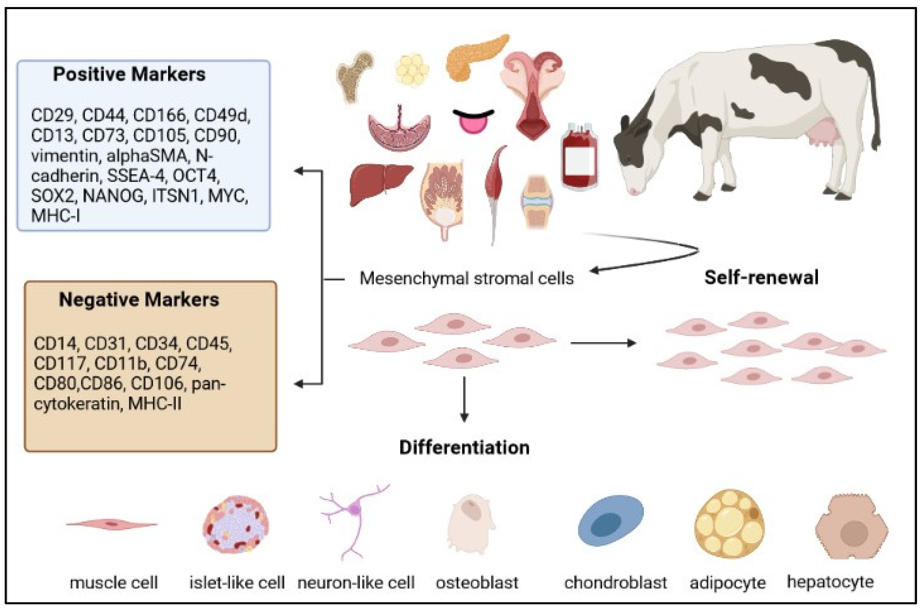
Bovine MCSs have been isolated and characterised (Figure 1) from different adult and foetal tissues, including bone marrow (BM) [3,4,5,6,7,8,9,10,11,12,13,14,15,16,17,18,19,20,21,22,23,24,25,26,27,28,29,30,31,32,33,34,35,36,37,38], endometrium (EN) [39,40,41,42,43,44,45,46,47,48], adipose tissue (AT) [29,30,31,32,34,37,49,50,51,52,53,54,55,56,57,58,59,60,61,62,63,64,65,66] and foetal liquid and adnexa, such as umbilical cord blood (UCB) [67,68,69,70], Wharton’s jelly (WJ) [58,71,72,73], umbilical cord matrix (UC) [74,75,76,77,78,79,80,81,82], amnion (AM) [83,84], amniotic fluid (AF) [57,83,85,86,87,88] and placenta (PL) [37,89,90]. Less common sources of bovine MSCs have been foetal liver [91], dermal tissue [92], foetal lung tissue [93], embryo yolk sack [94], synovial fluid [95], milk [96], pericardium membrane [97], pancreas [98], tongue epithelium [99], skeletal muscle [65,100] and peripheral blood [48,101].
The potential of MSCs for cell-based therapies has originally been based on their typical characteristics, which include the multipotentiality to differentiate in vitro into mesodermal-derived lineages, particularly osteogenic, chondrogenic and adipogenic cells [1]. Furthermore, it has been demonstrated that the paracrine activity of MSCs exerts therapeutical effects involving regeneration, immunomodulation, angiogenesis and antiapoptosis [102,103,104].
The immunomodulatory activity of MSCs depends on direct cell-to-cell contact and on contact-independent paracrine signalling, with the production of soluble factors regulating proliferation, differentiation, migration and apoptosis of several immune cells [105]. The reduced immunogenicity of MSCs is another aspect that strengthens their potential for cell therapy related in part to the low expression of major histocompatibility complexes I and II (MHC-I and II) and to the absence of expression of T-cell costimulatory molecules (CD40, CD80 and CD86) [106]. Taking together the immune regulatory abilities and reduced immunogenicity, allogeneic MSCs transplanted into recipients are able to escape direct recognition by natural killer cells and prevent activation of T lymphocytes, possibly also reducing the potential activation of the indirect pathway by the presentation of donor-derived MHC-I/II peptides by antigen-presenting cells to B cells and subsequent alloantibodies production [107]. Therefore, low immunogenicity may result in higher efficacy and lower risk of local inflammation following MSCs administration, reducing potential adverse effects [107]. In cattle, it has been demonstrated that foetal AT-MSCs and BM-MSCs respond to inflammatory stimulation with interferon γ (IFNγ) by increasing immune-related gene expression and activity in a dose-dependent manner and upregulating gene expression of IL-6 [30]. However, conditioned medium from IFNγ-stimulated and unstimulated BM-MSCs and AT-MSCs exerts similar suppression of proliferation of alloantigen-activated bovine peripheral blood lymphocytes [30]. Whereas immunomodulatory properties appear to be similar between BM-MSCs and AT-MSCs, higher expression of MHC-I and MHC-II in BM-MSCs suggested that the immunogenic potential of bovine foetal MSCs might be tissue-dependent and that AT-MSCs might be more suitable candidates for allogeneic therapy [30].
Autologous MSCs therapy implies cell isolation and expansion to achieve therapeutic doses. Consequently, there is a lag time between their collection and use, threatening the effectiveness of the treatment. In addition, critical parameters for MSCs isolation include donor variability, tissue of origin, amount of tissue and culture conditions [108]. On the other hand, foetal- and placental-derived MSCs have been found superior to adult MSCs as candidates for allogeneic therapeutic applications due to their lower immunogenicity [109,110]. Cryopreservation represents an efficient method for the preservation and pooling of MSCs to obtain the cell counts required for clinical applications. Samples can be harvested, and then cells can be isolated, expanded and stored for later use, optimising logistics from collection to transplantation. Accordingly, the ability of MSCs to survive long periods of storage and, at the same time, maintain their qualities is critical for the development of allogeneic cell therapies. Upon cryopreservation, it is important to preserve MSCs’ functional properties, including immunomodulatory properties and multilineage differentiation ability. Further, a biosafety evaluation of cryopreserved MSCs is essential prior to their clinical applications [111]. Considering cattle, Oyarzo et al. compared PL-MSCs and foetal MSCs originated from AT and BM in order to assess their ability to survive different cryoprotectant solutions exposure [37]. While the apoptotic potential was similar, foetal AT-MSCs and PL-MSCs presented consistently higher percentages of viability than did foetal BM-MSCs [37]. On the other hand, AT-MSCs were more resistant than PL-MSCs, but the latter have the advantage of coming from a readily available tissue usually considered waste, without ethical concerns [37].
Although in veterinary medicine, cell therapies are mainly focused on pets, regenerative medicine applications also involve farm animals, not only for their importance as a food source [112] but also as models [113]. Among livestock species, cows have a high economic impact, and reproductive biotechnologies are routinely applied [114,115]. The dairy and beef industries are essential for food production. Dairy products and ruminant meat provide essential elements for the human diet. According to the Food and Agriculture Organization (FAO), there are almost 1.5 billion cattle in the world. Cows produce 81 per cent of global milk production, and the world demand for beef is projected to increase to 75 million tonnes by 2030 [116]. Animal health is an important issue related not only to animal welfare itself but also to the One Health perspective, in which human, animal, plant and environmental health are interdependent. This review summarises the applications of MSCs in cattle to treat clinical conditions and improve reproductive biotechnologies.
2. Bovine MSCs for Clinical Treatments
So far, MSCs have been used in many experimental instances to treat various diseases in different animal species. Orthopaedic diseases were the primary field of regenerative veterinary medicine, and then the focus rapidly expanded to other areas. Dogs and horses were the species in which stem cell-based therapies were commonly used to treat different diseases of various organ systems, while for cats, they were used for renal, respiratory and inflammatory pathologies [117]. Bovine MSCs can be potentially used in various clinical conditions. Nevertheless, the application of novel MSCs therapies in large ruminants is still limited.
The major obstacles in livestock species are related to a minor interest in treating clinical conditions in these animals compared to pets and the higher maintenance costs in comparison with other animal models [118]. Laboratory animals or small animals are usually preferred as models to start any research for human pathologies due to the reasonable buying and care costs together with easier manageability and housing. However, for a better understanding and a thorough evaluation of cell-based therapies, various animal models are necessary to successfully move from the laboratory bench to human health applications. The development of products for animal use has the advantage that they can be immediately tested in the target species. This aspect not only allows to understand the potential of MSC-based products for clinical application in animals but may also provide models for similar human applications [119].
Although many studies have been published for animal MSCs, it is still not easy to evaluate the efficacy of MSC-based therapies because of the different sources of MSCs and variations in manufacturing processes, inconsistent characterisation and measure of potency, inappropriate controls and a lack of experimental power [119]. MSCs have been isolated from different sources and, depending on the tissue of origin, they may possess different properties, which should be taken into account when choosing the optimal stem cell therapy for a specific pathology in order to achieve successful results. On the other hand, there is no evidence for a favoured tissue as an MSCs source due to the presence of a wide variability between donors [108,120].
2.1. Chronic Wound Healing
In the last years, the application of regenerative medicine to skin lesions has been a focus for both human and veterinary medicine. The physiological healing process of cutaneous wounds is a well-orchestrated complex of molecular and biological activities. Even so, a chronic lesion can develop when the normal process fails. The regenerative potential of stromal cells has also been widely recognised for skin lesion repair [121]. Recent studies support the concept that MSCs can be appropriated for treating chronic wounds [122,123,124].
Even if the exact functions of stromal cells in wound healing have not yet been completely elucidated, they are involved in the removal of dead cells and necrotic tissue, angiogenesis, reduction in scar tissue formation, contraction of the wound and induction of re-epithelisation [121]. Consequently, wound healing is promoted, and local inflammation is reduced. Table 1 summarises the studies regarding MSC applications for wound healing in cattle.
The first report of a case study in which autologous BM-derived MSCs were used to treat a chronic ulcer in a heifer dates back to 2012 [10]. A 2-year-old Jersey heifer had been suffering from a chronic nonhealing ulcerative wound involving full-thickness skin and underlying muscle in the lumbar region for 4 months. Standard therapies were ineffective, so a clinical trial was made with autologous BM-MSCs. Bone marrow was aspirated from the tibia, and MSCs were isolated, expanded and then diluted in saline solution for intradermal and topical implantation in the wound. Various parameters and measures were monitored during the trial. At histopathology, the progression of the healing process was observed since neovascularisation appeared, as well as fibroblasts, sebaceous glands and epithelialisation. The content of collagen was increased after stem cell therapy, and the healed tissue was progressing towards physiological stretchability and tensile strength. The 4-month-old chronic wound healed within 18 days, indicating that MSCs application could be an effective therapeutic approach for nonhealing chronic wounds [10].
Another clinical study of the same research group concerns the successful treatment of an interdigital chronic ulcerative wound in a 6-year-old cross-bred Jersey cow [13]. The animal presented with a 4-month interdigital hoof lesion nonresponding to conventional treatments. Autologous BM-MSCs therapy was also used for this patient. Granulated tissue rapidly grew, and the healing process was completed in 18 days. The parameters analysed to assess the progression of the healing process confirmed the clinical process, and the pain-free walking distance evaluation was gradually increased over the study period [13].
In the last clinical trial [14], a bull was presenting a wound in a hind limb above the hock joint as a consequence of a car accident, which had happened 8 months before. Different local treatments and antibiotic courses turned out as unsuccessful as chemical and cryocauterisation. Autologous BM-MSCs application was performed. Similar protocols were used for the collection, isolation and expansion of BM-MSCs, but in this case, some cells were intravenously administered in addition to local treatment. Healing was completed within 4 weeks, and the evaluated parameters confirmed the outcome [14].
Despite the lack of controls and large-scale randomised studies and clinical trials, the promising results obtained from the applications of autologous BM-MSCs confirmed the potential of MSC-based therapy for treating chronic nonhealing wounds in bovines.
A weak immunogenicity and a vasculogenic effect are favourable properties for wound healing capacity. Bovine BM-MSCs are the most well-characterised cells, and recently their immunomodulatory properties [30] and proangiogenic potential [31] have been investigated. Comparing bovine foetal MSCs derived from bone marrow and adipose tissue, both upregulated the expression of immunomodulatory genes and showed similar in vitro immunomodulatory ability, while the lower expression of MHC-I and MHC-II suggested that AT-MSCs might be less immunogenic compared with BM-MSCs [30]. Furthermore, BM-MSCs displayed similar migratory ability, higher proliferative capacity and lower proangiogenic potential compared with AT-MSCs [31]. These results might suggest that bovine AT-MSCs could be even more promising than BM-MSCs in enhancing the treatment of chronic wound healing.
2.2. Mastitis
In the dairy industry, mastitis is a common problem, which implicates costs to treat the disease, and since antimicrobials are the standard therapy, this increases the possibility of developing antimicrobial resistance. Hence, alternative therapies are required.
The mammary gland contains stromal cells and precursors with high regenerative potential, which apparently are maintained during the productive life of dairy cows. The presence of such cells opens new research perspectives regarding the physiological mechanisms concerned with milk secretion and the possibility of enhancing or prolonging dairy cow production [125]. The presence of a subpopulation of adult stromal cells in the mammary gland was first demonstrated in human and mouse [126,127]. Then, in the cow, three different colony morphologies were isolated, suggesting the existence of different progenitor populations and of an epithelial cell hierarchy in the bovine mammary gland similar to humans [128]. Such stromal/progenitor cells have been largely investigated [125,129].
On the other hand, less research is available for MSCs and bovine mammary glands. As summarised in Table 2, different in vitro studies showed that UC-MSCs could promote milk protein and fat synthesis and the expression of key genes in bovine mammary gland epithelial cells via IGF-1 [75,76,78] and reduce their apoptosis rate [77]. Furthermore, it has been demonstrated that bovine MSCs have antibacterial activity [29]. The conditioned medium from bovine foetal MSCs obtained from bone marrow and adipose tissue showed in vitro antibacterial potential against S. aureus, a mastitis-causing pathogen, by reducing about 30% of relative bacterial growth [29]. The mechanisms that regulate the antibacterial activity of bovine MSCs have not been totally elucidated, but the expression of β–defensin 4 A and NK-lysine 1, two antibacterial peptides, was associated with the in vitro effect of such MSCs [29].
Dairy cows were experimentally infected to induce S. aureus clinical mastitis in order to evaluate the safety and efficacy of an allogenic MSC-based therapy [62]. Bovine foetal AT-MSCs were intramammary inoculated twice (days 1 and 10) during a 20-day experimental period. No clinical or immunological response was induced in healthy cows, and the bacterial count in milk was reduced in MSC-treated cows compared with controls [62]. A similar decrease in somatic cell count (SCC) in the milk of mastitic animals was observed in cows treated intramammary with a single administration of allogenic AT-MSCs during a 15-day experiment [64]. On days 3 and 7, maximum expression of anti-inflammatory cytokines (IL-6, IL-10), antimicrobial peptides (cathelicidin, lipocalin and cystatin) and angiogenic genes (angiopoietin) was observed [64]. With the aim of preventing subclinical mastitis, UCB-MSCs and extracellular vesicles (EVs) were injected locally and IV on days 0 and 7 in healthy (safety trial) and subclinical mastitis cows [68,69]. Both MSCs and EVs were safe, and all treated cows were cured permanently within 15 days [68]. Treated animals showed a reduced SCC in mastitic milk compared with the control (antibiotic) group, an enhancement in the expression of anti-inflammatory cytokines, antimicrobial peptides and angiogenic genes and a decrease in the expression of proinflammatory cytokines [68,69]. Finally, a conditioned medium from bovine AM-MSCs (2 h coincubation in phosphate-buffered saline (PBS)) was used to treat mastitis in comparison with conventional antibiotics [84]. Milk pH value and titratable acidity were similar between treatments, while the level of ionic calcium concentration decreased 3 days later in MSCs-treated cows compared with antibiotic-treated animals [84]. Moreover, the somatic cell number was similar in both groups, demonstrating that conditioned medium from bovine AM-MSCs has the therapeutic potential to treat bovine mastitis and might replace antibiotics in the future [84].
2.3. Reproductive System
In the last 50 years, the selection in the dairy industry has led to an improvement in average milk production by a single cow. However, the selection for milk yield has caused some unfavourable effects, such as a decrease in fertility. Despite an improvement in cow fertility in the last two decades, as a consequence of selection for fertility traits in breeding programmes and improvement in animal nutrition and comfort, reproductive performance is not optimal yet [130]. Reproductive disorders are directly correlated with low fertility in dairy cows.
The endometrium is characterised by an elevated and constant regeneration, and mesenchymal progenitor cells have also been identified in the cow endometrium [39]. Progenitor cells were isolated and characterised in cyclic cows [40,42,44] and heifers [41] and were able to respond after challenging with lipopolysaccharide (LPS) [43]. Furthermore, the presence of endometrial MSCs was also confirmed in the postpartum period in both healthy cows and those affected by endometritis [45]. In this period, uterine involution occurs, involving endometrial regeneration [131], and the presence of pathogenic bacteria needs to be controlled in the uterus for fertility restoration. However, pathogenic bacteria are not always rapidly eliminated and often generate uterine disease (metritis and endometritis), leading to reduced fertility [132]. Endometrial MSCs from bovine inflamed uteri showed modified characteristics, especially in clinical than in subclinical endometritis, and the in vitro exposure of endometrial MSCs to PGE2, a mediator of inflammation, modified their transcriptomic profile [45]. Bovine endometrial MSCs have also been immortalised from lines derived in different phases of the oestrous cycle [47]. Immortalised cells maintained mesenchymal and immunomodulatory characteristics, with an increased migratory capacity towards an inflammatory niche but a decreased answer to embryonic cytokine expression at implantation [47]. Interestingly, combined proinflammatory and implantation signals ensured the retention of endometrial MSCs in case of pregnancy, while they showed a mesenchymal to epithelial transition state in the absence of an embryo [47]. Despite research into bovine endometrial MSCs, no report exists about their application in treating cow uterine inflammations. On the other hand, bovine MSCs derived from adipose tissue showed an inhibitory effect on in vitro LPS challenge of endometrial epithelial cells [63]. When used in vivo to treat metritis, allogenic AT-MSCs did not induce any immunological rejection response in treated animals (IV, local, IV + local), and all cows were completely and permanently cured within 30 days after treatment [64]. Polymorphonuclear (PMN) cell count was reduced in cervical vaginal fluid and the expression of IL-6, IL-10, cathelicidin, lipocalin, cystatin and angiopoietin were observed at day 3 in the IV + local group [64]. More recently, UCB-MSCs and their EVs have also been successfully used for metritis treatment by the same research group [70]. Moreover, in this case, a higher decrease in PMN was observed for MSCs and EV-treated cows compared with antibiotic-treated ones, as well as an increase in the expression of anti-inflammatory cytokines [70].
Other pathologies, which can lead to considerable economic loss, are those involving the ovaries. Ovarian dysfunctions in dairy cattle have a high incidence and are responsible for a reduction in reproductive performance. The two major ovarian causes of infertility in dairy cows are inactive ovaries and ovarian cysts [133,134]. Chang et al. transplanted AF-MSCs into cows affected by bilateral ovarian dystrophy in an attempt to restore or improve ovarian function [87]. Each ovary was injected with 50 μL of PBS containing 0.58 million cells, and then cows were monitored for oestrus and inseminated [87]. Half (4/8) of the animals treated with AF-MSCs showed oestrus, and two of them delivered a calf, while no oestrus was observed in control animals, demonstrating that MSCs therapy is a potentially useful treatment to alleviate the impact of ovarian dystrophy in dairy cows [87]. Peng et al. injected PL-MSCs into ovarian cysts with or without fluid drainage and compared them to control animals and GnRH-treated animals [90]. The use of PL-MSCs allowed for recovery and conception [90], indicating a new therapeutic potential of these cells and a possible alternative to hormones in the treatment of cattle ovarian cysts. Finally, the intraovarian injection of MSCs was used to reduce the negative effects of repeated ovum pick-up (OPU) under acute and chronic scenarios in bovines [61]. In fact, this technique is generally considered a safe way to collect oocytes from live donors but inevitably causes trauma to the ovarian tissue, and repeated procedures over years are associated with a progressive decrease in oocyte yield [61]. For the experiment, one ovary was injected with 2.5 million AT-MSCs, and the other one was used as the control [61]. MSCs had beneficial effects on the fertility of acute OPU injured cows, but not in cows with chronic ovarian lesions [61]. In this case, it was speculated that MSCs could no longer restore the compromised follicular population or ovarian physiology in cows with chronic inflammatory processes in the ovaries due to repeated OPU over time [61]. The overall MSC clinical applications for the reproductive system are presented in Table 3.
3. Bovine MSCs for Reproductive Biotechnologies
The first successful nuclear transfer (NT) dates back to 1952, when the nucleus from an early tadpole embryo was transferred into an enucleated frog egg [135]. Then, in 1996, Dolly was the first mammalian cloned using an adult somatic cell as a nucleus donor [136]. Somatic cell nuclear transfer (SCNT) (Figure 2) is an important research tool since it permits a differentiated cell to be reprogrammed to a totipotent state [137]. The donor cell is a key factor in the process, and interest in bovine SCNT led to consider MSCs as appropriate candidates due to their characteristics. Studies using bovine MSCs from different sources for NT were carried out and are summarised in Table 4.
Firstly, it was demonstrated that bovine BM-MSCs had developmental totipotency after NT [3] and were better than adult fibroblasts in driving the preimplantation development of cloned embryos efficiently [5]. In another study investigating the epigenetic status of donor cells to improve SCNT [52], it was demonstrated that bovine AT-MSCs at passage 5 had the highest level of multipotency and the lowest level of chromatin compaction. Bovine AF and AT-MSCSs were then successfully used to produce embryos and calves after NT [53], and in vitro development of bovine embryos cloned using less methylated AF and AT-MSCS was improved using trichostatin A [57]. Pregnancies were also obtained after the transfer of blastocysts derived from WJ-MSCs NT [72]. A higher potential for AM and AT-MSCs than adult fibroblasts was observed in terms of blastocysts obtained after oocyte reconstruction [138]. More recently, epigenetic reprogramming events were investigated, and it was observed that the SCNT embryos derived from bovine AT-MSCs endured considerable nuclear reprogramming during early embryo development [56]. Finally, in an attempt to improve NT efficiency, the aggregation of two AT-MSC-derived embryos seemed to positively affect embryo quality, which may improve postimplantation development [66].
Another context of research into cells includes their ability to incorporate exogenous DNA for the production of transgenic animals. Bovine MSCs were transfected with pBC1-anti-CD3 vector, and while those derived from WJ were more sensitive to treatments, AT-MSCs showed a better response to transfection [58].
Bovine MSCs have also been used for in vitro embryo production. The traditional coculture system of bovine embryos with granulosa cells was less efficient than coculture with AT-MSCs [54]. In addition to increasing blastocyst rates, MSCs coculture also improved embryo quality, with an increase in total cell numbers and mRNA expression levels for POU5F1 and G6PDH [54]. It was speculated that the paracrine capacity of MSCs could be responsible for the positive effects observed [54].
Another application of MSCs is to produce germinal cells after differentiation. The in vitro production of germ cell lineages is a new intriguing strategy for obtaining gametes in order to treat infertility, disseminate the genetics of elite animals and preserve endangered species [139]. The in vitro effect of bone morphogenetic protein 4, transforming growth factor β1 and retinoic acid on the potential for germ cell differentiation of bovine foetal BM-MSCs was investigated [27]. The stimulated cells expressed pluripotent markers OCT4, NANOG and male germ cell gene DAZL, demonstrating their potential for early germ cell differentiation [27]. When coculturing bovine foetal BM and AT-MSCS with Sertoli cells, cell morphology modifications were induced, as well as variations in the expression profiles of mesenchymal, pluripotent and germ cell genes, suggesting progression of AT-MSC into early stages of germ cell differentiation and advancement of BM-MSCs into the multipotent state [34].
4. Conclusions
The development of stem cell technologies in species other than bovine can be seen as a useful background for developing and deepening similar advancements in livestock. MSC characteristics make them appealing for their potential in clinical applications, and the lack of ethical concern is the other factor that makes them ideal for laboratory studies. As for humans [140], for successful cell-based therapies, stem cells must be able to differentiate into specific targeting cells or must act via paracrine mechanisms. Their extraction and isolation must be feasible, and transplantation must be effective and safe. Furthermore, ex vivo cell expansion is required since a considerable number of cells is essential to optimise the therapeutic effects. However, the lifespan of MSCs is limited during in vitro culture, and their senescence is a limit from the viewpoint of clinical applications. On the one hand, the limited cell proliferation potency protects them from malignant transformation after transplantation; on the other, senescence can alter various cell functions essential for therapeutic efficacy, such as proliferation, differentiation and migration. Therefore, after in vitro expansion and before therapeutic use, it should be considered whether these cells still possess stemness properties.
The bovine model could be advantageous for the size and physiology when compared with traditional laboratory animals. In cattle, MSCs have been isolated from different tissues, and their pluripotency has been demonstrated, but there is still a lack of clinical applications and studies comparing MSCs from different sources to suggest which one is the best choice for cell therapy or for which specific pathology. The studies presented are promising for the possible applications of MSCs both in veterinary medicine and the livestock industry. However, more studies are required to develop bovine-specific protocols, and further investigation is needed to evaluate clinical responses after cell therapy applications. Attitudes in the livestock industry have shifted towards the preservation of the commercial viability of individual animals with high genetic value, leading, in turn, to an increase in medical expenditure to keep those animals healthy [141]. MSCs treatment has the potential to reduce animal recovery time and reduce economic loss associated with bone and joint injury, reducing the time for repair that can negatively influence milk and meat production and interfere with natural breeding [141]. Nevertheless, orthopaedic applications have not yet been applied clinically in cows. The antimicrobial activity of MSCs and their derivatives has great potential for the treatment of conditions such as mastitis. In addition to the direct impact on milk production in the dairy industry, it would provide an alternative to the use of antimicrobials, reducing the possibility of antimicrobial resistance and the presence of antibiotics in milk. MSCs treatment has the potential to decrease recovery from various diseases affecting production, thus increasing profitability.
Author Contributions
Conceptualisation, B.M.; writing—original draft preparation, B.M.; writing—review and editing, B.M., P.M.G. and E.I. All authors have read and agreed to the published version of the manuscript.
Funding
This review received no external funding.
Institutional Review Board Statement
Not applicable.
Informed Consent Statement
Not applicable.
Data Availability Statement
No new data were created or analysed in this study. Data sharing is not applicable to this article.
Conflicts of Interest
The authors declare no conflict of interest.
Abbreviations
| AF | Amniotic fluid | |
| AM | Amnion | |
| AT | Adipose tissue | |
| BM | Bone marrow | |
| EN | Endometrium | |
| EVs | Extracellular vesicles | |
| GnRH | Gonadotropin-releasing hormone | |
| IGF-1 | Insulin-like growth factor 1 | |
| IL-6 | Interleukin 6 | |
| IL-10 | Interleukin 10 | |
| INFγ | Interferon γ | |
| IV | Intravenous | |
| LPS | Lipopolysaccharide | |
| MHC-I | Major histocompatibility complex-I | |
| MHC-II | Major histocompatibility complex-II | |
| MSCs | Mesenchymal stromal cells | |
| NT | Nuclear transfer | |
| OPU | Ovum pick-up | |
| PBS | Phosphate-buffered saline | |
| PGE2 | Prostaglandin E2 | |
| PL | Placenta | |
| PMN | Polymorphonuclear | |
| SCC | Somatic cell count | |
| SCNT | Somatic cell nuclear transfer | |
| UC | Umbilical cord | |
| UCB | Umbilical cord blood | |
| WJ | Wharton’s jelly | |
References
- Dominici, M.; Le Blanc, K.; Mueller, I.; Slaper-Cortenbach, I.; Marini, F.C.; Krause, D.S.; Deans, R.J.; Keating, A.; Prockop, D.J.; Horwitz, E.M. Minimal Criteria for Defining Multipotent Mesenchymal Stromal Cells. The International Society for Cellular Therapy Position Statement. Cytotherapy 2006, 8, 315–317. [Google Scholar] [CrossRef]
- Stewart, M.C.; Stewart, A.A. Mesenchymal Stem Cells: Characteristics, Sources, and Mechanisms of Action. Vet. Clin. Equine Pract. 2011, 27, 243–261. [Google Scholar] [CrossRef] [PubMed]
- Kato, Y.; Imabayashi, H.; Mori, T.; Tani, T.; Taniguchi, M.; Higashi, M.; Matsumoto, M.; Umezawa, A.; Tsunoda, Y. Nuclear Transfer of Adult Bone Marrow Mesenchymal Stem Cells: Developmental Totipotency of Tissue-Specific Stem Cells from an Adult Mammal1. Biol. Reprod. 2004, 70, 415–418. [Google Scholar] [CrossRef] [Green Version]
- Bosnakovski, D.; Mizuno, M.; Kim, G.; Takagi, S.; Okumura, M.; Fujinaga, T. Isolation and Multilineage Differentiation of Bovine Bone Marrow Mesenchymal Stem Cells. Cell Tissue Res. 2005, 319, 243–253. [Google Scholar] [CrossRef] [PubMed]
- Colleoni, S.; Donofrio, G.; Lagutina, I.; Duchi, R.; Galli, C.; Lazzari, G. Establishment, Differentiation, Electroporation, Viral Transduction, and Nuclear Transfer of Bovine and Porcine Mesenchymal Stem Cells. Cloning Stem Cells 2005, 7, 154–166. [Google Scholar] [CrossRef] [PubMed] [Green Version]
- Donofrio, G.; Colleoni, S.; Galli, C.; Lazzari, G.; Cavirani, S.; Flammini, C.F. Susceptibility of Bovine Mesenchymal Stem Cells to Bovine Herpesvirus 4. J. Virol. Methods 2005, 127, 168–170. [Google Scholar] [CrossRef] [PubMed]
- Bosnakovski, D.; Mizuno, M.; Kim, G.; Takagi, S.; Okumur, M.; Fujinag, T. Gene Expression Profile of Bovine Bone Marrow Mesenchymal Stem Cell during Spontaneous Chondrogenic Differentiation in Pellet Culture System. Jpn. J. Vet. Res. 2006, 53, 127–139. [Google Scholar] [PubMed]
- Mauck, R.L.; Yuan, X.; Tuan, R.S. Chondrogenic Differentiation and Functional Maturation of Bovine Mesenchymal Stem Cells in Long-Term Agarose Culture. Osteoarthr. Cartil. 2006, 14, 179–189. [Google Scholar] [CrossRef] [Green Version]
- Erickson, I.E.; van Veen, S.C.; Sengupta, S.; Kestle, S.R.; Mauck, R.L. Cartilage Matrix Formation by Bovine Mesenchymal Stem Cells in Three-Dimensional Culture Is Age-Dependent. Clin. Orthop. 2011, 469, 2744–2753. [Google Scholar] [CrossRef] [Green Version]
- Das, J.; Nath, I.; Das, R.K.; Routray, P.; Behera, S.S. Autologous Stem Cell Therapy to Treat Chronic Ulcer in Heifer-A Case Study. Available online: https://scholar.google.com/citations?view_op=view_citation&hl=en&user=V8kdet8AAAAJ&citation_for_view=V8kdet8AAAAJ:UeHWp8X0CEIC (accessed on 15 July 2021).
- Cortes, Y.; Ojeda, M.; Araya, D.; Dueñas, F.; Fernández, M.S.; Peralta, O.A. Isolation and Multilineage Differentiation of Bone Marrow Mesenchymal Stem Cells from Abattoir-Derived Bovine Fetuses. BMC Vet. Res. 2013, 9, 133. [Google Scholar] [CrossRef] [Green Version]
- Randau, T.M.; Schildberg, F.A.; Alini, M.; Wimmer, M.D.; Haddouti, E.-M.; Gravius, S.; Ito, K.; Stoddart, M.J. The Effect of Dexamethasone and Triiodothyronine on Terminal Differentiation of Primary Bovine Chondrocytes and Chondrogenically Differentiated Mesenchymal Stem Cells. PLoS ONE 2013, 8, e72973. [Google Scholar] [CrossRef] [PubMed] [Green Version]
- Das, J.; Behera, S.S. Successful treatment of inter digital chronic ulcerative wound by mesenchymal stem cell therapy in a cow. Indian J. Vet. Sci. Biotechnol. 2013, 9, 66–67. [Google Scholar]
- Das, J.; Routray, N.P.; Das, R.K. Autologous Bone Marrow Derived Mesenchymal Stem Cells (BM-MSCs) Therapy for Treatment of Chronic Limb Wound in a Bull. Indian J. Available online: https://www.indianjournals.com/ijor.aspx?target=ijor:ipo&volume=14&issue=1&article=023 (accessed on 9 December 2021).
- Dueñas, F.; Becerra, V.; Cortes, Y.; Vidal, S.; Sáenz, L.; Palomino, J.; De los Reyes, M.; Peralta, O.A. Hepatogenic and Neurogenic Differentiation of Bone Marrow Mesenchymal Stem Cells from Abattoir-Derived Bovine Fetuses. BMC Vet. Res. 2014, 10, 154. [Google Scholar] [CrossRef] [Green Version]
- Jeong, J.Y.; Suresh, S.; Park, M.N.; Jang, M.; Park, S.; Gobianand, K.; You, S.; Yeon, S.-H.; Lee, H.-J. Effects of Capsaicin on Adipogenic Differentiation in Bovine Bone Marrow Mesenchymal Stem Cell. Asian-Australas. J. Anim. Sci. 2014, 27, 1783–1793. [Google Scholar] [CrossRef] [PubMed] [Green Version]
- Lu, T.; Huang, Y.; Wang, H.; Ma, Y.; Guan, W. Multi-Lineage Potential Research of Bone Marrow-Derived Stromal Cells (BMSCs) from Cattle. Appl. Biochem. Biotechnol. 2014, 172, 21–35. [Google Scholar] [CrossRef] [PubMed]
- Abou-Easa, K.F.; Ghazy, A.E.; Gewaily, M.S. Characterization of Behavior and Niche of Bovine Marrow Stem Cells In Vitro. Glob. Vet. 2015, 14, 922–928. [Google Scholar]
- Díaz, P.; Cuevas, F.; Peralta, O.A. GFP Labelling and Epigenetic Enzyme Expression of Bone Marrow-Derived Mesenchymal Stem Cells from Bovine Foetuses. Res. Vet. Sci. 2015, 99, 120–128. [Google Scholar] [CrossRef] [PubMed]
- Jeong, J.Y.; Park, M.N.; Cho, E.S.; Jang, H.-J.; Park, S.; Lee, H.-J. Epigallocatechin-3-Gallate-Induced Free-Radical Production upon Adipogenic Differentiation in Bovine Bone-Marrow Mesenchymal Stem Cells. Cell Tissue Res. 2015, 362, 87–96. [Google Scholar] [CrossRef]
- Jeong, J.Y.; Suresh, S.; Jang, M.; Park, M.N.; Gobianand, K.; You, S.; Yeon, S.; Lee, H. Epigallocatechin-3-gallate Suppresses the Lipid Deposition through the Apoptosis during Differentiation in Bovine Bone Marrow Mesenchymal Stem Cells. Cell Biol. Int. 2015, 39, 52–64. [Google Scholar] [CrossRef] [Green Version]
- Fisher, M.B.; Henning, E.A.; Söegaard, N.; Bostrom, M.; Esterhai, J.L.; Mauck, R.L. Engineering Meniscus Structure and Function via Multi-Layered Mesenchymal Stem Cell-Seeded Nanofibrous Scaffolds. J. Biomech. 2015, 48, 1412–1419. [Google Scholar] [CrossRef] [Green Version]
- Goldman, S.M.; Barabino, G.A. Hydrodynamic Loading in Concomitance with Exogenous Cytokine Stimulation Modulates Differentiation of Bovine Mesenchymal Stem Cells towards Osteochondral Lineages. BMC Biotechnol. 2016, 16, 10. [Google Scholar] [CrossRef] [Green Version]
- Ramírez-Espinosa, J.J.; González-Dávalos, L.; Shimada, A.; Piña, E.; Varela-Echavarria, A.; Mora, O. Bovine (Bos Taurus) Bone Marrow Mesenchymal Cell Differentiation to Adipogenic and Myogenic Lineages. Cells Tissues Organs 2016, 201, 51–64. [Google Scholar] [CrossRef]
- McCorry, M.C.; Bonassar, L.J. Fiber Development and Matrix Production in Tissue-Engineered Menisci Using Bovine Mesenchymal Stem Cells and Fibrochondrocytes. Connect. Tissue Res. 2017, 58, 329–341. [Google Scholar] [CrossRef] [Green Version]
- Okamura, L.H.; Cordero, P.; Palomino, J.; Parraguez, V.H.; Torres, C.G.; Peralta, O.A. Myogenic Differentiation Potential of Mesenchymal Stem Cells Derived from Fetal Bovine Bone Marrow. Anim. Biotechnol. 2018, 29, 1–11. [Google Scholar] [CrossRef] [PubMed]
- Cortez, J.; Bahamonde, J.; los Reyes, M.D.; Palomino, J.; Torres, C.G.; Peralta, O.A. In Vitro Differentiation of Bovine Bone Marrow-Derived Mesenchymal Stem Cells into Male Germ Cells by Exposure to Exogenous Bioactive Factors. Reprod. Domest. Anim. 2018, 53, 700–709. [Google Scholar] [CrossRef] [PubMed]
- Zhao, X.-X.; An, X.-L.; Zhu, X.-C.; Jiang, Y.; Zhai, Y.-H.; Zhang, S.; Cai, N.-N.; Tang, B.; Li, Z.-Y.; Zhang, X.-M. Inhibiting Transforming Growth Factor-β Signaling Regulates in Vitro Maintenance and Differentiation of Bovine Bone Marrow Mesenchymal Stem Cells. J. Exp. Zoolog. B Mol. Dev. Evol. 2018, 330, 406–416. [Google Scholar] [CrossRef] [PubMed]
- Cahuascanco, B.; Bahamonde, J.; Huaman, O.; Jervis, M.; Cortez, J.; Palomino, J.; Escobar, A.; Retamal, P.; Torres, C.G.; Peralta, O.A. Bovine Fetal Mesenchymal Stem Cells Exert Antiproliferative Effect against Mastitis Causing Pathogen Staphylococcus Aureus. Vet. Res. 2019, 50, 25. [Google Scholar] [CrossRef] [Green Version]
- Huaman, O.; Bahamonde, J.; Cahuascanco, B.; Jervis, M.; Palomino, J.; Torres, C.G.; Peralta, O.A. Immunomodulatory and Immunogenic Properties of Mesenchymal Stem Cells Derived from Bovine Fetal Bone Marrow and Adipose Tissue. Res. Vet. Sci. 2019, 124, 212–222. [Google Scholar] [CrossRef]
- Jervis, M.; Huaman, O.; Cahuascanco, B.; Bahamonde, J.; Cortez, J.; Arias, J.I.; Torres, C.G.; Peralta, O.A. Comparative Analysis of In Vitro Proliferative, Migratory and pro-Angiogenic Potentials of Bovine Fetal Mesenchymal Stem Cells Derived from Bone Marrow and Adipose Tissue. Vet. Res. Commun. 2019, 43, 165–178. [Google Scholar] [CrossRef]
- Korovina, D.G. The Use of Bovine Multipotent Mesenchymal Stem Cells Isolated from Bone Marrow and Adipose Tissue as Sources to Obtain Muscle Cells in Vitro. IOP Conf. Ser. Earth Environ. Sci. 2019, 315, 042040. [Google Scholar] [CrossRef]
- Pan, S.; Chen, Y.-C.; Zhao, N.; Feng, X.; Yang, D.-D.; Wang, Y.; Jin, Z.-B. A New Subset of Small Stem Cells in Bovine Bone Marrow Stromal Cell Populations. J. Cell. Biochem. 2019, 120, 13881–13892. [Google Scholar] [CrossRef] [PubMed]
- Segunda, M.N.; Bahamonde, J.; Muñoz, I.; Sepulveda, S.; Cortez, J.; De los Reyes, M.; Palomino, J.; Torres, C.G.; Peralta, O.A. Sertoli Cell-Mediated Differentiation of Bovine Fetal Mesenchymal Stem Cells into Germ Cell Lineage Using an in Vitro Co-Culture System. Theriogenology 2019, 130, 8–18. [Google Scholar] [CrossRef] [PubMed]
- Shi, M.; Li, Z.; Miao, Z.; Guo, Y.; Yi, L. Interleukin-15 Inhibits Adipogenic Differentiation of Cattle Bone Marrow-Derived Mesenchymal Stem Cells via Regulating the Crosstalk between Signal Transducer and Activator of Transcription 5A and Akt Signalling. Biochem. Biophys. Res. Commun. 2019, 517, 346–352. [Google Scholar] [CrossRef] [PubMed]
- Chen, J.; Chen, L.; Hua, J.; Song, W. Long-Term Dynamic Compression Enhancement TGF-Β3-Induced Chondrogenesis in Bovine Stem Cells: A Gene Expression Analysis. BMC Genom. Data 2021, 22, 13. [Google Scholar] [CrossRef] [PubMed]
- Oyarzo, R.; Valderrama, X.; Valenzuela, F.; Bahamonde, J. Bovine Fetal Mesenchymal Stem Cells Obtained From Omental Adipose Tissue and Placenta Are More Resistant to Cryoprotectant Exposure Than Those From Bone Marrow. Front. Vet. Sci. 2021, 8, 708972. [Google Scholar] [CrossRef]
- Abo-Aziza, F.A.M.; Zaki, A.K.A.; Alajaji, A.I.; Al barrak, S.M. Bone Marrow Mesenchymal Stem Cell Co-Adjuvant Therapy with Albendazole for Managing Toxocara Vitulorum-Rat Model. Vet. World 2021, 14, 347–363. [Google Scholar] [CrossRef] [PubMed]
- Donofrio, G.; Franceschi, V.; Capocefalo, A.; Cavirani, S.; Sheldon, I.M. Bovine Endometrial Stromal Cells Display Osteogenic Properties. Reprod. Biol. Endocrinol. 2008, 6, 65. [Google Scholar] [CrossRef] [Green Version]
- Cabezas, J.; Lara, E.; Pacha, P.; Rojas, D.; Veraguas, D.; Saravia, F.; Rodríguez-Alvarez, L.; Castro, F.O. The Endometrium of Cycling Cows Contains Populations of Putative Mesenchymal Progenitor Cells. Reprod. Domest. Anim. 2014, 49, 550–559. [Google Scholar] [CrossRef] [PubMed]
- Mehrabani, D.; Rahmanifar, F.; Mellinejad, M.; Tamadon, A.; Dianatpour, M.; Zare, S.; Jahromi, I.R.; Ghobadi, F. Isolation, Culture, Characterization, and Adipogenic Differentiation of Heifer Endometrial Mesenchymal Stem Cells. Comp. Clin. Pathol. 2015, 24, 1159–1164. [Google Scholar] [CrossRef]
- de Moraes, C.N.; Maia, L.; Dias, M.C.; Dell’Aqua, C.P.F.; da Mota, L.S.L.S.; Chapwanya, A.; da C. Landim-Alvarenga, F.; Oba, E. Bovine Endometrial Cells: A Source of Mesenchymal Stem/Progenitor Cells. Cell Biol. Int. 2016, 40, 1332–1339. [Google Scholar] [CrossRef]
- de Moraes, C.N.; Maia, L.; de Oliveira, E.; de Paula Freitas Dell’Aqua, C.; Chapwanya, A.; da Cruz Landim-Alvarenga, F.; Oba, E. Shotgun Proteomic Analysis of the Secretome of Bovine Endometrial Mesenchymal Progenitor/Stem Cells Challenged or Not with Bacterial Lipopolysaccharide. Vet. Immunol. Immunopathol. 2017, 187, 42–47. [Google Scholar] [CrossRef] [PubMed] [Green Version]
- Lara, E.; Rivera, N.; Rojas, D.; Rodríguez-Alvarez, L.L.; Castro, F.O. Characterization of Mesenchymal Stem Cells in Bovine Endometrium during Follicular Phase of Oestrous Cycle. Reprod. Domest. Anim. 2017, 52, 707–714. [Google Scholar] [CrossRef] [PubMed]
- Lara, E.; Velásquez, A.; Cabezas, J.; Rivera, N.; Pacha, P.; Rodríguez-Alvarez, L.; Saravia, F.; Castro, F.O. Endometritis and In Vitro PGE2 Challenge Modify Properties of Cattle Endometrial Mesenchymal Stem Cells and Their Transcriptomic Profile. Stem Cells Int. 2017, 2017, 4297639. [Google Scholar] [CrossRef] [Green Version]
- Lara, E.; Rivera, N.; Cabezas, J.; Navarrete, F.; Saravia, F.; Rodríguez-Alvarez, L.; Castro, F. Endometrial Stem Cells in Farm Animals: Potential Role in Uterine Physiology and Pathology. Bioengineering 2018, 5, 75. [Google Scholar] [CrossRef] [Green Version]
- Calle, A.; López-Martín, S.; Monguió-Tortajada, M.; Borràs, F.E.; Yáñez-Mó, M.; Ramírez, M.Á. Bovine Endometrial MSC: Mesenchymal to Epithelial Transition during Luteolysis and Tropism to Implantation Niche for Immunomodulation. Stem Cell Res. Ther. 2019, 10, 23. [Google Scholar] [CrossRef]
- Calle, A.; Toribio, V.; Yáñez-Mó, M.; Ramírez, M.Á. Embryonic Trophectoderm Secretomics Reveals Chemotactic Migration and Intercellular Communication of Endometrial and Circulating MSCs in Embryonic Implantation. Int. J. Mol. Sci. 2021, 22, 5638. [Google Scholar] [CrossRef]
- Ren, Y.; Wu, H.; Ma, Y.; Cang, M.; Wang, R.; Liu, D. Isolation, cultivation and identification of adipose-derived stem cell in bovines. Sheng Wu Gong Cheng Xue Bao Chin. J. Biotechnol. 2010, 26, 1645–1651. [Google Scholar]
- Lu, T.; Xiong, H.; Wang, K.; Wang, S.; Ma, Y.; Guan, W. Isolation and Characterization of Adipose-Derived Mesenchymal Stem Cells (ADSCs) from Cattle. Appl. Biochem. Biotechnol. 2014, 174, 719–728. [Google Scholar] [CrossRef] [PubMed]
- Sampaio, R.V.; Chiaratti, M.R.; Santos, D.C.N.; Bressan, F.F.; Sangalli, J.R.; Sá, A.L.A.; Silva, T.V.G.; Costa, N.N.; Cordeiro, M.S.; Santos, S.S.D.; et al. Generation of Bovine (Bos indicus) and Buffalo (Bubalus bubalis) Adipose Tissue Derived Stem Cells: Isolation, Characterization, and Multipotentiality. Available online: https://www.geneticsmr.com/articles/3881 (accessed on 15 July 2021).
- Abouhamzeh, B.; Salehi, M. DNA Methylation and Histone Acetylation Patterns in Cultured Bovine Adipose Tissue-Derived Stem Cells (BADSCs). CELL J. 2015, 16, 10. [Google Scholar]
- da Silva, C.G.; Martins, C.F.; Cardoso, T.C.; da Cunha, E.R.; Bessler, H.C.; Martins, G.H.L.; Pivato, I.; Báo, S.N. Production of Bovine Embryos and Calves Cloned by Nuclear Transfer Using Mesenchymal Stem Cells from Amniotic Fluid and Adipose Tissue. Cell Reprogram. 2016, 18, 127–136. [Google Scholar] [CrossRef] [Green Version]
- Miranda, M.S.; Nascimento, H.S.; Costa, M.P.R.; Costa, N.N.; Brito, K.N.L.; Lopes, C.T.A.; Santos, S.S.D.; Cordeiro, M.S.; Ohashi, O.M. Increasing of Blastocyst Rate and Gene Expression in Co-Culture of Bovine Embryos with Adult Adipose Tissue-Derived Mesenchymal Stem Cells. J. Assist. Reprod. Genet. 2016, 33, 1395–1403. [Google Scholar] [CrossRef] [PubMed] [Green Version]
- Yue, Y.; Zhang, L.; Zhang, X.; Li, X.; Yu, H. De Novo Lipogenesis and Desaturation of Fatty Acids during Adipogenesis in Bovine Adipose-Derived Mesenchymal Stem Cells. Vitro Cell. Dev. Biol. Anim. 2018, 54, 23–31. [Google Scholar] [CrossRef]
- Salehi, M.; Abouhamzeh, B.; Hosseini, A.; Zare, Z.; Bakhtari, A. Comparison of Epigenetic Modifier Genes in Bovine Adipose Tissue-Derived Stem Cell Based Embryos, as Donors, with In Vitro and Parthenogenesis Embryos. Cell J. 2019, 22, 149–157. [Google Scholar] [CrossRef] [PubMed]
- da Silva, C.G.; Martins, C.F.; Bessler, H.C.; da Fonseca Neto, Á.M.; Cardoso, T.C.; Franco, M.M.; dos S. Mendonça, A.; de O. Leme, L.; Borges, J.R.J.; Malaquias, J.V.; et al. Use of Trichostatin A Alters the Expression of HDAC3 and KAT2 and Improves in Vitro Development of Bovine Embryos Cloned Using Less Methylated Mesenchymal Stem Cells. Reprod. Domest. Anim. 2019, 54, 289–299. [Google Scholar] [CrossRef] [PubMed]
- Duarte, F.B.; Brigido, M.D.M.; de O. Melo, E.; Báo, S.N.; Martins, C.F. Strategies for Transfection of Bovine Mesenchymal Stem Cells with PBC1-Anti-CD3 Vector. Anim. Biotechnol. 2020, 30, 1–11. [Google Scholar] [CrossRef]
- HuiNa, L.; QuanBao, F.; WenKang, J.; MingMing, Z.; JingJing, W.; DongZhang, L.; BingYun, W. Isolation, culture and biological characteristics of dairy cow adipose-derived mesenchymal stem cells. J. South. Agric. 2020, 51, 1462–1469. [Google Scholar]
- Jurek, S.; Sandhu, M.A.; Trappe, S.; Bermúdez-Peña, M.C.; Kolisek, M.; Sponder, G.; Aschenbach, J.R. Optimizing Adipogenic Transdifferentiation of Bovine Mesenchymal Stem Cells: A Prominent Role of Ascorbic Acid in FABP4 Induction. Adipocyte 2020, 9, 35–50. [Google Scholar] [CrossRef] [Green Version]
- Malard, P.F.; Peixer, M.A.S.; Grazia, J.G.; Brunel, H.D.S.S.; Feres, L.F.; Villarroel, C.L.; Siqueira, L.G.B.; Dode, M.A.N.; Pogue, R.; Viana, J.H.M.; et al. Intraovarian Injection of Mesenchymal Stem Cells Improves Oocyte Yield and in Vitro Embryo Production in a Bovine Model of Fertility Loss. Sci. Rep. 2020, 10, 8018. [Google Scholar] [CrossRef]
- Peralta, O.A.; Carrasco, C.; Vieytes, C.; Tamayo, M.J.; Muñoz, I.; Sepulveda, S.; Tadich, T.; Duchens, M.; Melendez, P.; Mella, A.; et al. Safety and Efficacy of a Mesenchymal Stem Cell Intramammary Therapy in Dairy Cows with Experimentally Induced Staphylococcus Aureus Clinical Mastitis. Sci. Rep. 2020, 10, 2843. [Google Scholar] [CrossRef] [PubMed] [Green Version]
- Lu, W.; Xu, Z.-M.; Liu, Q.; Yu, N.-N.; Yu, J.-B.; Li, W.-L.; Mao, Y.-Y.; Du, Z.; Si, L.; Yuan, S.; et al. Inhibitory Effect of Bovine Adipose-Derived Mesenchymal Stem Cells on Lipopolysaccharide Induced Inflammation of Endometrial Epithelial Cells in Dairy Cows. Front. Vet. Sci. 2021, 8, 726328. [Google Scholar] [CrossRef]
- Singh, R.; Bhaskar, V.; Saini, S.; Kumar, A.; Thakur, A.; Kumar, S.; Malakar, D.; Singh, R.; Bhaskar, V.; Saini, S.; et al. 143 Therapeutic Efficacy and Safety of Adipose Tissue-Derived Mesenchymal Stem Cells in Treating Mastitis and Metritis in Dairy Cattle. Reprod. Fertil. Dev. 2021, 33, 179. [Google Scholar] [CrossRef]
- Naraoka, Y.; Mabuchi, Y.; Yoneyama, Y.; Suto, E.G.; Hisamatsu, D.; Ikeda, M.; Ito, R.; Nakamura, T.; Takebe, T.; Akazawa, C. Isolation and Characterization of Tissue Resident CD29-Positive Progenitor Cells in Livestock to Generate a Three-Dimensional Meat Bud. Cells 2021, 10, 2499. [Google Scholar] [CrossRef]
- Savy, V.; Alberio, V.; Vans Landschoot, G.; Moro, L.N.; Olea, F.D.; Rodríguez-Álvarez, L.; Salamone, D.F. Effect of Embryo Aggregation on In Vitro Development of Adipose-Derived Mesenchymal Stem Cell-Derived Bovine Clones. Cell Reprogram. 2021, 23, 277–289. [Google Scholar] [CrossRef] [PubMed]
- Raoufi, M.F.; Tajik, P.; Dehghan, M.M.; Eini, F.; Barin, A. Isolation and Differentiation of Mesenchymal Stem Cells From Bovine Umbilical Cord Blood. Reprod. Domest. Anim. 2011, 46, 95–99. [Google Scholar] [CrossRef] [PubMed]
- Ghai, S.; Verma, V.N.; Ansari, S.; Saini, S.; Thakur, A.; Kumar, A.; Kumar, S.; Malakar, D.; Ghai, S.; Verma, V.N.; et al. 157 Mesenchymal Stem Cells as a Regenerative Therapy for the Prevention of Subclinical Mastitis in Cattle. Reprod. Fertil. Dev. 2021, 34, 316–317. [Google Scholar] [CrossRef] [PubMed]
- Ghai, S.; Saini, S.; Ansari, S.; Verma, V.; Chopra, S.; Sharma, V.; Devi, P.; Malakar, D. Allogenic Umbilical Cord Blood-Mesenchymal Stem Cells Are More Effective than Antibiotics in Alleviating Subclinical Mastitis in Dairy Cows. Theriogenology 2022, 187, 141–151. [Google Scholar] [CrossRef]
- Verma, V.N.; Ghai, S.; Ansari, S.; Saini, S.; Thakur, A.; Kumar, A.; Kumar, S.; Malakar, D. 158 Umbilical Cord Blood-Derived Mesenchymal Stem Cells (UCB-MSC) Used for the Prevention of Metritis in Cattle. Reprod. Fertil. Dev. 2021, 34, 317. [Google Scholar] [CrossRef] [PubMed]
- Cardoso, T.C.; Ferrari, H.F.; Garcia, A.F.; Novais, J.B.; Silva-Frade, C.; Ferrarezi, M.C.; Andrade, A.L.; Gameiro, R. Isolation and Characterization of Wharton’s Jelly-Derived Multipotent Mesenchymal Stromal Cells Obtained from Bovine Umbilical Cord and Maintained in a Defined Serum-Free Three-Dimensional System. BMC Biotechnol. 2012, 12, 18. [Google Scholar] [CrossRef] [Green Version]
- da Silva, C.G.; Martins, C.F.; Cardoso, T.C.; da Cunha, E.R.; Bessler, H.C.; McManus, C.M.; Pivato, I.; Báo, S.N. Isolation and Characterization of Mesenchymal Stem Cells Derived from Bovine Wharton’s Jelly and Their Potential for Use in Cloning by Nuclear Transfer. Ciênc. Rural 2016, 46, 1830–1837. [Google Scholar] [CrossRef] [Green Version]
- Cardoso, T.C.; Okamura, L.H.; Baptistella, J.C.; Gameiro, R.; Ferreira, H.L.; Marinho, M.; Flores, E.F. Isolation, Characterization and Immunomodulatory-Associated Gene Transcription of Wharton’s Jelly-Derived Multipotent Mesenchymal Stromal Cells at Different Trimesters of Cow Pregnancy. Cell Tissue Res. 2017, 367, 243–256. [Google Scholar] [CrossRef] [Green Version]
- Xiong, H.; Bai, C.; Wu, S.; Gao, Y.; Lu, T.; Hu, Q.; Guan, W.; Ma, Y. Biological Characterization of Mesenchymal Stem Cells from Bovine Umbilical Cord. Anim. Cells Syst. 2014, 18, 59–67. [Google Scholar] [CrossRef] [Green Version]
- YanKun, Z.; Wei, S.; LiWen, W.; Xiong, Y. Effect of co-culturing with umbilical cord mesenchymal stem cells on milk fat synthesis and expression of key genes in bovine mammary gland epithelial cells. Acta Vet. Zootech. Sin. 2017, 48, 846–853. [Google Scholar]
- YanKun, Z.; Wei, S.; Xiong, Y. Regulation of umbilical cord mesenchymal stem cells (UC-MSCs) on milk protein synthesis in mammary epithelial cells (BMECs). Chin. J. Anim. Sci. 2017, 53, 55–59. [Google Scholar]
- YanKun, Z.; Wei, S.; ChengLong, L.; KaiLe, W.; Xiong, Y. An in vitro study of umbilical cord mesenchymal stem cells inhibit bovine mammary gland epithelial cells apoptosis by insulin like growth factor-I mediated Janus kinase/signal transducer and activator of transcription signaling pathway. Chin. J. Anim. Nutr. 2017, 29, 1335–1342. [Google Scholar]
- Zhao, Y.; Shao, W.; Luo, C.; Wu, K.; Yu, X. Co-culture with umbilical cord mesenchymal stem cells promotes the synthesis and mechnism of milk protein in bovine mammary epithelial cells. Xi Bao Yu Fen Zi Mian Yi Xue Za Zhi Chin. J. Cell. Mol. Immunol. 2017, 33, 185–189. [Google Scholar]
- LiWen, W.; Xiong, Y.; YanKun, Z.; Yang, L.; Wei, S. Study on the regulation of PI3K/Akt/mTOR signaling pathway mediated by umbilical cord mesenchymal stem cells of cow on the apoptosis of mammary epithelial cell. Chin. J. Anim. Sci. 2018, 54, 50–53. [Google Scholar]
- Debbarma, P.; Mondal, T.; Manna, C.; Kumar, K.; Mukherjee, J.; Das, B.C.; Bag, S.; Das, K. Post-Calving Umbilical Cord Tissue Offcut: A Potential Source for the Isolation of Bovine Mesenchymal Stem Cells. Vet. World 2020, 13, 2772–2779. [Google Scholar] [CrossRef] [PubMed]
- Shimoni, C.; Goldstein, M.; Ribarski-Chorev, I.; Schauten, I.; Nir, D.; Strauss, C.; Schlesinger, S. Heat Shock Alters Mesenchymal Stem Cell Identity and Induces Premature Senescence. Front. Cell Dev. Biol. 2020, 8, 565970. [Google Scholar] [CrossRef]
- Nir, D.; Ribarski-Chorev, I.; Shimoni, C.; Strauss, C.; Frank, J.; Schlesinger, S. Antioxidants Attenuate Heat Shock Induced Premature Senescence of Bovine Mesenchymal Stem Cells. Int. J. Mol. Sci. 2022, 23, 5750. [Google Scholar] [CrossRef]
- Corradetti, B.; Meucci, A.; Bizzaro, D.; Cremonesi, F.; Consiglio, A.L. Mesenchymal Stem Cells from Amnion and Amniotic Fluid in the Bovine. Reproduction 2013, 145, 391–400. [Google Scholar] [CrossRef]
- Ting, W.-J.; Shaw, S.W.; Hii, L.-Y.; Lin, T.-Y.; Chang, S.-C.; Liu, K.-Y.; Shen, P.-C.; Chen, T.-J.; Peng, S.-Y. Therapeutic Effects of Conditioned—DPBS from Amniotic Stem Cells on Lactating Cow Mastitis. Taiwan. J. Obstet. Gynecol. 2020, 59, 520–526. [Google Scholar] [CrossRef]
- Rossi, B.; Merlo, B.; Colleoni, S.; Iacono, E.; Tazzari, P.L.; Ricci, F.; Lazzari, G.; Galli, C. Isolation and in Vitro Characterization of Bovine Amniotic Fluid Derived Stem Cells at Different Trimesters of Pregnancy. Stem Cell Rev. Rep. 2014, 10, 712–724. [Google Scholar] [CrossRef] [PubMed]
- Gao, Y.; Zhu, Z.; Zhao, Y.; Hua, J.; Ma, Y.; Guan, W. Multilineage Potential Research of Bovine Amniotic Fluid Mesenchymal Stem Cells. Int. J. Mol. Sci. 2014, 15, 3698–3710. [Google Scholar] [CrossRef] [PubMed] [Green Version]
- Chang, L.-B.; Peng, S.-Y.; Chou, C.-J.; Chen, Y.-J.; Shiu, J.-S.; Tu, P.-A.; Gao, S.-X.; Chen, Y.-C.; Lin, T.-K.; Wu, S.-C. Therapeutic Potential of Amniotic Fluid Stem Cells to Treat Bilateral Ovarian Dystrophy in Dairy Cows in a Subtropical Region. Reprod. Domest. Anim. 2018, 53, 433–441. [Google Scholar] [CrossRef] [PubMed] [Green Version]
- Nawaz, S.; Özden Akkaya, Ö.; Dikmen, T.; Altunbaş, K.; Yağci, A.; Kibria, A.S.M.G.; Erdoğan, M.; Çelik, H.A. Molecular Characterization of Bovine Amniotic Fluid Derived Stem Cells with an Underlying Focus on Their Comparative Neuronal Potential at Different Passages. Ann. Anat. Anat. Anz. 2020, 228, 151452. [Google Scholar] [CrossRef]
- Peng, S.-Y.; Chou, C.; Kuo, Y.-H.; Shen, P.-C.; Shaw, S.W.S. Potential Differentiation of Islet-like Cells from Pregnant Cow-Derived Placental Stem Cells. Taiwan. J. Obstet. Gynecol. 2017, 56, 306–311. [Google Scholar] [CrossRef]
- Peng, S.-Y.; Wu, T.-H.; Lin, T.-Y.; Hii, L.-Y.; Chan, K.-S.; Fu, T.-Y.; Chang, S.-C.; Shen, P.-C.; Liu, K.-Y.; Shaw, S.W. Application of Cattle Placental Stem Cells for Treating Ovarian Follicular Cyst. World J. Stem Cells 2020, 12, 1366–1376. [Google Scholar] [CrossRef]
- Lu, T.; Hu, P.; Su, X.; Li, C.; Ma, Y.; Guan, W. Isolation and Characterization of Mesenchymal Stem Cells Derived from Fetal Bovine Liver. Cell Tissue Bank. 2014, 15, 439–450. [Google Scholar] [CrossRef] [PubMed]
- Sun, T.; Yu, C.; Gao, Y.; Zhao, C.; Hua, J.; Cai, L.; Guan, W.; Ma, Y. Establishment and Biological Characterization of a Dermal Mesenchymal Stem Cells Line from Bovine. Biosci. Rep. 2014, 34, e00101. [Google Scholar] [CrossRef] [Green Version]
- Hu, P.; Pu, Y.; Li, X.; Zhu, Z.; Zhao, Y.; Guan, W.; Ma, Y. Isolation, In Vitro Culture and Identification of a New Type of Mesenchymal Stem Cell Derived from Fetal Bovine Lung Tissues. Mol. Med. Rep. 2015, 12, 3331–3338. [Google Scholar] [CrossRef] [Green Version]
- Mançanares, C.A.F.; Oliveira, V.C.; Oliveira, L.J.; Carvalho, A.F.; Sampaio, R.V.; Mançanares, A.C.F.; Souza, A.F.; Perecin, F.; Meirelles, F.V.; Miglino, M.A.; et al. Isolation and Characterization of Mesenchymal Stem Cells from the Yolk Sacs of Bovine Embryos. Theriogenology 2015, 84, 887–898. [Google Scholar] [CrossRef]
- Ongaro, A.; Pellati, A.; Setti, S.; Masieri, F.F.; Aquila, G.; Fini, M.; Caruso, A.; De Mattei, M. Electromagnetic Fields Counteract IL-1β Activity during Chondrogenesis of Bovine Mesenchymal Stem Cells. J. Tissue Eng. Regen. Med. 2015, 9, E229–E238. [Google Scholar] [CrossRef] [PubMed]
- Pipino, C.; Mandatori, D.; Buccella, F.; Lanuti, P.; Preziuso, A.; Castellani, F.; Grotta, L.; di Tomo, P.; Marchetti, S.; di Pietro, N.; et al. Identification and Characterization of a Stem Cell-Like Population in Bovine Milk: A Potential New Source for Regenerative Medicine in Veterinary. Stem Cells Dev. 2018, 27, 1587–1597. [Google Scholar] [CrossRef] [PubMed]
- Ferroni, L.; Gardin, C.; Bellin, G.; Vindigni, V.; Mortellaro, C.; Zavan, B. Bovine Pericardium Membrane as New Tool for Mesenchymal Stem Cells Commitment. J. Tissue Eng. Regen. Med. 2019, 13, 1805–1814. [Google Scholar] [CrossRef]
- Gao, F.; Wu, Y.; Wen, H.; Zhu, W.; Ren, H.; Guan, W.; Tian, X. Multilineage Potential Research on Pancreatic Mesenchymal Stem Cells of Bovine. Tissue Cell 2019, 56, 60–70. [Google Scholar] [CrossRef]
- Lee, J.; Byeon, J.S.; Gu, N.-Y.; Lee, S.; Lee, S.-A.; Jeong, D.-U.; Ouh, I.-O.; Cho, I.-S.; Song, J.-Y.; Lee, Y.-H.; et al. Bovine Tongue Epithelium-Derived Cells: A New Source of Bovine Mesenchymal Stem Cells. Biosci. Rep. 2020, 40, BSR20181829. [Google Scholar] [CrossRef] [Green Version]
- Zhang, R.; Deng, Y.; Lv, Q.; Xing, Q.; Pan, Y.; Liang, J.; Jiang, M.; Wei, Y.; Shi, D.; Xie, B.; et al. SQLE Promotes Differentiation and Apoptosis of Bovine Skeletal Muscle-Derived Mesenchymal Stem Cells. Cell Reprogram. 2020, 22, 22–29. [Google Scholar] [CrossRef] [Green Version]
- Calle, A.; Gutiérrez-Reinoso, M.Á.; Re, M.; Blanco, J.; De la Fuente, J.; Monguió-Tortajada, M.; Borràs, F.E.; Yáñez-Mó, M.; Ramírez, M.Á. Bovine Peripheral Blood MSCs Chemotax towards Inflammation and Embryo Implantation Stimuli. J. Cell. Physiol. 2021, 236, 1054–1067. [Google Scholar] [CrossRef]
- Zhou, Y.; Yamamoto, Y.; Xiao, Z.; Ochiya, T. The Immunomodulatory Functions of Mesenchymal Stromal/Stem Cells Mediated via Paracrine Activity. J. Clin. Med. 2019, 8, 1025. [Google Scholar] [CrossRef] [Green Version]
- Maacha, S.; Sidahmed, H.; Jacob, S.; Gentilcore, G.; Calzone, R.; Grivel, J.-C.; Cugno, C. Paracrine Mechanisms of Mesenchymal Stromal Cells in Angiogenesis. Stem Cells Int. 2020, 2020, 4356359. [Google Scholar] [CrossRef]
- Alvites, R.; Branquinho, M.; Sousa, A.C.; Lopes, B.; Sousa, P.; Maurício, A.C. Mesenchymal Stem/Stromal Cells and Their Paracrine Activity—Immunomodulation Mechanisms and How to Influence the Therapeutic Potential. Pharmaceutics 2022, 14, 381. [Google Scholar] [CrossRef] [PubMed]
- Barrachina, L.; Remacha, A.R.; Romero, A.; Vázquez, F.J.; Albareda, J.; Prades, M.; Gosálvez, J.; Roy, R.; Zaragoza, P.; Martín-Burriel, I.; et al. Priming Equine Bone Marrow-Derived Mesenchymal Stem Cells with Proinflammatory Cytokines: Implications in Immunomodulation–Immunogenicity Balance, Cell Viability, and Differentiation Potential. Stem Cells Dev. 2017, 26, 15–24. [Google Scholar] [CrossRef] [PubMed]
- Zhang, J.; Huang, X.; Wang, H.; Liu, X.; Zhang, T.; Wang, Y.; Hu, D. The Challenges and Promises of Allogeneic Mesenchymal Stem Cells for Use as a Cell-Based Therapy. Stem Cell Res. Ther. 2015, 6, 234. [Google Scholar] [CrossRef] [Green Version]
- Consentius, C.; Reinke, P.; Volk, H.-D. Immunogenicity of Allogeneic Mesenchymal Stromal Cells: What Has Been Seen In Vitro and In Vivo? Available online: https://www.futuremedicine.com/doi/epub/10.2217/rme.15.14 (accessed on 19 July 2022).
- Colleoni, S.; Bottani, E.; Tessaro, I.; Mari, G.; Merlo, B.; Romagnoli, N.; Spadari, A.; Galli, C.; Lazzari, G. Isolation, Growth and Differentiation of Equine Mesenchymal Stem Cells: Effect of Donor, Source, Amount of Tissue and Supplementation with Basic Fibroblast Growth Factor. Vet. Res. Commun. 2009, 33, 811. [Google Scholar] [CrossRef]
- Papait, A.; Vertua, E.; Magatti, M.; Ceccariglia, S.; De Munari, S.; Silini, A.R.; Sheleg, M.; Ofir, R.; Parolini, O. Mesenchymal Stromal Cells from Fetal and Maternal Placenta Possess Key Similarities and Differences: Potential Implications for Their Applications in Regenerative Medicine. Cells 2020, 9, 127. [Google Scholar] [CrossRef] [PubMed] [Green Version]
- Yu, Y.; Valderrama, A.V.; Han, Z.; Uzan, G.; Naserian, S.; Oberlin, E. Human Fetal Liver MSCs Are More Effective than Adult Bone Marrow MSCs for Their Immunosuppressive, Immunomodulatory, and Foxp3+ T Reg Induction Capacity. Stem Cell Res. Ther. 2021, 12, 138. [Google Scholar] [CrossRef] [PubMed]
- Yong, K.W.; Wan Safwani, W.K.Z.; Xu, F.; Wan Abas, W.A.B.; Choi, J.R.; Pingguan-Murphy, B. Cryopreservation of Human Mesenchymal Stem Cells for Clinical Applications: Current Methods and Challenges. Biopreserv. Biobank. 2015, 13, 231–239. [Google Scholar] [CrossRef]
- Du, M.; Huang, Y.; Das, A.K.; Yang, Q.; Duarte, M.S.; Dodson, M.V.; Zhu, M.-J. MEAT SCIENCE AND MUSCLE BIOLOGY SYMPOSIUM: Manipulating Mesenchymal Progenitor Cell Differentiation to Optimize Performance and Carcass Value of Beef Cattle1,2. J. Anim. Sci. 2013, 91, 1419–1427. [Google Scholar] [CrossRef]
- Ribitsch, I.; Baptista, P.M.; Lange-Consiglio, A.; Melotti, L.; Patruno, M.; Jenner, F.; Schnabl-Feichter, E.; Dutton, L.C.; Connolly, D.J.; van Steenbeek, F.G.; et al. Large Animal Models in Regenerative Medicine and Tissue Engineering: To Do or Not to Do. Front. Bioeng. Biotechnol. 2020, 8, 972. [Google Scholar] [CrossRef] [PubMed]
- Strauss, S. Biotech Breeding Goes Bovine. Nat. Biotechnol. 2010, 28, 540–543. [Google Scholar] [CrossRef] [PubMed]
- Binyameen, M.; Saleem, M.; Riaz, A. Recent Trends in Bovine Reproductive Biotechnologies. CAB Rev. Perspect. Agric. Vet. Sci. Nutr. Nat. Resour. 2019, 14, 1–10. [Google Scholar] [CrossRef]
- OECD-FAO Agricultural Outlook 2021–2030. FAO|Food and Agriculture Organization of the United Nations. Available online: https://www.fao.org/publications/oecd-fao-agricultural-outlook/2021-2030/en/ (accessed on 13 July 2022).
- Voga, M.; Adamic, N.; Vengust, M.; Majdic, G. Stem Cells in Veterinary Medicine—Current State and Treatment Options. Front. Vet. Sci. 2020, 7, 278. [Google Scholar] [CrossRef] [PubMed]
- Gugjoo, M.B.; Amarpal; Fazili, M.R.; Shah, R.A.; Sharma, G.T. Mesenchymal Stem Cell: Basic Research and Potential Applications in Cattle and Buffalo. J. Cell. Physiol. 2019, 234, 8618–8635. [Google Scholar] [CrossRef]
- Devireddy, L.R.; Boxer, L.; Myers, M.J.; Skasko, M.; Screven, R. Questions and Challenges in the Development of Mesenchymal Stromal/Stem Cell-Based Therapies in Veterinary Medicine. Tissue Eng. Part B Rev. 2017, 23, 462–470. [Google Scholar] [CrossRef]
- Bagge, J.; MacLeod, J.N.; Berg, L.C. Cellular Proliferation of Equine Bone Marrow- and Adipose Tissue-Derived Mesenchymal Stem Cells Decline With Increasing Donor Age. Front. Vet. Sci. 2020, 7, 933. [Google Scholar] [CrossRef] [PubMed]
- Lopes, B.; Sousa, P.; Alvites, R.; Branquinho, M.; Sousa, A.; Mendonça, C.; Atayde, L.M.; Maurício, A.C. The Application of Mesenchymal Stem Cells on Wound Repair and Regeneration. Appl. Sci. 2021, 11, 3000. [Google Scholar] [CrossRef]
- Iacono, E.; Merlo, B.; Pirrone, A.; Antonelli, C.; Brunori, L.; Romagnoli, N.; Castagnetti, C. Effects of Mesenchymal Stem Cells Isolated from Amniotic Fluid and Platelet-Rich Plasma Gel on Severe Decubitus Ulcers in a Septic Neonatal Foal. Res. Vet. Sci. 2012, 93, 1439–1440. [Google Scholar] [CrossRef]
- Volk, S.W.; Theoret, C. Translating Stem Cell Therapies: The Role of Companion Animals in Regenerative Medicine. Wound Repair Regen. 2013, 21, 382–394. [Google Scholar] [CrossRef]
- Lanci, A.; Merlo, B.; Mariella, J.; Castagnetti, C.; Iacono, E. Heterologous Wharton’s Jelly Derived Mesenchymal Stem Cells Application on a Large Chronic Skin Wound in a 6-Month-Old Filly. Front. Vet. Sci. 2019, 6, 9. [Google Scholar] [CrossRef] [PubMed] [Green Version]
- Martignani, E.; Cravero, D.; Miretti, S.; Accornero, P.; Baratta, M. Bovine Mammary Stem Cells: New Perspective for Dairy Science. Vet. Q. 2014, 34, 52–58. [Google Scholar] [CrossRef] [PubMed] [Green Version]
- Stingl, J.; Eaves, C.J.; Zandieh, I.; Emerman, J.T. Characterization of Bipotent Mammary Epithelial Progenitor Cells in Normal Adult Human Breast Tissue. Breast Cancer Res. Treat. 2001, 67, 93–109. [Google Scholar] [CrossRef] [PubMed]
- Shackleton, M.; Vaillant, F.; Simpson, K.J.; Stingl, J.; Smyth, G.K.; Asselin-Labat, M.-L.; Wu, L.; Lindeman, G.J.; Visvader, J.E. Generation of a Functional Mammary Gland from a Single Stem Cell. Nature 2006, 439, 84–88. [Google Scholar] [CrossRef] [PubMed]
- Martignani, E.; Eirew, P.; Eaves, C.; Baratta, M. Functional Identification of Bovine Mammary Epithelial Stem/Progenitor Cells. Vet. Res. Commun. 2009, 33, 101–103. [Google Scholar] [CrossRef] [PubMed]
- Sharma, N.; Jeong, D.K. Stem Cell Research: A Novel Boulevard towards Improved Bovine Mastitis Management. Int. J. Biol. Sci. 2013, 9, 818–829. [Google Scholar] [CrossRef] [PubMed] [Green Version]
- Brito, L.F.; Bedere, N.; Douhard, F.; Oliveira, H.R.; Arnal, M.; Peñagaricano, F.; Schinckel, A.P.; Baes, C.F.; Miglior, F. Review: Genetic Selection of High-Yielding Dairy Cattle toward Sustainable Farming Systems in a Rapidly Changing World. Animal 2021, 15, 100292. [Google Scholar] [CrossRef]
- Gier, H.T.; Marion, G.B. Uterus of the Cow after Parturition: Involutional Changes. Am. J. Vet. Res. 1968, 29, 83–96. [Google Scholar]
- Sheldon & Owen Postpartum Uterine Infection and Endometritis in Dairy Cattle. Available online: http://www.animal-reproduction.org/journal/animreprod/article/doi/10.21451/1984-3143-AR1006 (accessed on 4 March 2022).
- Azarbayejani, R.; Mohammadsadegh, M. Glucose, Insulin, and Cortisol Concentrations and Glucose Tolerance Test in Holstein Cows with Inactive Ovaries. Trop. Anim. Health Prod. 2020, 53, 41. [Google Scholar] [CrossRef]
- Rodríguez, F.M.; Gareis, N.C.; Hein, G.J.; Salvetti, N.R.; Amweg, A.N.; Huber, E.; Stassi, A.F.; Ortega, H.H.; Rey, F. Role of Components of the Insulin-like Growth Factor System in the Early Stages of Ovarian Follicular Persistence in Cattle. Elsevier Enhanced Reader. Available online: https://reader.elsevier.com/reader/sd/pii/S0021997517300890?token=CAFD75A62ED09A8BC96BD735E4635151CA835435B23997BF17A2416CA74BA52D7942C35E65FB43FE36FC14EFA9B1DB3F&originRegion=eu-west-1&originCreation=20220505094657 (accessed on 5 May 2022).
- Briggs, R.; King, T.J. Transplantation of Living Nuclei From Blastula Cells into Enucleated Frogs’ Eggs. Proc. Natl. Acad. Sci. USA 1952, 38, 455–463. [Google Scholar] [CrossRef] [Green Version]
- Campbell, K.H.; McWhir, J.; Ritchie, W.A.; Wilmut, I. Sheep Cloned by Nuclear Transfer from a Cultured Cell Line. Nature 1996, 380, 64–66. [Google Scholar] [CrossRef]
- Ogura, A.; Inoue, K.; Wakayama, T. Recent Advancements in Cloning by Somatic Cell Nuclear Transfer. Philos. Trans. R. Soc. Lond. B Biol. Sci. 2013, 368, 20110329. [Google Scholar] [CrossRef] [Green Version]
- Nazari, H.; Shirazi, A.; Shams-Esfandabadi, N.; Afzali, A.; Ahmadi, E. The Effect of Amniotic Membrane Stem Cells as Donor Nucleus on Gene Expression in Reconstructed Bovine Oocytes. Int. J. Dev. Biol. 2016, 60, 95–102. [Google Scholar] [CrossRef] [PubMed]
- Hill, J.R.; Dobrinski, I.; Hill, J.R.; Dobrinski, I. Male Germ Cell Transplantation in Livestock. Reprod. Fertil. Dev. 2005, 18, 13–18. [Google Scholar] [CrossRef] [PubMed]
- Kim, H.J.; Park, J.-S. Usage of Human Mesenchymal Stem Cells in Cell-Based Therapy: Advantages and Disadvantages. Dev. Reprod. 2017, 21, 1–10. [Google Scholar] [CrossRef] [PubMed] [Green Version]
- Hill, A.B.T.; Bressan, F.F.; Murphy, B.D.; Garcia, J.M. Applications of Mesenchymal Stem Cell Technology in Bovine Species. Stem Cell Res. Ther. 2019, 10, 44. [Google Scholar] [CrossRef] [PubMed]
Figure 1.
Schematic diagram of the characteristics of bovine mesenchymal stromal cells (created in Biorender.com, accessed on 14 July 2022).
Figure 1.
Schematic diagram of the characteristics of bovine mesenchymal stromal cells (created in Biorender.com, accessed on 14 July 2022).

Figure 2.
Schematic diagram of the somatic cell nuclear transfer (SCNT) technology (created in Biorender.com, accessed on 14 July 2022).
Figure 2.
Schematic diagram of the somatic cell nuclear transfer (SCNT) technology (created in Biorender.com, accessed on 14 July 2022).


Table 1.
Bovine MSC applications for wound healing.
| Source | Application | References |
|---|---|---|
| Bone marrow | Autologous treatment of a chronic ulcer in a heifer | [10] |
| Bone marrow | Autologous treatment of an interdigital chronic ulcerative wound in a cow | [13] |
| Bone marrow | Autologous treatment of a wound in a hind limb of a bull | [14] |
Table 2.
Bovine MSC applications for the mammary gland.
| Source | Application | References |
|---|---|---|
| Umbilical cord | In vitro effects on mammary gland epithelial cells | [75,76,77,78] |
| Bone marrow, adipose tissue | In vitro effects on S. aureus | [29] |
| Adipose tissue | In vivo effects on S. aureus-induced mastitis | [62] |
| Adipose tissue | In vivo effects on mastitis | [64] |
| Umbilical cord blood | In vivo effects on subclinical mastitis | [68,69] |
| Amniotic membrane | In vivo effects of conditioned medium to treat mastitis | [84] |
Table 3.
Bovine MSCs from different sources for treatment of reproductive system diseases.
| Source | Application | References |
|---|---|---|
| Adipose tissue | Metritis | [64] |
| Umbilical cord blood | Metritis | [70] |
| Amniotic fluid | Bilateral ovarian dystrophy | [87] |
| Placenta | Ovarian cysts | [90] |
| Adipose tissue | Intraovarian injection for repeated OPU lesions | [61] |
Publisher’s Note: MDPI stays neutral with regard to jurisdictional claims in published maps and institutional affiliations. |
© 2022 by the authors. Licensee MDPI, Basel, Switzerland. This article is an open access article distributed under the terms and conditions of the Creative Commons Attribution (CC BY) license (https://creativecommons.org/licenses/by/4.0/).
Share and Cite
MDPI and ACS Style
Merlo, B.; Gugole, P.M.; Iacono, E. An Update on Applications of Cattle Mesenchymal Stromal Cells. Animals 2022, 12, 1956. https://doi.org/10.3390/ani12151956
AMA Style
Merlo B, Gugole PM, Iacono E. An Update on Applications of Cattle Mesenchymal Stromal Cells. Animals. 2022; 12(15):1956. https://doi.org/10.3390/ani12151956
Chicago/Turabian StyleMerlo, Barbara, Penelope Maria Gugole, and Eleonora Iacono. 2022. "An Update on Applications of Cattle Mesenchymal Stromal Cells" Animals 12, no. 15: 1956. https://doi.org/10.3390/ani12151956
Note that from the first issue of 2016, this journal uses article numbers instead of page numbers. See further details here.