
Towards Machine Recognition of Facial Expressions of Pain in Horses

 , , ,
, , ,  , and
, and 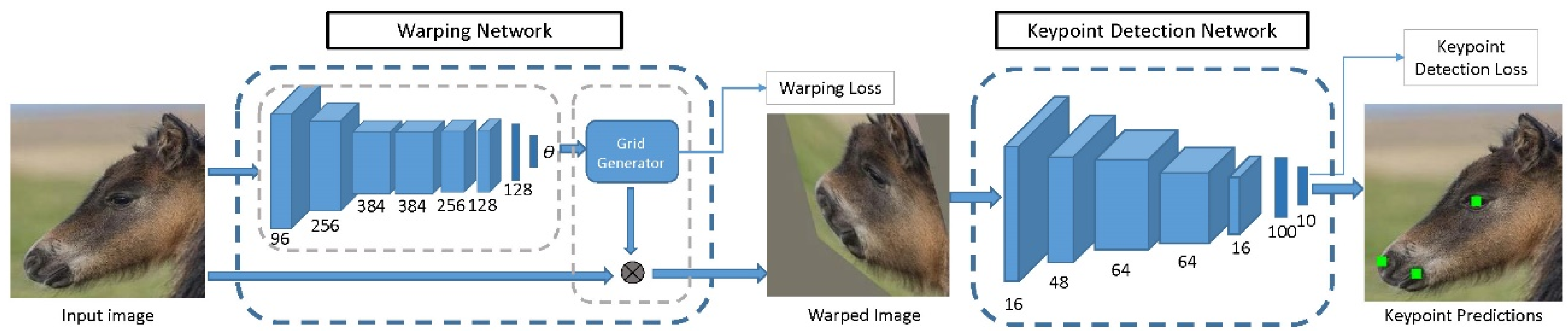
Abstract
:Simple Summary
Abstract
1. Background and Aim
2. Biological Challenges and Opportunities in Pain Assessment
3. Requirements on Video Recordings for Use in Computer Vision
4. Will a Pain Scale Deliver Ground Truth?
5. Analysis of EquiFACS Data
6. Automated Extraction of Facial Features from Images
6.1. Animal Facial Keypoint Detection
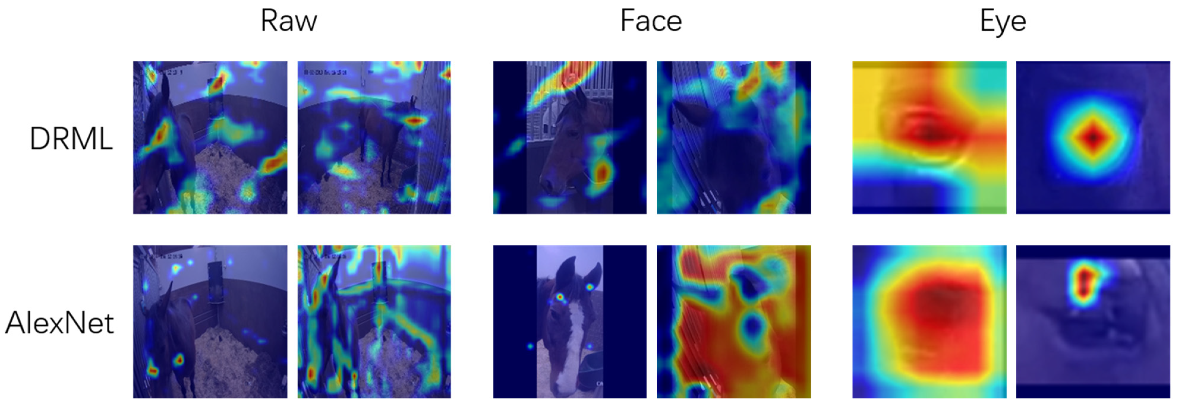
6.2. Automated Detection of Facial Action Units
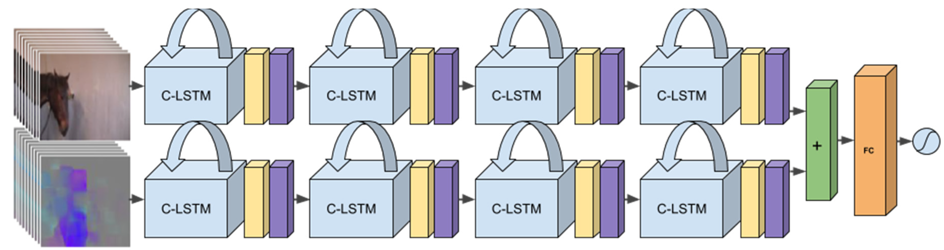
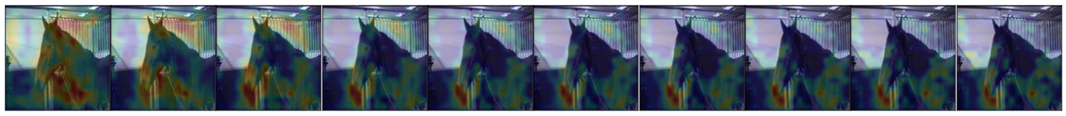
7. Automated Pain Detection Using Temporal Information
8. Discussion and Concluding Comments
Supplementary Materials
Author Contributions
Funding
Acknowledgments
Conflicts of Interest
References
- Egenvall, A.; Penell, J.C.; Bonnett, B.N.; Olson, P.; Pringle, J. Mortality of Swedish horses with complete life insurance between 1997 and 2000: Variations with sex, age, breed and diagnosis. Vet. Rec. 2006, 158, 397–406. [Google Scholar] [CrossRef]
- Stover, S.M. The epidemiology of Thoroughbred racehorse injuries. Clin. Tech. Equine Pract. 2003, 2, 312–322. [Google Scholar] [CrossRef]
- Logan, A.A.; Nielsen, B.D. Training Young Horses: The Science behind the Benefits. Animals 2021, 11, 13. [Google Scholar] [CrossRef]
- Price, J.; Marques, J.M.; Welsh, E.M.; Waran, N.K. Pilot epidemiological study of attitudes towards pain in horses. Vet. Rec. 2002, 151, 570–575. [Google Scholar] [CrossRef]
- Waran, N.; Williams, V.M.; Clarke, N.; Bridge, I.S. Recognition of pain and use of analgesia in horses by veterinarians in New Zealand. N. Z. Vet. J. 2010, 58, 274–280. [Google Scholar] [CrossRef]
- Bateson, P. Assessment of pain in animals. Anim. Behav. 1991, 42, 827–839. [Google Scholar] [CrossRef]
- Raekallio, M.; Heinonen, K.M.; Kuussaari, J.; Vainio, O. Pain Alleviation in Animals: Attitudes and Practices of Finnish Veterinarians. Vet. J. 2003, 165, 131–135. [Google Scholar] [CrossRef]
- Capner, C.A.; Lascelles, B.D.; Waterman-Pearson, A.E. Current British veterinary attitudes to perioperative analgesia for dogs. Vet. Rec. 1999, 145, 95–99. [Google Scholar] [CrossRef]
- Huxley, J.N.; Whay, H.R. Current attitudes of cattle practitioners to pain and the use of analgesics in cattle. Vet. Rec. 2006, 159, 662–668. [Google Scholar] [CrossRef] [PubMed]
- Fajt, V.R.; Wagner, S.A.; Norby, B. Analgesic drug administration and attitudes about analgesia in cattle among bovine practitioners in the United States. J. Am. Vet. Med. Assoc. 2011, 238, 755–767. [Google Scholar] [CrossRef]
- Norring, M.; Wikman, I.; Hokkanen, A.-H.; Kujala, M.V.; Hänninen, L. Empathic veterinarians score cattle pain higher. Vet. J. 2014, 200, 186–190. [Google Scholar] [CrossRef] [Green Version]
- Grégoire, M.; Coll, M.P.; Tremblay, M.P.B.; Prkachin, K.M.; Jackson, P.L. Repeated exposure to others’ pain reduces vicarious pain intensity estimation. Eur. J. Pain 2016, 20, 1644–1652. [Google Scholar] [CrossRef] [PubMed] [Green Version]
- Thomsen, P.T.; Anneberg, I.; Herskin, M.S. Differences in attitudes of farmers and veterinarians towards pain in dairy cows. Vet. J. 2012, 194, 94–97. [Google Scholar] [CrossRef] [PubMed]
- EU. Animal Welfare. Available online: https://ec.europa.eu/food/animals/welfare_en (accessed on 23 April 2021).
- FVE. European Veterinary Code of Conduct. 2019. Available online: fve.org/european-code-of.conduct-2019/2019 (accessed on 23 April 2021).
- Graubner, C.; Gerber, V.; Doherr, M.; Spadavecchia, C. Clinical application and reliability of a post abdominal surgery pain assessment scale (PASPAS) in horses. Vet. J. 2011, 188, 178–183. [Google Scholar] [CrossRef]
- van Loon, J.; Van Dierendonck, M.C. Monitoring acute equine visceral pain with the Equine Utrecht University Scale for Composite Pain Assessment (EQUUS-COMPASS) and the Equine Utrecht University Scale for Facial Assessment of Pain (EQUUS-FAP): A scale-construction study. Vet. J. 2015, 206, 356–364. [Google Scholar] [CrossRef]
- Bussieres, G.; Jacques, C.; Lainay, O.; Beauchamp, G.; Leblond, A.; Cadore, J.L.; Desmaizieres, L.M.; Cuvelliez, S.G.; Troncy, E. Development of a composite orthopaedic pain scale in horses. Res. Vet. Sci. 2008, 85, 294–306. [Google Scholar] [CrossRef]
- Lindegaard, C.; Gleerup, K.B.; Thomsen, M.H.; Martinussen, T.; Jacobsen, S.; Andersen, P.H. Anti-inflammatory effects of intra-articular administration of morphine in horses with experimentally induced synovitis. Am. J. Vet. Res. 2010, 71, 69–75. [Google Scholar] [CrossRef]
- Raekallio, M.; Taylor, P.M.; Bloomfield, M. A comparison of methods for evaluation of pain and distress after orthopaedic surgery in horses. J. Vet. Anaesth. 1997, 24, 17–20. [Google Scholar] [CrossRef]
- Price, J.; Catriona, S.; Welsh, E.M.; Waran, N.K. Preliminary evaluation of a behaviour-based system for assessment of post-operative pain in horses following arthroscopic surgery. Vet. Anaesth. Analg. 2003, 30, 124–137. [Google Scholar] [CrossRef]
- Sellon, D.C.; Roberts, M.C.; Blikslager, A.T.; Ulibarri, C.; Papich, M.G. Effects of Continuous Rate Intravenous Infusion of Butorphanol on Physiologic and Outcome Variables in Horses after Celiotomy. J. Vet. Intern. Med. 2004, 18. [Google Scholar] [CrossRef]
- Gleerup, K.B.; Lindegaard, C. Recognition and quantification of pain in horses: A tutorial review. Equine Vet. Educ. 2016, 28, 47–57. [Google Scholar] [CrossRef]
- Love, E.J. Assessment and management of pain in horses. Equine Vet. Educ. 2009, 21, 46–48. [Google Scholar] [CrossRef]
- de Grauw, J.C.; van Loon, J. Systematic pain assessment in horses. Vet. J. 2016, 209, 14–22. [Google Scholar] [CrossRef]
- Williams, A.C.D.C. Facial expression of pain: An evolutionary account. Behav. Brain Sci. 2002, 25. [Google Scholar] [CrossRef] [Green Version]
- Kadosh, K.C.; Johnson, M.H. Developing a cortex specialized for face perception. Trends Cogn. Sci. 2007, 11, 367–369. [Google Scholar] [CrossRef] [PubMed]
- Deyo, K.S.; Prkachin, K.M.; Mercer, S.R. Development of sensitivity to facial expression of pain. Pain 2004, 107, 16–21. [Google Scholar] [CrossRef]
- Poole, G.D.; Craig, K.D. Judgments of genuine, suppressed, and faked facial expressions of pain. J. Personal. Soc. Psychol. 1992, 63, 797–805. [Google Scholar] [CrossRef]
- Matsumoto, D.; Hwang, H.S. Evidence for training the ability to read microexpressions of emotion. Motiv. Emot. 2011, 35, 181–191. [Google Scholar] [CrossRef]
- Tate, A.J.; Fischer, H.; Leigh, A.E.; Kendrick, K.M. Behavioural and neurophysiological evidence for face identity and face emotion processing in animals. Philos. Trans. R. Soc. B Biol. Sci. 2006, 361, 2155–2172. [Google Scholar] [CrossRef] [Green Version]
- Correia-Caeiro, C.; Guo, K.; Mills, D.S. Perception of dynamic facial expressions of emotion between dogs and humans. Anim. Cogn. 2020, 23, 465–476. [Google Scholar] [CrossRef] [Green Version]
- Ekman, P.; Friesen, W.; Hagar, J. Facial Action Coding System; Research Nexus: Salt Lake City, UT, USA, 2002. [Google Scholar]
- Waller, B.M.; Vick, S.-J.; Parr, L.A.; Bard, K.A.; Pasqualini, M.C.S.; Gothard, K.M.; Fuglevand, A.J. Intramuscular electrical stimulation of facial muscles in humans and chimpanzees: Duchenne revisited and extended. Emotion 2006, 6, 367–382. [Google Scholar] [CrossRef] [Green Version]
- Sayette, M.A.; Cohn, J.F.; Wertz, J.M.; Perrott, M.A.; Parrott, D.J. A psychometric evaluation of the facial action coding system for assessing spontaneous expression. J. Nonverbal Behav. 2001, 25, 167–185. [Google Scholar] [CrossRef]
- Vick, S.-J.; Waller, B.M.; Parr, L.A.; Pasqualini, M.C.S.; Bard, K.A. A cross-species comparison of facial morphology and movement in humans and chimpanzees using the Facial Action Coding System (FACS). J. Nonverbal Behav. 2007, 31, 1–20. [Google Scholar] [CrossRef] [PubMed] [Green Version]
- Julle-Daniere, E.; Micheletta, J.; Whitehouse, J.; Joly, M.; Gass, C.; Burrows, A.M.; Waller, B.M. MaqFACS (Macaque Facial Action Coding System) can be used to document facial movements in Barbary macaques (Macaca sylvanus). PeerJ 2015, 3. [Google Scholar] [CrossRef] [PubMed] [Green Version]
- Caeiro, C.C.; Waller, B.M.; Zimmermann, E.; Burrows, A.M.; Davila-Ross, M. OrangFACS: A Muscle-Based Facial Movement Coding System for Orangutans (Pongo spp.). Int. J. Primatol. 2013, 34, 115–129. [Google Scholar] [CrossRef]
- Clark, P.R.; Waller, B.M.; Burrows, A.M.; Julle-Danière, E.; Agil, M.; Engelhardt, A.; Micheletta, J. Morphological variants of silent bared-teeth displays have different social interaction outcomes in crested macaques (Macaca nigra). Am. J. Phys. Anthropol. 2020, 173, 411–422. [Google Scholar] [CrossRef]
- Correia-Caeiro, C.; Holmes, K.; Miyabe-Nishiwaki, T. Extending the MaqFACS to measure facial movement in Japanese macaques (Macaca fuscata) reveals a wide repertoire potential. PLoS ONE 2021, 16, e0245117. [Google Scholar] [CrossRef] [PubMed]
- Waller, B.M.; Lembeck, M.; Kuchenbuch, P.; Burrows, A.M.; Liebal, K. GibbonFACS: A Muscle-Based Facial Movement Coding System for Hylobatids. Int. J. Primatol. 2012, 33, 809–821. [Google Scholar] [CrossRef]
- Waller, B.M.; Peirce, K.; Caeiro, C.C.; Scheider, L.; Burrows, A.M.; McCune, S.; Kaminski, J. Paedomorphic Facial Expressions Give Dogs a Selective Advantage. PLoS ONE 2013, 8, e82686. [Google Scholar] [CrossRef] [Green Version]
- Caeiro, C.C.; Burrows, A.M.; Waller, B.M. Development and application of CatFACS: Are human cat adopters influenced by cat facial expressions? Appl. Anim. Behav. Sci. 2017, 189, 66–78. [Google Scholar] [CrossRef]
- Wathan, J.; Burrows, A.M.; Waller, B.M.; McComb, K. EquiFACS: The Equine Facial Action Coding System. PLoS ONE 2015, 10, e0131738. [Google Scholar] [CrossRef] [Green Version]
- Burrows, A.; Diogo, R.; Waller, B.; Kaminski, J. Variation of Facial Musculature between Wolves and Domestic Dogs: Evolutionary Divergence in Facial Movement. Faseb J. 2017, 31, 577.3. [Google Scholar]
- Waller, B.M.; Parr, L.A.; Gothard, K.M.; Burrows, A.M.; Fuglevand, A.J. Mapping the contribution of single muscles to facial movements in the rhesus macaque. Physiol. Behav. 2008, 95, 93–100. [Google Scholar] [CrossRef] [Green Version]
- Prkachin, K.M.; Craig, K.D. Expressing pain: The communication and interpretation of facial pain signals. J. Nonverbal Behav. 1995, 19, 191–205. [Google Scholar] [CrossRef]
- Hill, M.L.; Craig, K.D. Detecting deception in facial expressions of pain—Accuracy and training. Clin. J. Pain 2004, 20, 415–422. [Google Scholar] [CrossRef]
- Lucey, P.; Cohn, J.F.; Prkachin, K.M.; Solomon, P.E.; Matthews, I. Painful data: The UNBC-McMaster shoulder pain expression archive database. In Proceedings of the 2011 IEEE International Conference on Automatic Face & Gesture Recognition (FG), Santa Barbera, CA, USA, 21–25 March 2011; pp. 57–64. [Google Scholar] [CrossRef]
- Rosenberg, E.L.; Zanesco, A.P.; King, B.G.; Aichele, S.R.; Jacobs, T.L.; Bridwell, D.A.; MacLean, K.A.; Shaver, P.R.; Ferrer, E.; Sahdra, B.K.; et al. Intensive Meditation Training Influences Emotional Responses to Suffering. Emotion 2015, 15, 775–790. [Google Scholar] [CrossRef]
- Rashid, M.; Silventoinen, A.; Gleerup, K.B.; Andersen, P.H. Equine Facial Action Coding System for determination of pain-related facial responses in videos of horses. PLoS ONE 2020, 15, e0231608. [Google Scholar] [CrossRef] [PubMed]
- Lundblad, J.; Rashid, M.; Rhodin, M.; Andersen, P.H. Effect of transportation and social isolation on facial expressions of healthy horses. PLoS ONE 2021. [Google Scholar] [CrossRef]
- Miller, A.L.; Leach, M.C. The Mouse Grimace Scale: A Clinically Useful Tool? PLoS ONE 2015, 10, e0136000. [Google Scholar] [CrossRef] [PubMed]
- McLennan, K.M.; Miller, A.L.; Dalla Costa, E.; Stucke, D.; Corke, M.J.; Broom, D.M.; Leach, M.C. Conceptual and methodological issues relating to pain assessment in mammals: The development and utilisation of pain facial expression scales. Appl. Anim. Behav. Sci. 2019, 217, 1–15. [Google Scholar] [CrossRef]
- Dyson, S.; Pollard, D. Application of a Ridden Horse Pain Ethogram and Its Relationship with Gait in a Convenience Sample of 60 Riding Horses. Animals 2020, 10, 1044. [Google Scholar] [CrossRef]
- Dyson, S.; Berger, J.M.; Ellis, A.D.; Mullard, J. Can the presence of musculoskeletal pain be determined from the facial expressions of ridden horses (FEReq)? J. Vet. Behav. Clin. Appl. Res. 2017, 19, 78–89. [Google Scholar] [CrossRef]
- Tuyttens, F.A.M.; Stadig, L.; Heerkens, J.L.T.; Van laer, E.; Buijs, S.; Ampe, B. Opinion of applied ethologists on expectation bias, blinding observers and other debiasing techniques. Appl. Anim. Behav. Sci. 2016, 181, 27–33. [Google Scholar] [CrossRef] [Green Version]
- Bartlett, M.S.; Littlewort, G.C.; Frank, M.G.; Lee, K. Automatic Decoding of Facial Movements Reveals Deceptive Pain Expressions. Curr. Biol. 2014, 24, 738–743. [Google Scholar] [CrossRef] [PubMed] [Green Version]
- Littlewort, G.C.; Bartlett, M.S.; Lee, K. Faces of Pain: Automated Measurement of Spontaneous Facial Expressions of Genuine and Posed Pain. In Proceedings of the ICMI’07, 9th International Conference on Multimodal Interfaces, Nagoya, Japan, 12–15 November 2007; pp. 15–21. [Google Scholar]
- Bartlett, M.S.; Littlewort, G.; Frank, M.; Lainscsek, C.; Fasel, I.; Movellan, J.; Soc, I.C. Fully automatic facial action recognition in spontaneous behavior. In Proceedings of the Seventh International Conference on Automatic Face and Gesture Recognition, Southampton, UK, 10–12 April 2006; pp. 223–228. [Google Scholar]
- Bartlett, M.S.; Littlewort, G.; Frank, M.; Lainscsek, C.; Fasel, I.; Movellan, J. Recognizing facial expression: Machine learning and application to spontaneous behavior. In Proceedings of the 2005 IEEE Computer Society Conference on Computer Vision and Pattern Recognition, San Diego, CA, USA, 20–25 June 2005; Volume 2, pp. 568–573. [Google Scholar]
- Huang, J.; Craig, K.; Diaz, D.; Sikka, K.; Ahmed, A.; Terrones, L.; Littlewort, G.; Goodwin, M.; Bartlett, M. Automated facial expression analysis can detect clinical pain in youth in the post-operative setting. J. Pain 2014, 15, S3. [Google Scholar] [CrossRef]
- Srinivasan, R.; Golomb, J.D.; Martinez, A.M. A Neural Basis of Facial Action Recognition in Humans. J. Neurosci. 2016, 36, 4434–4442. [Google Scholar] [CrossRef] [Green Version]
- Sikka, K.; Ahmed, A.A.; Diaz, D.; Goodwin, M.S.; Craig, K.D.; Bartlett, M.S.; Huang, J.S. Automated Assessment of Children’s Postoperative Pain Using Computer Vision. Pediatrics 2015, 136, e124–e131. [Google Scholar] [CrossRef] [PubMed] [Green Version]
- Zhang, X.; Yin, L.; Cohn, J.; Canavan, S.; Reale, M.; Horowitz, A.; Liu, P.; Girard, J. BP4D-Spontaneous: A High-Resolution Spontaneous 3D Dynamic Facial Expression Database. Image Vis. Comput. 2014, 32, 692–706. [Google Scholar] [CrossRef]
- Mavadati, S.M.; Mahoor, M.H.; Bartlett, K.; Trinh, P.; Cohn, J.F. DISFA: A Spontaneous Facial Action Intensity Database. IEEE Trans. Affect. Comput. 2013, 4, 151–160. [Google Scholar] [CrossRef]
- Rolnick, D.; Veit, A.; Belongie, S.; Shavit, N. Deep Learning is Robust to Massive Label Noise. arXiv 2018, arXiv:1705.10694. [Google Scholar]
- Erin Browne, M.; Hadjistavropoulos, T.; Prkachin, K.; Ashraf, A.; Taati, B. Pain Expressions in Dementia: Validity of Observers’ Pain Judgments as a Function of Angle of Observation. J. Nonverbal. Behav. 2019, 43, 309–327. [Google Scholar] [CrossRef] [PubMed] [Green Version]
- Sneddon, L.U.; Elwood, R.W.; Adamo, S.A.; Leach, M.C. Defining and assessing animal pain. Anim. Behav. 2014, 97, 201–212. [Google Scholar] [CrossRef] [Green Version]
- Seminowicz, D.A.; Laferriere, A.L.; Millecamps, M.; Yu, J.S.; Coderre, T.J.; Bushnell, M.C. MRI structural brain changes associated with sensory and emotional function in a rat model of long-term neuropathic pain. Neuroimage 2009, 47, 1007–1014. [Google Scholar] [CrossRef] [PubMed] [Green Version]
- Vila Pouca, C.; Brown, C. Contemporary topics in fish cognition and behaviour. Curr. Opin. Behav. Sci. 2017, 16, 46–52. [Google Scholar] [CrossRef]
- Descovich, K.A.; Wathan, J.; Leach, M.C.; Buchanan-Smith, H.M.; Flecknell, P.; Farningham, D.; Vick, S.J. Facial Expression: An Under-Utilized Tool for the Assessment of Welfare in Mammals. ALTEX Altern. Anim. Exp. 2017, 34, 409–429. [Google Scholar] [CrossRef] [PubMed] [Green Version]
- Raja, S.N.; Carr, D.B.; Cohen, M.; Finnerup, N.B.; Flor, H.; Gibson, S.; Keefe, F.J.; Mogil, J.S.; Ringkamp, M.; Sluka, K.A.; et al. The revised International Association for the Study of Pain definition of pain: Concepts, challenges, and compromises. Pain 2020, 161, 1976–1982. [Google Scholar] [CrossRef]
- Craig, K.D. Social communication model of pain. Pain 2015, 156, 1198–1199. [Google Scholar] [CrossRef] [Green Version]
- Rutherford, K.M.D. Assessing pain in animals. Anim. Welf. 2002, 11, 31–53. [Google Scholar]
- Ashley, F.H.; Waterman-Pearson, A.E.; Whay, H.R. Behavioural assessment of pain in horses and donkeys: Application to clinical practice and future studies. Equine Vet. J. 2005, 37, 565–575. [Google Scholar] [CrossRef]
- Coles, B.; Birgitsdottir, L.; Andersen, P.H. Out of Sight but Not out of Clinician’s Mind: Using Remote Video Surveillance to Disclose Concealed Pain Behavior in Hospitalized Horses. In Proceedings of the International Association for the Study of Pain 17th World Congress, Boston, MA, USA, 15–18 September 2018; p. 471121. [Google Scholar]
- Torcivia, C.; McDonnell, S. In-Person Caretaker Visits Disrupt Ongoing Discomfort Behavior in Hospitalized Equine Orthopedic Surgical Patients. Animals 2020, 10, 210. [Google Scholar] [CrossRef] [Green Version]
- Ask, K.; Rhodin, M.; Tamminen, L.M.; Hernlund, E.; Haubro Andersen, P. Identification of Body Behaviors and Facial Expressions Associated with Induced Orthopedic Pain in Four Equine Pain Scales. Animals 2020, 10, 2155. [Google Scholar] [CrossRef]
- Korshunov, P.; Ooi, W.T. Video quality for face detection, recognition, and tracking. ACM Trans. Multimed. Comput. Commun. Appl. 2011, 7, 14. [Google Scholar] [CrossRef]
- Gleerup, K.B.; Forkman, B.; Lindegaard, C.; Andersen, P.H. An equine pain face. Vet. Anaesth. Analg. 2015, 42, 103–114. [Google Scholar] [CrossRef] [Green Version]
- Dalla Costa, E.; Minero, M.; Lebelt, D.; Stucke, D.; Canali, E.; Leach, M.C. Development of the Horse Grimace Scale (HGS) as a Pain Assessment Tool in Horses Undergoing Routine Castration. PLoS ONE 2014, 9. [Google Scholar] [CrossRef] [Green Version]
- Dalla Costa, E.; Stucke, D.; Dai, F.; Minero, M.; Leach, M.C.; Lebelt, D. Using the Horse Grimace Scale (HGS) to Assess Pain Associated with Acute Laminitis in Horses (Equus caballus). Animals 2016, 6, 47. [Google Scholar] [CrossRef] [PubMed]
- van Loon, J.; Van Dierendonck, M.C. Monitoring equine head-related pain with the Equine Utrecht University scale for facial assessment of pain (EQUUS-FAP). Vet. J. 2017, 220, 88–90. [Google Scholar] [CrossRef] [PubMed]
- Langford, D.J.; Bailey, A.L.; Chanda, M.L.; Clarke, S.E.; Drummond, T.E.; Echols, S.; Glick, S.; Ingrao, J.; Klassen-Ross, T.; LaCroix-Fralish, M.L.; et al. Coding of facial expressions of pain in the laboratory mouse. Nat. Methods 2010, 7, 447–449. [Google Scholar] [CrossRef] [PubMed]
- Vandierendonck, M.C.; Van Loon, J.P.A.M. Monitoring acute equine visceral pain with the Equine Utrecht University Scale for Composite Pain Assessment (EQUUS-COMPASS) and the Equine Utrecht University Scale for Facial Assessment of Pain (EQUUS-FAP): A validation study. Vet. J. 2016, 216, 175–177. [Google Scholar] [CrossRef] [PubMed]
- Weary, D.M.; Niel, L.; Flower, F.C.; Fraser, D. Identifying and preventing pain in animals. Appl. Anim. Behav. Sci. 2006, 100, 64–76. [Google Scholar] [CrossRef] [Green Version]
- Dai, F.; Leach, M.; MacRae, A.M.; Minero, M.; Costa, E.D. Does Thirty-Minute Standardised Training Improve the Inter-Observer Reliability of the Horse Grimace Scale (HGS)? A Case Study. Animals 2020, 10, 781. [Google Scholar] [CrossRef]
- Gleerup, K.B.; Forkman, B.; Lindegaard, C.; Andersen, P.H. Facial expressions as a tool for pain recognition in horses. In Proceedings of the 10th International Equitation Science Conference, Bredsten, Denmark, 7–9 August 2014. [Google Scholar]
- Guesgen, M.J.; Beausoleil, N.J.; Minot, E.O.; Stewart, M.; Jones, G.; Stafford, K.J. The effects of age and sex on pain sensitivity in young lambs. Appl. Anim. Behav. Sci. 2011, 135, 51–56. [Google Scholar] [CrossRef]
- Reijgwart, M.L.; Schoemaker, N.J.; Pascuzzo, R.; Leach, M.C.; Stodel, M.; de Nies, L.; Hendriksen, C.F.M.; van der Meer, M.; Vinke, C.M.; van Zeeland, Y.R.A. The composition and initial evaluation of a grimace scale in ferrets after surgical implantation of a telemetry probe. PLoS ONE 2017, 12, e0187986. [Google Scholar] [CrossRef]
- Ijichi, C.; Collins, L.M.; Elwood, R.W. Pain expression is linked to personality in horses. Appl. Anim. Behav. Sci. 2014, 152, 38–43. [Google Scholar] [CrossRef] [Green Version]
- Guesgen, M.J.; Beausoleil, N.J.; Stewart, M. Effects of early human handling on the pain sensitivity of young lambs. Vet. Anaesth. Analg. 2013, 40, 55–62. [Google Scholar] [CrossRef] [PubMed]
- Clark, C.; Murrell, J.; Fernyhough, M.; O’Rourke, T.; Mendl, M. Long-term and trans-generational effects of neonatal experience on sheep behaviour. Biol. Lett. 2014, 10. [Google Scholar] [CrossRef] [PubMed]
- Rhodin, M.; Egenvall, A.; Andersen, P.H.; Pfau, T. Head and pelvic movement asymmetries at trot in riding horses in training and perceived as free from lameness by the owner. PLoS ONE 2017, 12. [Google Scholar] [CrossRef] [PubMed]
- Rhodin, M.; Persson-Sjodin, E.; Egenvall, A.; Serra Bragança, F.M.; Pfau, T.; Roepstorff, L.; Weishaupt, M.A.; Thomsen, M.H.; van Weeren, P.R.; Hernlund, E. Vertical movement symmetry of the withers in horses with induced forelimb and hindlimb lameness at trot. Equine Vet. J. 2018, 50, 818–824. [Google Scholar] [CrossRef]
- Van de Water, E.; Oosterlinck, M.; Korthagen, N.M.; Duchateau, L.; Dumoulin, M.; van Weeren, P.R.; Olijve, J.; van Doorn, D.A.; Pille, F. The lipopolysaccharide model for the experimental induction of transient lameness and synovitis in Standardbred horses. Vet. J. 2021, 270, 105626. [Google Scholar] [CrossRef]
- Lindegaard, C.; Frost, A.B.; Thomsen, M.H.; Larsen, C.; Hansen, S.H.; Andersen, P.H. Pharmacokinetics of intra-articular morphine in horses with lipopolysaccharide-induced synovitis. Vet. Anaesth. Analg. 2010, 37, 186–195. [Google Scholar] [CrossRef]
- Kunz, M.; Meixner, D.; Lautenbacher, S. Facial muscle movements encoding pain—A systematic review. Pain 2019, 160, 535–549. [Google Scholar] [CrossRef] [Green Version]
- Wagner, A.E. Effects of Stress on Pain in Horses and Incorporating Pain Scales for Equine Practice. Vet. Clin. N. Am. Equine Pract. 2010, 26, 481–492. [Google Scholar] [CrossRef]
- Trindade, P.H.E.; Hartmann, E.; Keeling, L.J.; Andersen, P.H.; Ferraz, G.d.C.; Paranhos da Costa, M.J.R. Effect of work on body language of ranch horses in Brazil. PLoS ONE 2020, 15, e0228130. [Google Scholar] [CrossRef]
- Kunz, M.; Lautenbacher, S. The faces of pain: A cluster analysis of individual differences in facial activity patterns of pain. Eur. J. Pain 2014, 18, 813–823. [Google Scholar] [CrossRef] [PubMed]
- Rashid, M.; Broomé, S.; Andersen, P.H.; Gleerup, K.B.; Lee, Y.J. What should I annotate? An automatic tool for finding video segments for EquiFACS annotation In Measuring Behaviour 2018 Conference Proceedings; Grant, R.A., Allen, T., Spink, A., Sullivan, M., Eds.; Manchester Metropolitan University: Manchester, UK, 2018; pp. 164–165. [Google Scholar]
- Lucey, P.; Cohn, J.F.; Kanade, T.; Saragih, J.; Ambadar, Z.; Matthews, I. The Extended Cohn-Kanade Dataset (CK+): A complete dataset for action unit and emotion-specified expression. In Proceedings of the 2010 IEEE Computer Society Conference on Computer Vision and Pattern Recognition—Workshops, San Francisco, CA, USA, 13–18 June 2010; pp. 94–101. [Google Scholar]
- Littlewort, G.; Whitehill, J.; Wu, T.; Fasel, I.; Frank, M.; Movellan, J.; Bartlett, M. The computer expression recognition toolbox (CERT). Face Gesture 2011. [Google Scholar] [CrossRef] [Green Version]
- Köstinger, M.; Wohlhart, P.; Roth, P.M.; Bischof, H. Annotated Facial Landmarks in the Wild: A large-scale, real-world database for facial landmark localization. In Proceedings of the 2011 IEEE International Conference on Computer Vision Workshops (ICCV Workshops), Barcelona, Spain, 6–13 November 2011; pp. 2144–2151. [Google Scholar]
- Yan, H.; Gao, W.; Pan, Z.; Zhang, F.; Fan, C. The expression of alpha-SMA in the painful traumatic neuroma: Potential role in the pathobiology of neuropathic pain. J. Neurotrauma 2012, 29, 2791–2797. [Google Scholar] [CrossRef]
- Li, J.; Wang, Y.; Wang, C.; Tai, Y.; Qian, J.; Yang, J.; Wang, C.; Li, J.; Huang, F. DSFD: Dual Shot Face Detector. In Proceedings of the 2019 IEEE/CVF Conference on Computer Vision and Pattern Recognition (CVPR), Long Beach, CA, USA, 15–20 June 2019; pp. 5055–5064. [Google Scholar]
- Rashid, M.; Gu, X.; Lee, Y.J. Interspecies Knowledge Transfer for Facial Keypoint Detection. In Proceedings of the 2017 IEEE Conference on Computer Vision and Pattern Recognition (CVPR), Honolulu, HI, USA, 21–26 July 2017; pp. 1600–1609. [Google Scholar]
- Li, Z.; Broome, S.; Andersen, P.H.; Kjellstrom, H. Automated Detection of Equine Facial Action Units. arXiv 2021, arXiv:2102.08983. [Google Scholar]
- Lu, Y.; Mahmoud, M.; Robinson, P. Estimating Sheep Pain Level Using Facial Action Unit Detection. In Proceedings of the 2017 12th Ieee International Conference on Automatic Face and Gesture Recognition, Washington, DC, USA, 30 May–3 June 2017; pp. 394–399. [Google Scholar] [CrossRef] [Green Version]
- Hummel, H.I.; Pessanha, F.; Salah, A.A.; van Loon, T.J.P.A.M.; Veltkamp, R.C. Automatic Pain Detection on Horse and Donkey Faces. In Proceedings of the 2020 15th IEEE International Conference on Automatic Face and Gesture Recognition (FG 2020), Buenos Aires, Argentina, 16–20 November 2020; pp. 793–800. [Google Scholar]
- Pessanha, F.; McLennan, K.; Mahmoud, M. Towards automatic monitoring of disease progression in sheep: A hierarchical model for sheep facial expressions analysis from video. In Proceedings of the 2020 15th IEEE International Conference on Automatic Face and Gesture Recognition (FG 2020), Buenos Aires, Argentina, 16–20 November 2020; pp. 387–393. [Google Scholar]
- Zhao, K.; Chu, W.; Zhang, H. Deep Region and Multi-label Learning for Facial Action Unit Detection. In Proceedings of the 2016 IEEE Conference on Computer Vision and Pattern Recognition (CVPR), Las Vegas, NV, USA, 27–30 June 2016; pp. 3391–3399. [Google Scholar]
- Rodriguez, P.; Cucurull, G.; Gonalez, J.; Gonfaus, J.M.; Nasrollahi, K.; Moeslund, T.B.; Roca, F.X. Deep Pain: Exploiting Long Short-Term Memory Networks for Facial Expression Classification. IEEE Trans. Cybern. 2017. [Google Scholar] [CrossRef] [PubMed] [Green Version]
- Krumhuber, E.G.; Kappas, A.; Manstead, A.S.R. Effects of Dynamic Aspects of Facial Expressions: A Review. Emot. Rev. 2013, 5, 41–46. [Google Scholar] [CrossRef]
- Broomé, S.; Gleerup, K.B.; Haubro Andersen, P.; Kjellström, H. Dynamics are Important for the Recognition of Equine Pain in Video. In Proceedings of the IEEE/CVF Conference on Computer Vision and Pattern Recognition (CVPR), Long Beach, CA, USA, 15–20 June 2019; pp. 12659–12668. [Google Scholar]
- Tuttle, A.H.; Molinaro, M.J.; Jethwa, J.F.; Sotocinal, S.G.; Prieto, J.C.; Styner, M.A.; Mogil, J.S.; Zylka, M.J. A deep neural network to assess spontaneous pain from mouse facial expressions. Mol. Pain 2018, 14, 1744806918763658. [Google Scholar] [CrossRef]
- Selvaraju, R.R.; Cogswell, M.; Das, A.; Vedantam, R.; Parikh, D.; Batra, D. Grad-CAM: Visual Explanations from Deep Networks via Gradient-Based Localization. In Proceedings of the 2017 IEEE International Conference on Computer Vision (ICCV), Venice, Italy, 22–29 October 2017; pp. 618–626. [Google Scholar]
- Lloyd, A.S.; Martin, J.E.; Bornett-Gauci, H.L.I.; Wilkinson, R.G. Horse personality: Variation between breeds. Appl. Anim. Behav. Sci. 2008, 112, 369–383. [Google Scholar] [CrossRef]
- Hausberger, M.; Fureix, C.; Lesimple, C. Detecting horses’ sickness: In search of visible signs. Appl. Anim. Behav. Sci. 2016, 175, 41–49. [Google Scholar] [CrossRef] [Green Version]
- Fureix, C.; Jego, P.; Henry, S.; Lansade, L.; Hausberger, M. Towards an Ethological Animal Model of Depression? A Study on Horses. PLoS ONE 2012, 7. [Google Scholar] [CrossRef] [PubMed]


Publisher’s Note: MDPI stays neutral with regard to jurisdictional claims in published maps and institutional affiliations. |
© 2021 by the authors. Licensee MDPI, Basel, Switzerland. This article is an open access article distributed under the terms and conditions of the Creative Commons Attribution (CC BY) license (https://creativecommons.org/licenses/by/4.0/).
Share and Cite
Andersen, P.H.; Broomé, S.; Rashid, M.; Lundblad, J.; Ask, K.; Li, Z.; Hernlund, E.; Rhodin, M.; Kjellström, H. Towards Machine Recognition of Facial Expressions of Pain in Horses. Animals 2021, 11, 1643. https://doi.org/10.3390/ani11061643
Andersen PH, Broomé S, Rashid M, Lundblad J, Ask K, Li Z, Hernlund E, Rhodin M, Kjellström H. Towards Machine Recognition of Facial Expressions of Pain in Horses. Animals. 2021; 11(6):1643. https://doi.org/10.3390/ani11061643
Chicago/Turabian StyleAndersen, Pia Haubro, Sofia Broomé, Maheen Rashid, Johan Lundblad, Katrina Ask, Zhenghong Li, Elin Hernlund, Marie Rhodin, and Hedvig Kjellström. 2021. "Towards Machine Recognition of Facial Expressions of Pain in Horses" Animals 11, no. 6: 1643. https://doi.org/10.3390/ani11061643