Article

Analysis of the Productivity, Immunity, and Health Performance of Nile Tilapia (Oreochromis niloticus) Broodstock-fed Dietary Fermented Extracts Sourced from Saccharomyces cerevisiae (Hilyses): A Field Trial

1
Department of Aquatic Animal Medicine and Management, Faculty of Veterinary Medicine, Cairo University, Giza 12211, Egypt
2
Department of Oceanography, Faculty of Science, Alexandria University, Alexandria 21568, Egypt
3
Department of Nutrition and Clinical Nutrition, Faculty of Veterinary Medicine, Cairo University, Giza 12211, Egypt
4
Department of Physiology, Faculty of Veterinary Medicine, Cairo University, Giza 12211, Egypt
5
Department of Pathology, Faculty of Veterinary Medicine, Cairo University, Giza 12211, Egypt
6
Research and Development, ICC Industrial Comércio Exportaçãoe Importação SA, São Paulo 01451-909, Brazil
7
Department of Animal Production, Faculty of Agriculture, Kafrelsheikh University, Kafrelsheikh 33516, Egypt
*
Authors to whom correspondence should be addressed.
Animals 2021, 11(3), 815; https://doi.org/10.3390/ani11030815
Submission received: 21 December 2020 / Revised: 9 March 2021 / Accepted: 10 March 2021 / Published: 14 March 2021

Abstract

:

Simple Summary

The low performance of Nile tilapia (Oreochromis niloticus) broodstock and high seed mortality along the spawning season are the major constraints which are directly reflected in hatchery profit. Broodstock nutrition is a key factor that can influence fish reproduction and subsequent larval quality. The objective of this study was to evaluate the effect of dietary fermented extracts sourced from Saccharomyces cerevisiae (nucleotides, β-glucans and MOS) (Hilyses®) on the seed production and health of Nile tilapia broodstock, as well as on seed survival and growth performance. The study was performed in the hatchery along the spawning season and continued in the laboratory to monitor the performance in fry and fingerlings. The results showed that dietary fermented yeast extracts could be used as a strategic approach to sustain tilapia production, as they improve the productivity and health of broodstock as well as seed survival and performance.

Abstract

The present study aimed to evaluate the effect of dietary fermented extracts sourced from Saccharomyces cerevisiae (nucleotides, β-glucans and MOS) (Hilyses®) on the production and health of Nile tilapia (Oreochromis niloticus) broodstock, as well as on seed survival and performance. The trial was performed in a hatchery along the spawning season and continued in the laboratory to monitor the performance in fry and fingerlings. The broodstock were divided into two groups, (C) fed a basal diet and (H) fed 0.4% Hilyses. Blood and histological parameters, antioxidant power, cortisol level and the expression of some immune-related (TLR-2, IL-1β and TNF-α) and growth-related genes (MUC-2 and IGF-1) were measured. The obtained seeds were subdivided into four treatments: (C-C) fed a basal diet, (C-H) fed 0.4% Hilyses, (H-C) fed a basal diet and (H-H) fed 0.4% Hilyses. Results revealed that the dietary inclusion of Hilyses in the broodstock increased seed production, survival, hematological parameters, and antioxidant power. Moreover, it improved the intestinal microstructure and upregulated the immune- and growth-related genes. The growth indices of fry and fingerlings were significantly increased in all Hilyses-treated groups (p < 0.05). The performance in the (H-H) group significantly surpassed those of all groups. Therefore, dietary fermented yeast could be used as a strategic solution to sustain tilapia production.

Graphical Abstract

1. Introduction

Fish farming in Egypt represents the largest part of production compared to captured fish and can reach up to 77% of the total production; the private sector share of this production is more than 99% [1]. Although several factors limit aquaculture development (e.g., feeding cost, diseases, bad water quality, low performance of broodstock, and the high mortality rate in seeds) [2], Egypt has the largest aquaculture industry in Africa and the third largest after China and Indonesia in Nile tilapia production [3,4]. Global tilapia culture has witnessed a sharp expansion during the past two decades and is being cultured in more than 130 countries worldwide. Tilapias are currently the second most important farmed finfish in the world, representing 125% of freshwater fish production and 107% of total fish culture [4]. Global production of farmed tilapia grew by 3.3 percent in 2020 to top 6 million tons for the first time, despite the impact of COVID-19 [5]. The expansion of tilapia production all over the world is due to its ability to be produced in various aquatic environments, selective breeding, and its potential to replace marine fish products [5]. There are several strategic approaches that have been recently adopted in aquaculture to sustain tilapia production. One of these approaches is the production of functional foods. A wide range of functional feed additives have been used [6]; certain additives, such as pellet binders, antimicrobials, antimycotoxins, antioxidants, and enzymes, act by improving feed quality. Other additives, such as probiotics, prebiotics, phytogenics, dietary acidifiers and immunostimulants, directly affect animal performance and health [6,7,8]. Functional feeds containing gut health promotors have become a key component of any strategy to prevent diseases in aquaculture, particularly when opportunistic bacteria are suspected to be a major cause of mortality [9,10]. Gut health promotors combine different modes of action, including modulating the intestinal microbial flora, stimulating the local immunity of the gut and improving the histological intestinal structure [11]. This approach directly correlates with the feed transformation into biomass gain and the profit of the farm.
Saccharomyces cerevisiae and its byproducts are the most common functional feed additives used in aquaculture [
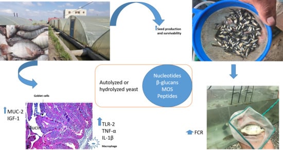
6]. Hydrolyzed, fully fermented yeast contains numerous nutrients and bioactive compounds, e.g., peptides, polypeptides glutamines, β-glucans, mannan oligosaccharides (MOS), chitin and nucleotides [12]. These compounds have different mechanisms of action and show benefits when used alone or in combination. Briefly, β-glucans are known by their immunostimulatory effect; they stimulate the innate immunity and enhance the resistance of fish and crustaceans against microbial diseases [13]. MOS is characterized by pathogen adsorption capacity, preventing the bacterial colonization, and stimulating the proliferation of beneficial gut flora. In addition, they increase the food palatability and improve the intestinal health [14]. Nucleotides are the building block of cell’s RNA. They are synthesized de novo in most tissues, but some tissues like, intestinal cells, hepatocytes, hemolymph, and immune cells have limited capacity for synthesis and need exogenous supply, especially under certain conditions, e.g., tissue injury, liver dysfunction, diseases, reproduction, and fast growth life stage [12]. Essential amino acids, such as glutamic acid, play a substantial role in tissue proliferation and regeneration [10].
Broodstock nutrition is recognized as a major factor that can influence fish reproduction and subsequent larval quality [15]. Many studies have indicated the positive effects of dietary fortification of whole yeast and/or its by-products on the growth performance, hematological indices, antioxidant defense systems, immune response, and disease resistance in Nile tilapia [9,16,17,18,19]. Abu-Elala et al. [5] recorded that dietary fermented yeast extracts sourced from Saccharomyces cerevisiae successfully enhanced the phagocytic activity/index, and lysozyme activity up-regulated the proinflammatory cytokines (TNF-α and IL-1β) and increased the resistance of fish against bacterial diseases. In addition, they improved the oxidant and antioxidant defense systems and reduced the lipid peroxidation. Moreover, authors observed a significant increase in RBCs count, WBCs count, Hb, and hematocrit, which could be related to the hepato-stimulatory and hepatoprotective effects of yeast by-products. Despite the progress made with tilapia growth and immunity in fry, fingerlings, and grow-out stages, few studies have examined the effects of additives on the performance, immunity, and health of broodstock and the subsequent effects on tilapia fry production and survival [16,17,18]. So, the current study is needed to fill the gaps regarding the potential effects of fermented yeast products (Hilyses, ICC Brazil) rich in nucleotides, β-glucans, MOS and essential amino acids to support the performance and productivity of Nile tilapia broodstock during the spawning season (from March until August) under prevailing Egyptian conditions. The hematological indices, antioxidant capacity and cortisol levels in broodstock were measured at the end of the spawning season. In Addition, the histological findings and relative expression of immune and growth-related genes e.g., toll like receptor TLR-2, tumor necrosis factor α TNF-α, interleukin IL-1β, insulin growth factor IGF-1, and mucin MUC-2 were assessed. Furthermore, we evaluated the influence of Hilyses-treated broodstock and/or Hilyses-treated diets on offspring survival and growth performance.

2. Materials and Methods

2.1. Hilyses Composition and Diet Preparation

Hilyses (Hilyses, ICC Industrial Comércio Exportaçãoe Importação SA, São Paulo, Brazil) is a commercial hydrolyzed fermented yeast product with 63 g/kg nucleotides, 235 g/kg β-glucans and 142 g/kg MOS. Three experimental diets (Table 1) were used along the feeding trial: a broodstock diet (Diet 1) with 36% crude protein and 460.67 Kcal GE/100 g diet. The feed ingredients were ground in a hammer mill (SWFL170, FAMSUN, Yangzhou Jiangsu, China), filtered through a 0.5 mm sieve, and pelletized to 3 mm in diameter. Broodstock were fed the experimental diets up to the satiation level twice daily (08:00 and 15:00).
Fry diet (Diet 2) (40% crude protein and 466.08 Kcal GE/100 g diet) was used during the nursery period and the first month of the laboratory experiment, and it was in powder form. Larvae were fed Diet 2 for 30 days seven times daily up to satiation level. A fingerling diet (Diet 3) of 30% crude protein and 454.46 Kcal GE/100 g diet, in a pelleted form with 1 mm diameter, was used in the second growth interval (30 days). From day 31 till day 90, fry and fingerlings fed the test diets up to the satiation level 3 to 5 times daily. All three experimental diets were formulated to satisfy the nutrient requirements of O. niloticus broodstock, larvae, fry and fingerlings [19], and Hilyses was added to the experimental diets (Diets 1, 2, and 3) at 0.4% during the coating stage of the pellets.

2.2. Experimental Design

This study was performed through two experiments:

2.2.1. Hatchery Trial: Evaluating the Effect of Dietary Inclusion of Hilyses on Production, Seed Mortality and Immune Response of Nile tilapia Broodstock

The hatchery phase began in mid-February. A total of 880 females (250 ± 26.3 g) and 352 males (400 ± 42.6 g) of Nile tilapia broodstock were netted from earthen ponds, selected manually, sexed, and transferred to conditioned concrete tanks under a greenhouse, where they were held and kept separately for 20 days for adaptation to the new environment.
The broodstock were divided into two treatment groups with 11 tanks for each, 40 females and 16 males/tank. Group (H) was fed a Hilyses-supplemented diet at 0.4%, and group (C) was fed a control diet (Table 1). Natural mating was practiced by placing the broodstock in breeding tanks (3 × 8 × 1 m3) and mating at a sex ratio of 1:3 (male: female). On the 12th day, broodstock were collected and transferred to earthen ponds for 3 weeks of rest and feeding. Then, tilapia seeds were collected, counted, and transferred to nursery ponds.
This experiment lasted for 6 months from March until August. In March and April, the water temperature was adjusted to 28 °C with a heater, and the water exchange rates varied according to the temperature outside the greenhouse. From May until August, the environmental temperature rose and reached more than 30 °C in a greenhouse. The environmental water parameters were measured daily and are recorded in Table 2.

Blood and Tissue Samples

At the end of ‘hatchery experiment, ten fish from each broodstock group were anaesthetized with commercial clove oil at 1 mL/10 L water (Ectyo-colve®, Deneuille-Les-Mines, France). Whole blood and serum samples were collected from the caudal vessels for hematological, biochemical, total antioxidant capacity and cortisol measurements. Samples from the intestine (middle part) and liver tissues were collected and stored at −80 °C until analysis for measuring the malondialdehyde (MDA) and glutathione reduced (GSH) levels and for relative expression of immune-related and growth-related genes. Samples from the liver, intestine, kidney, and spleen were collected at the end of the hatchery experiment and preserved in 10% formalin for histological evaluation.

Hematological and Biochemical Analysis

The hematocrit (HTC) % was estimated according to Dacie and Lewis [20]. The hemoglobin (Hb) concentration was determined using commercial kits (Biodiagnostic Company, Giza, Egypt) according to the method of Stoskopf [21]. Erythrocytes (RBCs) were counted manually using a hemocytometer counting chamber according to the method of Stoskopf [21]. Blood indices; MCV, MCH and MCHC were calculated following Dacie and Lewis [22]. Differential leukocytic counts were estimated using the method of Shaw [23]. One hundred cells were counted to determine the different types of leukocytes (granulocytes, lymphocytes, and monocytes) using an oil-immersion light microscope at 1000× magnification.
Biochemical parameters were determined using commercial kits supplied by Spectrum (Obour City, Egypt). The activities of the liver enzymes alanine aminotransferase (ALT) and aspartate aminotransferase (AST) were evaluated according to Lopes-Virella, et al. [24], the total protein level was determined using the Biuret method according to Tietz [25], the albumin level was estimated following the method of Tietz [26], and the globulin level was calculated by subtracting the albumin results from the total protein values according to Coles [27]. For kidney function tests, the urea and creatinine levels were measured following the methods of Tietz [25] and Tietz [26].

Antioxidant Biomarkers, Lipid Peroxidation and Cortisol Levels Measurements

Total antioxidant capacity (TAC) was determined calorimetrically in serum using a commercial kit (Biodiagnostic Company, Giza, Egypt) according to Koracevic, et al. [28]. Reduced glutathione (GSH) activity and malondialdehyde (MDA) levels were estimated in both the liver and intestine. The samples were homogenized in 5 mL of cold buffer (50 mM potassium phosphate, pH 7.5. 1 mM EDTA) for GSH and 50 mM potassium phosphate, pH 7.5, for MDA per gram tissue using an electric tissue homogenizer (DAIHAN Scientific Co., Ltd., Wonju-si, Korea). Then, the homogenates were centrifuged for 15 min at 1800× g. The supernatants were collected and used to measure the GSH activity and MDA levels using commercial kits (Biodiagnostic Company, Giza, Egypt) according to Beutler et al. [29] and Ohkawa, et al. [30], respectively. The serum cortisol levels were measured following the instructions of the VIDAS® Cortisol S Kit (Biomerieux. Biotechnology Company, Marcy-l’Étoile, France).

The Relative Expression of Immune- and Growth-Related Genes

RNA extraction from the tissue samples (liver and intestine) was applied using a QIAamp RNeasy Mini kit (Qiagen, GmbH, Düsseldorf, Germany) when 100 mg of the tissue sample was added to 600 µL of RLT buffer containing 10 μL of β-mercaptoethanol per 1 mL. For homogenization of the samples, tubes were placed into the adaptor sets, which are fixed into the clamps of the Qiagen TissueLyser. Disruption was performed in a 2-min high-speed (30 Hz) shaking step. One volume of 70% ethanol was added to the cleared lysate, and the steps were completed according to the Purification of Total RNA from Animal Tissues protocol of the QIAamp RNeasy Mini kit (Qiagen, GmbH, Düsseldorf, Germany). On-column DNase, digestion was performed to remove the residual DNA. Primers for the elongation factor EF-1α (housekeeping gene), insulin growth factor IGF-1, toll-like receptor TLR-2, mucin MUC-2, tumor necrosis factor TNF alpha and interleukin IL-1β genes used were supplied by Metabion (Germany). The primer sequences are listed in Table 3 [31,32,33,34]. Primers were utilized in a 25-µL reaction containing 12.5 µL of 2× QuantiTect SYBR Green PCR Master Mix (Qiagen, Germany, GmbH), 0.25 µL of RevertAid Reverse Transcriptase (200 U/µL) (Thermo Fisher Scientific, Inchinnan, UK), 0.5 µL of each primer at a 20 pmol concentration, 8.25 µL of water, and 3 µL of RNA template. The reaction was performed in a Stratagene MX3005P real-time PCR machine (Agilent Technologies, Santa Clara, CA, USA). The Stratagene MX3005P software version 4.10 determined the amplification curves and Ct values. For determination of the variation in gene expression with the RNA from the different samples, the CT of each sample was compared with that of the positive control group according to the “ΔΔCt” method described by Yuan et al. [35].

Histological Examination

For histological examination, samples from the liver, kidneys, spleen, and intestine were collected from both broodstock groups. The samples were fixed in 10% neutral buffered formalin (100 mL distilled water, 4 g sodium dihydrogen orthophosphate (monohydrate), 6.5 g disodium hydrogen orthophosphate (anhydrous), 100 mL formalin and 800 mL water), dehydrated using different series of ethyl alcohol (70% to 95% to 100%) and cleaned using xylene. The specimens were embedded in blocks of paraffin wax then sectioned using a microtome (Leica RM2125RTS, Leica Biosystems, Buffalo Grove, IL, USA) to obtain 3–4 µm tissue sections. The tissue sections were deparaffinized by running them in xylene to alcohol to water. Finally, the tissue sections were stained with stain hematoxylin and eosin (H&E) then examined under a microscope Leica Qwin 500 Image Analyzer (Leica, Cambridge, UK) [36]. The intestines of both groups were stained with Alcian blue-periodic acid-Schiff (PAS) staining to evaluate the acid and neutral mucin of goblet cells. In the morphometric analysis, the villi length and the crypt depth were analyzed. The villi length was calculated from tip to base, and the crypt depth was calculated from the base of the villi to the base of the crypt. In each group, ten fish were examined, and in each fish, 3 sections were examined. The heights of 5 villi and the depths of 5 crypts were measured per section.

2.2.2. The Laboratory Experiment: Evaluating the Effect of Hilyses Treated (Diet and Broodstock) on the Growth Performance and Feed Utilization of Nile Tilapia Fry

Seeds obtained from the two broodstock groups (H and C) were subjected to 2 × 2 factorial arrangements: two broodstocks (broodstock fed a control diet and broodstock fed the Hilyses diet) and two diets (0.4% Hilyses and basal diets). There were five replicates per group in nursery tanks (number of seeds = 50,000 seeds/pond in a 3 × 8 m concrete tank). Seeds obtained from the broodstock fed a basal diet (C) were divided into two treatments: treatment 1 (C-C) fed the basal diet, and treatment 2 (C-H) fed the basal diet supplemented with 0.4% Hilyses. Additionally, seeds obtained from the Hilyses-treated broodstock (H) were divided into two treatments: treatment 3 (H-C) fed a basal diet, and treatment 4 (H-H) fed a basal diet supplemented with 0.4% Hilyses. After 21 days of the nursery period, fry were collected and counted again to calculate the mortality percent over 6 months.
The laboratory phase was performed to evaluate the growth performance and feed utilization of Nile tilapia fry. The previously mentioned four treatment groups (C-C, C-H, H-C, H-H) with an average body weight of 118.8 ± 1.3 mg were allocated to 12 aquaria (30 × 40 × 100 cm) in triplicate at 20 fry/replicate. The aquaria were supplemented with aeriation and a thermostable thermometer adjusted to 26 °C. They were fed three times/day for three months [37]. Figure 1 is a summary of experimental design.

2.3. Statistical Analyses

Statistical analysis for broodstock hematological and biochemical parameters was performed using independent-samples t test, while seed production, seed mortality and growth performance data were subjected to two-way analysis of variance (ANOVA) with type of diets, time and/or broodstock as factors. Where an effect was significant, ANOVA was followed by Tukey’s HSD test for mean multi-comparison test. Significance was determined at p < 0.05. All statistical analysis was performed using SPSS (Version 21).

3. Results

3.1. Production of Nile Tilapia Broodstock and Percentage Mortality in Fry at the Hatchery during the Spawning Season

Figure 2 depicts the effect of both Hilyses supplementation and time on the seed production during the spawning season (March to August). Overall, the data revealed that the main effects of both factors Hilyses supplementation and time were significant (p < 0.05) for seed production of O. niloticus broodstock. Moreover, the interaction between the two factors was significant (p < 0.05). Generally, seed production is gradually increased at the beginning of the spawning season (March to May) followed by a sharp decrease in June, then maintaining the plateau till the end of the spawning season. Whereas Hilyses supplementation to broodstock diet during the spawning period caused a gradual decrease of seed production over the period from May to July. By the end of that period, a higher level than that of the control group was shown in August.
Regarding the seed mortality of O. niloticus during the spawning season, the results are illustrated in Figure 3. At first glance, the data showed that the main effects of both factors Hilyses supplementation and time were significant (p < 0.05) for the seed mortality of O. niloticus broodstock. Additionally, the interaction between the two factors was significant (p < 0.05). There is a direct relationship between time and seed mortality: the percentage of seed mortality increases over the period from May to August. Hilyses had the ability to decrease the percentage of seed mortality in all supplemented groups (C-H, H-C, and H-H) compared with C-C group over the period from May to June. Moreover, at the period from July till the end of spawning season the effect of Hilyses on seed mortality was clear in (H-C and H-H) groups rather than (C-H) group which had mortality % nearly the same as that of (C-C) group.

3.2. Hematological and Biochemical Parameters

The Hilyses-treated group showed a significant increase in erythrocyte count, hematocrit value, hemoglobin concentration and blood indices, such as the mean corpuscular volume (MCV) and mean corpuscular hemoglobin (MCH), compared to the control group. However, there was no significant difference in the mean corpuscular hemoglobin concentration (MCHC) and white blood cell count between the groups. The differential leukocyte count displayed a significant rise in lymphocyte % at the expense of granulocyte % in Hilyses group in comparison to control ones (Table 4). Regarding the results of the biochemical analyses, both groups revealed normal liver and kidney function (Table 5).

3.3. Oxidant and Antioxidant Biomarkers and Cortisol Level

There was a significant increase in the serum total antioxidant capacity and GSH activity in the liver and intestinal tissues of the Hilyses-treated broodstock compared to the controls (Figure 4B,C). The MDA activity showed no significant difference between the groups in intestine (p > 0.05) while, in liver, they significantly reduced in Hilyses group when compared to control group (p < 0.05) (Figure 4A). Furthermore, the Hilyses-treated broodstock showed a significant reduction in cortisol levels versus the controls (Figure 4D).

3.4. Effect of Hilyses on Immune-Related and Growth-Related Gene Expression

The expression of genes is represented as the fold increase in Hilyses treated group in comparison to control group. Genes such as IGF-1, TLR-2, MUC-2, TNF-α and IL-1β were upregulated in the liver and intestine tissues in response to Hilyses supplementation (Figure 5A,B). The highest expression levels of IGF-1, TLR-2, TNF-α and IL-1β were detected in liver (p < 0.05), while the highest expression level of MUC-2 was observed in the intestine.

3.5. Histological Findings

The control group showed a normal histological architecture of the liver, with mild to moderate vacuolar degeneration of hepatocytes (Figure 6A), while the treated group showed mild vacuolation of hepatocytes (Figure 6B). The kidneys revealed mild vacuolar degeneration of renal tubular epithelium with individual cell necrosis, mild depletion of hemopoietic elements and mild activation of melano-macrophage centers MMCs in the control group (Figure 6C). However, the treated group showed a normal histological structure with moderate activation of MMCs (Figure 6D). The spleen showed moderate activation of MMCs (Figure 6E), slight depletion of hemopoietic elements and lymphoidal necrosis. The treated group showed normal hepatic, renal and splenic architecture with marked activation of MMCs (Figure 6F). The intestines of both groups revealed intact mucosal lining of the intestinal villi, with some necrotic enterocytes observed in the control groups (Figure 7). In Alcian blue–PAS-stained intestinal sections, acid mucins-stained bright blue, while neutral mucins stained pink or purple in color. An intermediate color represents mixtures of the two types of mucin. The number of goblet cells stained bright blue with Alcian blue (Figure 8C,D) and pink with PAS (Figure 8A,B) was markedly increased in the Hilyses-treated groups compared with the control group. Figure 9 summarizes the results of villi length and crypt depth in both groups. In the treated group, the lengths of the intestinal villi and the lengths of the crypt depths showed significant increases compared to those of the control group.

3.6. Growth Performance and Feed Utilization in Nile Tilapia Fry and Fingerlings

Table 6 depicts the effect of Hilyses supplementation on growth performance parameters and feed utilization of tilapia fry. Overall, the main effects of both factors (Hilyses and broodstock on Hilyses) were significant (p < 0.05) for all growth parameters of O. niloticus. Meanwhile, the interactions between the main effects were not significant (p > 0.05) for the growth parameters. Hilyses dietary supplementation (either in the broodstock diet, the fry diet, or in both the broodstock and fry diet) resulted in higher fish performance parameters (including BWG, FI, and SGR) compared with those in the C-C group (negative control group). These changes resulted in lowered feed conversion ratios for all Hilyses supplemented groups. Moreover, the highest improvement was recorded in the H-H group (both broodstock and fry diets supplemented with 0.4% Hilyses) compared with other supplemented groups; however, the performance parameters of both H-C and C-H were nearly similar.

4. Discussion

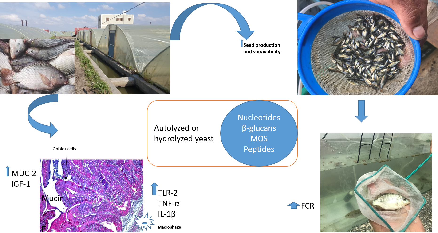
The reproductive performance of fish is influenced by many factors, e.g., nutrition, hatchery management and environmental conditions. Dietary supplementation of Hilyses in Nile tilapia broodstock showed higher seed production, immune response, and seed survival than the control group. In accordance with these findings, Abo-State and Tahoun [38] observed that 0.02% dietary supplementation with S. cerevisiae significantly improved both seed production (seed/m3) and seed/female comparatively to 0.01%; moreover, both 0.01 and 0.02% supplementations gave significantly higher values than the un-supplemented diet. In addition, Abasali and Mohamad [39] found a significant increase in fry production and fertility of Helleri females fed probiotics (containing Bifidobacterium thermophilum, Lactobacillus casei, Enterococcus faecium, and L. acidophilus) compared with non-supplemented females. Moreover, Li and Gatlin [40] first studied the nucleotide nutrition of broodstock haddock and observed that the first feeding success of larvae from nucleotide-fortified broodstock was significantly higher than that of larvae from broodstock fed a basal diet without supplemented nucleotides. In the present study, improved seed production of Nile tilapia may be related to the nutrients and bioactive materials in Hilyses that directly enhance broodstock nutrition and immunity. Izquierdo, et al. [15] stated that modification of the nutritional quality of broodstock diets in continuous spawners with short vitellogenic periods even during the spawning season improves gonadal development and seed quality.
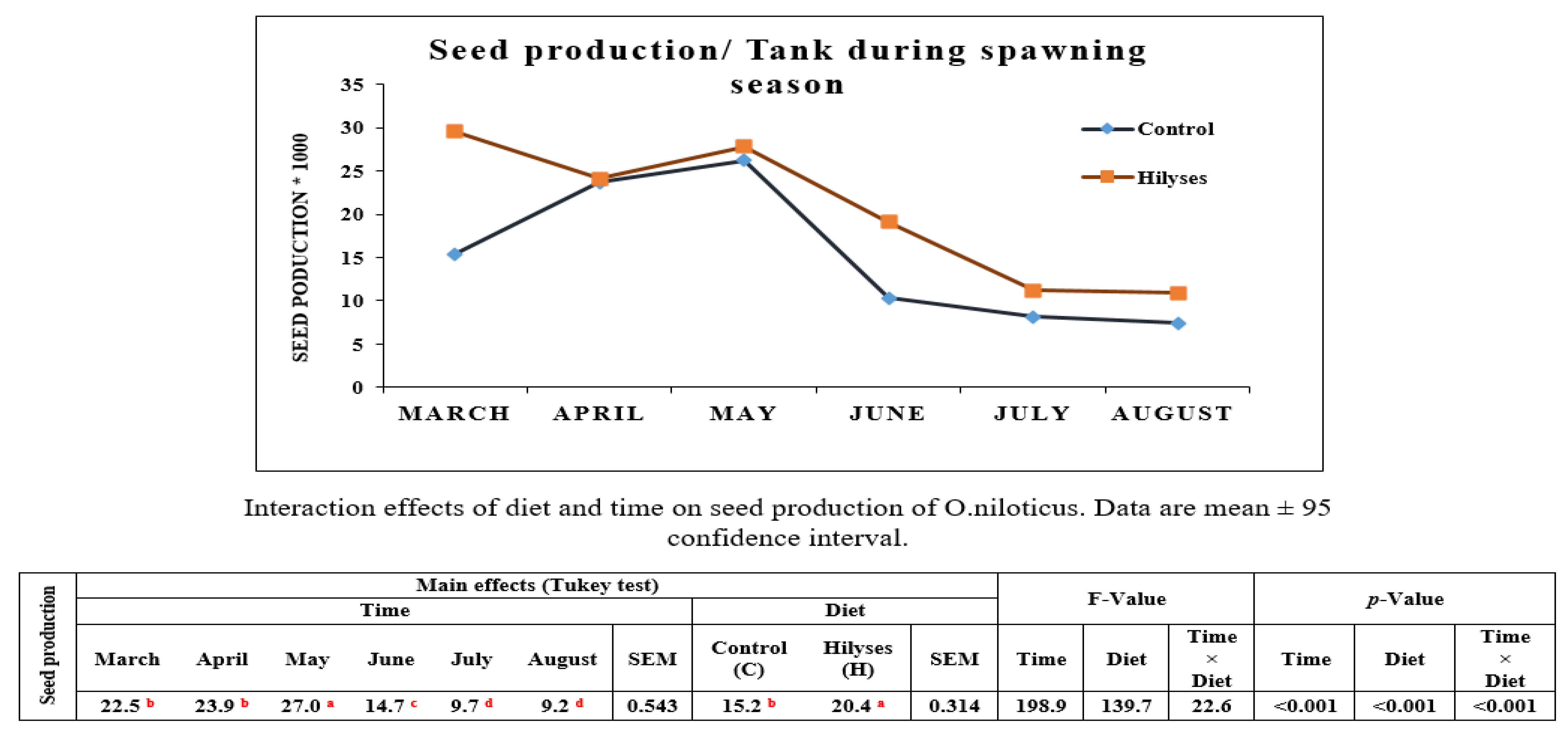
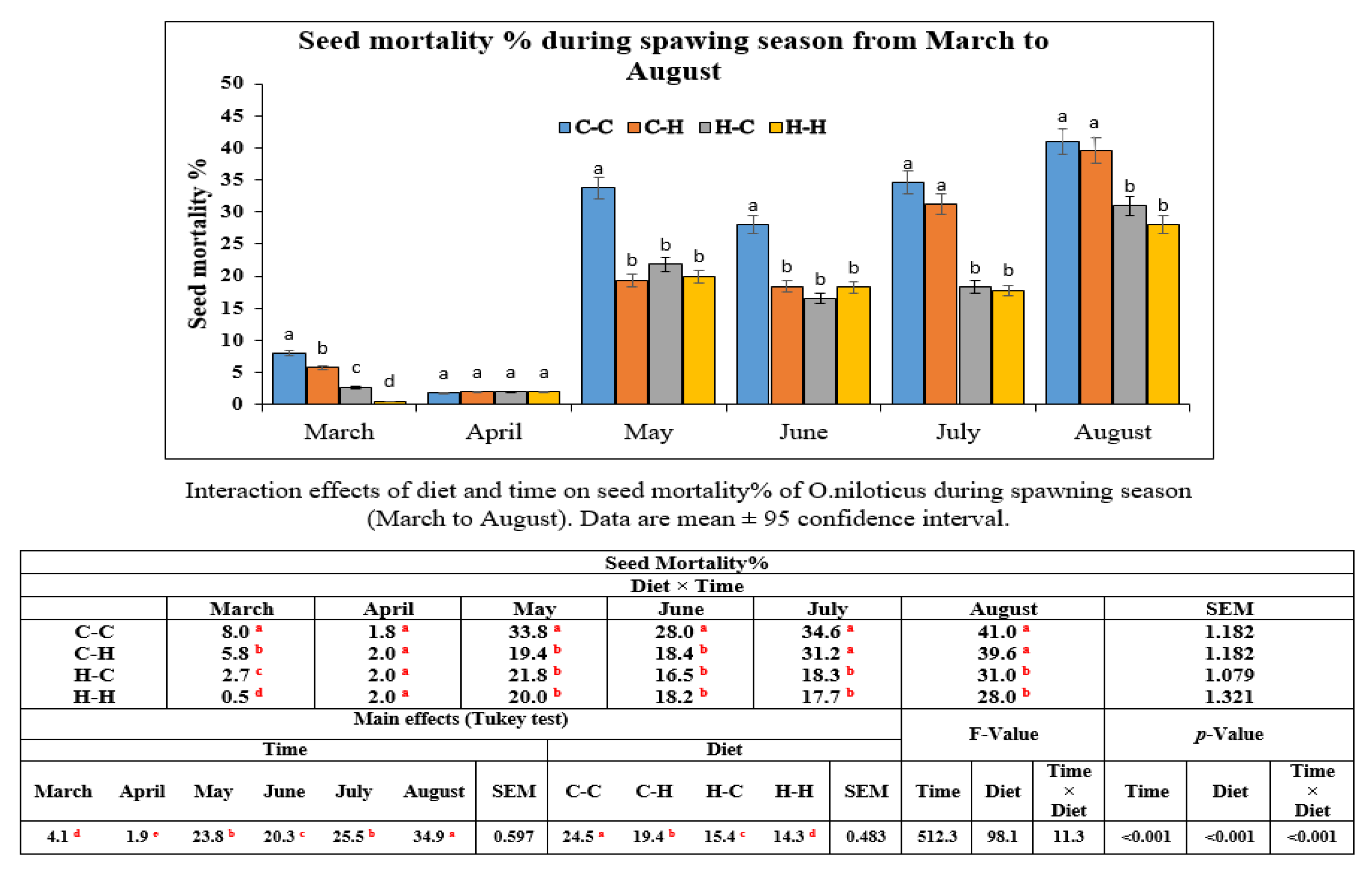
For seed survival, significant differences were found in seeds obtained from the Hilyses-treated broodstock groups (H-H and H-C), which showed the highest rate, followed by the C-H group, and the lowest survival rate was recorded in the C-C group. Transferring maternally trained innate immune elements from the Hilyses-treated broodstock into the yolk may increase the resistance of seeds against diseases and make them more resilient to adverse environmental conditions. The survival and size of larvae from haddock broodstock fed a nucleotide-supplemented diet were greater than that of larvae from broodstock fed the basal diet [15]. The mechanisms by which dietary nucleotides, β-glucans, and MOS promote growth and survivability include the improvement of intestinal morphology and the reduction of energy required for nucleotide synthesis, which allows more energy for the metabolic functions needed for the development and growth of fish larvae [41].
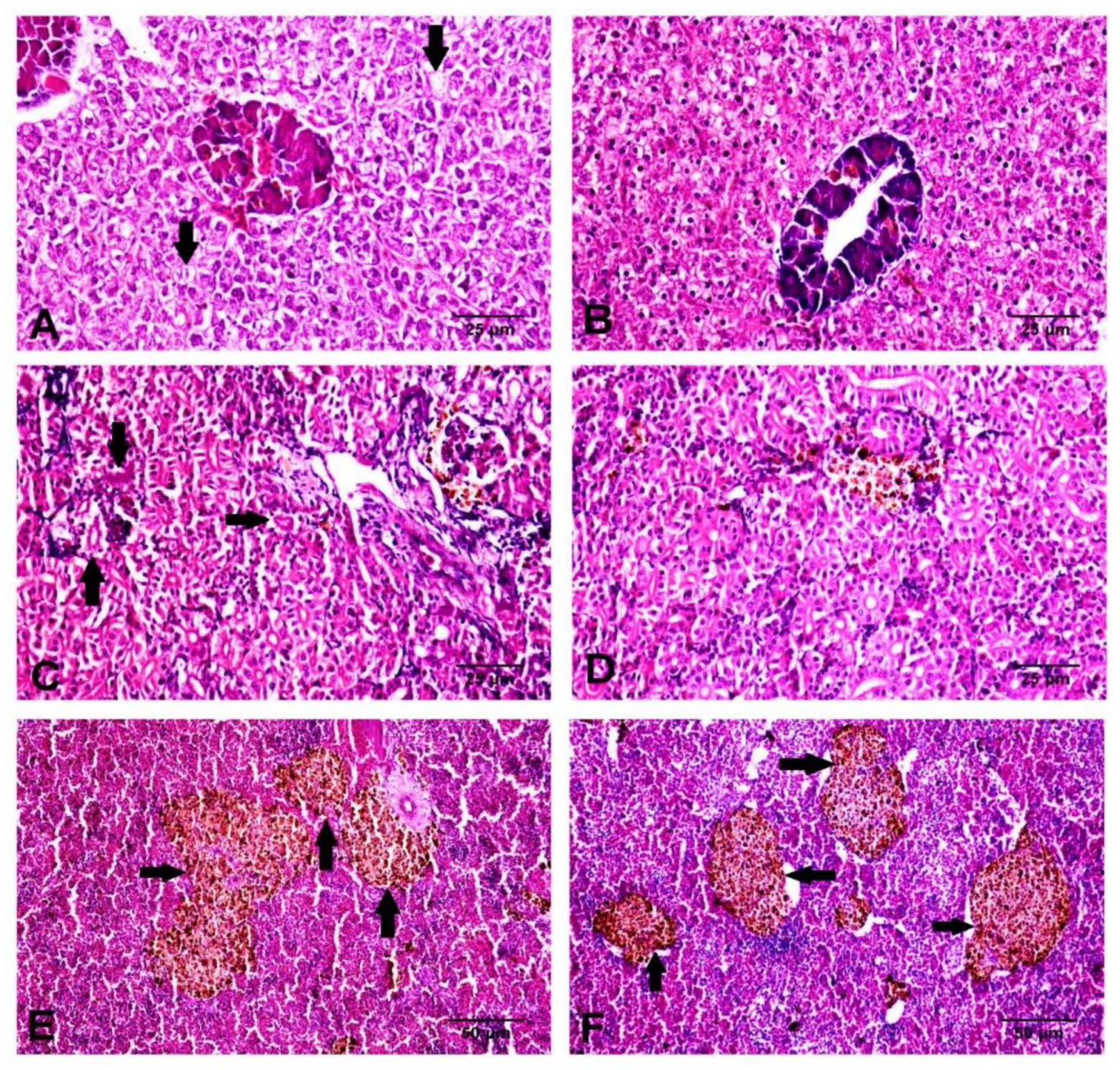
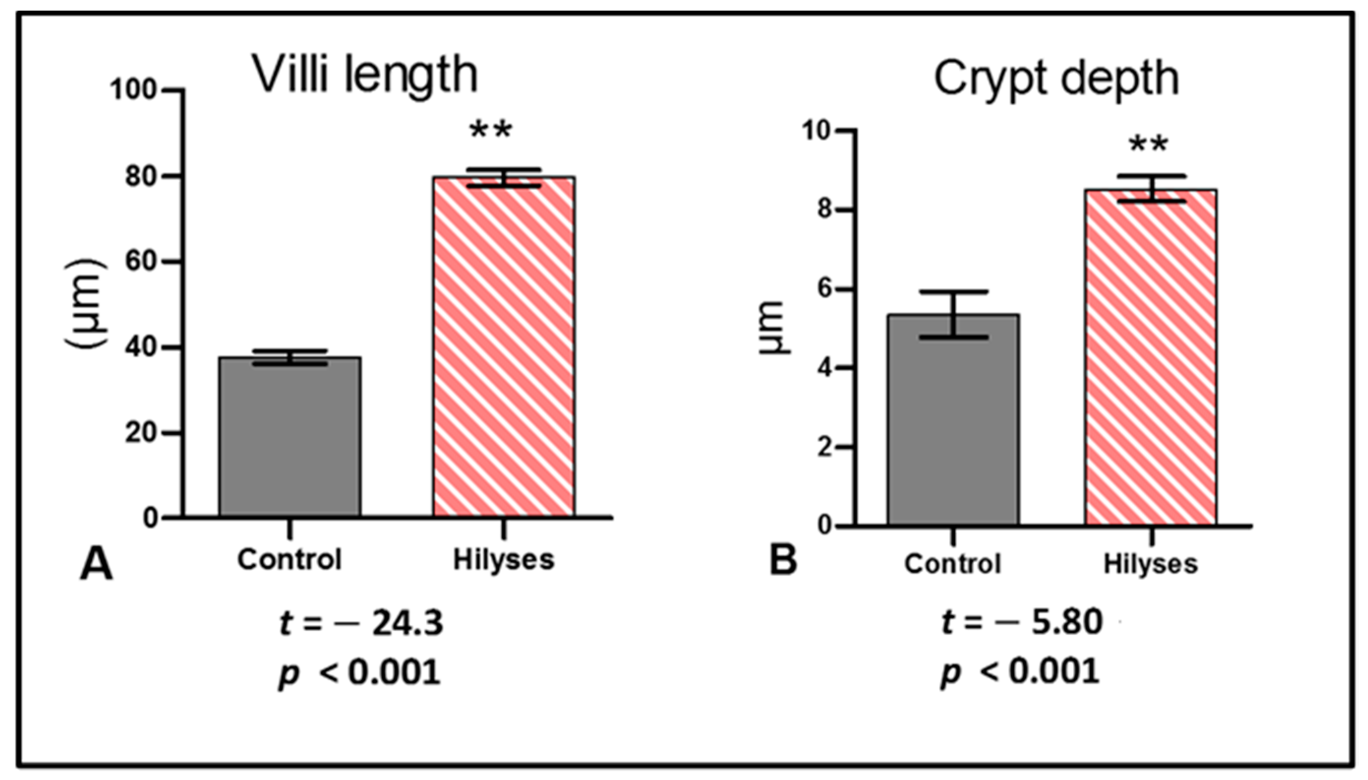
Gut health is considered the main factor that could affect the growth, reproductive performance, and immune status of aquatic animals [42,43]. Histological findings of the intestinal tract showed an improvement in intestinal microstructure in terms of increased fold height, enterocyte height and microvilli length, as well as the number of goblet cells in the Hilyses-treated group compared to the control group. This finding is consistent with the results of Dimitroglou et al. [44] and Xu et al. [45], who found that dietary MOS and nucleotides improve intestinal structure and health. Moreover, modulation of intestinal immunity is very important to maintain fish health. Mucins produced by goblet cells act as a barrier between the external environment and the internal environment of the body, select and transport essential nutrients through the epithelium, eliminate deleterious pathogens, and protect the mucosal layer from dehydration [46]. The current study revealed that dietary inclusion of Hilyses increased the expression of the MUC-2 gene in the intestine by 12.4-fold and in the liver 9.75-fold. Midhun et al. [32] reported that dietary probiotics could modulate MUC-2 gene expression in O. niloticus. Additionally, Nieves-Rodríguez et al. [47] showed that the expression of MUC-2 in Atractosteus tropicus tended to increase with the addition of a prebiotic, β-1,3/1,6-glucan derived from yeast at 1.5%, while there was a decrease in the fish fed 2% of the prebiotic, which indicated that high concentrations of this immune stimulant inhibited the expression of MUC-2. The elevated IGF-1 RNA levels in the liver and intestine of the Hilyses-treated group confirmed its potential growth-promoting effect. Midhun et al. [32] stated that hepatic IGF-1 mRNA expression could alter the growth rate in juvenile salmon.
Hematological and biochemical parameters usually reflect fish health status [48]. The recorded significant increase in hematological indices (including MCV and MCH) with a nonsignificant difference in MCHC in the Hilyses-supplemented group indicates that Hilyses has a positive impact on erythrogram, which directly reflects the health and productive status of Nile tilapia. In a similar sense, Abu-Elala et al. [49] observed a significant increase in RBC count, WBCs count, Hb concentration and PCV percent in Nile tilapia fed S. cerevisiae byproducts. The improved erythrogram could be related to the hepatostimulatory and hepatoprotective effects of Hilyses. The values of RBCs, hematocrit, and Hb were increased in the Hilyses-supplemented group. These results probably are associated with the regulation of the metabolic rate and wellbeing of Nile tilapia induced by S. cerevisiae feeding. Increasing of RBCs and Hb causes oxygen availability in the body tissues, owing to the absence of anemic features. The balanced levels of RBCs, hematocrit, and Hb were also attributed to the role of dietary S. cerevisiae in regulating the immune system. The antioxidant influence of the S. cerevisiae result in the balance of the protection of RBCs membranes and increases their life span by defending them against harmful impacts of oxygen-free radicals (ROS) and alleviating anemia and membrane disruption, as well as reducing cell hemolysis and degeneration [50,51]. In this regard Dawood et al. [46] reported that Nile tilapia fed dietary S. cerevisiae had enhanced RBCs, hematocrit, and Hb indices. Concurrent with the hematological indices, the results of related hepatic enzymes (ALT and AST), renal function (urea and creatinine), and blood protein profile (total protein, albumin, and globulin) of Nile tilapia serum following feeding, with or without Hilyses, refer to the stable metabolic rate under the current study conditions without apparent impairment of the fish health status [52].
The oxidative stress occurs during stressful environmental conditions, and pathogenic invasions that liberate ROS due to the imbalance of free radicals production and removal [53]. Excessive ROS also induces lipid peroxidation and damages RNA, which can be measured by the level of malondialdehyde (MDA) [54]. The total serum antioxidant capacity and GSH activity in the liver and intestine of fish fed Hilyses showed increased levels, while the MDA level was decreased which probably associated with the radical scavenge potential of the functional substances (nucleotides, β-glucans, and MOS) [55]. Similarly, Abu-Elala et al. [5] stated that the antioxidative response and MDA level were lowered in fish fed dietary S. cerevisiae.
High levels of cortisol indicate that the organism is suffering from stressful conditions and requires high glucose levels to afford the energy needed to cope with the stress [56,57]. Interestingly, the obtained results highlighted that tilapia-fed Hilyses had reduced cortisol level compared to fish fed the basal diet. Probiotics and prebiotics are known for the antistress influence, which is attributed to their role in improving the immunity and antioxidative capacity of aquatic organisms [58,59].
The enhancement of the innate immunity offers an interesting and attractive approach to increase the disease resistance of broodstock, newly hatched fish larvae and the first feeding fish, especially since the adaptive immune system is not fully developed [60]. Dietary inclusion of Hilyses in broodstock feeds upregulated some immune-related genes, such as TLR-2, IL-1β and TNF-α, in the liver and intestinal tissues. Zhang, et al. [60] suggested that to induce trained innate immunity by β-glucans, several different receptors must be engaged, such as Dectin-1 and TLRs. Stimulation of broodstock with β-glucans or any pathogen-associated molecular pattern (PAMP) at low doses increased the potential not only to resist pathogens, but also to transfer trained innate immunity to offspring. In addition to the direct innate immune training of broodstock, the molecules that are known to induce trained innate immunity may be maternally transferred and taken up by developing oocytes during vitellogenesis, potentially increasing the innate defense of developing embryos/larvae while the fish embryo or larvae are still in the eggs.
Gut-associated lymphoid tissue (GALT) is a diffusely arranged epithelium and includes immune cells along the teleost gut. This tissue expresses various pattern recognition receptors (PRRs), such as TLRs, which show significant expression when engaged with β-glucans. The ligation of TLR-2 with β-glucans led to the upregulation of proinflammatory cytokine genes, such as TNF-α and IL-1β, without any inflammatory signs in tissues. This finding may be due to the expression of anti-inflammatory cytokines and increased antioxidant enzyme activity [32]. Several articles have indicated that upregulation of proinflammatory cytokines improves immune promptness and increases disease resistance in tilapia [9,18,61].
Dietary fortification of Hilyses improved the growth performance and feed utilization of Nile tilapia fry. This finding may be due to the acceleration of digestive system maturation and to the increased nutrient digestion induced by the nutrients and bioactive compounds in Hilyses. Essential amino acids, such as glutamic acid, play a substantial role in tissue proliferation and regeneration that consequently improve nutrient digestion and feed utilization [62]. Modulation of intestinal microbiota by MOS increases villus integrity and resistance to pathogenic bacteria, which increases digestion and absorption efficiency [17,47,63]. β-glucans stimulate the proliferation of beneficial gut flora and eliminate pathogenic gut flora, resulting in increased weight gain [64]. In a similar sense, the use of prebiotics containing β-glucans and MOS improved the growth performance of Nile tilapia, common carp (Cyprinus carpio), beluga (Huso huso), and sea cucumbers (Apostichopus japonicus) [63,64,65,66,67]. In addition, the use of S. cerevisiae live cells (20 g/kg) markedly improved the growth performance and nutrient utilization in Nile tilapia broodstock [38]. Notably, the present study suggested the dose of 0.4% (4 g/kg diet) of Hilyses, whereas Abo-State and Tahoun [38] recommended a 20 g/kg diet of S. cerevisiae for enhancing the performance of tilapia broodstock. The low dose of Hilyses indicates that fermented yeast cell wall components (e.g., nucleotides, β-glucans, and MOS) in its pure form could be more effective than the incorporation of the whole yeast live cells.

5. Conclusions

In summary, boosting the nutrition and immunity of Nile tilapia broodstock during the spawning season by using dietary fermented yeast products improved fry production and survival. Dietary inclusion of Hilyses modulated some growth- and immune-related genes and improved the intestinal microstructure morphology. Furthermore, this treatment increased the antioxidant enzyme activity and reduced cortisol levels. Hilyses had major effects on the growth performance and feed utilization in fry, especially those obtained from the Hilyses-treated broodstock and fed 0.4% Hilyses over three months.

Author Contributions

Conceptualization, N.M.A.-E., T.E.-S.A., N.A.Y., D.A.A.-M., A.H.H., M.B. and M.A.O.D.; Data curation, T.E.-S.A.; Formal analysis, T.E.-S.A., N.M.R., S.E.A., R.M.A.-E., N.A.Y. and A.H.H.; Funding acquisition, N.M.A.-E., N.A.Y. and M.B.; Investigation, N.M.A.-E., N.M.R., S.E.A., R.M.A.-E. and N.A.Y.; Methodology, T.E.-S.A., N.M.R., S.E.A., R.M.A.-E., D.A.A.-M. and A.H.H.; Resources, N.M.R., S.E.A., D.A.A.-M. and M.B.; Writing—original draft, N.M.A.-E. and M.A.O.D. All authors have read and agreed to the published version of the manuscript.

Funding

This research received no external funding.

Institutional Review Board Statement

All experiments were conducted in accordance with the recommendations in the Guide for the Care and Use of Laboratory Animals approved by the Institutional Animal Use and Care Committee (Vet. CU. IACUC), Vet CU24112020238.

Data Availability Statement

The data presented in this study are available on request from the corresponding author.

Acknowledgments

Authors are very grateful to Engineer Ahmed El Sharaky, the owner of the hatchery, for his kind and continuous support to achieve our experiment.

Conflicts of Interest

The authors declare no conflict of interest.

References

  1. Shaalan, M.; El-Mahdy, M.; Saleh, M.; El-Matbouli, M. Aquaculture in Egypt: Insights on the Current Trends and Future Perspectives for Sustainable Development. Rev. Fish. Sci. Aquac. 2018, 26, 99–110. [Google Scholar] [CrossRef]
  2. Magouz, F.I.; Mahmoud, S.A.; El-Morsy, R.A.; Paray, B.A.; Soliman, A.A.; Zaineldin, A.I.; Dawood, M.A. Dietary menthol essential oil enhanced the growth performance, digestive enzyme activity, immune-related genes, and resistance against acute ammonia exposure in Nile tilapia (Oreochromis niloticus). Aquaculture 2021, 530, 735944. [Google Scholar] [CrossRef]
  3. Moffitt, C.M.; Cajas-Cano, L. Blue Growth: The 2014 FAO State of World Fisheries and Aquaculture. Fisheries 2014, 39, 552–553. [Google Scholar] [CrossRef]
  4. Food and Agriculture Organization of the United Nations (FAO). Aquaculture Department, the State of World Fisheries and Aquaculture; FAO: Rome, Italy, 2018. [Google Scholar]
  5. Abu-Elala, N.M.; Younis, N.A.; Abubakr, H.O.; Ragaa, N.M.; Borges, L.L.; Bonato, M.A. Influence of dietary fermented Saccharomyces cerevisiae on growth performance, oxidative stress parameters, and immune response of cultured Oreochromis niloticus. Fish Physiol. Biochem. 2020, 46, 533–545. [Google Scholar] [CrossRef]
  6. Dawood, M.A.; Koshio, S.; Esteban, M.Á. Beneficial roles of feed additives as immunostimulants in aquaculture: A review. Rev. Aquac. 2017, 10, 950–974. [Google Scholar] [CrossRef]
  7. Abu Elala, N.M.; Galal, M.K.; Abd-Elsalam, R.M.; Mohey-Elsaeed, O.; Ragaa, N.M. Effects of Dietary Supplementation of Spirulina platensis and Garlic on the Growth Performance and Expression Levels of Immune-related Genes in Nile tilapia (Oreochromis niloticus). J. Aquac. Res. Dev. 2016, 7, 433–442. [Google Scholar] [CrossRef] [Green Version]
  8. Dawood, M.; El Basuini, M.; Zaineldin, A.; Yilmaz, S.; Hasan, T.; Ahmadifar, E.; El Asely, A.; Abdel-Latif, H.; Alagawany, M.; Abu-Elala, N.; et al. Antiparasitic and Antibacterial Functionality of Essential Oils: An Alternative Approach for Sustainable Aquaculture. Pathogens 2021, 10, 185. [Google Scholar] [CrossRef] [PubMed]
  9. Yao, Y.; Yang, Y.; Gao, C.; Zhang, F.; Xia, R.; Li, D.; Hu, J.; Ran, C.; Zhang, Z.; Liu-Clarke, J.; et al. Surface display system for probiotics and its application in aquaculture. Rev. Aquac. 2020, 12, 2333–2350. [Google Scholar] [CrossRef]
  10. Dawood, M.A. Nutritional immunity of fish intestines: Important insights for sustainable aquaculture. Rev. Aquac. 2021, 13, 642–663. [Google Scholar] [CrossRef]
  11. Yukgehnaish, K.; Kumar, P.; Sivachandran, P.; Marimuthu, K.; Arshad, A.; Paray, B.A.; Arockiaraj, J. Gut microbiota metagenomics in aquaculture: Factors influencing gut microbiome and its physiological role in fish. Rev. Aquac. 2020, 12, 1903–1927. [Google Scholar] [CrossRef]
  12. Hossain, S.; Koshio, S.; Kestemont, P. Recent advances of nucleotide nutrition research in aquaculture: A review. Rev. Aquac. 2019, 12, 1028–1053. [Google Scholar] [CrossRef]
  13. Dawood, M.; Koshio, S.; Ishikawa, M.; Yokoyama, S.; El Basuini, M.; Hossain, M.; Nhu, T.; Moss, A.; Dossou, S.; Wei, H. Dietary supplementation of β-glucan improves growth performance, the innate immune response and stress resistance of red sea bream, Pagrus major. Aquac. Nutr. 2015, 23, 148–159. [Google Scholar] [CrossRef]
  14. Dawood, M.A.; Koshio, S.; Fadl, S.E.; Ahmed, H.A.; El Asely, A.; Abdel-Daim, M.M.; Alkahtani, S. The modulatory effect of mannanoligosaccharide on oxidative status, selected immune parameters and tolerance against low salinity stress in red sea bream (Pagrus major). Aquac. Rep. 2020, 16, 100278. [Google Scholar] [CrossRef]
  15. Izquierdo, M.; Fernández-Palacios, H.; Tacon, A. Effect of broodstock nutrition on reproductive performance of fish. Aquaculture 2001, 197, 25–42. [Google Scholar] [CrossRef]
  16. Mekawey, M. Incorporation of garlic meal (Allium sativum) as natural additive to enhance performance, immunity, gonad and larval survival of Nile tilapia (Oreochromis niloticus) broodstock. Afr. J. Biol. Sci. 2019, 15, 117–135. [Google Scholar] [CrossRef] [Green Version]
  17. Freccia, A.; Sousa, S.M.D.N.; Meurer, F.; Butzge, A.J.; Mewes, J.K.; Bombardelli, R.A. Essential oils in the initial phase of broodstock diets of Nile tilapia. Rev. Bras. Zootec. 2014, 43, 1–7. [Google Scholar] [CrossRef] [Green Version]
  18. Al-Feky, S.; El-Sayed, A.-F.; Ezzat, A. Dietary taurine improves reproductive performance of Nile tilapia (Oreochromis niloticus) broodstock. Aquac. Nutr. 2014, 22, 392–399. [Google Scholar] [CrossRef]
  19. National Research Council (NRC). Nutrient Requirements of Fish and Shrimp; National Academies Press: Washington, DC, USA, 2011. [Google Scholar] [CrossRef]
  20. Dacie, J.; Lewis, S. Practical Haematology, ELBS with Churchill Livingston; Churchil Livingstone: Edinburgh, England, 1991. [Google Scholar]
  21. Stoskopf, M.K. Fish Medicine Ed; WB Saunders Company: London, UK, 1993. [Google Scholar]
  22. Dacie, S.; Lewis, S. Practical Haematology, 11th ed.; Churchill Livingstone: London, UK, 2002. [Google Scholar]
  23. Shaw, A.F.B. A direct method for counting the leukocytes, thrombocytes and erythrocytes of birds’s blood. J. Pathol. Bacteriol. 1930, 33, 833–835. [Google Scholar] [CrossRef]
  24. Lopes-Virella, M.F.; Stone, P.; Ellis, S.; Colwell, J. Cholesterol determination in high-density lipoproteins separated by three different methods. Clin. Chem. 1977, 23, 882–884. [Google Scholar] [CrossRef]
  25. Tietz, N.W. Fundamentals of Clinical Chemistry Philadelphia; WB Saudners Co Ltd.: Philadelphia, PA, USA, 1976. [Google Scholar]
  26. Tietz, N. Clinical Guide to Laboratory Tests; WB Saudners Company: Philadelphia, PA, USA, 1990; Volume 554, p. 556. [Google Scholar]
  27. Coles, E. Veterinary Clinical Pathology, 2nd ed.; W.B. Saunders Company: Philadelphia, PA, USA, 1974. [Google Scholar]
  28. Koracevic, D.; Koracevic, G.; Djordjevic, V.; Andrejevic, S.; Cosic, V. Method for the measurement of antioxidant activity in human fluids. J. Clin. Pathol. 2001, 54, 356–361. [Google Scholar] [CrossRef] [PubMed] [Green Version]
  29. Beutler, E.; Duron, O.; Kelly, M. Glutathione reagent and method-patent. J. Lab. Clin. Med. 1963, 61, 882. [Google Scholar]
  30. Ohkawa, H.; Ohishi, N.; Yagi, K. Assay for lipid peroxides in animal tissues by thiobarbituric acid reaction. Anal. Biochem. 1979, 95, 351–358. [Google Scholar] [CrossRef]
  31. Gröner, F.; Ziková, A.; Kloas, W. Effects of the pharmaceuticals diclofenac and metoprolol on gene expression levels of enzymes of biotransformation, excretion pathways and estrogenicity in primary hepatocytes of Nile tilapia (Oreochromis niloticus). Comp. Biochem. Physiol. Part C Toxicol. Pharmacol. 2015, 167, 51–57. [Google Scholar] [CrossRef] [PubMed]
  32. Midhun, S.J.; Neethu, S.; Arun, D.; Vysakh, A.; Divya, L.; Radhakrishnan, E.; Jyothis, M. Dietary supplementation of Bacillus licheniformis HGA8B improves growth parameters, enzymatic profile and gene expression of Oreochromis niloticus. Aquaculture 2019, 505, 289–296. [Google Scholar] [CrossRef]
  33. Standen, B.; Peggs, D.; Rawling, M.; Foey, A.; Davies, S.; Santos, G.; Merrifield, D. Dietary administration of a commercial mixed-species probiotic improves growth performance and modulates the intestinal immunity of tilapia, Oreochromis niloticus. Fish Shellfish. Immunol. 2016, 49, 427–435. [Google Scholar] [CrossRef] [PubMed] [Green Version]
  34. Castro, R.; Zou, J.; Secombes, C.J.; Martin, S.A. Cortisol modulates the induction of inflammatory gene expression in a rainbow trout macrophage cell line. Fish Shellfish. Immunol. 2011, 30, 215–223. [Google Scholar] [CrossRef]
  35. Yuan, J.S.; Reed, A.; Chen, F.; Stewartjr, C.N. Statistical analysis of real-time PCR data. BMC Bioinform. 2006, 7, 85. [Google Scholar] [CrossRef] [PubMed] [Green Version]
  36. Abu-Elala, N.M.; Abd-Elsalam, R.M.; Marzouk, M.S. Molecular and Immunohistochemical Diagnosis of Photobacterium damselae Subspecies piscicida During Naturally Occurring Disease in Egypt. J. World Aquac. Soc. 2015, 46, 583–595. [Google Scholar] [CrossRef]
  37. Abu Elala, N.M.; Ragaa, N.M. Eubiotic effect of a dietary acidifier (potassium diformate) on the health status of cultured Oreochromis niloticus. J. Adv. Res. 2015, 6, 621–629. [Google Scholar] [CrossRef] [PubMed] [Green Version]
  38. Abo-State, H.A.; Tahoun, A.-A.M. Effect of Saccharomyces cerevisiae on seed production and fry performance of Oreochromis niloticus broodstock. Biosci. Res. 2017, 14, 1175–1181. [Google Scholar]
  39. Abasali, H.; Mohamad, S. Effect of Dietary Supplementation with Probiotic on Reproductive Performance of Female Livebearing Ornamental Fish. Res. J. Anim. Sci. 2010, 4, 103–107. [Google Scholar] [CrossRef] [Green Version]
  40. Li, P.; Gatlin, D.M. Nucleotide nutrition in fish: Current knowledge and future applications. Aquaculture 2006, 251, 141–152. [Google Scholar] [CrossRef]
  41. Guo, X.; Ran, C.; Zhang, Z.; He, S.; Jin, M.; Zhou, Z. The Growth-Promoting Effect of Dietary Nucleotides in Fish Is Associated with an Intestinal Microbiota-Mediated Reduction in Energy Expenditure. J. Nutr. 2017, 147, 781–788. [Google Scholar] [CrossRef] [PubMed] [Green Version]
  42. Magouz, F.I.; Dawood, M.A.; Salem, M.F.; El-Ghandour, M.; Van Doan, H.; Mohamed, A.A. The role of a digestive enhancer in improving the growth performance, digestive enzymes activity, and health condition of Nile tilapia (Oreochromis niloticus) reared under suboptimal temperature. Aquaculture 2020, 526, 735388. [Google Scholar] [CrossRef]
  43. Zaineldin, A.I.; Hegazi, S.; Koshio, S.; Ishikawa, M.; Dawood, M.A.; Dossou, S.; Yukun, Z.; Mzengereza, K. Singular effects of Bacillus subtilis C-3102 or Saccharomyces cerevisiae type 1 on the growth, gut morphology, immunity, and stress resistance of red sea bream (Pagrus major). Ann. Anim. Sci. 2020. [Google Scholar] [CrossRef]
  44. Dimitroglou, A.; Merrifield, D.L.; Moate, R.; Davies, S.J.; Spring, P.; Sweetman, J.; Bradley, G. Dietary mannan oligosaccharide supplementation modulates intestinal microbial ecology and improves gut morphology of rainbow trout, Oncorhynchus mykiss (Walbaum). J. Anim. Sci. 2009, 87, 3226–3234. [Google Scholar] [CrossRef] [Green Version]
  45. Xu, L.; Ran, C.; He, S.; Zhang, J.; Hu, J.; Yang, Y.; Du, Z.; Yang, Y.; Zhou, Z. Effects of dietary yeast nucleotides on growth, non-specific immunity, intestine growth and intestinal microbiota of juvenile hybrid tilapia Oreochromis niloticus ♀ × Oreochromis aureus ♂. Anim. Nutr. 2015, 1, 244–251. [Google Scholar] [CrossRef] [PubMed]
  46. Dawood, M.A.; Eweedah, N.M.; Khalafalla, M.M.; Khalid, A.; El Asely, A.; Fadl, S.E.; Amin, A.A.; Paray, B.A.; Ahmed, H.A. Saccharomyces cerevisiae increases the acceptability of Nile tilapia (Oreochromis niloticus) to date palm seed meal. Aquac. Rep. 2020, 17, 100314. [Google Scholar] [CrossRef]
  47. Nieves-Rodríguez, K.N.; Álvarez-González, C.A.; Peña-Marín, E.S.; Vega-Villasante, F.; Martínez-García, R.; Camarillo-Coop, S.; Tovar-Ramírez, D.; Guzmán-Villanueva, L.T.; Andree, K.B.; Gisbert, E. Effect of β-Glucans in Diets on Growth, Survival, Digestive Enzyme Activity, and Immune System and Intestinal Barrier Gene Expression for Tropical Gar (Atractosteus tropicus) Juveniles. Fishes 2018, 3, 27. [Google Scholar] [CrossRef] [Green Version]
  48. Fazio, F. Fish hematology analysis as an important tool of aquaculture: A review. Aquaculture 2019, 500, 237–242. [Google Scholar] [CrossRef]
  49. Abu-Elala, N.; Marzouk, M.; Moustafa, M. Use of different Saccharomyces cerevisiae biotic forms as immune-modulator and growth promoter for Oreochromis niloticus challenged with some fish pathogens. Int. J. Veter Sci. Med. 2013, 1, 21–29. [Google Scholar] [CrossRef] [Green Version]
  50. Mohapatra, S.; Chakraborty, T.; Kumar, V.; DeBoeck, G.; Mohanta, K.N. Aquaculture and stress management: A review of probiotic intervention. J. Anim. Physiol. Anim. Nutr. 2013, 97, 405–430. [Google Scholar] [CrossRef] [PubMed]
  51. Di Giulio, R.T.; Washburn, P.C.; Wenning, R.J.; Winston, G.W.; Jewell, C.S. Biochemical responses in aquatic animals: A review of determinants of oxidative stress. Environ. Toxicol. Chem. 1989, 8, 1103–1123. [Google Scholar] [CrossRef]
  52. Uribe, C.; Folch, H.; Enriquez, R.; Moran, G. Innate and adaptive immunity in teleost fish: A review. Veterinární Med. 2011, 56, 486–503. [Google Scholar] [CrossRef] [Green Version]
  53. Martínez-Álvarez, R.M.; Morales, A.E.; Sanz, A. Antioxidant Defenses in Fish: Biotic and Abiotic Factors. Rev. Fish Biol. Fish. 2005, 15, 75–88. [Google Scholar] [CrossRef]
  54. Brewer, M.S. Natural Antioxidants: Sources, Compounds, Mechanisms of Action, and Potential Applications. Compr. Rev. Food Sci. Food Saf. 2011, 10, 221–247. [Google Scholar] [CrossRef]
  55. Santacroce, M.P.; Merra, E.; Centoducati, G.; Zacchino, V.; Casalino, E. Effects of dietary yeast Saccaromyces cerevisiae on the antioxidant system in the liver of juvenile sea bass Dicentrarchus labrax. Fish Physiol. Biochem. 2012, 38, 1497–1505. [Google Scholar] [CrossRef] [PubMed]
  56. Odhiambo, E.; Angienda, P.O.; Okoth, P.; Onyango, D. Stocking Density Induced Stress on Plasma Cortisol and Whole Blood Glucose Concentration in Nile Tilapia Fish (Oreochromis niloticus) of Lake Victoria, Kenya. Int. J. Zool. 2020, 2020, 1–8. [Google Scholar] [CrossRef]
  57. Girao, P.M.; Pereira da Silva, E.M.; de Melo, M.P. Dietary lycopene supplementation on Nile tilapia (Oreochromis niloticus) juveniles submitted to confinement: Effects on cortisol level and antioxidant response. Aquac. Res. 2012, 43, 789–798. [Google Scholar] [CrossRef]
  58. Huynh, T.-G.; Shiu, Y.-L.; Nguyen, T.-P.; Truong, Q.-P.; Chen, J.-C.; Liu, C.-H. Current applications, selection, and possible mechanisms of actions of synbiotics in improving the growth and health status in aquaculture: A review. Fish Shellfish. Immunol. 2017, 64, 367–382. [Google Scholar] [CrossRef] [PubMed]
  59. Dawood, M.A.; Abo-Al-Ela, H.G.; Hasan, T. Modulation of transcriptomic profile in aquatic animals: Probiotics, prebiotics and synbiotics scenarios. Fish Shellfish. Immunol. 2020, 97, 268–282. [Google Scholar] [CrossRef]
  60. Zhang, Z.; Chi, H.; Dalmo, R.A. Trained Innate Immunity of Fish Is a Viable Approach in Larval Aquaculture. Front. Immunol. 2019, 10, 42. [Google Scholar] [CrossRef] [Green Version]
  61. Welker, T.L.; Lim, C.; Yildirim-Aksoy, M.; Klesius, P.H. Use of diet crossover to determine the effects of β-glucan supplementation on immunity and growth of Nile tilapia, Oreochromis niloticus. J. World Aquac. Soc. 2012, 43, 335–348. [Google Scholar] [CrossRef]
  62. Cruzat, V.; Rogero, M.M.; Keane, K.N.; Curi, R.; Newsholme, P. Glutamine: Metabolism and Immune Function, Supplementation and Clinical Translation. Nutrients 2018, 10, 1564. [Google Scholar] [CrossRef] [PubMed] [Green Version]
  63. Abu-Elala, N.M.; Younis, N.A.; AbuBakr, H.O.; Ragaa, N.M.; Borges, L.L.; Bonato, M.A. Efficacy of dietary yeast cell wall supplementation on the nutrition and immune response of Nile tilapia. Egypt. J. Aquat. Res. 2018, 44, 333–341. [Google Scholar] [CrossRef]
  64. Aramli, M.S.; Kamangar, B.; Nazari, R.M. Effects of dietary β-glucan on the growth and innate immune response of juvenile Persian sturgeon, Acipenser persicus. Fish Shellfish. Immunol. 2015, 47, 606–610. [Google Scholar] [CrossRef] [PubMed]
  65. Gu, M.; Ma, H.; Mai, K.; Zhang, W.; Bai, N.; Wang, X. Effects of dietary β-glucan, mannan oligosaccharide and their combinations on growth performance, immunity and resistance against Vibrio splendidus of sea cucumber, Apostichopus japonicus. Fish Shellfish. Immunol. 2011, 31, 303–309. [Google Scholar] [CrossRef] [PubMed]
  66. Ta’Ati, R.; Soltani, M.; Bahmani, M.; Zamini, A.A. Effects of the prebiotics Immunoster and Immunowall on growth performance of juvenile beluga (Huso huso). J. Appl. Ichthyol. 2011, 27, 796–798. [Google Scholar] [CrossRef]
  67. Ebrahimi, G.; Ouraji, H.; Khalesi, M.K.; Sudagar, M.; Barari, A.; Zarei Dangesaraki, M.; Jani Khalili, K.H. Effects of a prebiotic, immunogen®, on feed utilization, body composition, immunity and resistance to Aeromonas hydrophila infection in the common carp Cyprinus carpio (linnaeus) fingerlings. J. Anim. Physiol. Anim. Nutr. 2012, 96, 591–599. [Google Scholar] [CrossRef] [PubMed]
Figure 1. Summary of experimental design.
Animals 11 00815 g001
Figure 2. Seed production in tilapia broodstock fed a Hilyses (H) and basal diet (C) during the spawning season from March to August. The cement tank area was 3 × 8 m2 stocked with 40 females/16 males in each tank (n = 11 tanks), Temp. = 30 ± 1 °C, pH = 8.6 ± 0.2. The results were statistically analyzed using two-way analysis of variance (ANOVA) with type of diets and time as factors, followed by Tukey’s HSD test for mean multi-comparison test. Significance was determined at p < 0.05. a, b, c, d Means within a row with different superscripts are significantly different (p < 0.05).
Animals 11 00815 g002
Figure 3. Mortality in Hilyses and control groups over the period of six months (spawning season) starting from March to August. The results were statistically analyzed by using two-way analysis of variance (ANOVA) with type of diets and time as factors, followed by Tukey’s HSD test for mean multi-comparison test. Significance was determined at p < 0.05. a, b, c, d Means with different superscripts are significantly different (p < 0.05).
Animals 11 00815 g003
Figure 4. Oxidant and antioxidant biomarkers and cortisol levels in both broodstock groups. (A). Malondialdehyde lipid peroxide (MDA) levels in the intestine and liver of the control and Hilyses groups; (B). Intestinal and hepatic reduced glutathione (GSH) activity of the control and Hilyses groups; (C). Serum total antioxidant capacity of the control and Hilyses groups; (D). Cortisol activity of the control and Hilyses groups. The results were statistically analyzed by independent samples’ t test and expressed as the mean ± S.E. (n = 10). * statistically significant (p < 0.05), ** Statistically highly significant (p < 0.01) compared to the control group.
Animals 11 00815 g004
Figure 5. Relative expression of IGF1, TLR-2, Muc-2, TNF alpha and IL-1β. (A) gene expression of liver samples of Hilyses-treated fish (H): IGF1 (p = 0.019), TLR-2 (p = 0.010), Muc-2 (p = 0.008), TNF alpha (p = 0.016), and IL-1β (p = 0.034) are up-regulated (p < 0.05) in comparison to control group (C); (B) gene expression of intestinal samples of Hilyses-treated fish (H): IGF1 (p = 0.015), TLR-2 (p = 0.011), MUC-2 (p = 0.040), TNF alpha (p = 0.019), and IL-1β (p = 0.010) are up-regulated (p < 0.05) in comparison to control group (C). Data are represented as means ± S.E. (n = 10).
Animals 11 00815 g005
Figure 6. Histological examination of the liver, kidney, and spleen in the control and Hilyses-treated groups. (A). Liver of the control group showing vacuolation of hepatocytes (arrow) (H&E ×400); (B). Liver of the Hilyses-treated group showing normal histological characteristics of hepatocytes (H&E ×400); (C). Kidney of the control group showing necrosis of some renal tubular epithelium (arrow) (H&E ×400); (D). The Hilyses-treated group showing normal histological architecture of the kidney (H&E ×400); (E). Spleen of the control group showing moderate activation of MMCs (arrow) (H&E ×200); (F). Spleen of the treated group showing marked activation of MMCs (arrow) (H&E ×200).
Animals 11 00815 g006
Figure 7. Histological examination of the intestines of the control and Hilyses-treated groups. (A). Control group showing intestinal villi (IV) (H&E ×100); (B). Higher magnification of the previous picture showing sloughing and necrosis of some enterocytes (arrow) (H&E ×200); (C). Hilyses-treated group showing intact intestinal villi (IV) (H&E ×100); (D). Hilyses-treated group showing an intestine with severe hyperplasia of enterocytes (arrow) (H&E ×200).
Animals 11 00815 g007
Figure 8. Histological examination of the intestines of the control and Hilyses-treated groups stained with Alcian blue-PAS techniques (×200). (A). Control group showing few goblet cells containing neutral mucins (arrow); (B). Hilyses-treated group showing numerous goblet cells containing neutral mucins (arrow); (C). Control group showing small numbers of goblet cells containing acid mucins (arrow); (D). Hilyses-treated group showing large numbers of goblet cells containing acid mucins (arrow).
Animals 11 00815 g008
Figure 9. Morphometric analysis of the intestine. (A). Villi length; (B). Crypt depth. The results are expressed as the mean ± S.E. (n = 10) and were analyzed statistically by independent samples t tests, ** statistically highly significant (p < 0.01).
Animals 11 00815 g009
Table 1. Ingredients (%) and chemical composition of the experimental diets.
Ingredient %Diet 1 (36% CP 5)Diet 2 (40% CP)Diet 3 (30% CP)
Fish meal (67%)21.823.5516
Soybean meal (48%)37.344.8430.5
Yellow corn29.1821.3338.22
Wheat flour6.515.758.5
Soybean oil333.5
Mono calcium phosphate (23.7%)0.80.281.3
Calcium carbonate0.420.340.87
Common salt0.50.50.5
Premix 10.30.30.3
Vitamin C0.10.10.1
DL-Methionine0.0800.2
BHT 20.010.010.01
Total100100100
Calculated Analysis (%)
Crude protein36.044030.03
Dry matter90.1890.4189.9
Ash8.658.758.2
Ether extract8.48.638.39
Crude fiber3.583.953.26
NFE 343.3338.6650.1
Gross energy (kcal/100 g) 4460.67466.08454.46
Calcium1.221.191.23
Total phosphorus1.0611.01
Methionine0.760.750.75
Lysine2.252.541.79
Threonine1.441.611.174
Linoleic acid2.362.222.78
Chemical Analysis (%)
Crude protein36.1140.0830.07
Dry matter90.0790.3789.93
Ash8.678.738.18
Ether extract8.438.568.36
Crude fiber3.613.983.37
Calcium1.201.211.25
Total phosphorus1.041.011.03
1 Each kg vitamin and mineral mixture premix contained vitamin A, 4.8 million IU; D3, 0.8 million IU; E, 4 g; K, 0.8 g; B1, 0.4 g; riboflavin, 1.6 g; B6, 0.6 g, B12, 4 mg; pantothenic acid, 4 g; nicotinic acid, 8 g; folic acid, 0.4 g; biotin, 20 mg; Mn, 22 g; Zn, 22 g; Fe, 12 g; Cu, 4 g; I, 0.4 g, selenium, 0.4 g; and Co, 4.8 mg. 2 Antioxidant–butylated hydroxytoluene. 3 Nitrogen-free extract. 4 Gross energy. 5 Crude protein. Based on 5.65 Kcal/g protein, 9.45 Kcal/g fat, and 4.1 carbohydrate Kcal/g [19].
Table 2. Environmental water parameters measured daily in the hatchery during the trial.
ParametersMarchAprilMayJuneJulyAugust
Temperature (°C)28 ± 128 ± 130 ± 130 ± 131 ± 132 ± 1
Dissolved oxygen (mg/L)5.9 ± 0.26 ± 0.36.3 ± 0.26.2 ± 0.16.5 ± 0.26.5 ± 0.1
pH8.3 ± 0.28.6 ± 0.28.6 ± 0.28.7 ± 0.18.7 ± 0.38.7 ± 0.2
Total ammonia (mg/L)0.1 ± 0.020.1 ± 0.010.2 ± 0.020.2 ± 0.020.2 ± 0.010.2 ± 0.01
Table 3. Primer sequences and target genes for SYBR Green RT-PCR.
GenesPrimer SequencesReference
EF-1α (housekeeping)CCTTCAACGCTCAGGTCATCGröner et al. [31]
TGTGGGCAGTGTGGCAATC
IGF1CATCGTGGACGAGTGCTGMidhun et al. [32]
ACAGGTGCACAGTACATCTCAAG
TLR-2CCCACAATGGATTCACCAG
AAAGATCAAGACTCAAGGCACTG
MUC-2CAACTGTTTTTGAGACAACTTCAGA
CTGAAGTGACCGTGGAAGG
TNF alphaCCAGAAGCACTAAAGGCGAAGAStanden et al. [33]
CCTTGGCTTTGCTGCTGATC
IL-1βGCTGGAGAGTGCTGTGGAAGAACATATAGCastro et al. [34]
CCTGGAGCATCATGGCGTG
Table 4. Hematological parameters of Nile tilapia broodstock fed Hilyses and the control diets.
ParametersControlHilysest-Valuep-Value
RBCs count (×106 cell/mm3)1.52 ± 0.021.68 ± 0.05−2.8360.02 *
Hematocrit value (%)30.80 ± 1.3938.80 ± 1.62−3.7380.006 **
Hb (g/dL)5.79 ± 0.156.93 ± 0.17−4.9500.001 **
MCV (fl)202.95 ± 7.83230.71 ± 3.14−3.2880.01 **
MCH (pg)38.22 ± 0.8541.29 ± 0.48−3.1340.01 **
MCHC (%)18.92 ± 0.6617.92 ± 0.451.2410.25
WBCs count (×103 cell/mm3)61.60 ± 1.4363.80 ± 0.80−1.3390.2
Granulocyte %38.00 ± 0.4535.40 ± 0.683.2000.01 **
Lymphocyte %58.00 ± 0.7160.80 ± 0.86−2.5140.04 *
Monocyte %4.00 ± 0.453.80 ± 0.370.3430.74
The results are expressed as the mean ± S.E. (n = 10) and were analyzed statistically by independent samples t tests, * statistically significant (p < 0.05), ** statistically highly significant (p < 0.01).
Table 5. Serum biochemical parameters of Nile tilapia broodstock-fed Hilyses and the control diets.
Parameters ControlHilysest-Valuep-Value
ALT (U/L)5.44 ± 0.434.54 ± 0.281.7760.11
AST (U/L)42.24 ± 3.6638.58 ± 3.450.7300.49
Total protein (g/dL)3.10 ± 0.092.97 ± 0.140.7620.47
Albumin (g/dL)1.89 ± 0.051.71 ± 0.072.0200.08
Globulin (g/dL)1.21 ± 0.081.26 ± 0.11−0.3870.71
Urea (mg/dL)2.82 ± 0.292.91 ± 0.22−0.2590.80
Creatinine (mg/dL)0.36 ± 0.010.36 ± 0.01−0.0650.95
The results are expressed as the mean ± S.E. (n = 10) and were analyzed statistically by independent samples t-tests.
Table 6. Growth performance and feed utilization in Nile tilapia fry after three months.
TreatmentsVariables
GroupsHilyses% 1Broodstock on HilysesFinal BW at 3 m, gTotal BW gain, gTotal Feed Intake/fish, gFinal FCR 3, g/gFinal PER 4, g/gFinal SGR 5
C-C0.015.4 c15.3 c20.5 d1.3 a2.5 c5.4 c
C-H0.419.8 b19.7 b23.8 c1.2 b2.8 b5.7 b
H-C0.0+20.8 b20.7 b25.2 b1.2 b2.7 b5.7 b
H-H0.4+26.2 a26.0 a28.9 a1.1 c3.0 a5.9 a
--SEM 20.3720.3720.1150.0120.0120.012
Main Effects
Hilyses0.0%18.1 b17.9 b22.9 b1.3 a2.6 b5.5 b
-0.4%22.9 a22.9 a26.4 a1.1 b2.9 a5.8 a
-SEM0.2560.2560.0820.0080.0080.008
Broodstock on Hilyses18.3 b18.2 b22.7 b1.2 a2.6 b5.5 b
-+24.4 a24.3 a27.7 a1.1 b2.9 a5.9 a
-SEM0.2590.2590.0820.0080.0080.008
p-Value
Hilyses <0.001<0.001<0.001<0.001<0.001<0.001
Broodstock <0.001<0.001<0.001<0.001<0.001<0.001
Hilyses × broodstock on Hilyses0.2350.2350.1370.2530.5630.213
F-Value
Hilyses 179.203179.203906.941111.447557.261524.053
Broodstock 261.810261.8101810.73593.995468.853764.349
Hilyses × broodstock on Hilyses1.4371.4372.7281.5140.3651.827
The results were statistically analyzed by using two-way analysis of variance (ANOVA) with type of diets and broodstock followed by Tukey’s HSD test for mean multi-comparison test. Significance was determined at p < 0.05. a, b, c Means within a column with different superscripts are significantly different (p < 0.05). 1 Hilyses®, nucleotides, MOS, and β-glucan (ICC Company, Brazil). 2 SEM: standard error of the mean. 3 Feed conversion ratio = feed intake (g)/body weight gain. 4 Protein efficiency ratio = weight gain (g)/protein intake (g). 5 Specific growth rate = (Ln. Final body weight–Ln. Initial body weight) × 100/experimental period.
Publisher’s Note: MDPI stays neutral with regard to jurisdictional claims in published maps and institutional affiliations.

