Proceeding Paper

Immunosensing Cancer Markers through Surface-Enhanced Photoluminescence on Nanostructured Silver Substrates †

by
Georgia Geka
1,2,
Anastasia Kanioura
1,
Ioannis Kochylas
3,
Vlassis Likodimos
3,
Spiros Gardelis
3,
Kalliopi Chatzantonaki
4,
Ekaterina Charvalos
4,
Anastasios Dimitriou
5,
Nikolaos Papanikolaou
5,
Anastasios Economou
2,
Sotirios Kakabakos
1 and
Panagiota Petrou
1,*
1
Immunoassays/Immunosensors Lab, Institute of Nuclear & Radiological Sciences & Technology, Energy & Safety, National Centre for Scientific Research “Demokritos”, 15341 Aghia Paraskevi, Greece
2
Department of Chemistry, National and Kapodistrian University of Athens, 15771 Athens, Greece
3
Section of Condensed Matter Physics, Department of Physics, National and Kapodistrian University of Athens, 15784 Athens, Greece
4
Molecular Diagnosis Department, Invitrolabs S.A., 12251 Peristeri, Greece
5
Institute of Nanoscience & Nanotechnology, National Centre for Scientific Research “Demokritos”, 15341 Aghia Paraskevi, Greece
*
Author to whom correspondence should be addressed.
Presented at the 3rd International Electronic Conference on Biosensors, 8–21 May 2023; Available online: https://iecb2023.sciforum.net.
Eng. Proc. 2023, 35(1), 7; https://doi.org/10.3390/IECB2023-14583
Published: 8 May 2023
(This article belongs to the Proceedings of The 3rd International Electronic Conference on Biosensors)

Abstract

:
Noble metal nanostructured substrates enhance photoluminescence emitted from molecules immobilized onto their surface, allowing for the development of highly sensitive immunoassays employing fluorescent labels. In this work, nanostructured silver surfaces were implemented as substrates for the immunochemical detection of two ovarian cancer markers, carbohydrate antigen 125 (CA125) and human epididymis protein 4 (HE4). Biotinylated detection antibodies were used to allow for the detection of immunocomplexes through a reaction with streptavidin conjugated to Rhodamine Red-X. The detection limits achieved were 2.5 U/mL and 0.06 ng/mL for CA125 and HE4, respectively, with linear dynamic ranges, covering the concentration ranges of both healthy and ovarian cancer patients.

1. Introduction

Cancer is one of the main causes of death worldwide, with ovarian carcinoma being one of the most common gynecologic malignancies characterized by a significantly high mortality rate [1]. The reason for this is the fact that epithelial ovarian carcinomas remain undiagnosed until patients reach late stages of the disease, due to a lack of symptoms during tumor growth [2]. Thus, the development and use of reliable, sensitive, and specific serum biomarkers is of great importance for the identification of carcinogenesis at an early stage, during which it can be more efficiently treated. One of the most widely used biomarkers, also referred to as the “gold standard” for ovarian cancer, is cancer antigen 125 or CA125 [3,4]. In addition to CA125, a novel biomarker known as human epididymis protein 4 (HE4) has lately received attention in the field of ovarian cancer diagnosis [5]. In healthy individuals the values of CA125 and HE4 are lower than 35 U/mL and 1.2 ng/mL, respectively, and are rapidly elevated in the case of ovarian cancer. The determination of both biomarkers can increase the accuracy of ovarian cancer diagnosis and the efficiency of the therapeutic scheme followed.
Several methods have been developed for the quantitative and sensitive detection of cancer biomarkers in biological fluids, most of which rely on immunochemical techniques, i.e., they involve specific antibodies developed against the targeted analytes. The most widely used immunochemical method is enzyme-linked immunosorbent assay (ELISA), which is mostly a laboratory-based method that can detect one analyte per run. To surpass the limitations of standard immunochemical methods, new detection approaches have emerged, aiming at the multiplexed determination of biomarkers, preferably at the point-of-care [6,7]. Immunosensors seem to satisfy both criteria since they combine the specificity of the antibody–antigen reaction, with multiplexity, portability and high analytical sensitivity [7]. In the past decade, nanomaterials have been incorporated into biosensors, mainly the electrochemical and optical ones, aiming to improve their detection capabilities [8]. A typical example of an optical biosensing approach where nanomaterials play the major role is the implementation of nanostructured metal surfaces for detection through surface-enhanced photoluminescence or Raman scattering [9,10,11]. Both approaches rely on phenomena related to the interaction of light with the metal nanostructures, such as the excitation of surface plasmons, which are highly sensitive on changes occurring on the surface, thus allowing for the detection of biomolecular interactions taking place on them. This high sensitivity is one of the reasons that nanostructured metal surfaces have been employed to develop highly sensitive immunosensors for the detection of cancer markers [9,10,11], including the detection of CA125 and HE4, either separately [12,13,14], together [15], or in combination with other markers [16].
In this direction, the aim of this work was to evaluate nanostructured silver surfaces as substrates for the immunochemical detection of two ovarian cancer markers, namely CA125 and HE4. The substrates were prepared following a single-step method for the creation of nanostructured silicon substrates decorated with silver nanoparticles known as the metal-assisted chemical etching (MACE) procedure [17]. Non-competitive immunoassays for the two biomarkers were developed on those substrates using pairs of highly specific mouse monoclonal antibodies, with one as capture and the other as detection antibody. The detection antibodies were biotinylated to allow for the detection of immunocomplexes through a reaction with streptavidin labeled with a fluorescent label for photoluminescence measurements. The measurements were performed employing an in-house-developed low-cost optical setup.

2. Materials and Methods

2.1. Materials

Hydrofluoric acid (HF; 50% in water) was obtained from Technic Inc. (Saint-Denis, France). The silver nitrate (AgNO3; purity ≥ 99.0%) was from Sigma-Aldrich (Taufkirchen, Germany). Human cancer antigen 125 (CA125) isolated from human adenocarcinoma (250 kIU) was obtained from HyTest (Turku, Finland). Recombinant human epididymis protein 4 antigen (100 μg; purity ≥ 85%) was from EastCoast Bio (Maryland Heights, MO, USA). Mouse monoclonal antibodies against CA125 (clone 4601 as the capture and 4602 as detection antibody) and HE4 (clone 4501 as the capture and 4505 as detection antibody) were purchased from Medix Biochemica (Espoo, Finland). The bovine serum albumin (BSA) was from Acros Organics (Geel, Belgium), and streptavidin conjugated with Rhodamine Red-X was from ThermoFischer Scientific (Waltham, MA, USA). The water used throughout the study was distilled. All other chemicals were of analytical grade from Merck (Darmstadt, Germany).

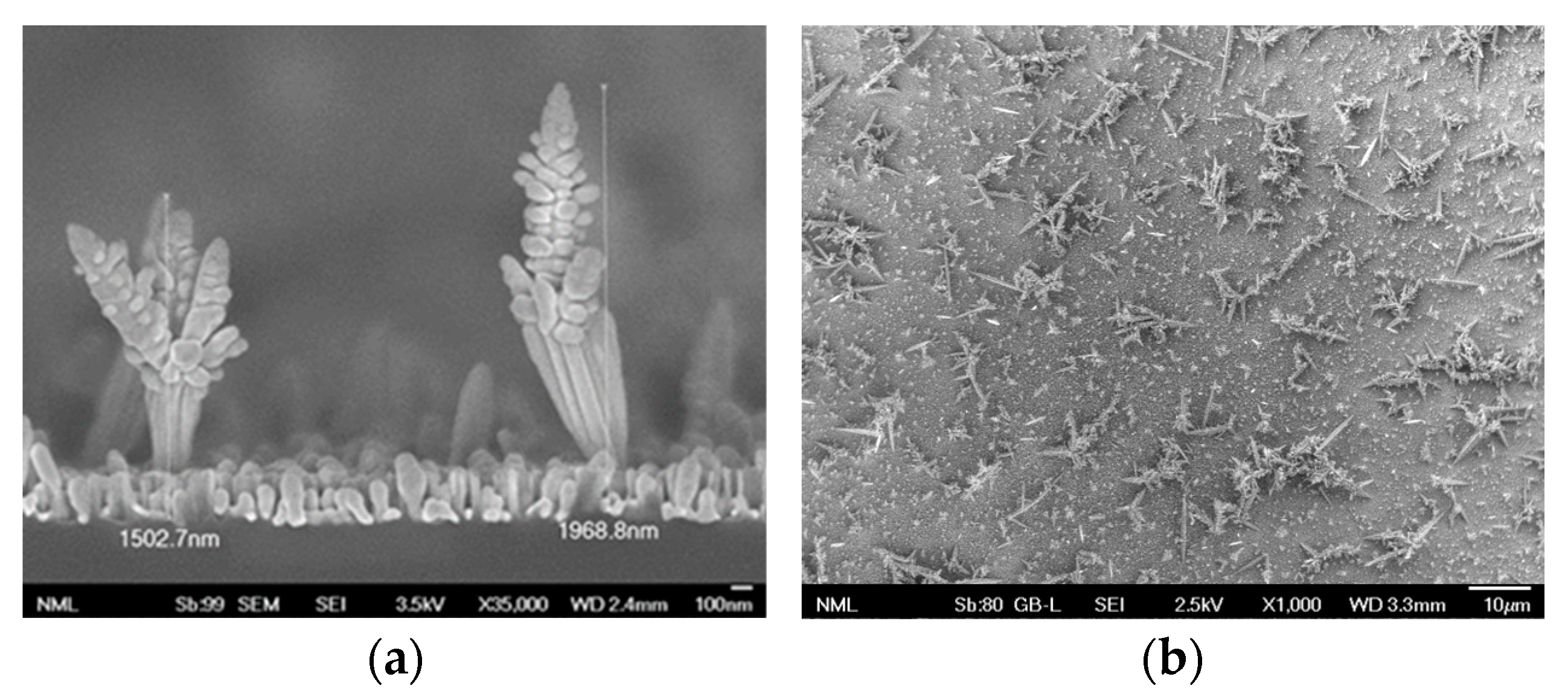
2.2. Synthesis of SERS Substrates

The SERS substrates were fabricated by single-step metal-assisted chemical etching method (MACE), following a previously published protocol [17]. Briefly, a p-type (100)-oriented monocrystalline Si wafer was used as a substrate for the growth of silver dendrites by immersion into an aqueous solutions containing 0.02 M AgNO3 and 4.8 M HF for 6 min. The SERS substrates were characterized by results from scanning electron microscopy (SEM) employing a JSM- 7401F SEM instrument (JEOL Europe bv; Zaventem, Belgium), working at 30 kV.

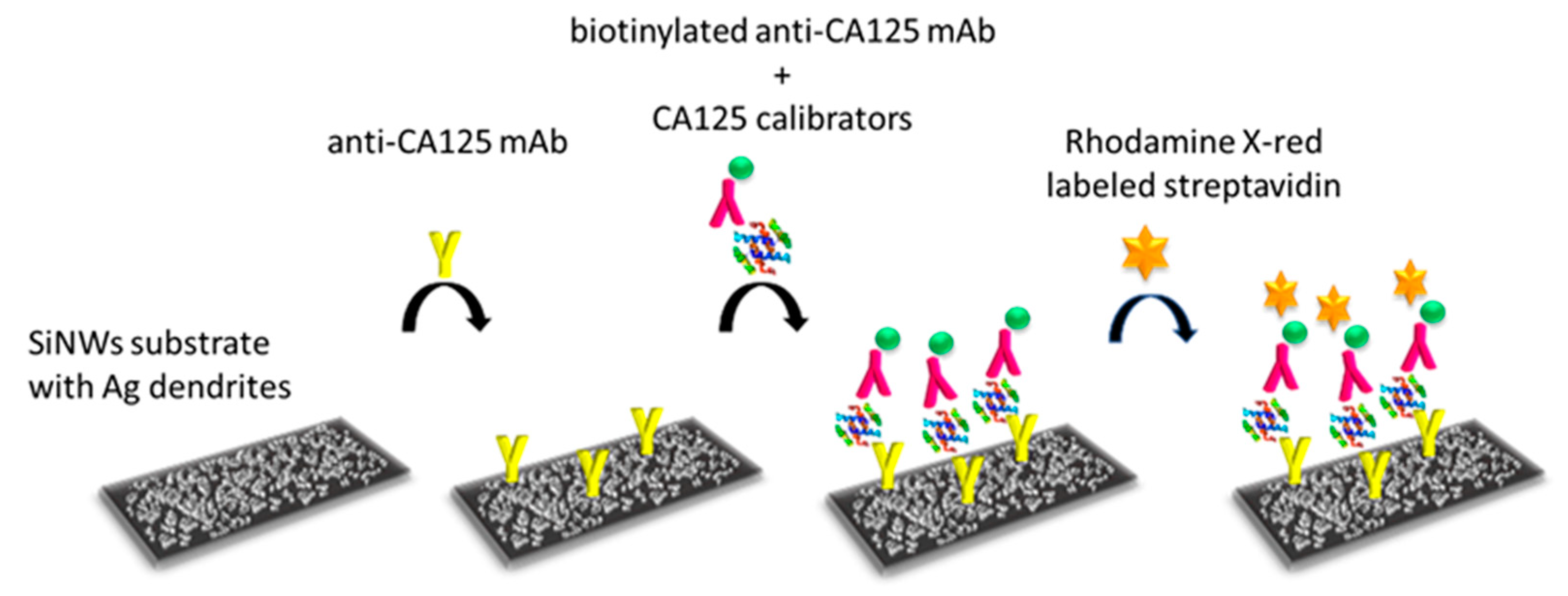
2.3. Immunoassay Protocol

Immunoassays for CA125 and HE4 were performed on SERS substrates with dimensions 0.5 × 0.5 cm2. Firstly, 20 μL of a 200 μg/mL anti-CA125 antibody solution in 10 mM Tris-HCl buffer, pH 8.25, 0.1 M NaCl, or 20 μL of a 200 μg/mL anti-HE4 in 50 mM carbonate buffer, pH 9.2, were spotted onto the substrates and incubated overnight at 4 °C. The antibody excess was removed by washing twice with 300 μL of a 10 mM Tris-HCl, pH 8.25, 0.15 M NaCl or a 10 mM PBS, pH 7.4, for CA125 and HE4, respectively. The non-specific binding sites were blocked after the incubation with 500 μL of a 10 mg/mL BSA solution in 0.1 M NaHCO3, pH 8.5, for 2 h at room temperature. After washing, as carried out previously, 200 μL of solutions containing equal volumes of the CA125 and HE4 calibrators and the biotinylated detection antibodies (2.5 and 5 μg/mL for CA125 and HE4, respectively) in 50 mM Tris-HCl, pH 7.8, 9 mg/mL NaCl, 10 mg/mL BSA for CA125 or 50 mM PBS, pH 7.4, 10 mg/mL BSA for HE4 were added, followed by incubation for 1 h under gentle shaking at room temperature. The surfaces were washed four times with 300 μL of the respective washing buffer containing 0.05% v/v Tween. Then, 200 μL of a 5 μg/mL streptavidin–Rhodamine Red-X solution in 50 mM PBS buffer, pH 6.5, containing 10 mg/mL BSA, were added onto the surfaces and incubated at room temperature for 30 min under gentle shaking. Finally, the substrates were washed three times with 300 μL of the respective washing solution and once with 300 μL DI water, and dried prior to photoluminescence (PL) signal detection, with a PL experimental setup which has been described previously [18]. The setup consists of a 532 nm diode laser for illuminating the sample at a 45-degree angle and a focusing lens to collimate the laser beam. The illuminated spot on the sample was approximately 2 mm wide. The blocking of the excitation laser was achieved using a long pass filter, and the PL emission was collected with a fiber vertical to the surface and analyzed using a spectrophotometer (Ocean Optics, Hong Kong). The laser power on the sample was 8 mW. The acquisition time was kept as short as possible, never exceeding 1 s. At this laser power and acquisition time, there was no indication of heating or quenching of the PL signal.

3. Results and Discussion

3.1. Design and Characterization of PL Substrates

The substrates used in the current study were prepared in silicon wafers following the MACE method [17]. In this method, the silicon wafer was immersed in an AgNO3/HF solution and two different processes take place: the etching of silicon wafer to create silicon nanowires and the growth of silver nanoparticles in the form of dendrites on top of the silicon nanowires. This is evident from the cross-section and top-view SEM images of the nanostructured substrates prepared via MACE provided in Figure 1. As shown, the length of the silver dendrites is about 2 μm.

3.2. Optimization of CA125 and HE4 Immunoassay Conditions

The detection of CA125 and HE4 was performed through non-competitive immunoassays, the main steps of which are illustrated in Figure 2. Several assay parameters were optimized for both CA125 and HE4, such as the capture antibody concentration used for the modification of nanostructured silver substrates, the biotinylated detection antibody concentration, the assay duration and the incubation time with the streptavidin–Rhodamine Red-X conjugate, as well as the composition of all the solutions used, including the coating, the blocking, the washing and the assay buffer. In short, the first parameter optimized was the concentration of capture antibodies. For this reason, antibody concentrations of 50, 100, 200, and 400 μg/mL were tested for a fixed concentration of detection antibody, and the concentration of 200 μg/mL was selected for both analytes. Furthermore, the effect of the concentration of detection antibodies was evaluated by incubating mixtures of CA125 or HE4 calibrators with the respective biotinylated antibodies at concentrations ranging from 1.0 to 10 μg/mL. The concentrations which provided maximum plateau signal values were 2.5 μg/mL for CA125 and 5.0 μg/mL for HE4. Once the detection and capture antibody concentrations for each analyte were selected, the duration of the immunoreaction was also optimized and 1 h was selected for both CA125 and HE4. The last parameter optimized was the duration of substrate incubation with the streptavidin–Rhodamine conjugate (15, 30 and 60 min). It was found that adequate signal was provided after 30 min of incubation and this was selected for the final protocol. In Table 1, the selected immunoassay conditions for each analyte are presented.

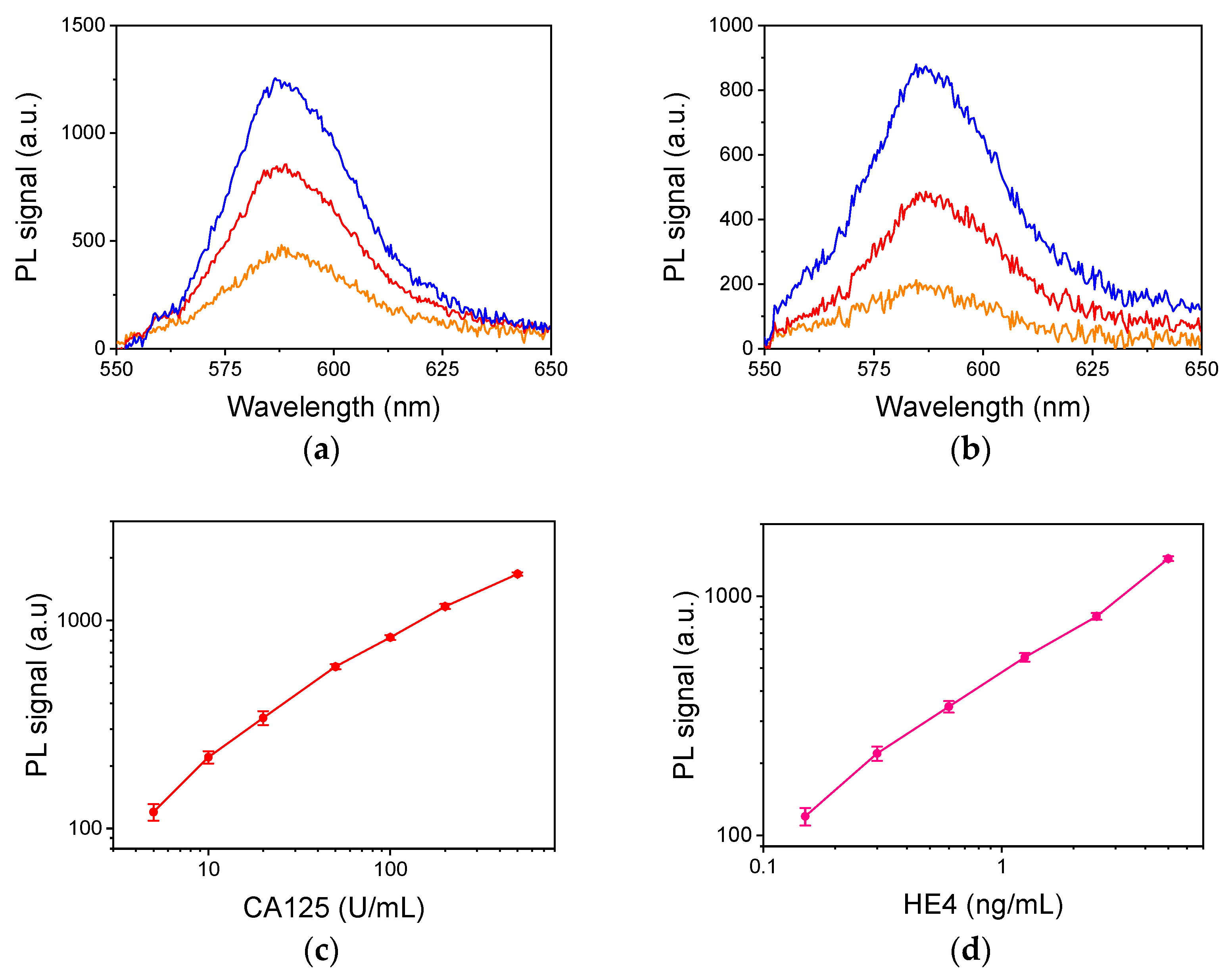
3.3. Analytical Characterisis of CA125 and HE4 Assays

Using the optimum for each immunoassay condition, the respective calibration curves were obtained and the analytical characteristics of both immunoassays were determined. In Figure 3a,b, characteristic PL spectra corresponding to zero calibrators (orange line) and two other calibrators (red and blue line) for CA125 and HE4, respectively, are presented. In addition, the full calibration curve for CA125 is shown in Figure 3c. The detection limit was determined as the concentration corresponding to signals equal to five standard deviations of five measurement of zero calibrator, and was 2.5 U/mL and the dynamic range was from 5 to 500 U/mL. Accordingly, for HE4, the detection limit was calculated to be 0.06 ng/mL and the dynamic range extended up to 5.0 ng/mL, as also indicated by the calibration curve provided in Figure 3d. Both assays were repeatable with an intra- and inter-coefficient of variation < 8.5 and 10.5, respectively.

4. Conclusions

In conclusion, nanostructured silicon surfaces decorated with silver dendrites were evaluated as substrates for the sensitive detection through photoluminescence of the ovarian cancer biomarkers, CA125 and HE4, following a non-competitive immunoassay protocol. The detection limit of CA125 and HE4 was 2.5 U/mL and 0.06 ng/mL, respectively, with a linear dynamic range extending up to 500 U/mL for CA125 and 5.0 ng/mL for HE4. The working ranges of both assays cover the concentrations encountered in both healthy individuals and ovarian cancer patients, and they can thus be implemented for the early diagnosis of ovarian cancer. Currently, the nanostructured silver surfaces are evaluated as substrates for the immunochemical detection of additional ovarian cancer markers, e.g., survivin [19]. In addition, a multiplexed determination of these markers in serum samples by a portable system at the point-of-need will be targeted, aiming to increase their diagnostic and prognostic value with respect to ovarian cancer.

Author Contributions

Conceptualization, V.L., S.G., N.P. and P.P.; methodology, G.G., A.K., A.D. and P.P.; formal analysis, G.G., A.K. and A.D.; investigation, G.G., A.K., I.K., K.C. and A.D.; resources, V.L., S.G., K.C., E.C., A.E., S.K. and P.P.; data curation, G.G., A.K., I.K. and A.D.; writing—original draft preparation, G.G., A.K., I.K. and A.D.; writing—review and editing, V.L., S.G., E.C., N.P., A.E., S.K., E.C. and P.P.; visualization, G.G., A.K., I.K. and A.D.; supervision, N.P., A.E. and P.P.; project administration, V.L., S.G., N.P. and P.P.; funding acquisition, S.G., N.P. and P.P. All authors have read and agreed to the published version of the manuscript.

Funding

This research has been co-financed by the European Regional Development Fund of the European Union and Greek national funds through the Operational Program Competitiveness, Entrepreneurship and Innovation, under the call RESEARCH–CREATE–INNOVATE (project code: T2EΔK-03746 BioNanoDiagnostiki).

Institutional Review Board Statement

Not applicable.

Informed Consent Statement

Not applicable.

Data Availability Statement

The data presented in this study are available on request from the corresponding author. The data are not publicly available due to privacy issues.

Acknowledgments

I.K. acknowledges the financial support for his doctoral thesis research by Greece and the European Union (European Social Fund-ESF) through the Operational Program “Human Resources Development, Education and Lifelong Learning” in the context of the Act “Enhancing Human Re-sources Research Potential by undertaking a Doctoral Research” Sub-action 2: IKY Scholarship Program for PhD candidates at Greek Universities.

Conflicts of Interest

The authors declare no conflict of interest.

References

  1. Kossaï, M.; Leary, A.; Scoazec, J.Y.; Genestie, C. Ovarian Cancer: A Heterogeneous Disease. Pathobiology 2018, 85, 41–49. [Google Scholar] [CrossRef] [PubMed]
  2. Stewart, C.; Ralyea, C.; Lockwood, S. Ovarian Cancer: An Integrated Review. Semin. Oncol. Nurs. 2019, 35, 151–156. [Google Scholar] [CrossRef] [PubMed]
  3. Scholler, N.; Urban, N. CA125 in ovarian cancer. Biomark. Med. 2007, 1, 513–523. [Google Scholar] [CrossRef] [PubMed]
  4. Bottoni, P.; Scatena, R. The role of CA 125 as tumor marker: Biochemical and clinical aspects. Adv. Exp. Med. Biol. 2015, 867, 229–244. [Google Scholar] [CrossRef] [PubMed]
  5. Anastasi, E.; Giovanna Marchei, G.; Viggiani, V.; Gennarini, G.; Frati, L.; Reale, M.G. HE4: A new potential early biomarker for the recurrence of ovarian cancer. Tumor Biol. 2010, 31, 113–119. [Google Scholar] [CrossRef] [PubMed]
  6. Van Gool, A.; Corrales, F.; Čolović, M.; Krstić, D.; Oliver-Martos, B.; Martínez-Cáceres, E.; Jakasa, I.; Gajski, G.; Brun, V.; Kyriacou, K.; et al. Analytical techniques for multiplex analysis of protein biomarkers. Expert Rev. Proteom. 2020, 17, 257–273. [Google Scholar] [CrossRef] [PubMed]
  7. Suntornsuk, W.; Suntornsuk, L. Recent applications of paper-based point-of-care devices for biomarker detection. Electrophoresis 2020, 41, 287–305. [Google Scholar] [CrossRef] [PubMed]
  8. Sha, R.; Badhulika, S. Recent advancements in fabrication of nanomaterial based biosensors for diagnosis of ovarian cancer: A comprehensive review. Microchim. Acta 2020, 187, 181. [Google Scholar] [CrossRef] [PubMed]
  9. Pollap, A.; Swit, P. Recent Advances in Sandwich SERS Immunosensors for Cancer Detection. Int. J. Mol. Sci. 2022, 23, 4740. [Google Scholar] [CrossRef] [PubMed]
  10. Jahn, I.J.; Žukovskaja, O.; Zheng, X.-S.; Weber, K.; Bocklitz, T.W.; Cialla-Maya, D.; Popp, J. Surface-enhanced Raman spectroscopy and microfluidic platforms: Challenges, solutions and potential applications. Analyst 2017, 142, 1022. [Google Scholar] [CrossRef] [PubMed]
  11. Kant, K.; Abalde-Cela, S. Surface-Enhanced Raman Scattering Spectroscopy and Microfluidics: Towards Ultrasensitive Label-Free Sensing. Biosensors 2018, 8, 62. [Google Scholar] [CrossRef]
  12. Tunc, I.; Susapto, H.H. Label-free detection of ovarian cancer antigen CA125 by surface enhanced Raman scattering. J. Nanosci. Nanotechnol. 2019, 20, 1358–1365. [Google Scholar] [CrossRef]
  13. Ge, M.; Wei, C.; Xu, M.; Fang, C.; Yuan, Y.; Gu, R.; Yao, J. Ultra-sensitive magnetic immunoassay of HE4 based on surface enhanced Raman spectroscopy. Anal. Methods 2015, 7, 6489–6495. [Google Scholar] [CrossRef]
  14. Eom, G.; Hwang, A.; Kim, H.; Moon, J.; Kang, H.; Jung, J.; Lim, E.-K.; Jeong, J.; Park, H.G.; Kang, T. Ultrasensitive detection of ovarian cancer biomarker using Au nanoplate SERS immunoassay. BioChip J. 2021, 15, 348–355. [Google Scholar] [CrossRef]
  15. Nguyen, A.H.; Lee, J.; Choi, H.I.; Kwak, H.S.; Sim, S.J. Fabrication of plasmon length-based surface enhanced Raman scattering for multiplex detection on microfluidic device. Biosens. Bioelectron. 2015, 70, 358–365. [Google Scholar] [CrossRef] [PubMed]
  16. Zheng, Z.; Wu, L.; Li, L.; Zong, S.; Wang, Z.; Cui, Y. Simultaneous and highly sensitive detection of multiple breast cancer biomarkers in real samples using a SERS microfluidic chip. Talanta 2018, 188, 507–515. [Google Scholar] [CrossRef] [PubMed]
  17. Kochylas, I.; Spiros Gardelis, S.; Likodimos, V.; Giannakopoulos, K.P.; Falaras, P.; Nassiopoulou, A.G. Improved surface-enhanced-Raman scattering sensitivity using Si nanowires/Silver nanostructures by a single step metal-assisted chemical etching. Nanomaterials 2021, 11, 1760. [Google Scholar] [CrossRef] [PubMed]
  18. Kochylas, I.; Dimitriou, A.; Apostolaki, M.A.; Skoulikidou, M.C.; Likodimos, V.; Gardelis, S.; Papanikolaou, N. Enhanced Photoluminescence of R6G Dyes from Metal Decorated Silicon Nanowires Fabricated through Metal Assisted Chemical Etching. Materials 2023, 16, 1386. [Google Scholar] [CrossRef] [PubMed]
  19. Gąsowska-Bajger, B.; Gąsowska-Bodnar, A.; Knapp, P.; Bodnar, L. Prognostic significance of survivin expression in patients with ovarian carcinoma: A meta-analysis. J. Clin. Med. 2021, 10, 879. [Google Scholar] [CrossRef] [PubMed]
Figure 1. (a) Cross-section (scale bar 100 nm, magnification ×35,000) and (b) top-view (scale bar 10 μm, magnification ×1000) SEM images of silicon nanowires decorated with Ag dendrites.
Engproc 35 00007 g001
Figure 2. Schematic of non-competitive immunoassay for CA125 detection on the nanostructured silver substrate. The same procedure was followed for HE4.
Engproc 35 00007 g002
Figure 3. (a) Photoluminescence signal at the spectral range of 550–650 nm received from surfaces with silver dendrites functionalized with an anti-CA125 antibody. The orange line corresponds to zero calibrator (non-specific binding), the red line to a calibrator with a concentration of 20 U/mL, and the blue line to a calibrator with a concentration of 100 U/mL. (b) Photoluminescence signal at the spectral range of 550–650 nm received from surfaces with silver dendrites functionalized with an anti-HE4 antibody. The orange line corresponds to zero calibrator, the red line to a calibrator with a concentration of 0.6 ng/mL, and the blue line to a calibrator with a concentration of 1.25 ng/mL. (c) Typical CA125 calibration curve. (d) Typical HE4 calibration curve. Each point is the mean value of three measurements ± 3 SD.
Engproc 35 00007 g003
Table 1. Selected conditions for CA125 and HE4 immunoassay performance on the nanostructured silver surfaces.
ParameterCA125HE4
Capture antibody concentration200 μg/mL200 μg/mL
Detection antibody concentration2.5 μg/mL5.0 μg/mL
Immunoreaction duration60 min
Streptavidin–Rhodamine incubation30 min
Disclaimer/Publisher’s Note: The statements, opinions and data contained in all publications are solely those of the individual author(s) and contributor(s) and not of MDPI and/or the editor(s). MDPI and/or the editor(s) disclaim responsibility for any injury to people or property resulting from any ideas, methods, instructions or products referred to in the content.

