 Jingyuan Yang
 Shan Wu3†
 Weihong Yu
 Youxin Chen
- 1Department of Ophthalmology, Peking Union Medical College Hospital, Chinese Academy of Medical Sciences, and Peking Union Medical College, Beijing, China
- 2Key Laboratory of Ocular Fundus Diseases, Chinese Academy of Medical Sciences, and Peking Union Medical College, Beijing, China
- 3Department of Anaesthesiology, Beijing Hospital, National Center of Gerontology, Institute of Geriatric Medicine, Chinese Academy of Medical Sciences, Beijing, China
Background and purpose: Pathologic myopia (PM) is an international public health issue. This study aimed to analyze PM research trends by reporting on publication trends since 2000 and identifying influential journals, countries, authors, and keywords involved in PM.
Methods: A bibliometric analysis was performed to evaluate global production and development trends in PM since 2000 and the keywords associated with PM.
Results: A total of 1,435 publications were retrieved. PM has become a fascinating topic (with relative research interest ranging from 0.0018% in 2000 to 0.0044% in 2021) and a global public health issue. The top three countries with the highest number of publications were China, the USA, and Japan. The journals, authors, and institutions that published the most relevant literature came from these three countries. China exhibited the most rapid increase in the number of publications (from 0 in 2000 to 69 in 2021). Retina published the most papers on PM. Kyoko Ohno-Matsui and Tokyo Medical and Dental University contributed the most publications among authors and institutions, respectively. Based on keyword analysis, previous research emphasized myopic choroidal neovascularization and treatment, while recent hotspots include PM changes based on multimodal imaging, treatment, and pathogenesis. Keyword analysis also revealed that deep learning was the latest hotspot and has been used for the detection of PM.
Conclusion: Our results can help researchers understand the current status and future trends of PM. China, the USA, and Japan have the greatest influence, based on the number of publications, top journals, authors, and institutions. Current research on PM highlights the pathogenesis and application of novel technologies, including multimodal imaging and artificial intelligence.
Introduction
Myopia is a leading cause of vision loss worldwide, with an increasing trend; it can be divided into simple myopia and pathologic myopia (PM). There is no standardized definition for PM. However, most researchers and clinicians agree that PM is a subtype of myopia that accompanies characteristic myopic fundus changes that usually occur in eyes with high myopia, including myopic maculopathy equal to or more serious than diffuse choroidal atrophy or the presence of posterior staphyloma (1). The prevalence of myopic retinopathy in PM was reported to range from 1.2 to 3.7% in the population (2–6). Moreover, the visual prognosis for eyes with myopic maculopathy is much worse than that for those without maculopathy (7–9). A bibliometric analysis of publications is important due to the growing number of pathologic myopic eyes and their influence on public health, which has prompted researchers to analyze the development trend and research hotspots of PM.
Bibliometric analyses were first proposed in 1922 by Hulme; however (10), these analyses are not equal to reviews. Bibliometric analyses provide an overview of publications using mathematical and statistical methods and predict possible development trends based on citation reports and academic outputs. Bibliometric analysis, which has a century-long history, has been applied in medicine to investigate its development and trends.
Research on PM has received increasing attention in recent years, which makes it difficult for many researchers and clinicians to determine the research focus and frontiers of PM from the growing number of publications. However, to our knowledge, no bibliometric analysis has specifically focused on PM. Therefore, the current study aimed to investigate frontier research and PM trends across the international scientific literature based on the Web of Science (WOS) Core Collection over the past two decades. We also aimed to predict trends for the next few years, noting that the increase in the number of publications on PM is expected to lead to a valuable reference for clinicians and researchers.
Methods
Search strategy
The WOS Core Collection is the most suitable database for bibliometric analysis, particularly because of the high quality of the included publications. The search for papers to be included in the current study was carried out on 7 August 2022, and all the included publications were published from 1 January 2000, to 1 August 2022. The search strategy was “TS = pathologic myopia OR TS = myopic degeneration OR TS = myopic maculopathy.” One thousand and thirty-seven pieces of literature were identified. Two published poems and news items were excluded according to the document type. One thousand and thirty-five publications were finally included.
Data collection
All the data were extracted and downloaded from the WOS Core Collection databases, including metrics of publication numbers, countries and regions, authors, citations, self-citations, and H-indexes. We also investigated the relationship between the global productivity of PM and the human development index (HDI), which measures the level of human development based on knowledge, life expectancy, and income per capita indicators rather than economic growth alone. The United Nations Development Programme published the Human Development Report 2020, in which countries and areas were divided into four categories based on HDI: very high human development, high human development, medium human development, and low human development (11). Countries and regions were classified according to the default classification in the WOS and the HDI. Prism 9, R (R. app. GUI 1.79) and its tools Biblioshiny, VOSviewer 1.6.18, and SPSS 26 were used to input and analyze the data.
Bibliometric analysis
Descriptive indices were extracted from the WOS Core Collection and calculated using SPSS. The co-occurrence network was constructed using the VOSviewer software. Keywords were extracted from the titles and abstracts of the included publications. R and its tool Biblioshiny were used to generate word-cloud maps. To provide a better classification of keyword clusters and a better summarization of research trends, frequencies over 30 were the criteria for the inclusion of these analyses. H-indices were collected from the WOS database and can partially reflect the impact of researchers. Relative research interest (RRI) was measured as the number of publications in a specific field divided by the number of all publications in all fields. The value of this metric reflects the global attention and interest in a specific field. Higher RRI values represent more research interest and hotspots in this field. A third-order polynomial method was used in the prediction model using the Prism software. The average year of appearance was used to assess the novelty of the keywords.
Results
Contributions of various countries and regions to global publications
A total of 1,435 publications were analyzed. Since 2000, Mainland China has contributed to the majority of publications (294, 20.5%), followed by the USA (280, 19.5%) and Japan (239, 16.7%) (Figure 1A). The remaining countries had published fewer than 200 publications. The USA, Japan, China, Germany, Italy, and Singapore were the top five countries with the highest number of publications and H-index. The total number of publications on PM has grown over the past 22 years, especially in recent years (Figure 1B). Additionally, the RRI of PM has increased from 0.0018% in 2000 to 0.0044% in 2021, indicating that research interest in PM has continued to increase worldwide over the past two decades. In the first 8 months of 2022, the RRI of PM reaches 0.0046%. According to the HDI category, we noticed that most publications were from very high HDI countries or regions, and the numbers of publications on PM were consistent with the HDI classification (Figure 1C).
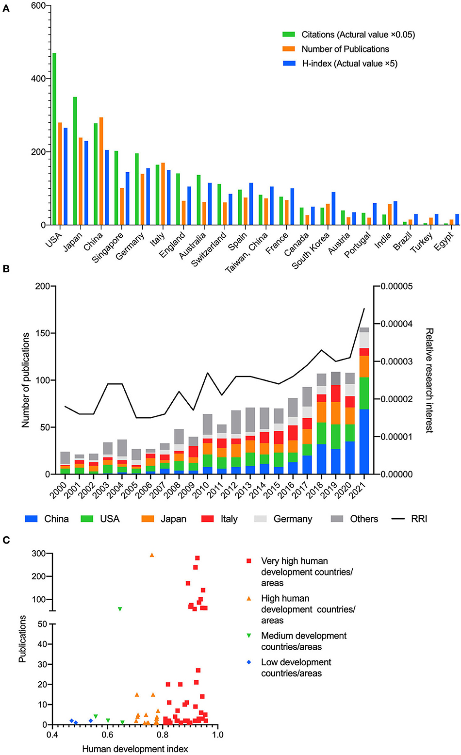
Figure 1. (A) Top 20 countries/regions in the publications on pathologic myopia. The green bar shows the number of citations (actual value multiply by 0.05), the orange bar shows the number of publications, and the blue bar shows the H-index (actual value multiply by 5). (B) The proportion of publications of China Mainland, USA, Japan, Italy, Germany, and other countries/regions and relative research interest (RRI) in each year on the field of pathologic myopia. (C) The number of publications on pathologic myopia in countries or areas of various levels of human development. Very high human development countries or areas contributed to most publications.
We analyzed the co-occurrence of 34 countries and regions (Supplementary Figure 1). The analysis suggests six clusters: (1) Mainland China, Australia, and Iran; (2) USA, Switzerland, France, South Korea, Canada, Austria, Brazil, Turkey, Greece, and Venezuela; (3) Japan, Singapore, Spain, and Portugal; (4) Germany, England, the Netherlands, Russia, Scotland, Northern Ireland, Poland, Wales, Ireland, and Egypt; (5) Italy, India, Tunisia, and Israel; and (6) Taiwan, Denmark, and Saudi Arabia.
The publication rate of papers on PM has been increasing in the past two decades, and predictions for the coming years reflect this increase (Figure 2). China exhibited the most rapid increase in the number of publications in the last 5 years. China is also projected to maintain its leading position and show steady growth in the number of publications.
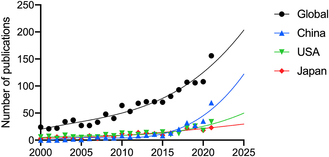
Figure 2. The publication trends and prediction curve of global and countries which had the most publications of pathologic myopia since 2000. (A) Global. (B) China Mainland. (C) USA. (D) Japan.
Citations and H-index
WOS citation reports revealed a total of 21,432 citations without self-citations of the 30,234 relevant citations since 2000 (details for top countries and regions in Supplementary Table 1). Each paper cited an average of 21.07 times. The USA contributed the most citations (9,395 citations, 9,062 without self-citations) and the highest H-index (53) (Figure 1A) from 2000. Japan ranked second in terms of both citations (6,999 citations, 6,026 without self-citations) and H-index (46), and Mainland China ranked third in both citations (5,559 citations, 4,895 without self-citations) and H-index (41). The most-cited publication was cited 618 times. We divided the publications into three groups according to citation frequency: high frequency (more than 100 citations), medium frequency (>50 and <100 citations), and low frequency (<50 citations). Most publications were in a low-frequency group. One hundred and ten publications were cited with a medium frequency, and 46 were cited with a high frequency.
To investigate the distribution of citation numbers each year, we drew heat maps of each group of citation frequency (Supplementary Figures 2B–D). Every row in the heatmap represents a publication, the x-axis represents the year, and the color represents the citation number. The timespan of citations with high and medium frequency is similar and longer than most publications with low citation frequency. Moreover, most publications with high and medium citation frequencies have been published in the last 10 years (Supplementary Figure 2A).
The leading institutions, journals, and authors
We investigated the top institutions in this field; the Tokyo Medical and Dental University in Japan (136, 9.5%), Singapore National Eye Center in Singapore (93, 6.5%), Ruprecht-Karls-Universität Heidelberg in Germany (78, 5.4%), and Sun Yat-Sen University (55, 3.8%) and Capital Medical University (54, 3.8%) in China published the most publications (Figure 3A).
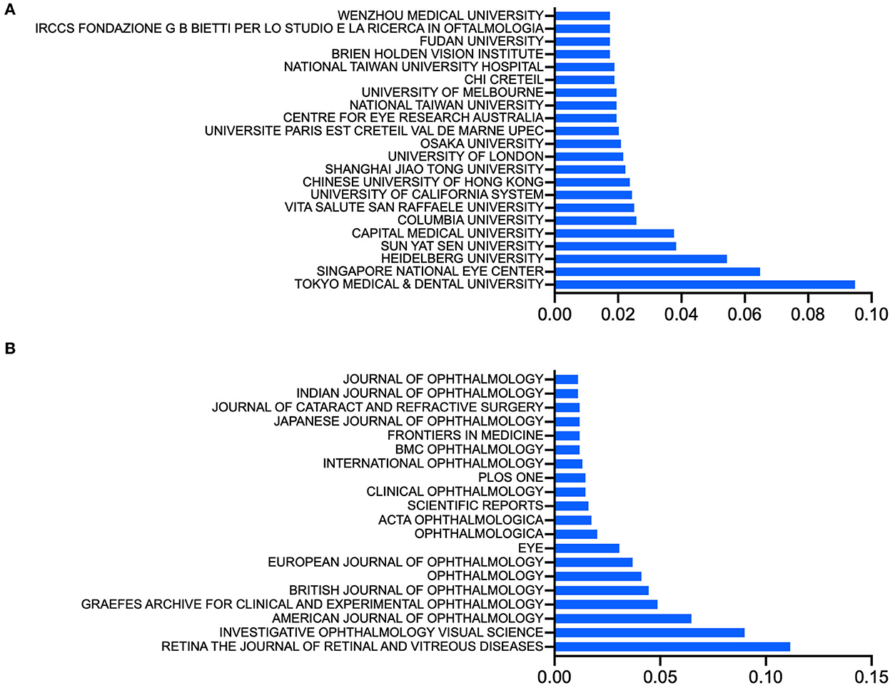
Figure 3. (A) Top institutions, ranked by the ratio of the number of publications from an institution to the total number of included publications about pathologic myopia. (B) Top journals, ranked by the ratio of the number of publications from a journal to the total number of included publications about pathologic myopia.
Approximately half (726, 50.6%) of the papers on PM were published in 10 journals, including Retina, which published the maximum number of relevant publications (160). Investigative Ophthalmology and Visual Science and the American Journal of Ophthalmology published the second and third-most publications, with 129 and 93, respectively (Figure 3B).
The 10 papers with the most citations are listed in Table 1. The most cited paper was published in Ophthalmic and Physiological Optics, an ophthalmic periodical that has ceased publication, and was called Myopia and Associated Pathological Complications. The corresponding author was Seang Mei Saw. Most publications on PM were published in ophthalmology journals (Table 2).
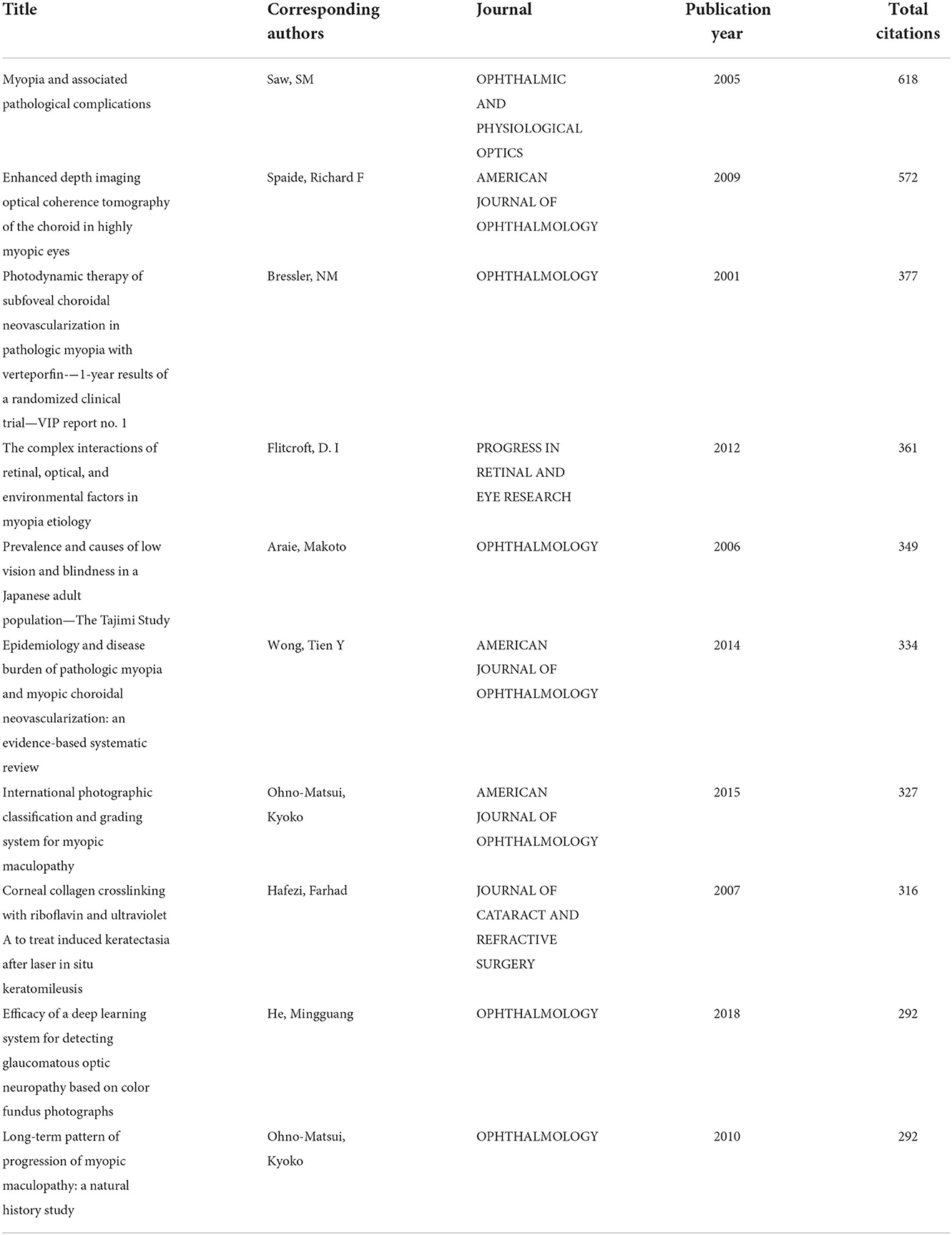
Table 1. Top 10 papers with the most citations relevant to pathologic myopia.
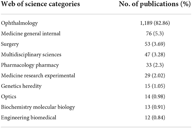
Table 2. Top 10 Web of Science categories of journals on pathologic myopia research.
The top 10 authors in this field are listed in Table 3 according to the number of publications. The works of Kyoko Ohno-Matsui from Tokyo Medical and Dental University have been published the most since 2000, with 139 papers and 4,710 citations (4,033 without self-citations). The H-indexes of both the author and the institution were 39. Jost B. Jonas, from Ruprecht-Karls-Universität Heidelberg, ranked second, with 77 publications and 2,224 citations (2,025 without self-citations). Tien Y Wong ranked third with 44 publications and 2,135 citations (2,045 without self-citations) (Table 3).
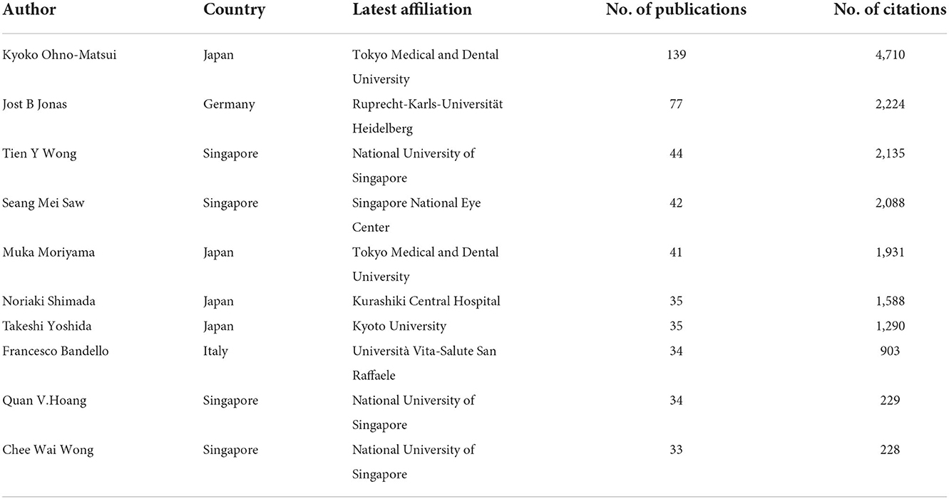
Table 3. Top 10 authors who published most in the field of pathologic myopia.
We also analyzed the cooperation between investigators (Supplementary Figure 3). The node size within a collaboration network indicates the strength of the connections between each author. Several authors, including Kyoko Ohno-Matsui, Takeshi Yoshida, Muka Moriyama, Jost B. Jonas, and Noriaki Shimada, closely cooperated with other researchers and teams.
Research hotspots in PM
Keyword analysis identified the most frequently used words and their linkages within the field of PM research. We analyzed keywords that appeared more than 30 times across the included publications. Merging repeated words, excluding meaningless ones, resulted in 70 keywords that could be divided into three primary clusters by co-occurrence frequency, including an epidemiology-related cluster (in red), a treatment-related cluster (in green), and a lesion-related cluster (in blue) (Figure 4A). Keywords with high link strength were assigned to the same cluster.
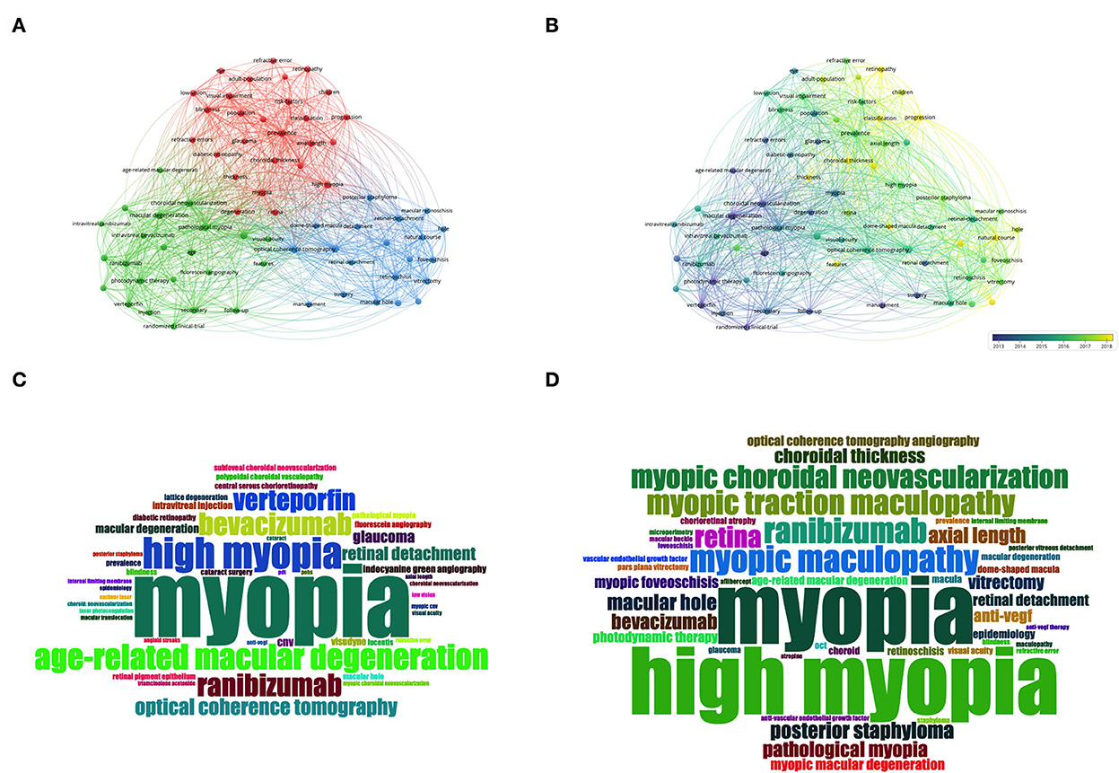
Figure 4. Keywords analysis by VOSviewer and R. (A) Co-occurrence map of keywords in titles and abstracts. Keywords were classified into 3 clusters by co-occurrence frequency, including an epidemiology-related cluster (in red), a treatment-related cluster (in green), and a lesion-related cluster (in blue). (B) Color-coded map of keywords by the average time of appearance. Blue keywords appeared earlier, while yellow keywords appeared more recently. (C) The word-cloud map of the most frequent keywords from 2000 to 2010. (D) The word-cloud map of the most frequent keywords from 2011 to August 2022.
We also color-coded the keywords by the average time of appearance and found that most of the keywords appeared in recent years (Figure 4B). Figure 4B shows the temporal evolution of these keywords. The classification and progression keywords were the latest keywords with high occurrence.
To attain an intuitive impression of the most frequent keywords, we listed the most used keywords in 2000–2010 and 2011–2022 in word-cloud images, respectively (Figure 4C for keywords from 2000 to 2010 and Figure 4D for keywords from 2011 to August 2022). Besides the keywords of myopia and high myopia, keywords related to pathologic lesions and treatments, such as myopic maculopathy, myopic choroidal neovascularization (mCNV), and ranibizumab, were the most frequent words. We extracted the most frequent keywords to explore changes in hotspots and keywords in the field (Supplementary Figure 4). A changing trend was noticed from treatment for choroidal neovascularization to myopic lesions and complications and to deep learning.
Discussion
This study involved a bibliometric analysis focusing on PM over the last two decades. We identified an increasing trend in publications on PM since 2000 (from 24 publications in 2000 to 156 publications in 2021). Although China and the USA had the most publications (294 and 280, respectively), authors from Japan, Singapore, and Germany had the greatest influence (4, 4, and 1 of the top 10 authors, respectively). To our knowledge, this is the first bibliometric analysis to focus on PM.
Country- or region-based contributions to PM research
The prevalence of PM in a country or region did not completely parallel its number of publications (Table 4). For example, although the USA contributed a large number of PM publications, only Asian and Pacific Islanders in the USA had a greater prevalence (ethnicity OR 1.64) (12). Conversely, Asian countries, especially some countries in East Asia, including Japan and China (1.7% and 3.7%, respectively), had a relatively higher prevalence of myopic retinopathy than some Western countries, such as Australia (1.2%) (2–6). Therefore, the factors influencing the number of publications on PM are not solely the prevalence of the disease.
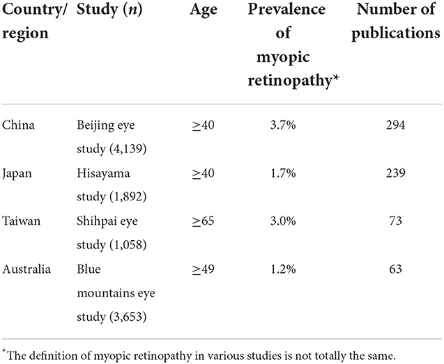
Table 4. The population-based prevalence of myopic retinopathy and the number of publications since 2020.
The number of publications by country or region is usually associated with the interest in and knowledge of a certain field. The global number of publications related to PM has continued to increase since the millennium, with the greatest growth occurring in 2021 (increased by 48 publications compared to 2020), in which China contributed 70.8% of the global growth (34 publications). This increasing trend might have benefited from the state policy of prevention and control of myopia in China, which has been set in recent years (13). Therefore, not only research interest but also administrative encouragement was an important motivation for PM research. State policy can create an encouraging environment for researchers with adequate funding, advanced techniques, and equipment, which are essential for conducting relevant studies.
As PM is a global healthcare challenge, international collaboration has become a mainstream research pattern. In the present study, the USA had the most international collaboration (total link strength of 306 in VOSviewer), followed by China (total link strength of 236), Germany (total link strength of 227), Singapore (total link strength of 213), and Japan (total link strength of 206) (Supplementary Figure 1). The USA is also the second-most productive country, in which authors published ~51% of studies from at least two countries or regions. It can be speculated that the USA was the most productive country. Collaboration and exchange are important in the academic community, and the USA performs well in international research. Although none of the top 10 authors hailed from the USA, the country had the most citations (9,395 total citations) and the highest H-index (53), indicating that the USA's research had a relatively high quality with good international collaboration and communication.
Therefore, not only is the burden of PM on patients, local economics, and public health a significant determinant of promoting PM research, but state policy and international collaboration are also essential factors contributing to global PM research.
We used the metric of the H-index, which has been widely used, to evaluate a country/area's productivity and citation impact of publications. However, this metric has some limitations (14). We did not evaluate the number of authors in an article or a specific author's position in an article (15), which could result in the distribution of citations from an article to all authors equally. Similar to the distribution of authors, the H-index of a country or region does not consider that the contributions from each country or region in the research were usually heterogeneous, and the equal distribution of citations of a publication to all countries and regions might be unjustified. Moreover, this metric does not differentiate between different cited sections of a publication (e.g., results or discussion, etc.) or between different types of publications (e.g., a review or original research). Additionally, this metric counts self-citations, and some intentional citations might have influenced it; for example, citing the publications of potential reviewers or editors (16, 17).
We also investigated the associations between HDI and publications; all of the top 10 authors came from countries with very high HDI. In addition to China, countries with very high HDI contributed the most publications. Patients from medium- and low-HDI countries and regions also suffer from PM; however, the number and influence of publications from these countries and regions need to be improved due to financial-, technical-, and equipment-related restrictions. The application of advanced technology, such as artificial intelligence, telemedicine, and advanced communication technology, might enhance the diagnosis, screening, and regular examinations in these underdeveloped areas.
Publication tendency in PM research
We analyzed the top papers with the most citations; 80% of the publications were published in clinical ophthalmology journals, indicating that PM research mainly focuses on clinical medicine rather than basic medical research. Similarly, most publications belong to the WOS category of ophthalmology (82.86%). The analysis revealed that this research field involves more clinicians than scientists and also explained the phenomenon that many PM-related manifestations were noticed by applying multimodal imaging methods, but their pathogenesis was not fully understood, such as dome-shaped maculopathy. In the future, greater efforts should be made to investigate the pathogenesis of PM using animal models.
In PM research, the RRI metric has maintained growth from 0.0018% in 2001 to 0.0044% in 2021, and the number of publications has also increased from 24 in 2000 to 156 in 2021. These results indicate a great interest in PM raised in the medical community, which also conforms with the increasing worldwide prevalence of PM (Figure 1B).
The study trends based on keywords can be considered indicators of basic and clinical research directions. Based on the analysis of keywords in the current study, most studies on PM correlate with epidemiology and risk factors, treatments, lesions, and abnormalities. The color-coded average time of appearance map of co-occurrence keywords showed that the research hotspots transformed from treatment for mCNV to epidemiologic and public health studies and then to more manifestations of PM and their treatments. In more detail (Supplementary Figure 4), we noticed that besides the keywords “myopic, high myopic, and pathologic myopia,” the keywords of “photodynamic therapy, ranibizumab, (myopic) choroidal neovascularization” (year of the median from 2006 to 2014), and “optical coherence tomography, myopic maculopathy, myopic traction maculopathy” (year of the median from 2013 to 2017) had a frequency no < 50, which indicates that the main interest of researches focused on treatment for mCNV, followed by PM-related maculopathy and its imaging findings. The latest hotspot keyword was “deep learning,” revealing a novel research direction. Automated diagnosis, screening, and regular examinations with artificial intelligence and advanced communication technology might enhance teleophthalmology and improve practice patterns in these underdeveloped areas.
The changes in keywords (Figures 3A, 4B) in various periods indicate increasing and emerging research themes in the PM field. Based on the bibliometric analysis, especially the keyword and research hotspots analysis, the following themes have reported the latest outcomes.
Multimodal imaging and pathologic manifestations
Since the millennium, optical coherence tomography (OCT), optical coherence tomography angiography (OCTA), magnetic resonance imaging (MRI), and other imaging methods have been applied in clinical practice, enabling researchers to evaluate pathologic myopic alterations and detect myopic complications (18). For example, current ultrawide-field imaging can show peripheral retinal abnormalities, and swept-source OCT can exhibit vitreous and retinal changes, such as the formation of posterior vitreous detachment, paravascular cystic lesions, and paravascular lamellar holes (19, 20).
Posterior staphyloma
Posterior staphyloma is defined as an outpouching of the eye wall with a radius of curvature less than the surrounding curvature of the eye wall (21). However, its formation is not yet fully understood. Currently, ultrawide-field OCT, ultrawide-field photography and MRI can visualize and evaluate the structure of staphyloma and its complications (21–23). Relevant complications include tractional maculopathy (such as retinoschisis), lacquer cracks, mCNV, and chorioretinal atrophy. Traditionally, posterior staphyloma was divided into 10 subtypes by Curtin (24). Recently, five types of posterior staphylomas have been proposed using MRI by Ohno-Matsui (21). Compared with MRI, ultrawide-field OCT provides visualization of retinal tissues simultaneously with scleral changes (usually when the choroid is not too thick, based on our experience).
Myopic choroidal neovascularization
Based on the analysis of keywords, we can observe that mCNV has always been the focus of PM research. Approximately 5.2–11.3% of eyes with PM develop mCNV (25). Ninety percent of eyes with PM progress to legal blindness in 10 years without treatment (26). Ischemia and breaks in Bruch's membrane may contribute to the development of mCNV (27, 28). In addition to fluorescein angiography (FA) and OCT, which can determine the activity of mCNV, non-invasive OCTA can demonstrate neovascularization volumetrically and can be performed repeatedly during follow-up. Anti-vascular endothelial growth factor (VEGF) agents are the first-line therapy (29–31).
Myopic maculopathy
According to several population-based studies, the frequency of myopic maculopathy varies from 1.2 to 3.8% (32–35). The classification of myopic maculopathy in PM was established using fundus photography (FP) and OCT images (Supplementary Table 2) (36–40). Myopic traction maculopathy can be evaluated using OCT for macular status, as well as surgical indications (41). However, these classification systems do not include some manifestations, such as the dome-shaped macula.
Dome-shaped maculopathy
Dome-shaped maculopathy refers to an inward convex protrusion of the macula within the posterior concavity of the eye that is mainly visualized using OCT and MRI (42, 43). The alteration can be detected only in horizontal scanning images, only in vertical images, or in both horizontal and vertical images because of the morphologic pattern of the round dome (43, 44). However, domes in children only appear in vertical OCT scanning images (45). Dome-shaped maculopathy can be identified quantitatively as a macular inward bulge height of more than 50 μm in the most convex image (46). Serous retinal detachment was detected in 2–67% of eyes with dome-shaped maculopathy (42, 46–49), and CNV was observed in 41.2% of the affected eyes (46). A bulge height of more than 400 μm is purportedly associated with decreased visual acuity, serous detachment, and greater retinal pigment epithelial atrophy (50). However, the mechanism of dome formation remains unclear.
Myopic glaucoma-like optic neuropathy
In PM, expansion, tilting, torsion, and effacement of the optic nerve head had been noticed, which resembled glaucoma-like neuropathy with loss of the neuroretinal rim (51). However, the irregularity of the optic nerve head shape and macula makes it challenging to determine the neuroretinal rim and to evaluate the thickness of the retinal nerve fiber layers and ganglion cell layers using OCT (52, 53). Fortunately, OCTA might be helpful in the future to differentiate myopia from glaucoma by exploring vascular changes under various conditions (54).
Impact on public health and management
The global prevalence of PM-related visual impairment is estimated to rise from 0.1% in 2020 to 0.6% in 2050 (55); therefore, the public health burden and impact on the quality of life will likely increase. PM affects patients' reading, mobility, and emotional wellbeing (56). For PM patients who suffer from visual loss or low vision, care and rehabilitation with adaptive technologies are recommended (57, 58).
Patients with myopia are usually unaware that the development of pathological complications might result in irreversible vision impairment (59). Public education campaigns on the increased risk of PM and regular eye examinations are urgently needed, particularly for high-risk individuals. Myopia control and prevention programs, national-level policies, and healthcare providers should be available to PM patients to provide timely medical care (60–63). Moreover, more research was performed on adult subjects (808 publications involving adults vs. 56 publications involving juveniles), and more research on juvenile patients is expected.
Increasing outdoor time (at least 3 h per day) and decreasing near-work activities are beneficial to the prevention and control of myopia (61, 64–66), which also implicitly controls the progression of simple myopia on PM. Optical aids with myopia control properties, including orthokeratology and soft multifocal lenses, can be used in children with myopia (67–69). Atropine and pirenzepine are alternative interventions for myopic control (70–74). Regular measurement of refraction and axial length helps monitor the progression of PM.
Pathogenesis mechanism
Both environmental and genetic factors contribute to the pathogenesis of PM, and the sclera has profound effects on the development of PM. Environmental factors include educational stress, economic level, outdoor time, and near-work time and intensity. Regarding genetic factors, CCDC102B is a susceptibility gene for myopic maculopathy in the Japanese population (75). However, other studies have not found an association between candidate genes and PM.
Animal models with PM features have mainly been established using mice, chicks, and monkeys (76–79). However, no animal models precisely matched the characteristic patterns of PM in human eyes. Therefore, extrapolation from animal models to human beings needs to be done cautiously, and mimicking the formation of PM in human eyes in developing animal models is not promising.
Scleral remolding plays an important role in the pathogenesis of PM (80). Pathologic visual stimulation influences choroidal blood and initiates scleral hypoxia, resulting in the development of myopia and axial elongation (81–83). However, its pathogenesis at the molecular level remains largely unknown.
The sclera has been a treatment target for PM, and several surgical approaches have been reported (84–96). However, no sclera-targeted treatment regimens have been proven safe and effective for the long-term management of PM.
Artificial intelligence and future direction
Deep learning, as the main component of artificial intelligence, has a great ability to manipulate multiple-dimensional data and perform complex tasks on medical data. Several studies have investigated the diagnostic performance of deep-learning models for identifying PM based on fundus photos or OCT images and have achieved an AUC of more than 0.95 (97–100). Other studies have used deep-learning methods to automatically detect PM-related lesions, such as myopic maculopathy (101–104). However, the performance of these models in real-world clinical practice and population-based screening remains unclear and requires further validation.
Moreover, three directions require further research. First, a deep-learning approach based on multimodal imaging for diagnosing PM and detecting lesions has not yet been studied. The latest ATN classification system comprehensively evaluates myopic maculopathy based on fundus photographs and OCT images (105, 106). Deep-learning methods based on bimodal or multimodal imaging can provide a more precise evaluation of PM. Second, PM is a disease requiring timely diagnosis and regular checks and examinations, which consume large amounts of medical resources unavailable in many underdeveloped countries and regions. Therefore, teleophthalmology systems with embedded automated deep-learning models for PM diagnosis and follow-up could help solve this dilemma in the real world. Third, deep learning in PM may serve additional tasks, including designing treatment regimens and predicting the prognosis of PM.
Strengths and limitations
The current study was the first bibliometric analysis of PM based on publications since 2000, reflecting the latest updates in this field. Data were extracted from the authoritative WOS Core Collection, and VOSviewer and Biblioshiny were used for bibliometric analysis. However, this study has several limitations. The nature of selection bias existed in the methods; only papers published in authoritative and influential journals that were listed in the WOS Core Collection were included in our analysis, and publications from other databases such as Medline and Scopus were not included. Medline and Scopus did not provide complete records of citations as the WOS Core Collection did. WOS provides better accuracy in document type assignment than Scopus and more comprehensive citation data than Medline (107, 108). Therefore, the WOS database is the most commonly used reference database for bibliometric analysis.
Moreover, no perfect and comprehensive metrics exist for analyzing and predicting future trends in PM research, and the drawbacks of the H-index have been explained. The metric of impact factor is also not perfect. It is only calculated for journals by Clarivate, and this metric is not available for authors or institutions. A comprehensive and consistent metric to evaluate academic influence for authors, institutions, journals, and countries is always needed and under investigation. Furthermore, although we evaluated the number of publications each author participated in, we did not identify the author's positions in individual publications. However, the author's position in an article does not necessarily correlate with their specific contribution to PM research.
Conclusion
This study comprehensively analyzed published research on PM since the millennium and presents the current status of mainstream studies on PM. PM is a topic of interest for both scientific and clinical research. China, the USA, and Japan contributed the greatest number of publications; the journals, authors, and institutions that published the most relevant literature also came from these three countries. More pathologic changes in the macula have been observed using multimodal imaging methods, and their pathogenesis is under investigation. With the increasing prevalence of PM, interventions for PM have become a public health issue and a research hotspot. Combined with the latest technology, including artificial intelligence, automated diagnosis, and screening of PM is a novel field. Taken together, our results should help researchers understand the current status and provide future directions.
Data availability statement
The original contributions presented in the study are included in the article/Supplementary material, further inquiries can be directed to the corresponding author.
Author contributions
JY: design, definition of intellectual content, data acquisition, data analysis, manuscript preparation, and manuscript editing. SW: design, definition of intellectual content, data acquisition, data analysis, funding, manuscript preparation, and manuscript editing. CZ: data analysis and manuscript review. All authors contributed to the article and approved the submitted version.
Funding
This work was funded by National High Level Hospital Clinical Research Funding (BJ-2021-208) and National Natural Science Foundation of China (82200730).
Conflict of interest
The authors declare that the research was conducted in the absence of any commercial or financial relationships that could be construed as a potential conflict of interest.
Publisher's note
All claims expressed in this article are solely those of the authors and do not necessarily represent those of their affiliated organizations, or those of the publisher, the editors and the reviewers. Any product that may be evaluated in this article, or claim that may be made by its manufacturer, is not guaranteed or endorsed by the publisher.
Supplementary material
The Supplementary Material for this article can be found online at: https://www.frontiersin.org/articles/10.3389/fpubh.2022.1047787/full#supplementary-material
Supplementary Figure 1. The co-occurrence map of 34 countries and regions, which showed the international collaboration among countries/regions.
Supplementary Figure 2. The distribution of publication year for publications of various citation frequency (A). High frequency: more than 100 citations; medium frequency: more than 50 citations and <100 citations; low frequency <50 citations. The heatmaps of high citation frequency (more than 100 citations) group (B), medium frequency (more than 50 citations and <100 citations) group (C), and low frequency (<50 citations) group (D) in each year, respectively. Every row in the heatmap represents a publication. The color represents the total citation number in each year (x axis).
Supplementary Figure 3. The co-occurrence map of scholars who published papers of pathologic myopia, which showed the cooperation among researchers.
Supplementary Figure 4. The top keywords with strongest burst in pathologic myopia research since 2,000 based on the authors' keywords lists. On the base timeline, the location of the circles represents the median year of the duration when keywords were used frequently, and the size of the circles represents the frequency. The blue segments represent the first quartile time point to the third quartile time point of the duration when keywords were used frequently.
Abbreviations
FA, fluorescein angiography; FP, fundus photograph; HDI: human development index; mCNV, myopic choroidal neovascularization; MRI, magnetic resonance imaging; OCT, optical coherence tomography; OCTA, optical coherence tomography angiography; PM, pathologic myopia; RRI, relative research interest; VEGF, vascular endothelial growth factor; WOS, web of science.
References
1. Flitcroft DI, He M, Jonas JB, Jong M, Naidoo K, Ohno-Matsui K, et al. IMI-defining and classifying myopia: a proposed set of standards for clinical and epidemiologic studies. Invest Ophthalmol Vis Sci. (2019) 60:M20–m30. doi: 10.1167/iovs.18-25957
2. Xu L, Wang Y, Li Y, Wang Y, Cui T, Li J, Jonas JB. Causes of blindness and visual impairment in urban and rural areas in Beijing: the Beijing Eye Study. Ophthalmology. (2006) 113:1134.e1–11. doi: 10.1016/j.ophtha.2006.01.035
3. Hsu WM, Cheng CY, Liu JH, Tsai SY, Chou P. Prevalence and causes of visual impairment in an elderly Chinese population in Taiwan: the shihpai eye study. Ophthalmology. (2004) 111:62–9. doi: 10.1016/j.ophtha.2003.05.011
4. Liu HH, Xu L, Wang YX, Wang S, You QS, Jonas JB. Prevalence and progression of myopic retinopathy in Chinese adults: the Beijing eye study. Ophthalmology. (2010) 117:1763–8. doi: 10.1016/j.ophtha.2010.01.020
5. Asakuma T, Yasuda M, Ninomiya T, Noda Y, Arakawa S, Hashimoto S, et al. Prevalence and risk factors for myopic retinopathy in a Japanese population: the Hisayama study. Ophthalmology. (2012) 119:1760–5. doi: 10.1016/j.ophtha.2012.02.034
6. Chen SJ, Cheng CY Li AF, Peng KL, Chou P, Chiou SH, et al. Prevalence and associated risk factors of myopic maculopathy in elderly Chinese: the Shihpai eye study. Invest Ophthalmol Vis Sci. (2012) 53:4868–73. doi: 10.1167/iovs.12-9919
7. Hayashi K, Ohno-Matsui K, Shimada N, Moriyama M, Kojima A, Hayashi W, et al. Long-term pattern of progression of myopic maculopathy: a natural history study. Ophthalmology. (2010) 117:1595–611. doi: 10.1016/j.ophtha.2009.11.003
8. Lin C, Li SM, Ohno-Matsui K, Wang BS, Fang YX, Cao K, et al. Five-year incidence and progression of myopic maculopathy in a rural Chinese adult population: the handan eye study. Ophthalmic Physiol Opt. (2018) 38:337–45. doi: 10.1111/opo.12456
9. Cheung CM, Arnold JJ, Holz FG, Park KH, Lai TY, Larsen M, et al. Myopic choroidal neovascularization: review, guidance, and consensus statement on management. Ophthalmology. (2017) 124:1690–711. doi: 10.1016/j.ophtha.2017.04.028
10. Hulme EW. Statistical bibliography in relation to the growth of modern civilization. Nature. (1923) 112:585–6. doi: 10.1038/112585a0
12. Theophanous C, Modjtahedi BS, Batech M, Marlin DS, Luong TQ, Fong DS. Myopia prevalence and risk factors in children. Clin Ophthalmol. (2018) 12:1581–87. doi: 10.2147/OPTH.S164641
13. Ministry of Education ea. Ministry of Education and Other Eight Departments on the Issuance of the Comprehensive Prevention and Control of Children's Youth Myopia Implementation Plan: Central People's Government of the People's Republic of China. Beltsville, MD: Ministry of Education ea (2018). Available online at: http://www.gov.cn/gongbao/content/2019/content_5361801.htm (accessed August 30, 2018).
14. Wendl MC. H-index: however ranked, citations need context. Nature. (2007) 449:403. doi: 10.1038/449403b
15. Tscharntke T, Hochberg ME, Rand TA, Resh VH, Krauss J. Author sequence and credit for contributions in multiauthored publications. PLoS Biol. (2007) 5:e18. doi: 10.1371/journal.pbio.0050018
16. Ferrara E, Romero AE. Scientific impact evaluation and the effect of self-citations: mitigating the bias by discounting the h-index. J Am Soc Inform Sci Technol. (2013) 64:2332–39. doi: 10.1002/asi.22976
17. Leimu R, Koricheva J. What determines the citation frequency of ecological papers? Trends Ecol Evol. (2005) 20:28–32. doi: 10.1016/j.tree.2004.10.010
18. Ang M, Wong CW, Hoang QV, Cheung GC, Lee SY, Chia A, et al. Imaging in myopia: potential biomarkers, current challenges and future developments. Br J Ophthalmol. (2019) 103:855–62. doi: 10.1136/bjophthalmol-2018-312866
19. Takahashi H, Tanaka N, Shinohara K, Yokoi T, Yoshida T, Uramoto K, et al. Ultra-widefield optical coherence tomographic imaging of posterior vitreous in eyes with high myopia. Am J Ophthalmol. (2019) 206:102–12. doi: 10.1016/j.ajo.2019.03.011
20. Song M, Shen M, Zhou Y, Zheng K, Zhai Y, Xiao M, et al. Observation of vitreous features using enhanced vitreous imaging optical coherence tomography in highly myopic retinoschisis. Retina. (2019) 39:1732–41. doi: 10.1097/IAE.0000000000002226
21. Ohno-Matsui K. Proposed classification of posterior staphylomas based on analyses of eye shape by three-dimensional magnetic resonance imaging and wide-field fundus imaging. Ophthalmology. (2014) 121:1798–809. doi: 10.1016/j.ophtha.2014.03.035
22. Ohno-Matsui K, Akiba M, Modegi T, Tomita M, Ishibashi T, Tokoro T, et al. Association between shape of sclera and myopic retinochoroidal lesions in patients with pathologic myopia. Invest Ophthalmol Vis Sci. (2012) 53:6046–61. doi: 10.1167/iovs.12-10161
23. Ohno-Matsui K, Alkabes M, Salinas C, Mateo C, Moriyama M, Cao K, et al. Features of posterior staphylomas analyzed in wide-field fundus images in patients with unilateral and bilateral pathologic myopia. Retina. (2017) 37:477–86. doi: 10.1097/IAE.0000000000001327
24. Curtin BJ, Karlin DB. Axial length measurements and fundus changes of the myopic eye. I. The posterior fundus. Trans Am Ophthalmol Soc. (1970) 68:312–34.
25. Wong Tien Y, Alberto F, Rowena H, Carter G, Mitchell P. Epidemiology and disease burden of pathologic myopia and myopic choroidal neovascularization: an evidence-based systematic review. Am J Ophthalmol. (2014) 157:9–25.e12. doi: 10.1016/j.ajo.2013.08.010
26. Yoshida T, Ohno-Matsui K, Yasuzumi K, Kojima A, Shimada N, Futagami S, et al. Myopic choroidal neovascularization: a 10-year follow-up. Ophthalmology. (2003) 110:1297–305. doi: 10.1016/S0161-6420(03)00461-5
27. Neelam K, Cheung CM, Ohno-Matsui K, Lai TY, Wong TY. Choroidal neovascularization in pathological myopia. Prog Retin Eye Res. (2012) 31:495–525. doi: 10.1016/j.preteyeres.2012.04.001
28. Fang Y, Du R, Nagaoka N, Yokoi T, Shinohara K, Xu X, et al. OCT-based diagnostic criteria for different stages of myopic maculopathy. Ophthalmology. (2019) 126:1018–32. doi: 10.1016/j.ophtha.2019.01.012
29. Wolf S, Balciuniene VJ, Laganovska G, RADIANCE Study Group. RADIANCE: a randomized controlled study of ranibizumab in patients with choroidal neovascularization secondary to pathologic myopia. Ophthalmology. (2014) 121:682–92.e2. doi: 10.1016/j.ophtha.2013.10.023
30. Ikuno Y, Ohno-Matsui K, Wong TY, Korobelnik JF, Vitti R, Li T, et al. Intravitreal aflibercept injection in patients with myopic choroidal neovascularization: the MYRROR study. Ophthalmology. (2015) 122:1220–7. doi: 10.1016/j.ophtha.2015.01.025
31. Chen Y, Sharma T, Li X, Song Y, Chang Q, Lin R, et al. Ranibizumab vs. verteporfin photodynamic therapy in asian patients with myopic choroidal neovascularization: brilliance, a 12-month, randomized, double-masked study. Retina. (2019) 39:1985–94. doi: 10.1097/IAE.0000000000002292
32. Vongphanit J, Mitchell P, Wang JJ. Prevalence and progression of myopic retinopathy in an older population. Ophthalmology. (2002) 109:704–11. doi: 10.1016/S0161-6420(01)01024-7
33. Wong YL, Sabanayagam C, Ding Y, Wong CW, Yeo AC, Cheung YB, et al. Prevalence, risk factors, and impact of myopic macular degeneration on visual impairment and functioning among adults in Singapore. Invest Ophthalmol Vis Sci. (2018) 59:4603–13. doi: 10.1167/iovs.18-24032
34. Ueda E, Yasuda M, Fujiwara K, Hashimoto S, Ohno-Matsui K, Hata J, et al. Trends in the prevalence of myopia and myopic maculopathy in a Japanese population: the hisayama study. Invest Ophthalmol Vis Sci. (2019) 60:2781–86. doi: 10.1167/iovs.19-26580
35. Li Z, Liu R, Jin G, Ha J, Ding X, Xiao W, et al. Prevalence and risk factors of myopic maculopathy in rural southern China: the Yangxi eye study. Br J Ophthalmol. (2019) 103:1797–802. doi: 10.1136/bjophthalmol-2018-313057
36. Ruiz-Medrano J, Montero JA, Flores-Moreno I, Arias L, García-Layana A, Ruiz-Moreno JM. Myopic maculopathy: current status and proposal for a new classification and grading system (ATN). Prog Retin Eye Res. (2019) 69:80–115. doi: 10.1016/j.preteyeres.2018.10.005
37. Ohno-Matsui K, Kawasaki R, Jonas JB, Gemmy-Cheung CM, Saw SM, Verhoeven V, et al. International photographic classification and grading system for myopic maculopathy. Am J Ophthalmol. (2015) 159:877–83.e7. doi: 10.1016/j.ajo.2015.01.022
38. Shimada N, Tanaka Y, Tokoro T, Ohno-Matsui K. Natural course of myopic traction maculopathy and factors associated with progression or resolution. Am J Ophthalmol. (2013) 156:948–57.e1. doi: 10.1016/j.ajo.2013.06.031
39. Fujimoto M, Hangai M, Suda K, Yoshimura N. Features associated with foveal retinal detachment in myopic macular retinoschisis. Am J Ophthalmol. (2010) 150:863–70. doi: 10.1016/j.ajo.2010.06.023
40. Ohno-Matsui K, Wu PC, Yamashiro K, Vutipongsatorn K, Fang Y, Cheung CM, et al. IMI pathologic myopia. Invest Ophthalmol Vis Sci. (2021) 62:5. doi: 10.1167/iovs.62.5.5
41. Parolini B, Palmieri M, Finzi A, Besozzi G, Lucente A, Nava U, et al. The new myopic traction maculopathy staging system. Eur J Ophthalmol. (2021) 31:1299–312. doi: 10.1177/1120672120930590
42. Gaucher D, Erginay A, Lecleire-Collet A, Haouchine B, Puech M, Cohen SY, et al. Dome-shaped macula in eyes with myopic posterior staphyloma. Am J Ophthalmol. (2008) 145:909–14. doi: 10.1016/j.ajo.2008.01.012
43. Dai F, Li S, Wang Y, Li S, Han J, Li M, et al. Correlation between posterior staphyloma and dome-shaped macula in high myopic eyes. Retina. (2020) 40:2119–26. doi: 10.1097/IAE.0000000000002722
44. Caillaux V, Gaucher D, Gualino V, Massin P, Tadayoni R, Gaudric A. Morphologic characterization of dome-shaped macula in myopic eyes with serous macular detachment. Am J Ophthalmol. (2013) 156:958-67.e1. doi: 10.1016/j.ajo.2013.06.032
45. Xu X, Fang Y, Jonas JB, Du R, Shinohara K, Tanaka N, et al. Ridge-shaped macula in young myopic patients and its differentiation from typical dome-shaped macula in elderly myopic patients. Retina. (2020) 40:225–32. doi: 10.1097/IAE.0000000000002395
46. Ellabban AA, Tsujikawa A, Matsumoto A, Yamashiro K, Oishi A, Ooto S, et al. Three-dimensional tomographic features of dome-shaped macula by swept-source optical coherence tomography. Am J Ophthalmol. (2013) 155:320–28.e2. doi: 10.1016/j.ajo.2012.08.007
47. Viola F, Dell'Arti L, Benatti E, Invernizzi A, Mapelli C, Ferrari F, et al. Choroidal findings in dome-shaped macula in highly myopic eyes: a longitudinal study. Am J Ophthalmol. (2015) 159:44–52. doi: 10.1016/j.ajo.2014.09.026
48. Liang IC, Shimada N, Tanaka Y, Nagaoka N, Moriyama M, Yoshida T, et al. Comparison of clinical features in highly myopic eyes with and without a dome-shaped macula. Ophthalmology. (2015) 122:1591–600. doi: 10.1016/j.ophtha.2015.04.012
49. Zhao X, Ding X, Lyu C, Li S, Lian Y, Chen X, et al. Observational study of clinical characteristics of dome-shaped macula in Chinese Han with high myopia at Zhongshan Ophthalmic Centre. BMJ Open. (2018) 8:e021887. doi: 10.1136/bmjopen-2018-021887
50. Sánchez JF, Ramos CC, Fernández JR, Segura JU. Clinical, fundoscopic, tomographic and angiographic characteristics of dome shaped macula classified by bulge height. Arch Soc Esp Oftalmol. (2017) 92:458–63. doi: 10.1016/j.oftale.2017.07.001
51. Tan NY, Sng CC, Jonas JB, Wong TY, Jansonius NM, Ang M. Glaucoma in myopia: diagnostic dilemmas. Br J Ophthalmol. (2019) 103:1347–55. doi: 10.1136/bjophthalmol-2018-313530
52. Tan NYQ, Sng CCA, Ang M. Myopic optic disc changes and its role in glaucoma. Curr Opin Ophthalmol. (2019) 30:89–96. doi: 10.1097/ICU.0000000000000548
53. Shinohara K, Tanaka N, Jonas JB, Shimada N, Moriyama M, Yoshida T, et al. Ultrawide-field OCT to investigate relationships between myopic macular retinoschisis and posterior staphyloma. Ophthalmology. (2018) 125:1575–86. doi: 10.1016/j.ophtha.2018.03.053
54. Ang M, Sng C, Milea D. Optical coherence tomography angiography in dural carotid-cavernous sinus fistula. BMC Ophthalmol. (2016) 16:93. doi: 10.1186/s12886-016-0278-1
55. Fricke TR, Jong M, Naidoo KS, Sankaridurg P, Naduvilath TJ, Ho SM, et al. Global prevalence of visual impairment associated with myopic macular degeneration and temporal trends from 2000 through 2050: systematic review, meta-analysis and modelling. Br J Ophthalmol. (2018) 102:855–62. doi: 10.1136/bjophthalmol-2017-311266
56. Wong YL, Sabanayagam C, Wong CW, Cheung YB, Man RE, Yeo AC, et al. Six-year changes in myopic macular degeneration in adults of the singapore epidemiology of eye diseases study. Invest Ophthalmol Vis Sci. (2020) 61:14. doi: 10.1167/iovs.61.4.14
57. Vincent SJ. The use of contact lenses in low vision rehabilitation: optical and therapeutic applications. Clin Exp Optom. (2017) 100:513–21. doi: 10.1111/cxo.12562
58. Tey CS, Man RE, Fenwick EK, Aw AT, Drury V, Chiang PP, et al. Effectiveness of the “living successfully with low vision” self-management program: results from a randomized controlled trial in Singaporeans with low vision. Patient Educ Couns. (2019) 102:1150–56. doi: 10.1016/j.pec.2019.01.014
59. Morgan IG, Ohno-Matsui K, Saw SM. Myopia. Lancet. (2012) 379:1739–48. doi: 10.1016/S0140-6736(12)60272-4
60. Seet B, Wong TY, Tan DT, Saw SM, Balakrishnan V, Lee LK, et al. Myopia in Singapore: taking a public health approach. Br J Ophthalmol. (2001) 85:521–6. doi: 10.1136/bjo.85.5.521
61. He M, Xiang F, Zeng Y, Mai J, Chen Q, Zhang J, et al. Effect of time spent outdoors at school on the development of myopia among children in China: a randomized clinical trial. JAMA. (2015) 314:1142–8. doi: 10.1001/jama.2015.10803
62. Morgan IG. Myopia prevention and outdoor light intensity in a school-based cluster randomized trial. Ophthalmology. (2018) 125:1251–52. doi: 10.1016/j.ophtha.2018.04.016
63. Saw SM, Matsumura S, Hoang QV. Prevention and management of myopia and myopic pathology. Invest Ophthalmol Vis Sci. (2019) 60:488–99. doi: 10.1167/iovs.18-25221
64. Wu PC, Tsai CL, Wu HL, Yang YH, Kuo HK. Outdoor activity during class recess reduces myopia onset and progression in school children. Ophthalmology. (2013) 120:1080–5. doi: 10.1016/j.ophtha.2012.11.009
65. Wu PC, Chang LC, Niu YZ, Chen ML, Liao LL, Chen CT. Myopia prevention and outdoor light intensity in a school-based cluster randomized trial. Ophthalmology. (2018) 125:1239–50. doi: 10.1016/j.ophtha.2017.12.011
66. Jin JX, Hua WJ, Jiang X, Wu XY, Yang JW, Gao GP, et al. Effect of outdoor activity on myopia onset and progression in school-aged children in northeast China: the Sujiatun eye care study. BMC Ophthalmol. (2015) 15:73. doi: 10.1186/s12886-015-0052-9
67. Wolffsohn JS, Kollbaum PS, Berntsen DA, Atchison DA, Benavente A, Bradley A, et al. IMI-clinical myopia control trials and instrumentation report. Invest Ophthalmol Vis Sci. (2019) 60:M132–m60. doi: 10.1167/iovs.18-25955
68. Huang J, Wen D, Wang Q, McAlinden C, Flitcroft I, Chen H, et al. Efficacy comparison of 16 interventions for myopia control in children: a network meta-analysis. Ophthalmology. (2016) 123:697–708. doi: 10.1016/j.ophtha.2015.11.010
69. Li SM, Kang MT, Wu SS, Liu LR Li H, Chen Z, et al. Efficacy, safety and acceptability of orthokeratology on slowing axial elongation in myopic children by meta-analysis. Curr Eye Res. (2016) 41:600–8. doi: 10.3109/02713683.2015.1050743
70. Chia A, Chua WH, Wen L, Fong A, Goon YY, Tan D. Atropine for the treatment of childhood myopia: changes after stopping atropine 0.01, 0.1, and 0.5%. Am J Ophthalmol. (2014) 157:451–57.e1. doi: 10.1016/j.ajo.2013.09.020
71. Gong Q, Janowski M, Luo M, Wei H, Chen B, Yang G, et al. Efficacy and adverse effects of atropine in childhood myopia: a meta-analysis. JAMA Ophthalmol. (2017) 135:624–30. doi: 10.1001/jamaophthalmol.2017.1091
72. Yam JC, Jiang Y, Tang SM, Law AK, Chan JJ, Wong E, et al. Low-concentration atropine for myopia progression (LAMP) study: a randomized, double-blinded, placebo-controlled trial of 005%, 0025%, and 001% atropine eye drops in myopia control. Ophthalmology. (2019) 126:113–24. doi: 10.1016/j.ophtha.2018.05.029
73. Tan DT, Lam DS, Chua WH, Shu-Ping DF, Crockett RS, Asian Pirenzepine Study Group. One-year multicenter, double-masked, placebo-controlled, parallel safety and efficacy study of 2% pirenzepine ophthalmic gel in children with myopia. Ophthalmology. (2005) 112:84–91. doi: 10.1016/j.ophtha.2004.06.038
74. Siatkowski RM, Cotter SA, Crockett RS, Miller JM, Novack GD, Zadnik K, et al. Two-year multicenter, randomized, double-masked, placebo-controlled, parallel safety and efficacy study of 2% pirenzepine ophthalmic gel in children with myopia. J Aapos. (2008) 12:332–9. doi: 10.1016/j.jaapos.2007.10.014
75. Hosoda Y, Yoshikawa M, Miyake M, Tabara Y, Shimada N, Zhao W, et al. CCDC102B confers risk of low vision and blindness in high myopia. Nat Commun. (2018) 9:1782. doi: 10.1038/s41467-018-03649-3
76. Kantarci S, Al-Gazali L, Hill RS. Mutations in LRP2, which encodes the multiligand receptor megalin, cause Donnai-Barrow and facio-oculo-acoustico-renal syndromes. Nat Genet. (2007) 39:957–9. doi: 10.1038/ng2063
77. Montiani-Ferreira F, Kiupel M, Petersen-Jones SM. Spontaneous lacquer crack lesions in the retinopathy, globe enlarged (rge) chick. J Comp Pathol. (2004) 131:105–11. doi: 10.1016/j.jcpa.2004.01.009
78. Mao J, Liu S, Wen D, Tan X, Fu C. Basic fibroblast growth factor suppresses retinal neuronal apoptosis in form-deprivation myopia in chicks. Curr Eye Res. (2006) 31:983–7. doi: 10.1080/02713680600910510
79. Zhu X, McBrien NA, Smith EL, Troilo D, Wallman J. Eyes in various species can shorten to compensate for myopic defocus. Invest Ophthalmol Vis Sci. (2013) 54:2634–44. doi: 10.1167/iovs.12-10514
80. Wallman J, Winawer J. Homeostasis of eye growth and the question of myopia. Neuron. (2004) 43:447–68. doi: 10.1016/j.neuron.2004.08.008
81. McBrien NA, Gentle A. Role of the sclera in the development and pathological complications of myopia. Prog Retin Eye Res. (2003) 22:307–38. doi: 10.1016/S1350-9462(02)00063-0
82. Nickla DL, Wallman J. The multifunctional choroid. Prog Retin Eye Res. (2010) 29:144–68. doi: 10.1016/j.preteyeres.2009.12.002
83. Wu H, Chen W, Zhao F, Zhou Q, Reinach PS, Deng L, et al. Scleral hypoxia is a target for myopia control. Proc Natl Acad Sci U S A. (2018) 115:E7091–e100. doi: 10.1073/pnas.1721443115
84. Avetisov ES, Tarutta EP, Iomdina EN, Vinetskaya MI, Andreyeva LD. Nonsurgical and surgical methods of sclera reinforcement in progressive myopia. Acta Ophthalmol Scand. (1997) 75:618–23. doi: 10.1111/j.1600-0420.1997.tb00617.x
85. Cui D, Trier K, Zeng J, Wu K, Yu M, Hu J, et al. Effects of 7-methylxanthine on the sclera in form deprivation myopia in guinea pigs. Acta Ophthalmol. (2011) 89:328–34. doi: 10.1111/j.1755-3768.2009.01688.x
86. Curtin BJ, Whitmore WG. Long-term results of scleral reinforcement surgery. Am J Ophthalmol. (1987) 103:544–8. doi: 10.1016/S0002-9394(14)74278-3
87. Jobling A, Nguyen M, Gentle A, McBrien NA. Isoform-specific changes in scleral transforming growth factor-beta expression and the regulation of collagen synthesis during myopia progression. J Biol Chem. (2004) 279:18121–6. doi: 10.1074/jbc.M400381200
88. Kim T, Krishnasamy S, Meyer CH, Toth CA. Induced corneal astigmatism after macular translocation surgery with scleral infolding. Ophthalmology. (2001) 108:1203–8. doi: 10.1016/S0161-6420(01)00578-4
89. Oshita T, Hayashi S, Inoue T, Hayashi A, Maeda N, Kusaka S, et al. Topographic analysis of astigmatism induced by scleral shortening in pig eyes. Graefes Arch Clin Exp Ophthalmol. (2001) 239:382–6. doi: 10.1007/s004170100265
90. Peng C, Xu J, Ding X, Lu Y, Zhang J, Wang F, et al. Effects of posterior scleral reinforcement in pathological myopia: a 3-year follow-up study. Graefes Arch Clin Exp Ophthalmol. (2019) 257:607–17. doi: 10.1007/s00417-018-04212-y
91. Shinohara K, Yoshida T, Liu H, Ichinose S, Ishida T, Nakahama KI, et al. Establishment of novel therapy to reduce progression of myopia in rats with experimental myopia by fibroblast transplantation on sclera. J Tissue Eng Regen Med. (2018) 12:e451–e61. doi: 10.1002/term.2275
92. Su J, Iomdina E, Tarutta E, Ward B, Song J, Wildsoet CF. Effects of poly(2-hydroxyethyl methacrylate) and poly(vinyl-pyrrolidone) hydrogel implants on myopic and normal chick sclera. Exp Eye Res. (2009) 88:445–57. doi: 10.1016/j.exer.2008.10.029
93. Wang M, Zhang F, Qian X, Zhao X. Regional Biomechanical properties of human sclera after crosslinking by riboflavin/ultraviolet A. J Refract Surg. (2012) 28:723–8. doi: 10.3928/1081597X-20120921-08
94. Wollensak G, Spoerl E. Collagen crosslinking of human and porcine sclera. J Cataract Refract Surg. (2004) 30:689–95. doi: 10.1016/j.jcrs.2003.11.032
95. Wong FF, Lari DR, Schultz DS, Stewart JM. Whole globe inflation testing of exogenously crosslinked sclera using genipin and methylglyoxal. Exp Eye Res. (2012) 103:17–21. doi: 10.1016/j.exer.2012.06.010
96. Xue A, Zheng L, Tan G, Wu S, Wu Y, Cheng L, et al. Genipin-crosslinked donor sclera for posterior scleral contraction/reinforcement to fight progressive myopia. Invest Ophthalmol Vis Sci. (2018) 59:3564–73. doi: 10.1167/iovs.17-23707
97. Lu L, Ren P, Tang X, Yang M, Yuan M, Yu W, et al. AI-model for identifying pathologic myopia based on deep learning algorithms of myopic maculopathy classification and “plus” lesion detection in fundus images. Front Cell Dev Biol. (2021) 9:719262. doi: 10.3389/fcell.2021.719262
98. Li J, Wang L, Gao Y, Liang Q, Chen L, Sun X, et al. Automated detection of myopic maculopathy from color fundus photographs using deep convolutional neural networks. Eye Vis. (2022) 9:13. doi: 10.1186/s40662-022-00285-3
99. Park SJ, Ko T, Park CK, Kim YC, Choi IY. Deep learning model based on 3D optical coherence tomography images for the automated detection of pathologic myopia. Diagnostics. (2022) 12:742. doi: 10.3390/diagnostics12030742
100. Hemelings R, Elen B, Blaschko MB, Jacob J, Stalmans I, De Boever P, et al. Pathological myopia classification with simultaneous lesion segmentation using deep learning. Comput Methods Prog Biomed. (2021) 199:105920. doi: 10.1016/j.cmpb.2020.105920
101. Du R, Xie S, Fang Y, Igarashi-Yokoi T, Moriyama M, Ogata S, et al. Deep learning approach for automated detection of myopic maculopathy and pathologic myopia in fundus images. Ophthalmol Retina. (2021) 5:1235–44. doi: 10.1016/j.oret.2021.02.006
102. Shao L, Zhang QL, Long TF, Dong L, Zhang C, Da Zhou W, et al. Quantitative assessment of fundus tessellated density and associated factors in fundus images using artificial intelligence. Transl Vis Sci Technol. (2021) 10:23. doi: 10.1167/tvst.10.9.23
103. Sogawa T, Tabuchi H, Nagasato D, Masumoto H, Ikuno Y, Ohsugi H, et al. Accuracy of a deep convolutional neural network in the detection of myopic macular diseases using swept-source optical coherence tomography. PLoS ONE. (2020) 15:e0227240. doi: 10.1371/journal.pone.0227240
104. Du R, Xie S, Fang Y, Hagino S, Yamamoto S, Moriyama M, et al. Validation of soft labels in developing deep learning algorithms for detecting lesions of myopic maculopathy from optical coherence tomographic images. Asia Pac J Ophthalmol. (2022) 11:227–36. doi: 10.1097/APO.0000000000000466
105. RuizRuiz-Medrano J, Flores-Moreno I, Ohno-Matsui K, Cheung CM, Silva R, Ruiz-Moreno JM. Validation of the recently developed atn classification and grading system for myopic maculopathy. Retina. (2020) 40:2113–18. doi: 10.1097/IAE.0000000000002725
106. Ruiz-Medrano J, Flores-Moreno I, Ohno-Matsui K, Cheung CM, Silva R, Ruiz-Moreno JM. Correlation between atrophy-traction-neovascularization grade for myopic maculopathy and clinical severity. Retina. (2021) 41:1867–73. doi: 10.1097/IAE.0000000000003129
107. Falagas ME, Kouranos VD, Arencibia-Jorge R, Karageorgopoulos DE. Comparison of PubMed, Scopus, Web of Science, and Google Scholar: strengths and weaknesses. FASEB J. (2008) 22:338–42. doi: 10.1096/fj.07-9492LSF
Keywords: pathologic myopia, bibliometric analysis, maculopathy, myopic degeneration, myopia control
Citation: Yang J, Wu S, Zhang C, Yu W, Dai R and Chen Y (2022) Global trends and frontiers of research on pathologic myopia since the millennium: A bibliometric analysis. Front. Public Health 10:1047787. doi: 10.3389/fpubh.2022.1047787
Received: 18 September 2022; Accepted: 10 November 2022;
Published: 06 December 2022.
Edited by:
Carla Lanca, Escola Superior de Tecnologia da Saúde de Lisboa (ESTeSL), PortugalReviewed by:
André Ferreira, Centro Hospitalar Universitário do Porto, PortugalSayantan Biswas, Aston University, United Kingdom
Copyright © 2022 Yang, Wu, Zhang, Yu, Dai and Chen. This is an open-access article distributed under the terms of the Creative Commons Attribution License (CC BY). The use, distribution or reproduction in other forums is permitted, provided the original author(s) and the copyright owner(s) are credited and that the original publication in this journal is cited, in accordance with accepted academic practice. No use, distribution or reproduction is permitted which does not comply with these terms.
*Correspondence: Youxin Chen, chenyx@pumch.cn
†These authors share first authorship