 Silvia Aguilar-Rodríguez1
 Ma. Edith López-Villafranco2
 María Patricia Jácquez-Ríos2
 Claudia Tzasna Hernández-Delgado3
 María Fernanda Mata-Pimentel3
 Edgar Antonio Estrella-Parra4
 Adriana Montserrat Espinosa-González4
 Erick Nolasco-Ontiveros3
 José Guillermo Avila-Acevedo4
 Ana María García-Bores4*
- 1Laboratory of Botany, UMF, FES-Iztacala, National Autonomous University of Mexico, Mexico City, Mexico
- 2Herbarium IZTA, FES-Iztacala, National Autonomous University of Mexico, Mexico City, Mexico
- 3Laboratory of Bioactivity of Natural Products, UBIPRO, FES-Iztacala, National Autonomous University of Mexico, Mexico City, Mexico
- 4Laboratory of Phytochemistry, UBIPRO, FES-Iztacala, National Autonomous University of Mexico, Mexico City, Mexico
Adenophyllum porophyllum var. cancellatum, known as “árnica del monte” in Mexico, is an aromatic annual plant belonging to the Asteraceae family that grows from southern Arizona to central Mexico. The aerial parts of the plant are used in traditional medicine to treat skin diseases such as irritations, infections, and wounds. In this study, the essential oil of this plant was characterized, and its antimicrobial activity was evaluated. This species has large glands in its leaves; therefore, for quality control purposes, an anatomical study of the leaves was performed. The essential oil was isolated from the aerial parts of the plant through hydro-distillation and analyzed using a gas chromatography/mass spectrometry (GC/MS) system. Its anti-yeast activity was evaluated against three Candida species and ten bacterial strains using the disk diffusion technique. The minimum inhibitory concentration (MIC), minimum fungicidal concentration (MFC), and minimum bactericidal concentration (MBC) were determined using broth microdilution. Anatomical study was performed on the middle part of the leaf. A yield of 0.5% of the essential oil was obtained from the herb, and Eighteen compounds in the essential oil were identified, within them trans pinocamphone (29.5%), limonene (24.7%), pinocarvone (21.8%), and cis pinocamphone (8.0%) were the main components. The inhibition zones were between 10 mm and 20 mm, and the MIC and MFC against the three Candida species ranged from 60 to 500 μg/ml. The leaf anatomy showed anisocytic stomata, simple and glandular trichomes of different types, and large and elliptical-shaped lysigenous glands, which can be used for taxonomic identification. The A. porophyllum var. cancellatum essential oil can serve as an alternative source of natural antimicrobial agents as an affordable approach to control infectious diseases. This is the first study that reports the chemical composition and antimicrobial activity of the essential oil, as well as the leaf anatomy of this species.
Introduction
Antimicrobial agents are essential to reduce the global burden of infectious diseases (Manandhar et al., 2019). Fungal and bacterial pathogens affect over a billion people worldwide, particularly those causing skin, nail, and hair infections (Siscar-Lewin et al., 2022). Infectious diseases are complicated by the presence of drug-resistant microorganisms (Sharma et al., 2017). The World Health Organization (2017) published a list of bacterial pathogens for which new antimicrobial developments are urgently needed. Within this broad list, ESKAPE (Enterococcus faecium, Staphylococcus aureus, Klebsiella pneumoniae, Acinetobacter baumannii, Pseudomonas aeruginosa, and Enterobacter species) pathogens were designated as priority. Candida spp., are among the most common human pathogenic fungi (Kölher et al., 2017), which cause a mortality of at least 50% in hospitalized patients (De Moraes, 2022). There is an urgent need for the development of new antimicrobial drugs due to increase in the incidence of antibiotic-resistant infections (Wu et al., 2019).
Plant secondary metabolites, which are widely used to treat various infectious diseases, are known for their unique chemical diversity and bioactivity (Soliman et al., 2017). The World Health Organization estimates that 80% of the world’s population relies on traditional medicine based on plant remedies. The study of medicinal plants has led to the discovery of compounds that can be used to modify existing drugs or design new therapeutic alternatives (Rolnik and Olas, 2021). Traditional medicine has enabled the discovery of plant metabolites with therapeutic properties against pathogens associated with human diseases (Lerato et al., 2021). The Asteraceae family is extensively used in traditional medicine and is one of the largest botanical families. Its members have several morphological characteristics in common and a wide variety of specialized metabolites. This family is a very important source of medicinal species with several bioactive compounds, such as terpenes from essential oils that have great potential as antimicrobial agents (Kostić et al., 2020). However, owing to the demand for natural products in international trade, the possibility of adulteration of plant species has been increasing. This has resulted in various adverse consequences; adulteration has put the health and safety of consumers at risk. Currently, there are no standard protocols or practices for identifying and evaluating the different plant species or parts used in natural herbal products (Srirama et al., 2017). Different tools have been used to obtain information that helps in the authentication of plant samples, among them are chemical markers and microscopic characteristics (Ramzan et al., 2019). In the case of the anatomical aspects of Asteraceae, several reports using different approaches are published (Rivera et al., 2019). Some of these investigations describe species used in traditional medicine; for example, Younis et al. (2020) characterized the epidermal attributes of 20 medicinally important species in Asteroideae.
The genus Adenophyllum belongs to the Asteraceae family and includes 12 species; some of their traditional use, chemical composition, and biological properties have been evaluated. A. appendiculatum (Lag.) Strother (= Dyssodia appendiculata Lag.) is used in Oaxaca, Mexico to treat candidiasis and pain (head, stomach, and gums) and has antibacterial activity (Frei et al., 1998; Cilia-López et al., 2021). A. aurantium (L.) Strother is used as an infusion to treat intestinal diseases (amoebiasis). The ethyl acetate root extract is effective against Entamoeba histolytica trophozoites and to prevent different steps of the parasite’s pathogenic process, including encystment, liver abscess development, fibronectin adhesion, and erythrophagocytosis. This effect may be due to the action of thiophenes such as α-terthienyl and 5-(4″-hydroxy-1″-butynyl)-2-2′-bithiophene, which are the main components of the extract (Herrera-Martínez et al., 2016). In addition, A. aurantium roots exhibit activity against phytopathogenic mycelial fungi (Lira-De León et al., 2014), and the aerial parts and roots are nematocidal herbal resources (Nacobbus aberrans) (Velasco-Azorsa et al., 2021). However, there are no studies on the anatomical characteristics of the vegetative organs of any of the species in the genus Adenophyllum.
Adenophyllum porophyllum var. cancellatum (Cass.) Strother is the accepted name for an infraspecific taxon of the species A. porophyllum (Cav.) Hemsl (Tropicos, 1982). It is located within the subfamily Asteroideae and the tribe Tageteae (Loockerman et al., 2003; Villarreal, 2003; Villaseñor, 2018). A. porophyllum var. cancellatum is an aromatic annual herb that grows from southern Arizona to central Mexico. This herb is known as “árnica del monte, alcanfor, cardo santo del monte, cimpasúchil, and coronilla” (Villarreal, 2003). The inhabitants from Tonatico, Estado de México, use the aerial organs of the plant (stems with leaves), with or without flowers, to treat skin affections, such as irritation, infections, wounds, and ulcers. The plant is applied topically to the skin in the form of plasters, poultices, and washes. There is only one report on the activity of this species against fungi of agricultural interest (Hernández-Ceja et al., 2021). However, there are no scientific publications that validate the traditional use of this herb or determine its chemical composition or anatomical features.
The objective of this study was to characterize chemically the essential oil, evaluate its activity against Candida and bacteria, and conduct an anatomical study of plant leaf to contribute to the pharmacognostic knowledge of A. porophyllum var. cancellatum, and the development of novel alternatives for the treatment of infectious diseases.
Materials and methods
Plant material and isolation of essential oil
The aerial parts of A. porophyllum var. cancellatum were collected from San José de los Amates, Tonatico, State of Mexico (September 2021). This zone is located at 18°47′4″ NL and 99°41′12″ WL, at 1677 masl. The vegetation corresponds to a deciduous tropical forest and forms part of the Balsas River Basin. Herborized samples of this species were identified and registered in the ethnobotanical collection of the Herbarium IZTA, Facultad de Estudios Superiores Iztacala, with Registration No. 3504-IZTA (Figure 1A).
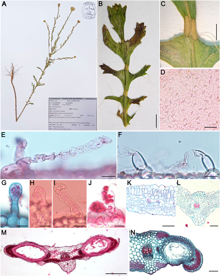
FIGURE 1. Morpho-anatomical characters of Adenophyllum porophyllum var. cancellatum: Herbarium specimen No. 3504-IZTA (A), leaf morphology (B), elliptical glands (C), superficial view of abaxial epidermis (D), uniseriate simple trichomes (E,F), glandular trichomes (G–J), transverse view of leaf anatomy (K,L), glands anatomy (M,N). Bars: (B) = 3 mm, (C) = 1 mm, (D,K,L,N) = 100 μm, (E−J) = 25 μm, (M) = 250 µm.
A total of 1,500 g of fresh aerial parts were collected, and the essential oil was obtained once through hydro-distillation using a Clevenger-type apparatus. The plant material was heated in water for 3 h in a mantle (6 L), and the steam/oil mix was condensed and collected in a 50 ml funnel. The oil layer was separated and dried over anhydrous sodium sulfate. The isolated essential oil was stored in the dark in a hermetically sealed glass container at −13°C prior to analysis. The yield was calculated as the ratio of the weight of the essential oil obtained to the weight of the plant used (w/w) x100.
Gas chromatography/mass spectrometry analysis of the essential oil
The essential oil of A. porophyllum var. cancellatum was analyzed through GC/MS using an Agilent Technologies 7890B Gas Chromatograph (GC) coupled to Agilent Technologies 5977D Mass Spectrometry (MS) Detector under the following conditions: a HP-5MS capillary column (30 m, 0.25 mm, 0.25 μm film thickness), helium (99.999%) as the carrier gas at a constant flow of 1.0 ml/min, an injection volume of 1 μl, an injector temperature of 280°C, and an ion source temperature of 200°C. The oven temperature was programmed to increase from 40°C to 300°C at a rate of 8°C/min, and was then held at 300°C for 5 min. A mass detector was operated in electron impact mode at an ionization energy of 70 eV, with a mass range of 30–600 (m/z).
The oil components were identified through comparative analysis of their retention indices (RI) and mass spectra. The RI values were calculated through linear interpolation relative to the retention times of a series of n-alkanes (C8−C20 alkane standards, Sigma–Aldrich). Then, the RIs and mass spectra were compared against values in the NIST14 and Mass Spectral Library 15 (National Institute of Standards and Technology, 2022 WebBook, Match > 90%) and literature (Adams, 2007; García-Roja et al., 2009; Lei et al., 2018). The compound concentrations (as % content) were calculated by integrating the corresponding chromatographic peak areas. α -pinene, β-pinene, and trans-farnesol were used as standards to verify the correct functioning of the CG/MS system.
Antimicrobial activity
The anti-Candida activity of the essential oil of A. porophyllum var. cancellatum was evaluated using three strains isolated from clinical cases: C. albicans 17MR (donated by the Clinical Analysis Laboratory of FES Iztacala, UNAM); C. glabrata, and C. tropicalis (isolated from a clinical case and donated by Hospital Angeles Metropolitano, México). The stock culture was maintained on potato dextrose agar (PDA Bioxon) and subcultured twice prior to performing bioassays. Antibacterial activity tests were performed using the following strains: gram-positive bacteria, Micrococcus luteus ATCC 10240, S. aureus ATCC 29213, S. aureus cc, and S. aureus CUSI (donated by the Clinical Analysis Laboratory of FES-Iztacala), and S. epidermidis FES-C (donated by the Microbiology Laboratory of FES-Cuautitlán), isolated from clinical cases; gram-negative bacteria, K. pneumoniae ATCC 13884, K. oxytoca ATCC 8724, Serratia marcescens ATCC 14756, Salmonella typhi ATCC 19430, Escherichia coli 82MR, and E. coli CUSI (donated by the Clinical Analysis Laboratory of FES-Iztacala), isolated from clinical cases. These strains were maintained at 4°C on Mueller Hinton agar (Bioxon), subjected to sensitivity tests, and sub-cultured twice, before and after the bioassays were performed.
The antifungal and antibacterial activities of the essential oil were evaluated according to the M100 guidelines of the Clinical and Laboratory Standard Institute (CLSI, 2020). A diffusion test was performed using 5 μl (3.25 mg/per disk) of essential oil. Nystatin (50 μg/ml) and chloramphenicol (25 μg/ml) were used as positive controls. The minimum inhibitory concentration (MIC), minimum fungicidal concentration (MFC), and minimum bactericidal concentration (MBC) were determined using microdilution tests with serial dilutions ranging from 3 to 0.06 mg/ml. The experiments were performed in triplicates.
The antibiofilm activity of the A. porophyllum var. cancellatum essential oil was evaluated using the crystal violet method described by Gómez-Sequeda et al. (2020). Due to the sensitivity to the oil and the fact that they were strains isolated from clinical cases, C. albicans 17MR, S. aureus cc, and E. coli 82MR were used to evaluate the antibiofilm effect. The MIC, MFC, or MBC values were used in all cases. The experiments were performed in triplicates. The percentage biofilm inhibition was calculated as follows:
OD control: optical density without essential oil; OD experimental: optical density with essential oil.
Three independent experiments were performed for all tests, and the mean and standard deviation of the results were reported. The antibiofilm activity results were analyzed using one-way analysis of variance (ANOVA), where values of p < 0.05 were considered statistically significant. In addition, the data were analyzed using Tukey’s test at 95% confidence. Statistical analysis was performed on the collected data using Excel software.
Anatomical study of the leaf of A. porophyllum var. cancellatum
An anatomical study was performed on the middle part of the leaf blade, including both wide and narrow areas, and floral involucre bracts. Free-hand sectioning was carried out, and some tissues were cleared with 20% sodium hydroxide and 50% sodium hypochlorite, stained, and mounted in glycerin jelly with safranin (Aguilar-Rodríguez, 1998). Other samples were processed using the paraffin inclusion method (Ruzin, 1999); 15-µm thick cross-sections were obtained using a rotary microtome. Tissue staining was performed using safranin-fast green (Johansen, 1940). Finally, the sections were mounted in a synthetic resin. Images and measurements were obtained using a Nikon E200 microscope attached to an image analyzer (NIS-Elements BR5.21.01; Nikon Instruments). The anatomical descriptions were made according to Metcalfe and Chalk (1979) and Fahn (1985). The bars and brightness of the images were improved using Adobe Photoshop CC 2020.
Results
Yield and gas chromatography/mass spectrometry analysis of the essential oil
The yield of essential oil from the aerial parts of A. porophyllum var. cancellatum was 0.5% (7.5 g); the oil density was 0.7 g/ml. Eighteen compounds were identified in the essential oil using GC/MS analysis. The main components were trans-pinocamphone (29.5%), limonene (24.7%), pinocarvone (21.9%), and cis-pinocamphone (8.0%) (Supplementary Figure S1). The essential oil of the plant contained only hydrocarbons (31.8%) and oxygenated monoterpenoids (62.1%) but did not sesquiterpenes (Table 1).
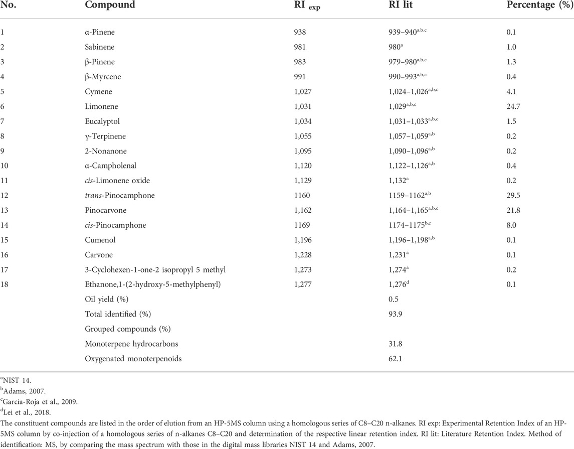
TABLE 1. The chemical composition of the essential oil of A. porophyllum var. cancellatum.
Antimicrobial activity
The antifungal activity of the essential oil of A. porophyllum var. cancellatum was demonstrated by inhibition of the growth of C. glabrata (10 ± 0.0 inhibition zone), C. albicans (20 ± 0.0 inhibition zone), and C. tropicalis (15 ± 0.0 inhibition zone), all of which were isolated from clinical cases. C. glabrata was determined to be the most sensitive species to the essential oil, with an MIC of <0.06 mg/ml compared to the 0.125 and 0.250 mg/ml for the other species, respectively. Furthermore, the essential oil inhibited the growth of ten bacterial strains, five gram-positive, and five gram-negative strains. The most sensitive bacterial strains were S. aureus cc (9.33 ± 0.6 inhibition zone; MIC 1 mg/ml), E. coli 82MR (8.00 ± 0.0 inhibition zone; MIC 0.75 mg/ml), and K. pneumoniae (16.00 ± 0.0 inhibition zone; MIC 0.75 mg/ml) (Table 2). The essential oil substantially prevented the formation of biofilms in C. albicans, S. aureus cc, and E. coli 82MR by 46.3%, 47.6%, and 33.1%, respectively, with respect to the untreated control (Table 3).
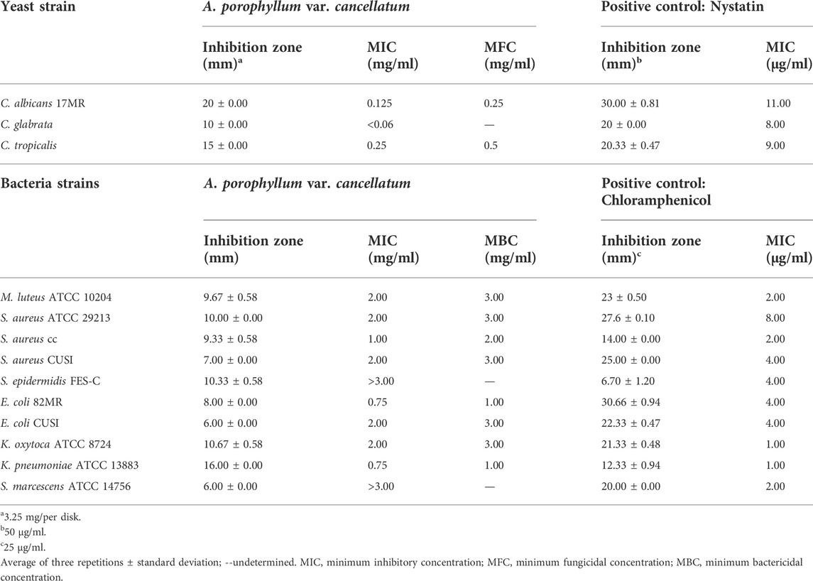
TABLE 2. Anti-Candida and antibacterial activities of the essential oil of A. porophyllum var. cancellatum.
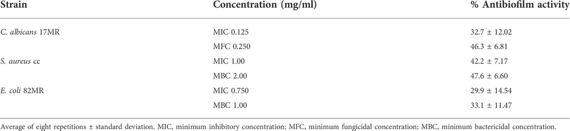
TABLE 3. Antibiofilm activity of the essential oil of A. porophyllum var. cancellatum.
Anatomical study of the leaf
According to Villarreal (2003), A. porophyllum var. cancellatum is an annual herbaceous plant with oval oil glands in its leaves (Figure 1B,C) and floral involucre bracts. Leaves were 2–6 cm long, pinnatisect (7–13 lobes linear to obovate), with the lobes toothed to split, and some teeth ending in long bristles (Figure 1B). The leaf anatomy of the species is shown in Figure 1D–N. In the superficial view of the leaf epidermis, the epidermal cells had undulated anticlinal walls, anisocytic and a few anomocytic stomata, (Figure 1D). The main types of trichomes were as follows: 1) uniseriate simple with a foot formed by 1–2 rounded thick-walled basal cells, and a head containing up to seven thin-walled cells, some of which showed collapsed walls or contents, frequently oriented parallel to the epidermal surface. In some cases, trichomes were observed with a flexuous, wavy, and ribbon-like appearance (Figure 1E,F); 2) glandular with a uniseriate foot formed by 1–4 cells and a head consisting of 2–5 irregularly shaped or rounded cells (Figures 1G–I); 3) glandular with a uniseriate foot made up of up to four cells and a club-shaped or rounded head, formed by four rows of cells with dense cytoplasm; these were scarce (Figure 1J).
In the transverse view (Figure 1K,L), the leaves were amphistomatic. The cuticle was smooth, with a simple epidermis; bifacial mesophyll, with uni-bistratified palisade parenchyma and compact spongy with 5–6 strata of quadrangular to rounded cells; collateral vascular bundles, those of the second level (secondary veins) surrounded by a parenchymal sheath (Figure 1K). The middle vein had a triangular outline with protuberances toward both surfaces, the lower one more prominent and thinner toward the underside; trichomes were scarce and of the same type as those described for the rest of the leaf blade; cuticle was crenate, with cells of the abaxial epidermis more voluminous than those of the rest of the lamina. Parenchymatous or collenchymatous cells with slightly thickened walls were observed under the adaxial and abaxial epidermis. These walls were thinner toward the center of the midvein, where they surrounded the only collateral-type vascular bundle, which was circular and encased in a parenchyma sheath. The palisade and spongy parenchyma penetrate part of the midvein (Figure 1L).
In the narrowest areas of the leaf blade, two large glands, approximately 1–2 mm long, were located on each side of the midvein (Figure 1C,M). In the transverse view, the cuticle was a smooth, simple epidermis, with rounded dome-like cells on both surfaces; below which there were 4–5 layers of tangentially elongated cells with thickened walls and narrow lumens surrounding the glands. The glands were elliptical in shape and almost occupied the entire mesophyll (Figure 1N). They were of lysigenous origin; each gland was associated with a pair of vascular bundles at its ends. Toward the margin of the lamina there was chlorenchyma, which sometimes partially surrounded each gland and extended into the midvein. The cuticle was crenate below which there were several layers of collenchyma associated with the central and semicircular collateral vascular bundle.
Discussion
A. porophyllum var. cancellatum is one of the 12 species of the genus Adenophyllum distributed in the southwestern United States, Mexico, Central America, and the Antilles (Villarreal, 2003). Phytochemical studies on the genus Adenophyllum indicate that thiophenes are a constituent compound of this herb in organic extracts, with terthiophenes being the most abundant (Downum et al., 1985). Hexanedioic acid and bis (2-ethylhexyl) ester are the main component of the ethyl acetate extract of A. porophyllum and exhibits activity against the phytopathogenic fungi, Pestalotiopsis clavispora, Colletotrichum gloeosporioides, and Lasiodiplodia pseudotheobromae (Hernández-Ceja et al., 2021). In addition, methanolic and ethyl acetate extracts of the roots of A. aurantium have antifungal activity against the phytopathogenic mycelial fungi, Alternaria alternata and Fusarium solani (Lira-De León et al., 2014). For other species of the genus antifungal activity are reported but no its antibacterial activity as well as their essential oils.
The essential oil of A. porophyllum var. cancellatum exhibits broad-spectrum antimicrobial activity, inhibiting the growth of both yeasts and bacteria. Candida species are the most sensitive to this oil. To the best of our knowledge, this is the first report on the chemical composition and antimicrobial potential of the essential oil, and the anatomy of the plant.
A. porophyllum var. cancellatum produces mainly monoterpenoids, the most abundant are trans-pinocamphone, limonene, pinocarvone, and cis-pinocamphone, which make up 84% of the essential oil. There are no studies on the essential oils from the other species of the genus Adenophyllum for comparison; however, it is known that other species in the Tageteae tribe, such as Dyssodia acerosa (Tellez et al., 1997), D. tagetiflora (García-Bores et al., 2018), D. decipiens (Pacheco-Hernández et al., 2020), and several species of the genus Tagetes (Ali et al., 2014) mainly produce monoterpenoids. In all of them, limonene is one of the main components of the essential oils.
There are species that mainly produce pinocamphone and pinocarvone in essential oils. In Hyssopus officinalis (Lamiaceae), cis-pinocamphone, trans-pinocamphone, pinocarvone, pinene, and phellandrene are the most abundant constituents; there is quantitative variability in the chromatographic profiles depending on variety, genotype, region, and culture conditions (Chalchat et al., 1999; Fraternale et al., 2004; Hristova et al., 2015; Stappen et al., 2015; Sharifi-Rad et al., 2022). The composition of hyssop oil is linked to its medicinal properties. The plant is traditionally used for its antiseptic properties in the treatment of infectious diseases, chronic bronchitis, and asthma (Stankovic' et al., 2016). The essential oil of H. officinalis has antibacterial activity, with an MIC of 4,000 μg/ml against S. aureus and 2000 μg/ml against E. coli (Stappen et al., 2015). In addition, the oil exhibits antifungal activity against Fusarium (Fraternale et al., 2004) and Candida (Hristova et al., 2015) strains.
The essential oil from the aerial parts of Cedronella canariensis var. canariensis (Lamiaceae) contains mainly pinocarvone, 46.8%–58.0% (López-García et al., 1992; Zorzetto et al., 2015). The oil inhibits the growth of Gram-positive and Gram-negative bacteria; however, it is more active against fungi of the genera Candida, Cryptococcus, and Saccharomyces, with zones of inhibition between 46 mm and 60 mm (López-García et al., 1992). The essential oil of Myrothamnus moschatus (Myrothamnaceae) mainly contains oxygenated monoterpenoids, trans-pinocarveol (35.6%), and pinocarvone (20.0%); coincidentally, the essential oil of this species exhibits antimicrobial activity against C. albicans (with an inhibition zone diameter of 18 mm) (Nicoletti et al., 2012). However, the antimicrobial activity of pure pinocarvone remains unknown.
Pinocamphones (cis and trans) and limonene are the main monoterpenoids in the essential oil of A. porophyllum var. cancellatum. The presence and quantity of these three monoterpenoids could be responsible for the antimicrobial activity of the essential oil of the plant because they constitute around 59.3% of the essential oil. Hristova et al. (2015) studied the anti-Candida activities of trans-pinocamphone and cis-pinocamphone. Both isomers have MICs of 1,000 μg/ml, indicating that isomerization does not influence activity. Thakre et al. (2018) reported that limonene exhibits antimicrobial activity against C. albicans and inhibits planktonic growth (yeast), morphogenesis (hyphae), and biofilm growth. Furthermore, the application of this compound resulted in a concentration-dependent reduction in the biofilm formation by several species of Streptococcus (Subramenium et al., 2015). In cells of the yeast, Zygosaccharomyces rouxii, limonene destroys yeast proteins, inhibits their synthesis, produces nucleic acid leakage, and alters membrane permeability (Cai et al., 2019). In E. coli, (-)-limonene acts by producing oxidative stress through the formation of significant amounts of hydrogen peroxide and superoxide anion radicals, which damage DNA and the cell membrane, increase permeability, resulting in cell death (Melkina et al., 2021).
The antimicrobial activity of essential oils is generally attributed to their major components, in part because it is difficult to discriminate the biological activities of minor compounds. Oxygenated terpenoids with groups aldehydes, and alcohols are the most active, followed by those containing esters and ketones, while hydrocarbons are generally ineffective. However, there is a synergistic effect between compounds with strong, moderate, and low antimicrobial activities (Nikkhah et al., 2017; Pinto et al., 2020). Hydrocarbon-type terpenoids could facilitate the transmembrane transport of oxygenates, resulting in a synergistic effect on antimicrobial activity (Nguefack et al., 2009). This could explain why essential oils are more active than pure compounds. For instance, the H. officinalis oil is more active in several species of Candida than its main components cis- and trans-pinocamphone, α- and β-pinene, and β-phellandrene. The MICs values of the pinocamphones were 28% and 21% higher than that of hyssop oil, against C. albicans and C. glabrata, respectively (Hristova et al., 2015). In the case of A. porophyllum var. cancellatum essential oil, it is possible that the hydrocarbon-type monoterpenes, such as limonene, have a synergistic effect with pinocamphones. However, further studies are needed to verify this hypothesis as well as the mechanism of action.
The anatomical characteristics of other Adenophyllum species have not yet been recorded. Therefore, as part of the pharmacognosy investigation of A. porophyllum var. cancellatum, the leaf microstructure was compared with that in the other taxa of the Tageteae tribe (Supplementary Table S1), because there are shared characteristics, such as amphiestomatic leaves, anisocytic/anomocytic stomata, and parenchyma sheath surrounding veins. Parenchyma sheaths are associated with Kranz-type photosynthesis, which appears to occur in dry climates, such as tropical deciduous forests, scrubs, grasslands, and disturbed areas where this species grows (Villarreal, 2003). Kranz-type anatomy is reported in different Asteraceae (Sánchez et al., 1986; Peter and Katinas, 2003). The anatomical characters are more homogeneous within the Tageteae tribe than at the level of the Asteraceae family (Rivera et al., 2017). Nevertheless, trichomes can contribute to the delimitation of species or genera belonging to the Tageteae tribe (Milan et al., 2006; Azevedo, 2007; García-Sánchez et al., 2012; Ferraro and Scremin-Dias, 2018; Rivera et al., 2019; Younis et al., 2020). In addition, a single central vascular bundle in the midvein and the size and shape of the glands could differentiate A. porphyllum var. cancellatum from the other closely related taxa.
Considering the high essential oil production of this taxon, further comparative analyzes of chemistry, antimicrobial activity, and leaf anatomy between different Adenophyllum species are needed.
Conclusion
This study validated the medicinal use of A. porophyllum var. cancellatum for the treatment of infections. The essential oil from its aerial parts showed antimicrobial properties against bacteria and yeasts of medical importance. The main chemical components of the oil were trans- and cis-pinocamphone, limonene, and pinocarvone, which could be responsible for the activity and could potentially be chemical markers of the species. An anatomical study of the leaf enabled characterization of the oil-producing glands and other histological attributes to support the identification of this species for quality control purposes.
Data availability statement
The original contributions presented in the study are included in the article/Supplementary Material, further inquiries can be directed to the corresponding author.
Author contributions
SA-R Validation, formal analysis and writing—original and review and editing; MEL-V Data curation, funding acquisition, methodology, and writing—original and review and editing; MPJ-R data curation, methodology, and visualization; CTH-D methodology and formal analysis; MFM-P methodology, EAE-P visualization, formal analysis, AME-G methodology, and visualization; EN-O methodology, JGA-A validation, formal analysis, and writing—original and review and editing; AMG-B conceptualization, supervision, and writing—original and review and editing. All the authors contributed to revising the manuscript revision and have read and approved the submitted version.
Funding
This study was funded by UNAM DGAPA-PAPIIT IN220920.
Acknowledgments
We thank Dalia Grego-Valencia for laboratory work and images improvement of plant anatomy section.
Conflict of interest
The authors declare that the research was conducted in the absence of any commercial or financial relationships that could be construed as a potential conflict of interest.
Publisher’s note
All claims expressed in this article are solely those of the authors and do not necessarily represent those of their affiliated organizations, or those of the publisher, the editors and the reviewers. Any product that may be evaluated in this article, or claim that may be made by its manufacturer, is not guaranteed or endorsed by the publisher.
Supplementary material
The Supplementary Material for this article can be found online at: https://www.frontiersin.org/articles/10.3389/fphar.2022.981959/full#supplementary-material
References
Adams, R. P. (2007). Identification of essential oil components by gas chromatography/mass spectrometry. 4th Edition. ILCarol Stream: Allured Publ.
Aguilar-Rodríguez, S. (1998). “Apéndice I. “Técnicas de laboratorio para el estudio de las embriofitas”,” in Plantae. Introducción al Estudio de las plantas con embrión. Editors J. D. Tejero-Díez, and M. P. Granillo V (México: Universidad Nacional Autónoma de México, Facultad de Estudios Superiores Iztacala), 247–272.
Ali, N. A., Sharopov, F. S., Al-Kaf, A. G., Hill, G. M., Arnold, N., Al-Sokari, S. S., et al. (2014). Composition of essential oil from Tagetes minuta and its cytotoxic, antioxidant and antimicrobial activities. Nat. Prod. Commun. 9 (2), 1934578X1400900–8. doi:10.1177/1934578x1400900233
Azevedo, C. S. (2007). “Leaf anatomy and ultrastructure of Pectis brevipedunculata: Secretory cavity development and Kranz structure,” in Dissertação/mestrado em Botânica estrutural Ecologia e Sistemática. [Viçosa](Minas Gerais, Brasil: Universidade Federal de Viçosa), 53.
Cai, R., Hu, M., Zhang, Y., Niu, Ch., Yue, T., Yuan, Y., et al. (2019). Antifungal activity and mechanism of citral, limonene and eugenol against Zygosaccharomyces rouxii. LWT 106, 50–56. doi:10.1016/j.lwt.2019.02.059
Chalchat, J. C., Adamovic, D., and Gorunovic, M. S. (1999). Composition of oils of three cultivated forms of Hyssopus officinalis endemic in yugoslavia: F. albus alef., f. cyaneus alef. and f. ruber mill. J. Essent. Oil Res. 13, 419–421. doi:10.1080/10412905.2001.9699712
Cilia-López, V. G., Cariño-Cortés, R., and Zurita-Salinas, L. R. (2021). Ethnopharmacology of the Asteraceae family in Mexico. Bot. Sci. 99 (3), 455–486. doi:10.17129/botsci.2715
CLSI (2020). Performance standards for antimicrobial susceptibility testing. 30th ed. Wayne, Pennsylvania 19087 USA: West Valley.
De Moraes, D. C. (2022). Current scenario of the search for new antifungal agents to treat Candida auris infections: An integrative review. J. Mycol. Med. 32 (1), 101232. doi:10.1016/j.mycmed.2021.101232
Downum, K. R., Keil, D. J., and Rodríguez, E. (1985). Distribution of acetylenic thiophenes in the Pectidinae. Biochem. Syst. Ecol. 13 (2), 109–113. doi:10.1016/0305-1978(85)90067-5
Ferraro, A., and Scremin-Dias, E. (2018). Structural features of species of Asteraceae that arouse discussions about adaptation to seasonally dry environments of the Neotropics. Acta Bot. Bras. 32, 113–127. doi:10.1590/0102-33062017abb0246
Fraternale, D., Ricci, D., Epifano, F., and Curini, M. (2004). Composition and antifungal activity of two essential oils of Hyssop (Hyssopus officinalis L.) J. Essent. Oil Res. 16, 617–622. doi:10.1080/10412905.2004.9698810
Frei, B., Heinrich, M., Bork, P. M., Herrmann, D., Jaki, B., Kato, T., et al. (1998). Multiple screening of medicinal plants from Oaxaca, Mexico: Ethnobotany and bioassays as a basis for phytochemical investigation. Phytomedicine 5 (3), 177–186. doi:10.1016/S0944-7113(98)80025-1
García-Bores, A. M., Arciniegas-Arciniegas, A., Reyna-Campos, A., Céspedes-Acuña, C., Avila-Suárez, B., Alarcón-Enos, J., et al. (2018). Phytochemical composition and biological activities of Dyssodia tagetiflora Lag. Chem. Biodivers. 15 (2), e1700415. doi:10.1002/cbdv.201700415
García-Roja, A., Quijano, C. E., Morales, G., and Pino, J. A. (2009). Composition of the essential oil from leaves and fruits of Margyricarpus pinnatus (Lam.) O. Kuntze grown in Colombia. J. Essent. Oil Res. 21 (6), 547–549. doi:10.1080/10412905.2009.9700240
García-Sánchez, F., López-Villafranco, M. E., Aguilar-Rodríguez, S., and Aguilar-Contreras, A. (2012). Etnobotánica y morfoanatomía comparada de tres especies de Tagetes que se utilizan en Nicolás Romero. Bot. Sci. 90, 221–232. doi:10.17129/botsci.388
Gómez-Sequeda, N., Cáceres, M., Stashenko, E. E., Hidalgo, W., and Ortiz, C. (2020). Antimicrobial and antibiofilm activities of essential oils against Escherichia coli O157:H7 and methicillin-resistant Staphylococcus aureus (MRSA). Antibiot. (Basel) 9 (11), 730. doi:10.3390/antibiotics9110730
Hernández-Ceja, A., Loeza-Lara, P. D., Espinosa-García, F. J., García-Rodríguez, Y. M., Medina-Medrano, J. R., Gutiérrez-Hernández, G. F., et al. (2021). In vitro antifungal activity of plant extracts on pathogenic fungi of blueberry (Vaccinium sp.) Plants (Basel) 10 (5), 852. doi:10.3390/plants10050852
Herrera-Martínez, M., Hernández-Ramírez, V. I., Hernández-Carlos, B., Chávez-Munguía, B., Calderón-Oropeza, M. A., and Talamás-Rohana, P. (2016). Antiamoebic activity of Adenophyllum aurantium (L.) Strother and its effect on the actin cytoskeleton of Entamoeba histolytica. Front. Pharmacol. 7, 169. doi:10.3389/fphar.2016.00169
Hristova, Y., Wanner, J., Jirovetz, L., Stappen, I., Iliev, I., and Gochev, V. (2015). Chemical composition and antifungal activity of essential oil of Hyssopus officinalis L. from Bulgaria against clinical isolates of Candida species. Biotechnol. Biotechnol. Equip. 29 (3), 592–601. doi:10.1080/13102818.2015.1020341
Köhler, J. R., Hube, B., Puccia, R., Casadevall, A., and Perfect, J. R. (2017). Fungi that infect humans. Microbiol. Spectr. 5 (3). doi:10.1128/microbiolspec.FUNK-0014-2016
Kostić, A. Ž., Janaćković, P., Kolašinac, S. M., and Dajić Stevanović, Z. P. (2020). Balkans' Asteraceae species as a source of biologically active compounds for the pharmaceutical and food industry. Chem. Biodivers. 17 (6), e2000097. doi:10.1002/cbdv.202000097
Lei, A., Yuchen, S., Jia, H., Yuping, L., Hua, Y., Runjun, Z., et al. (2018). Chemical compositions and in vitro antioxidant activity of the essential oil from Coreopsis tinctoria Nutt. Flower. J. Essent. Oil Bear. Plants 21 (4), 876–885. doi:10.1080/0972060X.2018.1510792
Lerato, S. K., Moteetee, A., and Van Vuuren, S. (2021). Ethnobotany, toxicity, and antibacterial activity of medicinal plants used in the Maseru District of Lesotho for the treatment of selected infectious diseases. S. Afr. J. Bot. 143, 141–154. doi:10.1016/j.sajb.2021.07.048
Lira-De León, K. I., Ramírez-Mares, M. V., Sánchez-López, V., Ramírez-Lepe, M., Salas-Coronado, R., Santos-Sánchez, N. F., et al. (2014). Effect of crude plant extracts from some Oaxacan flora on two deleterious fungal phytopathogens and extract compatibility with a biofertilizer strain. Front. Microbiol. 5, 383. doi:10.3389/fmicb.2014.00383
Loockerman, D. J., Turner, B. L., and Jansen, R. K. (2003). Phylogenetic relationships within Tageteae (Asteraceae) based on nuclear ribosomal ITS and chloroplast ndhF gene sequences. Syst. Bot. 28, 191–207. doi:10.1043/0363-6445-28.1.191
López-García, R. E., Hernández-Pérez, M., Rabanal, R. M., Darias, V., Martín-Herrera, D., Arias, A., et al. (1992). Essential oils and antimicrobial activity of two varieties of Cedronella canariensis (L.) W. et B. J. Ethnopharmacol. 36 (3), 207–211. doi:10.1016/0378-8741(92)90045-s
Manandhar, S., Luitel, S., and Dahal, R. K. (2019). In vitro antimicrobial activity of some medicinal plants against human pathogenic bacteria. J. Trop. Med. 2019, 1895340. doi:10.1155/2019/1895340
Melkina, O. E., Plyuta, V. A., Khmel, I. A., and Zavilgelsky, G. B. (2021). The mode of action of cyclic monoterpenes (-)-limonene and (+)-α-pinene on bacterial cells. Biomolecules 11 (6), 806. doi:10.3390/biom11060806
Metcalfe, C. R. C., and Chalk, L. (1979). “Anatomy of the dicotyledons,” in Systematic anatomy of leaf and stem, with a brief history of the subject (Oxford: Clarendon Press).
Milan, P., Hissae, H. A., and Appezzato-da-Glória, B. (2006). Comparative leaf morphology and anatomy of three Asteraceae species. Braz. Arch. Biol. Technol. 49 (1), 135–144. doi:10.1590/S1516-89132006000100016
National Institute of Standards and Technology (2022). webbook. AvaliableAt: https://webbook.nist.gov/chemistry/(Accessed June 25, 2022).
Nguefack, J., Dongmo, J. B., Dakole, C. D., Leth, V., Vismer, H. F., Torp, J., et al. (2009). Food preservative potential of essential oils and fractions from Cymbopogon citratus, Ocimum gratissimum and Thymus vulgaris against mycotoxigenic fungi. Int. J. Food Microbiol. 131 (2-3), 151–156. doi:10.1016/j.ijfoodmicro.2009.02.009
Nicoletti, M., Maggi, F., Papa, F., Vittori, S., Quassinti, L., Bramucci, M., et al. (2012). In vitro biological activities of the essential oil from the ‘resurrection plant’ Myrothamnus moschatus (Baillon) Niedenzu endemic to Madagascar. Nat. Prod. Res. 26 (24), 2291–2300. doi:10.1080/14786419.2012.665916
Nikkhah, M., Hashemi, M., Habibi Najafi, M. B., and Farhoosh, R. (2017). Synergistic effects of some essential oils against fungal spoilage on pear fruit. Int. J. Food Microbiol. 257, 285–294. doi:10.1016/j.ijfoodmicro.2017.06.021
Pacheco-Hernández, Y., Sánchez-Hernández, G. R., Reyes-Cervantes, E., Romero-Arenas, O., Pérez-Xochipa, I., and Villa-Ruano, N. (2020). Chemical variation and pharmacological properties of Dyssodia decipiens essential oil. Chem. Biodivers. 17 (10), e2000487. doi:10.1002/cbdv.202000487
Peter, G., and Katinas, L. (2003). A new type of Kranz anatomy in Asteraceae. Aust. J. Bot. 51 (2), 217–226. doi:10.1071/BT02080
Pinto, L., Bonifacio, M. A., De Giglio, E., Cometa, S., Logrieco, A. F., and Baruzzi, F. (2020). Unravelling the antifungal effect of red thyme oil (Thymus vulgaris L.) compounds in vapor phase. Molecules 25 (20), 4761. doi:10.3390/molecules25204761
Ramzan, S., Shaheen, S., Hussain, K., Khan, M. A., Sohail, J., and Khan, F. (2019). Significance of leaf morphoanatomical markers for the authentication of adultered drugs sold in herbal markets of district Lahore, Pakistan. Microsc. Res. Tech. 82 (11), 1911–1921. doi:10.1002/jemt.23359
Rivera, P., Terrazas, T., Rojas-Leal, A., and Villaseñor, J. L. (2019). Leaf architecture and anatomy of Asteraceae species in a xerophytic scrub in Mexico City, Mexico. Acta Bot. Mex. 126, 1–26. doi:10.21829/abm126.2019.1515
Rivera, P., Villaseñor, J. L., and Terrazas, T. (2017). Meso- or xeromorphic? Foliar characters of Asteraceae in a xeric scrub of Mexico. Bot. Stud. 58, 12–16. doi:10.1186/s40529-017-0166-x
Rolnik, A., and Olas, B. (2021). The plants of the Asteraceae family as agents in the protection of human health. Int. J. Mol. Sci. 22 (6), 3009. doi:10.3390/ijms22063009
Sánchez, E., Arriaga, O. M., and Panarello, O. H. (1986). El síndrome de “Kranz” en Asteraceae de la flora ArArgentinaBol. Soc. Argent. Bot. 24 (3-4), 249–259.
Sharifi-Rad, J., Quispe, C., Kumar, M., Akram, M., Amin, M., Iqbal, M., et al. (2022). Hyssopus essential oil: An update of its phytochemistry, biological activities, and safety profile. Oxid. Med. Cell. Longev. 2022, 8442734. doi:10.1155/2022/8442734
Sharma, A., Flores-Vallejo, R. D. C., Cardoso-Taketa, A., and Villarreal, M. L. (2017). Antibacterial activities of medicinal plants used in Mexican traditional medicine. J. Ethnopharmacol. 208, 264–329. doi:10.1016/j.jep.2016.04.045
Siscar-Lewin, S., Hube, B., and Brunke, S. (2022). Emergence and evolution of virulence in human pathogenic fungi. Trends Microbiol. 30 (7), 693–704. doi:10.1016/j.tim.2021.12.013
Soliman, S., Alnajdy, D., El-Keblawy, A. A., Mosa, K. A., Khoder, G., and Noreddin, A. M. (2017). Plants natural products as alternative promising anti-Candida drugs. Pharmacogn. Rev. 11 (22), 104–122. doi:10.4103/phrev.phrev_8_17
Srirama, R., Santhosh Kumar, J. U., Seethapathy, G. S., Newmaster, S. G., Ragupathy, S., Ganeshaiah, K. N., et al. (2017). Species adulteration in the herbal trade: Causes, consequences, and mitigation. Drug Saf. 40, 651–661. doi:10.1007/s40264-017-0527-0
Stankovic´, N., Mihajilov-Krstev, T., Zlatkovic´, B., Stankov-Jovanovic´, V., Mitic´, V., Jovic´, J., et al. (2016). Antibacterial and antioxidant activity of traditional medicinal plants from the Balkan Peninsula. NJAS Wageningen J. Life Sci. 78 (1), 21–28. doi:10.1016/j.njas.2015.12.006
Stappen, I., Wanner, J., Tabanca, N., Wedge, D. E., Ali, A., Kaul, V. K., et al. (2015). Chemical composition and biological activity of essential oils of Dracocephalum heterophyllum and Hyssopus officinalis from Western Himalaya. Nat. Prod. Commun. 10 (1), 1934578X1501000–8. doi:10.1177/1934578X1501000131
Subramenium, G. A., Vijayakumar, K., and Pandian, S. K. (2015). Limonene inhibits streptococcal biofilm formation by targeting surface-associated virulence factors. J. Med. Microbiol. 64, 879–890. doi:10.1099/jmm.0.000105
Tellez, M. R., Rick, E., Estell, R. E., Fredrickson, E. L., and Havstad, K. M. (1997). Essential oil of Dyssodia acerosa DC. J. Agric. Food Chem. 45, 3276–3278. doi:10.1021/jf9701502
Thakre, A., Zore, G., Kodgire, S., Kazi, R., Mulange, S., Patil, R., et al. (2018). Limonene inhibits Candida albicans growth by inducing apoptosis. Med. Mycol. 56, 565–578. doi:10.1093/mmy/myx074
Tropicos (1982). Adenophyllum porophyllum var. cancellatum (Cass.) Strother. AvaliableAt: https://tropicos.org/name/2734534 (Accessed April 12, 2022).
Velasco-Azorsa, R., Cruz-Santiago, H., Cid Del Prado-Vera, I., Ramírez-Mares, M. V., Gutiérrez-Ortiz, M. D. R., Santos-Sánchez, N. F., et al. (2021). Chemical characterization of plant extracts and evaluation of their nematicidal and phytotoxic potential. Molecules 26 (8), 2216. doi:10.3390/molecules26082216
Villarreal, Q. J. A. (2003). “Tribu Tageteae,” in Flora del Bajío y de Regiones Adyacentes. Editor J. Rzedowski (Saltillo, Coahuila, Pátzcuaro, Michoacán: Universidad Autónoma Agraria Antonio Narro, Instituto de Ecología, A.C. Centro Regional del Bajío. México), 85. doi:10.21829/fb.200.2003.113
Villaseñor, J. L. (2018). Diversidad y distribución de la familia Asteraceae en México. Bot. Sci. 96 (2), 332–358. doi:10.17129/botsci.1872
World Health Organization (2017). Global priority list of antibiotic-resistant bacteria to guide research, discovery, and development of new antibiotics. AvaliableAt: http://www.who.int/medicines/publications/WHO-PPL-Short_Summary_25Feb-ET_NM_WHO.pdf?ua=1 (Accessed July 25, 2022).
Wu, S. C., Liu, F., Zhu, K., and Shen, J. Z. (2019). Natural products that target virulence factors in antibiotic-resistant Staphylococcus aureus. J. Agric. Food Chem. 67 (48), 13195–13211. doi:10.1021/acs.jafc.9b05595
Younis, S., Shaheen, S., Zaib, M., Harun, N., Khalid, S., Hussain, K., et al. (2020). Scanning electron microscopic screening of 20 medicinally important Asteroideae taxa. Microsc. Res. Tech. 83 (8), 988–1006. doi:10.1002/jemt.23492
Zorzetto, C., Sánchez-Mateo, C. C., Rabanal, R. M., Lupidi, G., Bramucci, M., Quassinti, L., et al. (2015). Antioxidant activity and cytotoxicity on tumour cells of the essential oil from Cedronella canariensis var. canariensis (L.) Webb & Berthel. (Lamiaceae). Nat. Prod. Res. 29 (17), 1641–1649. doi:10.1080/14786419.2014.994213
Keywords: Adenophyllum porophyllum var. cancellatum, antifungal activity, aromatic plant, Candida, oil gland, oxidized monoterpenes
Citation: Aguilar-Rodríguez S, López-Villafranco ME, Jácquez-Ríos MP, Hernández-Delgado CT, Mata-Pimentel MF, Estrella-Parra EA, Espinosa-González AM, Nolasco-Ontiveros E, Avila-Acevedo JG and García-Bores AM (2022) Chemical profile, antimicrobial activity, and leaf anatomy of Adenophyllum porophyllum var. cancellatum. Front. Pharmacol. 13:981959. doi: 10.3389/fphar.2022.981959
Received: 29 June 2022; Accepted: 20 September 2022;
Published: 11 October 2022.
Edited by:
Milan Mladenovic, University of Kragujevac, SerbiaReviewed by:
Maria Yolanda Rios, Universidad Autónoma del Estado de Morelos, MexicoAmner Muñoz-Acevedo, Universidad del Norte, Colombia
Copyright © 2022 Aguilar-Rodríguez, López-Villafranco, Jácquez-Ríos, Hernández-Delgado, Mata-Pimentel, Estrella-Parra, Espinosa-González, Nolasco-Ontiveros, Avila-Acevedo, García-Bores. This is an open-access article distributed under the terms of the Creative Commons Attribution License (CC BY). The use, distribution or reproduction in other forums is permitted, provided the original author(s) and the copyright owner(s) are credited and that the original publication in this journal is cited, in accordance with accepted academic practice. No use, distribution or reproduction is permitted which does not comply with these terms.
*Correspondence: Ana María García-Bores, boresana@yahoo.com