 Zsuzsa Bagoly
 István Szegedi
 Rita Kálmándi
 Noémi Klára Tóth
 László Csiba2,3*†
- 1Division of Clinical Laboratory Sciences, Department of Laboratory Medicine, Faculty of Medicine, University of Debrecen, Debrecen, Hungary
- 2MTA-DE Cerebrovascular and Neurodegenerative Research Group, University of Debrecen, Debrecen, Hungary
- 3Department of Neurology, Clinical Centre, University of Debrecen, Debrecen, Hungary
Intravenous administration of recombinant tissue plasminogen activator (rt-PA) has been proven to be safe and effective in the treatment of acute ischemic stroke. Little is known, however, why this treatment is less effective in some patients while in others life-threatening side-effects, e.g., symptomatic intracerebral hemorrhage might occur. Clinical failure of thrombolysis related to absent or partial recanalization or reocclusion as well as hemorrhagic complications of thrombolysis are possibly related to hemostatic events. Data on markers of coagulation and/or fibrinolysis in acute stroke patients are numerous and may provide indications regarding therapy outcomes. Better understanding of the hemostatic and fibrinolytic system during rt-PA therapy might be clinically useful and ultimately might lead to an improvement in the efficacy or safety of this treatment. Studies on thrombus composition retrieved from cerebral arteries may also advance our knowledge and provide a key to improve acute stroke therapy. Here we provide a comprehensive review on a wide range of factors and markers of coagulation and fibrinolysis that have been studied in the context of thrombolysis outcome in ischemic stroke patients. Moreover, a brief summary is given on the most recent research on thrombus composition having a potential influence on outcomes.
Introduction
Ischemic stroke is one of the most important and serious vascular diseases affecting millions of people all over the world (1). By causing paresis, gait disturbance, speech disturbances and other symptoms stroke increases morbidity and mortality significantly (2). Despite the emerging importance of endovascular stroke therapy, e.g., mechanical thrombectomy (3), the primary treatment of acute ischemic stroke today is thrombolysis using recombinant tissue plasminogen activator within the therapeutic time window. The 1995 National Institute of Neurological Disorders and Stroke rt-PA (recombinant tissue plasminogen activator) Stroke Study (NINDS trial) was a milestone for stroke therapy as it proved that treating ischemic stroke patients with rt-PA within 3 h of the onset of stroke symptoms results in a considerable neurological improvement at 3 months as compared to the placebo group without significant change in mortality (4). The ECASS (European Cooperative Acute Stroke Study) III trial showed that intravenous rt-PA treatment is efficient beyond the time window of 3 h as compared to placebo in a certain group of ischemic stroke patients (5). By extending the time window to up to 4.5 h, 52.4% of patients had a favorable outcome as compared to the placebo group. Despite the unquestionable effectiveness of rt-PA in the treatment of acute ischemic stroke, this treatment is not a remedy for all. The rate of early recanalization using rt-PA is ~25% in patients with a proximal middle cerebral artery occlusion and only 10% in patients with an internal carotid occlusion (6). Additionally, the rate of re-occlusion is as high as 30% (7). Besides recanalization failure, about 6–8% of patients develop intracranial hemorrhage as side-effect, despite taking all precautionary steps to minimize bleeding risk (8). Little is known why in some patients recanalization failure occurs while in others bleeding will take place. In theory, the effect of rt-PA might depend on hemostasis factors affecting clot structure. The aim of this review article is to provide an insight on coagulation and fibrinolysis parameters that can have an impact on or were shown to have an association with the outcome of thrombolysis in ischemic stroke patients. Recent studies on thrombus composition obtained via endovascular treatment from the cerebral arteries of ischemic stroke patients are also summarized.
Brief Overview of the Non-hemostasis Factors Affecting Thrombolysis Outcome
Although the Safe Implementation of Thrombolysis in Stroke-MOnitoring STudy (SITS-MOST) proved the efficiency and safety of intravenous rt-PA (8), our understanding of predictive factors affecting thrombolysis outcomes is still limited. Poor outcome of thrombolysis is generally defined at 3 months post-event as a modified Rankin score (mRS) of 3 or more (less frequently 2 or more), which includes mortality (mRS = 6) (9). Thrombolytic therapy-related hemorrhage has been defined by the NINDS, ECASS I, ECASS II, ECASS III, and the SITS-MOST studies. (10) Symptomatic intracranial hemorrhage (sICH) was defined as any neurological deterioration associated with hemorrhage within 36 h of rt-PA therapy in the NINDS trial, while in ECASS III and SITS-MOST studies intracranial hemorrhage and ≥4 point increase in baseline NIHSS was considered as sICH [for a comprehensive review, see (10)]. There are several baseline clinical factors which may affect the outcomes of rt-PA therapy including male gender, stroke severity on admission, infarct size, advanced age, hyperglycaemia, etc., nevertheless, due to their low predictive value, these factors are mostly unhelpful for individual treatment decisions (9). Wahlgren et al. adjusted the outcomes of the SITS-MOST to the baseline characteristics of randomized, controlled trials in order to find parameters that can predict the outcome of the thrombolysis (11). In a multivariable analysis it was found that older age, high blood glucose, high National Institutes of Health Stroke Scale (NIHSS) score and definitive ischemic lesion on imaging scans were related to poor outcome (mRS 3–6). Male sex, disability before the actual stroke (modified Rankin Score 2–5), congestive heart failure, diastolic blood pressure, antiplatelet therapy other than aspirin, treatment in new centers were associated with higher mortality at the third month of the follow-up. Atrial fibrillation, systolic blood pressure, and weight were predictors of the occurrence of sICH, but current smokers had a lower rate of sICH.
Among standard baseline laboratory parameters, few were found to be associated with thrombolysis outcomes. In a systematic review based on 54 previous reports unadjusted and adjusted meta-analysis of high admission glucose level showed association with poor outcome (mRS≥2) and sICH (12). In a relatively large study, admission neutrophil count and neutrophil to lymphocyte ratio was found to be independently associated with increased risk of sICH and worse outcomes (mRS≥3) at 3 months (13).
The disruption of blood-brain barrier (BBB) is thought to be associated with post-lysis ICH (14). Early detection of BBB changes using imaging techniques (contrast-enhanced MRI or CT) are promising tools to predict hemorrhagic transformation (15, 16). The emerging role of imaging techniques in identifying therapy failure and safety is out of the scope of this review, interested readers should consult recent publications (17, 18).
Unfortunately, despite all efforts to predict patient subgroups that are more likely to clearly benefit or not benefit from intravenous thrombolysis, it is not yet possible to predict individual outcomes infallibly based on the above listed baseline clinical, standard laboratory or imaging variables (19). Moreover, known predictors of worse functional outcome due to inefficacy of therapy and the predictors of symptomatic intracranial hemorrhage show an overlap. This suggests a potential role for other factors in the diverse pathomechanisms ultimately leading to poor clinical outcomes.
Here we give a comprehensive list of hemostasis parameters that were reported to be associated with thrombolysis outcomes. Hemostasis parameters showing significant association with hemorrhagic transformation after thrombolysis are summarized in Table 1, while markers with a significant association regarding poor outcome based on mRS are listed in Table 2. A simplified overview of the coagulation and fibrinolytic system and cited literature investigating the potential role of certain factors/markers in the outcome of thrombolytic therapy is provided on Figure 1.
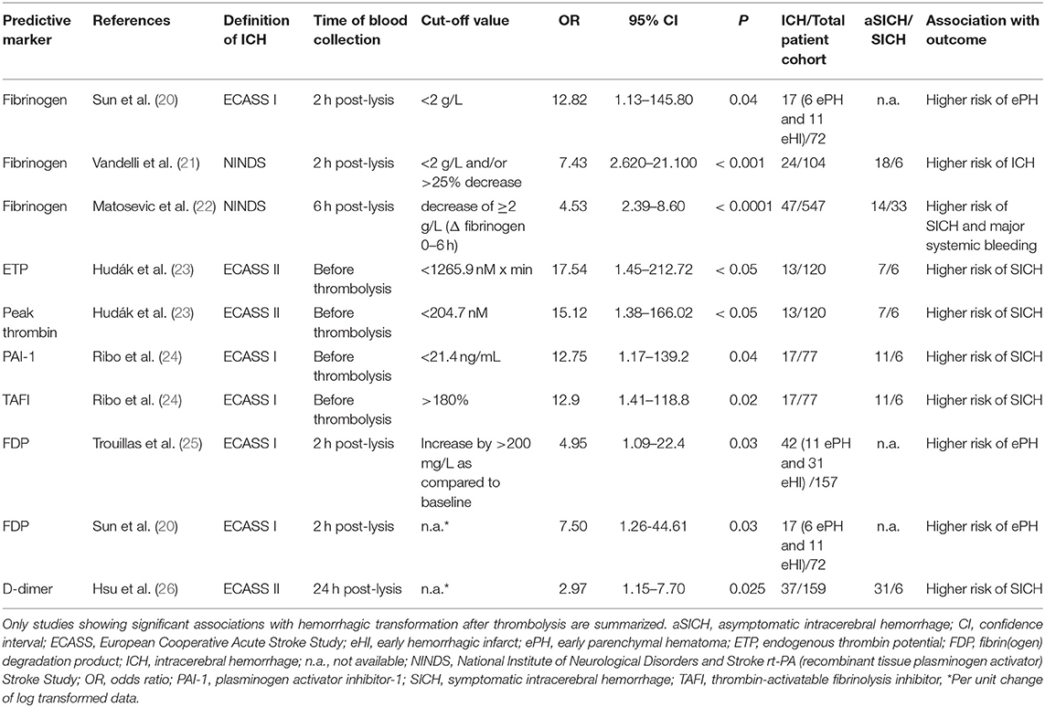
Table 1. Hemostasis markers associated with hemorrhagic transformation after acute ischemic stroke thrombolysis.
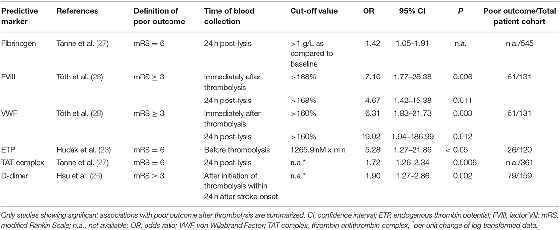
Table 2. Hemostasis markers associated with poor outcome at 3 months after acute ischemic stroke thrombolysis.
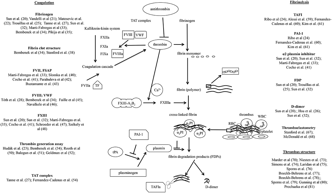
Figure 1. Simplified scheme demonstrating the main elements of coagulation, fibrinolysis, and thrombus formation, depicting publications that investigated the relationship of these elements with the outcome of thrombolytic therapy applied in acute ischemic stroke. α2PI, α2 plasmin inhibitor; FVII, factor VII; FVIIa, activated factor VII; FVIII, factor VIII; FVIIIa, activated factor VIII; FIXa, activated factor IX; FXIa, activated factor XI; FXIIa, activated factor XII; FXIII, factor XIII; FXIIIa, activated factor XIII; FXIII-A2B2, plasma (tetramer) form of factor XIII; PAI-1, plasminogen activator inhibitor-1; RBC, red blood cell; TAFI, thrombin-activatable fibrinolysis inhibitor TAFIa, activated thrombin-activatable fibrinolysis inhibitor; TAT complex, thrombin-antithrombin complex; TF, tissue factor; tPA, tissue plasminogen activator; VWF, von Willebrand Factor; WBC, white blood cell.
Coagulation Factors/Markers of Coagulation Activation Associated With Thrombolysis Outcomes
Fibrinogen
Fibrinogen is a central protein of hemostasis, although it has considerable non-hemostasis related functions as well: it plays a role in cellular and matrix interactions, inflammatory response, wound healing, cancer progression (29). Fibrinogen is the substrate of the procoagulant enzyme thrombin that cleaves it into fibrin. Fibrin forms a branched three-dimensional network that is cross-linked by activated factor XIII (FXIIIa) in the last step of the coagulation cascade (30). Cross-linked fibrin forms a network providing a stable biochemical and biophysical support to blood clots. Fibrin acts as a cofactor in tPA-induced plasminogen activation thus linking fibrin formation and fibrinolysis. Variations in fibrin properties and fibrin clot structure ultimately affect its degradation by plasmin (29, 31).
Low circulating fibrinogen levels (or a significant early decrease in fibrinogen levels after therapy) have been associated with post-thrombolysis hemorrhage in ischemic stroke patients (Table 1), although some studies showed contradictory results. In 2004, the phenomena of “early fibrinogen degradation coagulopathy” was proposed, describing a biological syndrome predictive of cerebral bleeding post-thrombolysis related to an early loss of fibrinogen and thus characterized by the increase of fibrin(ogen) degradation products (FDPs) (25). These results were later confirmed by other studies, including that of Sun et al. (20) (Table 1). In that study, 72 consecutive ischemic stroke patients treated with rt-PA were enrolled, and it was shown that a decrease in fibrinogen levels at 2 h post-lysis to <2 g/l multiplies the odds of early parenchymal hemorrhage by a factor of 12.82. It has to be noted, however, that in another study by the same authors somewhat different conclusions were reached (32). In that study 80 consecutive ischemic stroke patients were involved and changes in coagulation and fibrinolytic parameters were studied after rt-PA induced thrombolysis. Fibrinogen levels showed a 20% mean decrease, which was statistically linked to a decrease in plasminogen levels and it was also related to decreases of factors inhibiting thrombolysis (FXIII, α2-plasmin inhibitor). Nevertheless, fibrinogen levels didn't show a direct correlation with outcomes.
The concept of low fibrinogen levels being associated with post-lysis intracranial hemorrhage was confirmed by two other studies (21, 22) (Table 1). One hundred and four stroke patients, treated with intravenous thrombolysis, were included in the study by Vandelli et al. (21). Fibrinogen levels were measured on admission and 2 h after the thrombolysis. After 7 days of follow-up 24 patients presented with intracranial hemorrhage, of whom 6 patients were symptomatic. Among the 24 patients with intracerebral hemorrhage, 18 belonged to the “low fibrinogen group” (classified as levels decreased to <2 g/L and/or by 25% or more). Multivariate logistic regression analysis confirmed that patients belonging to the “low fibrinogen group” had a significantly increased risk for therapy-associated intracranial hemorrhage (OR: 7.47, 95%CI: 2.26–24.74, p < 0.001) and among the conventional risk factors only baseline NIHSS score conferred a statistically significant risk (OR: 1.15, 95%CI: 1.06–1.25, p < 0.001). On the other hand, they also demonstrated that higher baseline fibrinogen levels seem to enhance the risk of more significant decrease after thrombolysis. These data are in line with results published by Matosevic et al. who also showed that a reduction of at least 2 g/L in fibrinogen levels at 6 h post-lysis increases the risk of symptomatic intracranial hemorrhage (22). In this study a relatively large (n = 547) consecutive stroke patient cohort was investigated, and it was found that quantification of fibrinogen depletion after stroke thrombolysis significantly improved routine risk prediction of bleeding complications.
The association between admission fibrinogen levels and poor outcomes at 3 months post-lysis is not entirely clear. Tanne et al. investigated fibrinogen levels among other hemostasis parameters in patients with acute ischemic stroke from the NINDS rt-PA Stroke Study (27) (Table 2). Fibrinogen levels were measured at baseline, at 2 and 24 h after the thrombolysis. Of the 624 patients of the trial (consisting of rt-PA treatment and placebo group) the plasma samples of 465 patients were available at baseline, after 2 h or after 24 h and in the case of 281 patients all 3 plasma samples were available and were used for further evaluation. Fibrinogen levels showed a decrease at 2 h and remained low after 24 h in the treatment group and the placebo group as well. Higher levels of fibrinogen at baseline were associated with infarct lesion volume as detected by CT at 3 months within the entire study cohort (n = 570; p = 0.05). Higher 24 h fibrinogen levels were associated with an approximately 40% increase in the odds of death by 90 days in the whole study group (OR: 1.42; 95% CI: 1.05–1.91 per 1 g/L increment), but no other significant associations with outcomes were detected.
On the other hand, in an earlier study by Marti-Fabregas et al. including 83 patients treated by intravenous rt-PA, admission fibrinogen levels showed no association with long-term outcomes as determined by mRS>2 at 3 months (33). Similarly, in a most recent paper, fibrinogen levels on admission, at 24 h and 3 months post-event did not differ significantly in patients with poor outcomes (mRS>2) as compared to those with favorable outcomes at 3 months (34).
The association of fibrinogen levels on admission with imaging results found by Tanne et al. were confirmed in a hyperdense artery study Pikija et al. (35). The authors assessed on-admission fibrinogen level and clot burden in relation with the severity of the stroke, the volume of the infarct and in-hospital mortality in 132 ischemic stroke patients with hyperdense artery sign admitted within 6 h from the onset of the symptoms. Thrombolysis was performed in 60% of the patients and thrombectomy in 44% of them. Increased fibrinogen levels on admission showed association with smaller clot burden and lower NIHSS on admission, while patients with decreased fibrinogen had a higher clot burden and bigger volume of the infarct. However, in the adjusted statistical model, admission fibrinogen levels were not associated significantly with in hospital survival or death.
As conclusion, according to these data assessment of plasma fibrinogen levels pre- and post-lysis could be potentially useful to predict post-lysis intracranial hemorrhage but more data is needed regarding its associations with poor outcomes. It must be noted that determination of fibrinogen levels by the Clauss method is a relatively quick, easy and cheap measurement that can be performed in most clinical laboratories in any hour of the day, which is a clear advantage as compared to many other potentially useful biomarkers. Nevertheless, future studies involving more patients are needed to clearly define its predictive value and to validate its use as a potential biomarker for rt-PA associated bleeding events.
Fibrin Clot Structure
The structure of the fibrin clots is determined by genetic and environmental factors. Fibrin clots composed of thin, highly branched fibrin fibers are more rigid, less permeable, and less susceptible to dissolution by fibrinolysis (36). It has been shown that patients with ischemic stroke have reduced clot permeability and have features of prothrombotic clot phenotype as compared to healthy individuals (37). On the other hand, changes in clot microstructure of ischemic stroke patients who underwent thrombolysis have been studied scarcely. In a prospective cohort study by Stanford et al. clot microstructure was compared in ischemic stroke patients before, at 2–4 h and at 24 h after thrombolysis to assess the effects of thrombolysis (38). It was shown that thrombolysis itself has an effect on clot structure, but due to the relatively small number of patients who received rt-PA (n = 32) no conclusions were reached regarding patient outcomes. In a most recent paper by Bembenek et al. clot properties of 74 ischemic stroke patients undergoing thrombolysis were studied and their impact on clinical outcome was assessed (34). Ex vivo plasma fibrin clot formation was investigated on admission, at 24 h and at 3 months post-lysis. Compared with the pretreatment values, fibrin networks assessed at 24 h post-lysis were formed more slowly, were less compact, composed of thinner fibers, which lysed more rapidly. Logistic regression adjusted for potential confounders showed that pretreatment clot lysis time predicted excellent outcome as measured by mRS (0–1). In conclusion, formation of denser fibrin clots that show impaired lysability and the pattern of their changes might be indicative of clinical outcomes in acute stroke patients treated with thrombolysis.
Factor VII (FVII), Factor VII Activating Protease (FSAP)
The complex of activated factor VII (FVIIa) and tissue factor is the most potent activator that initiates the blood clotting cascade in normal hemostasis (39). FVII levels have been investigated in a handful of studies involving ischemic stroke patients undergoing thrombolysis but no relevant association was found between FVII levels and treatment outcomes or safety (33, 40, 41).
Factor VII activating protease (FSAP)—predominantly expressed in the liver—is a plasma serine protease that activates coagulation factor VII as well as pro-urokinase. Besides, FSAP inactivates tissue factor pathway inhibitor (TFPI), which would have a procoagulant effect on hemostasis as well (42). One hundred and twenty acute stroke patients were prospectively studied by Bustamente et al. in order to determine whether plasma FSAP levels have an association with recanalization after thrombolysis (43). The authors found that lower FSAP antigen levels correlates with a higher chance of recanalization after thrombolysis, suggesting the involvement of FSAP in rt-PA induced clot lysis, however, FSAP levels showed no significant association with poor outcomes (mRS>2) at 3 months post-event.
Factor VIII (FVIII), Von Willebrand Factor (VWF)
Factor VIII (FVIII), a key component of the blood coagulation system, is a cofactor for factor IXa that converts factor X to the activated form (FXa). In plasma, it circulates with von Willebrand factor (VWF) in a non-covalent complex (44). Associations between FVIII or VWF levels and stroke thrombolysis outcomes were investigated in four recent studies (28, 34, 45, 46). Faille et al. studied VWF levels before rt-PA treatment in 64 acute ischemic stroke patients (45). No association was found between VWF levels and poor outcome (mRS>2). In the study by Tóth et al. 131 consecutive acute ischemic stroke patients undergoing thrombolysis were investigated and FVIII activity and VWF levels were determined on admission, immediately after and 24 h after rt-PA treatment. FVIII levels decreased significantly immediately after lysis, that was attributed to plasmin-mediated FVIII degradation. In this study, VWF levels at all investigated time points and FVIII levels on admission and 24 h after thrombolysis were associated with worse imaging results (24 h post-lysis ASPECTS scores). In a binary backward logistic regression analysis elevated FVIII and VWF levels after thrombolysis were independently associated with poor long-term functional outcomes as defined by mRS>2 (Table 2). No association was found between FVIII levels during thrombolysis and post-lysis ICH. Navalkele et al. tested FVIII levels of 29 acute ischemic stroke patients on arrival, at 6 h and followed by 12-h intervals up to 72 h. (46) Similarly to previous findings, a significant decrease in median FVIII level from baseline to 6 h after thrombolysis was found. Baseline FVIII level and change in FVIII levels were not associated with recanalization or sICH. In another study including 74 acute ischemic stroke patients by Bembenek et al. FVIII levels on admission, at 24 h and 3 months did not differ significantly in patients with favorable or poor outcome (mRS>2) (34). To summarize these findings, elevated FVIII, and VWF levels after thrombolysis treatment might have a potential prognostic value regarding poor outcomes, but more studies are warranted on this topic.
Factor XIII (FXIII)
Activated factor XIII (FXIIIa) cross-links fibrin chains in the last step of the clotting cascade. FXIIIa plays a crucial role in protecting the fibrin clot against prompt fibrinolysis not only via fibrin cross-linking but also by cross-linking α2-plasmin inhibitor (α2-PI) and perhaps other plasma components to the fibrin clot, thus effectively hindering its proteolysis by plasmin (17). The association of FXIII levels with thrombolysis outcomes in ischemic stroke patients has been studied in several papers (20, 32, 33, 41, 47, 48). Although it might be hypothesized that a decrease in FXIII level could be a predictor of post-lysis ICH, no such association was found in any of the investigated study cohorts. In 3 studies, a decrease of FXIII level was observed after thrombolytic treatment but it was not related to hemorrhagic complications (20, 47, 48).
In an early multicenter study investigating 63 acute ischemic stroke patients receiving intravenous thrombolysis, FXIII levels on admission were not related to poor outcomes (mRS>2) (33). In the study by Sun et al. investigating a panel of hemostasis markers in 80 acute stroke patients treated with rt-PA, FXIII activity as measured on admission, at 2 h and 24 h post-lysis was found to have no significant predictive value on recanalization or poor functional outcome (32). In the study by Schroeder et al. investigating 66 acute ischemic stroke patients, it was suggested that the decrease in FXIII levels post-stroke might be associated with an unfavorable short-term outcome and the authors expressed the need for larger studies investigating FXIII as a candidate prognostic marker (47). In a most recent study by our group FXIII levels were investigated on admission, immediately after and 24 h after thrombolysis in 132 consecutive patients with acute ischemic stroke (48). In a backward multiple regression analysis it was revealed that a FXIII level in the lowest quartile 24 h after thrombolysis is an independent predictor of short-term mortality (14 days). However, as found by previous studies, FXIII level was not a predictor of long-term outcome or mortality at 3 months. Major FXIII-A and FXIII-B polymorphisms had no impact on therapeutic outcomes.
To conclude, in the above mentioned studies, although interesting trends were seen on low FXIII levels post-event and short-term outcomes, FXIII levels showed no association with unfavorable outcomes at 3 months after the event and were not associated with post-lysis ICH.
Global Markers of Coagulation Activation (Thrombin Generation, Thrombin-Antithrombin Complex)
Thrombin generation test is a global hemostasis assay that provides information on the speed and the amount of generated thrombin in plasma. The assay in its present form is a relatively new test but it is a potentially promising laboratory tool to elucidate coagulation mechanisms in various clinical conditions (49). Due to this fact the first studies on thrombin generation in ischemic stroke patients treated with intravenous thrombolysis were published in the past few years (23, 34, 50, 51) and patient outcomes were only assessed in two papers (23, 34). In an early study investigating thrombin generation in stroke patients, significantly higher peak thrombin concentration was found as compared to controls, but unfortunately in this study therapy outcomes were not investigated (50). In the study by Balogun et al. thrombin generation was assessed in 154 ischemic stroke patients of which 57 were treated with rt-PA (51). The authors concluded that endogen thrombin potential (ETP) or peak thrombin concentrations were not different between stroke subtypes or as compared to healthy controls, but results were not correlated with outcomes. Ninety five patients who suffered acute ischemic stroke were studied by Goldman et al. (52). Seventy one patients recevied intravenous thrombolysis, the remaining 24 served as control patients. Thrombolysed patients had markedly decreased thrombin generation parameters measured after 24 h, with the strongest impact on lag time compared to the baseline values, but again, results were not correlated with outcomes.
Our group investigated the thrombin generation test in 120 consecutive acute ischemic stroke patients before the administration of intravenous thrombolysis (23). We found that symptomatic intracranial hemorrhage was significantly associated with low ETP and peak thrombin levels (Table 1). Moreover, in a multiple logistic regression analysis it was shown that a low ETP result is an independent predictor of mortality within the first 2 weeks (OR: 6.03; 95%CI: 1.2–30.16, p < 0.05) and 3 months after the event (OR: 5.28; 95%CI: 1.27–21.86, p < 0.05) (Table 2). In the studied patient cohort, ETP was not significantly lower in those patients who had worse functional outcomes (mRS 2-5) but survived by the end of the 3rd month. In the study by Bembenek et al. it was found that a higher peak thrombin result at baseline is significantly associated with poor outcome at 3 months post-lysis (34).
Based on these interesting observations, one can conclude that the thrombin generation test might serve in the future as a useful tool to predict outcomes and safety of thrombolysis treatment, but future prospective studies involving large cohorts of patients are needed.
Antithrombin is a major inhibitor of thrombin, forming a 1:1 stable complex with it (thrombin-antithrombin complex: TAT). TAT complex is a sensitive marker for the activation of intravascular coagulation, thus useful for the risk assessment and diagnosis of thromboembolic events (53). Levels of TAT has been investigated in the population of the NINDS trial from samples collected at baseline, at 2 h after treatment and after 24 h post-lysis (27). TAT levels peaked at 2 h post-treatment selectively in the rt-PA treatment group. Increased levels of TAT (in the entire cohort) were associated with higher mortality at the third month of the follow-up that could have been due to resistance to recanalization (Table 2).
The association between pretreatment TAT levels and the outcome of thrombolysis was assessed in an interesting study by Fernandez-Cadenas et al. as well (54). TAT levels of 89 patients with middle cerebral artery occlusion were measured before the administration of rt-PA and the results correlated with the outcomes. No association was found between the measured TAT concentrations and the occurrence of hemorrhagic transformation. Decreased levels of TAT showed a significant association with better recanalization rates at all time-points (1 h: OR: 24.8, 95% CI 1.4–434.8, p = 0.028; 2 h: OR: 6.3 95% CI 1.5–27, p = 0.014; 6 h: OR: 6.4 95% CI 1.5–26.5, p = 0.011) after adjustment for stroke risk factors. Nevertheless, as compared to the study of Tanne, statistically significant correlation wasn't found between pre-thrombolysis levels of TAT and mortality rates.
Fibrinolytic Parameters/Markers of Fibrinolysis Associated With Thrombolysis Outcomes
Thrombin-Activatable Fibrinolysis Inhibitor (TAFI), Plasminogen Activator Inhibitor-1 (PAI-1)
Endogenous fibrinolysis inhibitors (e.g., TAFI, PAI-1) play an important role in the balance of coagulation and fibrinolysis and in theory may be involved in the hemorrhagic transformation after thrombolysis. After its activation TAFI removes lysine residues from partially degraded fibrin thus suppresses fibrinolysis (55). PAI-1 is the main inhibitor of t-PA: by forming stable complexes with t-PA it effectively blocks plasminogen activation (56). Moreover, it has been proposed that t-PA facilitates the disruption of BBB and PAI-1 could prevent t-PA-induced neuronal degeneration and BBB impairment (57, 58). Thus, the potential role of PAI-1 in preventing post-lysis hemorrhagic complications is intriguing.
In a prospective, longitudinal, multicenter, observational study Alessi et al. investigated the correlation between consumption of TAFI, activated/inactivated TAFI (TAFIa/ai) and the severity and outcome of stroke (59). Two groups of 109 stroke patients were enrolled in the study: one treated (68 patients) and one untreated with rt-PA (41 patients), furthermore there was a reference group of 20 patients without stroke. TAFI levels were sequentially measured in treated and not treated patients. On admission, patients had higher level of TAFIa/ai than the healthy reference group matched for age and gender. TAFIa/ai levels significantly increased at the end of thrombolysis that lasted up to 4 h. Higher levels of TAFIa/ai showed an association with a more severe day 2 NIHSS score and an unfavorable mRS score at day.
The authors concluded that their data demonstrate a considerable relationship between TAFI levels and early clinical severity during thrombolysis.
The aim of a study by Fernandez-Cadenas et al. was to evaluate whether the presence of two relatively common functional polymorphisms of the PAI-1 and TAFI genes (influencing PAI-1 and TAFI levels) have impact on recanalization rates of the middle cerebral artery among stroke patients treated with rt-PA (60). One hundred and thirty nine patients were enrolled in the study who all underwent the classic post-stroke diagnostic check-up (Doppler sonography, echocardiography, long-term ECG monitoring, complete blood cell count, and special coagulation tests) and the severity of the stroke was determined using NIHSS score. Occlusion and recanalization was diagnosed by the means of transcranial Doppler. There was no association between PAI-1 4G/5G polymorphism and the rate of recanalization, on the other hand, TAFI Thr325Ile polymorphism was significantly associated with recanalization resistance. The combination of the two polymorphisms doubled the risk of recanalization failure (OR: 11.1; 95% CI: 1.4–89.8%, p = 0.025).
Kim et al. also aimed to analyze pretreatment fibrinolysis inhibitor levels in patients who underwent intravenous thrombolysis and to investigate their potential association with thrombolysis failure (61). Forty three stroke patients were enrolled in the study: 17 patients were treated with intravenous t-PA, 11 with intra-arterial urokinase, and 15 with combined intravenous t-PA and intra-arterial urokinase. Patients were categorized into 2 groups according to recanalization (n = 30) or non-recanalization (n = 13), and a group of healthy volunteers (n = 34) were used as controls. It was found that plasma PAI-1 levels were increased in patients with acute stroke, and the increased pretreatment plasma PAI-1 levels were associated with the failure of the thrombolysis based on post-lysis angiography. TAFI levels did not differ among the groups of this cohort.
Admission PAI-1 and TAFI levels of consecutive rt-PA treated stroke patients with middle cerebral artery occlusion were studied by Ribo et al. (24). Seventy seven patients were involved in the study who had middle cerebral artery occlusion proved by transcranial Doppler. Patients with hemorrhagic transformation had lower baseline PAI-1 and higher TAFI levels. The combination of admission PAI <21.4 ng/mL and TAFI>180% had a sensitivity of 75% and a specificity of 97.6% predicting symptomatic intracranial hemorrhage (Table 1), indicating the need of further studies testing whether these biomarkers could be useful to improve the safety of thrombolysis.
α2-Plasmin Inhibitor (α2-PI)
α2-PI is one of the most important regulators of fibrinolysis. It has an inhibitory effect on the fibrinolytic pathway by three ways: it forms a complex with plasmin; inhibits the binding of plasminogen to fibrin; and makes fibrin more resistant toward the effect of plasmin via its own cross-linking to fibrin mediated by FXIIIa (62).
The levels of α2-PI were found to correlate well with the rate of recanalization in ischemic stroke patients treated with rt-PA (33). In this study by Marti-Fabregas et al., α2-PI level was proved to be an independent predictor of recanalization, although it didn't have a relation with long-term outcome. In studies by others, although a decrease in α2-PI levels post-lysis was found, α2-PI levels showed no association with the occurrence of hemorrhagic transformation or poor outcomes (20, 32, 41).
Fibrin(ogen) Degradation Products (FDPs) and D-Dimer
The in vivo formation of cross-linked fibrin and its subsequent secondary fibrinolytic digestion yields to a variety of soluble cross-linked FDPs. One of these products is known as D-dimer (63). It has been proved by many clinical studies that D-dimer is a valuable marker of coagulation activation and fibrinolysis (64, 65).
An increase in FDP levels 2 h post-lysis was found to be a predictor of post-lysis ICH in two studies. In an early study by Trouillas et al. FDPs were studied in 157 patients of the Lyon rt-PA trial in order to determine whether early fibrin(ogen) degradation might be indicative of hemorrhagic lesions (25). It was found that an FDP level at 2 h was a strong predictive factor for early parenchymal hematomas. An increase of FDP level >200 mg/L at 2 h post-lysis multiplied the probability of parenchymal hematoma by 4.95 (95%CI: 1.09–22.4) (Table 1). Based on the results of this single-centered study it was concluded that early fibrin(ogen) degradation can be indicative of early parenchymal hematomas that may be recognized by the detection of FDPs.
Sun et al. investigated the correlations between the presence of early intracerebral hemorrhage and the post-thrombolytic changes of hemostasis parameters (20). Seventy two patients were enrolled in the study. Similarly to the study by Trouillas et al. the presence of early parenchymal hematomas were associated with an increase in FDPs at 2 h post-lysis, indicating a massive lysis of fibrin and fibrinogen (Table 1). These results suggest that measuring not only fibrinogen but also FDPs might be useful in predicting the possibility of thrombolysis-induced intracerebral hematoma in acute stroke patients. In another study of the same authors, FDP and D-dimer levels were measured before rt-PA treatment, at 2 h and 24 h after thrombolysis in 80 stroke patients, and the results were correlated with global outcome (32). As expected, FDP and D-dimer levels increased between h0 and h2 and showed a tendency to return to the initial values at 24 h post-lysis. However, none of these values were predictive of poor outcome at 3 months.
In a study by Hsu et al. plasma D-dimer levels were evaluated after the initiation of rt-PA but within 24 h of stroke onset in 159 patients (26). Plasma D-dimer levels were significantly associated with unfavorable outcome at 3 months. Elevated D-dimer level was an independent parameter of symptomatic intracerebral hemorrhage after treatment with intravenous rt-PA. After adjustment for clinical variables in the statistical model, a higher level of D-dimer remained significantly associated with an unfavorable outcome (OR 1.90, 95% CI 1.27–2.86, p = 0.002) and with the occurrence of symptomatic intracranial hemorrhage (OR 2.97, 95% CI 1.15–7.70, p = 0.025) (Tables 1, 2).
Global Testing of Coagulation and Fibrinolysis: Thromboelastography (TEG) and Rotational Thromboelastometry (ROTEM)
Thromboelastography (TEG) and rotational thromboelastometry (ROTEM) are global viscoelasticity-based hemostasis tests of similar methodology providing information about coagulation and fibrinolysis from whole blood (66). The viscoelastic properties of blood monitored using TEG or ROTEM were found to be associated with bleeding and thrombotic predisposition in a variety of clinical conditions. According to the present research so far, however, its usefulness to detect hypercoagulability or therapy resistance might be questionable in the ischemic stroke patient population. In a study involving 72 patients with ischemic stroke and 71 healthy subjects, the ROTEM method did not seem to be able to detect a hypercoagulable state in patients with ischemic stroke as compared to controls (67). Moreover, in another study involving 171 acute ischemic stroke patients treated with rt-PA, no robust association was found between TEG results and clinical response to therapy. Baseline and post-lysis TEG data showed no association neither with rapid improvement as detected by a change of NIHSS, nor with hemorrhagic transformation (68).
Therefore, according to these results it is not proved as yet that the above listed viscoelasticity-based point-of-care tests could be useful to guide clinical decisions in acute stroke patients, nevertheless, future prospective studies involving large number of patients are needed to confirm such negative associations.
Studies on the Structure of Thrombi
The origin of thrombi causing ischemic stroke can be traced back to either atherosclerosis, cardioembolism or, rarely, to dissection (69). As mechanical thrombectomy became more widespread it became possible to analyze thrombi derived from the site of ischemic lesions. This technique opened a new, unique and interesting field of research providing the possibility to analyze fresh pathological thrombi of acute ischemic stroke patients. The main components of the thrombi are platelets, red blood cells, white blood cells, and cholesterol crystals (70). Knowledge of the composition of these thrombi may advance our knowledge on thrombolysis failure/therapy outcomes, as differently structured thrombi respond potentially differently to thrombolytic therapy. For instance, platelet-rich thrombi seem to be more resistant to lysis by thrombolytic therapy in rat models (71).
Most recently, a consensus statement on the current knowledge and future directions on the analysis of thrombi in acute stroke patients has been published on behalf of an international group of well-recognized clinicians and scientists (72). In this consensus paper recent research, current opportunities and limitations were published, which interested readers should consult. Here we provide a brief overview of studies that investigated thrombus composition and its relation to stroke or therapy outcomes.
The first, ground breaking study demonstrating systematic histological analysis of thrombi removed from the cerebral artery network of patients with acute ischemic stroke was published by Marder et al. (70). In that report, the authors analyzed the histology of 25 thrombi retrieved from the cerebral circulation of ischemic stroke patients and correlated the findings with clinical data. They found that the retrieved thrombi had similar histological components, independent on whether they derived from cardiac or arterial sources. Nevertheless, histological examination of these thrombi indicated that none of them were similar in overall appearance, each demonstrating a distinctive pattern. Yet the components of most cerebral thrombi were remarkably similar, 75% of them showing the pattern of lightly stained fibrin and platelet areas, interspersed with deposits of nucleated cells, often with intervening collections of erythrocytes.
In the paper by Niesten et al. the composition of the 22 extracted thrombi were found to be related to stroke subtype (73). They found that 73% of the thrombi were fresh, 18% were lytic and 9% were organized. Four thrombi were red blood cell rich, 4 thrombi were platelet rich while the rest were mixed thrombi. They found that most of the examined thrombi were fresh and the ones from the large artery atherosclerosis had the highest rate of red blood cell while the cardioembolism and cryptogenic subtype had the lowest. The large artery atherosclerosis was the only subtype with red thrombi. They found no correlation between subtype of the stroke and platelet and fibrin content. There was correlation between the red blood cell component and thrombus attenuation that can improve the attenuation on plain CT, pointing out the possibility of this as a useful imaging marker of stroke management.
In a study by Simons et al. the correlation between thrombus composition and hyperdense artery sign was investigated (74). Forty stroke patients were involved of which 28 patients underwent intravenous thrombolysis prior to the mechanical thrombectomy. Besides the basic histopathologic evaluation the samples were stained with CD34 immunostain, then they were categorized into 4 phases of thrombus formation: red blood cell dominant (11 samples), red blood cell proportion equal to fibrin (11 samples), fibrin dominant (7 samples), and organized fibrin pathology (11 samples). The hyperdense artery sign was defined by a neuroradiologist as an asymmetrical, increased density in one of the intracranial arteries. In 29 cases the radiologist was able to assess hyperdense artery sign on the pre-treatment CT. The conclusion of the examination was that thrombi were composed of early phase pathology. The impact of this data is that the presence or absence of hyperdense artery sign might enable neurologists to predict the composition of the thrombus, which might provide the benefit of different treatment options. They didn't find statistically significant connection between thrombus composition and cardioembolic stroke data.
The presence of white blood cells with an emphasis on neutrophil extracellular traps (NETs) was studied in a particularly interesting paper by Laridan et al. (75). Sixty-eight thrombi were extracted from ischemic stroke patients undergoing endovascular treatment. The thrombi were immunostained and characterized for the presence of neutrophils and NETs. Both neutrophils and NETs were detected in all thrombi. Older thrombi and those of cardioembolic origin contained more NETs. Ex vivo lysis of thrombi indicated that adding DNase might have prothrombolytic effect, which is an interesting observation with potential future therapeutic value.
In a study by Sporns et al. involving a remarkably large cohort (n = 187) of patients histopathologic investigation of clots retracted with mechanical thrombectomy was used in order to find specific clot patterns that might help to differentiate between causes of ischemic stroke (76). Of the 187 patients included 77 had cardioembolic source of stroke, 35 had large artery atherosclerosis, 11 were of other origin (dissection, radiogenic stenosis, tumor associated) and 64 cases were identified as cryptogenic stroke. Erythrocyte-rich thrombi were shown to have a significant correlation with non-cardioembolic stroke sources, while cardioembolic cases showed a significant association with fibrin-rich thrombi. The immunhistochemical markers CD68/KiM1P were higher in cardioembolic cases as compared to noncardioembolic strokes. Based on this study, it was concluded that histological thrombus features vary significantly according to the underlying cause and might be used to help to differentiate between cardioembolic and non-cardioembolic stroke types.
In another study with a relatively large number of clot samples (n = 137) histological clot composition was also studied in the relation of ischemic stroke causes (77). In this paper the authors aimed to find specific patterns that may help to differentiate between the causes of cryptogenic stroke. They found significant differences between the composition of thrombi of cardioembolic and noncardioembolic stroke patients. Cardioembolic thrombi consisted of significantly higher rate of fibrin/platelets, more leukocytes and less erythrocytes than noncardioembolic thrombi. Thrombi received from cryptogenic stroke patients had the same basic pattern as cardioembolic thrombi, with higher proportions of fibrin/platelets and smaller fractions of red blood cells. Similarities were also found between cryptogenic and cardioembolic strokes in terms of interventional and outcome parameters.
Despite the significant achievements in the efficacy of mechanic thrombectomy, little is known why in some patients recanalization is less successful. The composition of thrombi and its relation to the outcomes after thrombectomy were studied in only few studies as yet (78–81).
In a prospective study by Boeckh-Behrens et al. thrombus composition was analyzed with respect to the etiology of stroke, recanalization and clinical outcome (78). Thirty-four patients with acute ischemic stroke were included in the study, with distal internal carotid artery/carotid-T, anterior cerebral artery or middle cerebral artery occlusion. Histopathological examination of the extracted thrombi was carried out and it was found that the higher rate of white blood cells in the thrombus was associated with extended mechanical recanalization time, less favorable recanalization and worse clinical outcome (mRS>2). The percentage of leukocytes within the thrombi and the mRS scores showed a positive correlation (r = 0.358, p = 0.057) with a borderline statistical significance. The authors found significant relation between the white blood cell fraction of the thrombi and cardioembolic etiology of stroke. These data suggest that white blood cell-mediated immunological or hemostatic processes may play an important role in the development of stroke and might have impact on outcomes.
The aim of the prospective study by Sporns was to identify the effect of thrombus composition on the time and grade of revascularization and the risk of procedure-related secondary embolisms (79). Moreover, the authors aimed to evaluate the correlation of pre-interventional CT thrombus attenuation with the histological analysis of thrombi and other outcome parameters. Samples of 180 patients with complete diagnostic and histological workup were included in the study. The cause of the stroke as determined by the TOAST-classification was arterioembolic in 34 patients, cardioembolic in 75 patients, of other determined cause in 11 patients, and cryptogenic in 60 patients. In 168 patients recanalization was complete and 27 patients (15%) suffered secondary embolism. Besides the basic histopatological examination, detailed immunohistochemistry staining was also performed in the extracted thrombi samples (CD3, CD20, and CD68/KiM1P CD3; CD20, and CD68/KiM1P). Based on their observations fibrin-rich thrombi with low erythrocyte rate had significantly longer intervention times, while thrombi with a low rate of red blood cells and low CT-density caused embolisms in the thrombectomy process more often suggesting that these thrombi have higher fragility.
Similar results were obtained in the study of Gunning et al. (80). In this paper, the relationship between clot composition and the resistance to sliding (friction) was studied, which might contribute to resistance to clot removal. They found that fibrin-rich clots with <20% red cell content have significantly higher friction coefficient that might contribute to resistance to clot removal.
Prochazka et al. investigated thrombus composition in a prospective cohort study including 131 patients with ischemic stroke (81). Recanalization was successful in 115 patients from whom 90 samples were extracted and analyzed histologically. They found a significant relationship between plasma VWF and the VWF found in the thromboembolus, platelets, or fibrin. There was a correlation between the area of immunostained VWF and platelet count, CD31- positive cells and fibrin. The amount of all CD31-positive cells correlated with the number of neutrophils in the thrombus. D-dimer levels showed a significantly positive relationship with plasma VWF levels and with long-term prognosis (mRS>2). Significant correlations were found between the numbers of NK (natural killer) cells and the fibrin content of the thrombus, blood neutrophil levels and VWF levels within the thrombus and the number of lymphocytes and fibrin content of the thrombus.
To conclude, insights into thrombus composition might reveal stroke etiology and might provide relations to clinical outcomes. Understanding thrombus composition might serve as a key in advancing our knowledge and improving acute stroke care.
Conclusion
Despite advances in understanding the mechanisms leading to poor clinical outcomes and hemorrhagic complications following rt-PA treatment of acute ischemic stroke, prediction of individual patient risks is still unavailable. Clinical failure of thrombolysis as well as hemorrhagic complications are possibly related to hemostatic events. Data on markers of coagulation and/or fibrinolysis in acute stroke patients are numerous but most studies include relatively few patients thus they lack statistical power. Based on these data clinical recommendations regarding treatment and monitoring cannot be given as yet. Nevertheless, few markers seem to be potentially useful in predicting therapeutic outcomes. A simplified evaluation regarding the prognostic values of hemostasis markers/tests on thrombolysis safety and outcome is summarized in Table 3.
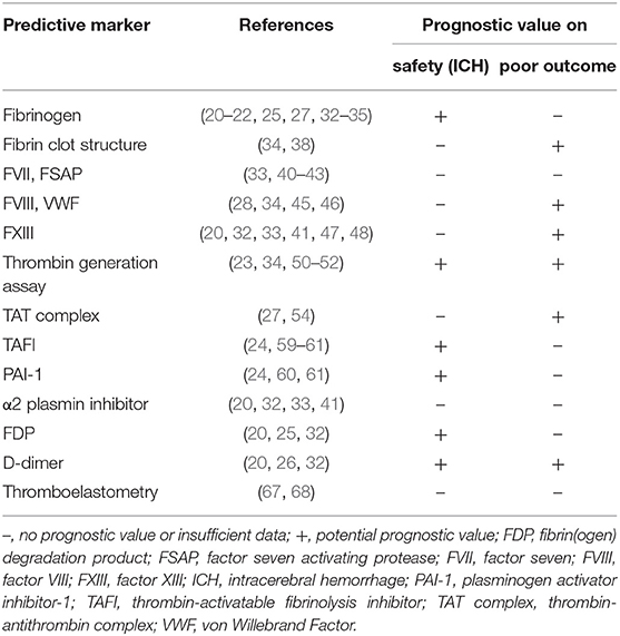
Table 3. A simplified evaluation regarding the prognostic values of hemostasis markers/tests on thrombolysis safety and outcome.
Well-designed, prospective clinical studies involving large cohorts of patients are awaited to better understand the hemostatic and fibrinolytic system during rt-PA therapy and to validate the prognostic value of promising markers in clinical decision making. Conclusions of such studies might lead to an improvement of the efficacy and/or safety of thrombolysis treatment. Studies on thrombus composition also seem useful in advancing our knowledge that could ultimately provide a key to improve acute stroke therapy.
Author Contributions
RK and NT were responsible for literature screening. ZB, IS, and LC screened literature, processed the articles and wrote the paper.
Funding
Supported by grants from the National Research, Development and Innovation Fund (K109712, K120042, FK128582), by GINOP-2.3.2-15-2016-00048 and GINOP-2.3.2-15-2016-00043 and the Hungarian Academy of Sciences (MTA-DE Cerebrovascular and Neurodegenerative Research Group). ZB is the recipient of János Bólyai fellowship.
Conflict of Interest Statement
The authors declare that the research was conducted in the absence of any commercial or financial relationships that could be construed as a potential conflict of interest.
References
1. Strong K, Mathers C, Bonita R. Preventing stroke: saving lives around the world. Lancet Neurol. (2007) 6:182–7. doi: 10.1016/S1474-4422(07)70031-5
3. Nogueira RG, Jadhav AP, Haussen DC, Bonafe A, Budzik RF, Bhuva P, et al. Thrombectomy 6 to 24 hours after stroke with a mismatch between deficit and infarct. N Engl J Med. (2017) 378:11–21. doi: 10.1056/NEJMoa1706442
4. National Institute of Neurological D Stroke rt PASSG: Tissue plasminogen activator for acute ischemic stroke. N Engl J Med. (1995) 333:1581–7. doi: 10.1056/NEJM199512143332401
5. Hacke W, Kaste M, Bluhmki E, Brozman M, Davalos A, Guidetti D, et al. Thrombolysis with alteplase 3 to 4.5 hours after acute ischemic stroke. N Engl J Med. (2008) 359:1317–29. doi: 10.1056/NEJMoa0804656
6. del Zoppo GJ, Poeck K, Pessin MS, Wolpert SM, Furlan AJ, Ferbert A, et al. Recombinant tissue plasminogen activator in acute thrombotic and embolic stroke. Ann Neurol. (1992) 32:78–86. doi: 10.1002/ana.410320113
7. Alexandrov AV, Grotta JC. Arterial reocclusion in stroke patients treated with intravenous tissue plasminogen activator. Neurology. (2002) 59:862–7. doi: 10.1212/WNL.59.6.862
8. Wahlgren N, Ahmed N, Davalos A, Ford GA, Grond M, Hacke W, et al. Thrombolysis with alteplase for acute ischaemic stroke in the safe implementation of thrombolysis in stroke-monitoring study (SITS-MOST): an observational study. Lancet. (2007) 369:275–82. doi: 10.1016/S0140-6736(07)60149-4
9. Miller DJ, Simpson JR, Silver B. Safety of thrombolysis in acute ischemic stroke: a review of complications, risk factors, and newer technologies. Neurohospitalist. (2011) 1:138–47. doi: 10.1177/1941875211408731
10. Chapman SN, Mehndiratta P, Johansen MC, McMurry TL, Johnston KC, Southerland AM. Current perspectives on the use of intravenous recombinant tissue plasminogen activator (tPA) for treatment of acute ischemic stroke. Vasc Health Risk Manag. (2014) 10:75–87. doi: 10.2147/VHRM.S39213
11. Wahlgren N, Ahmed N, Eriksson N, Aichner F, Bluhmki E, Davalos A, Erila T, et al. Multivariable analysis of outcome predictors and adjustment of main outcome results to baseline data profile in randomized controlled trials: safe Implementation of Thrombolysis in Stroke-MOnitoring STudy (SITS-MOST). Stroke. (2008) 39:3316–22. doi: 10.1161/STROKEAHA.107.510768
12. Desilles JP, Meseguer E, Labreuche J, Lapergue B, Sirimarco G, Gonzalez-Valcarcel J, et al. Diabetes mellitus, admission glucose, and outcomes after stroke thrombolysis: a registry and systematic review. Stroke. (2013) 44:1915–23. doi: 10.1161/STROKEAHA.111.000813
13. Maestrini I, Strbian D, Gautier S, Haapaniemi E, Moulin S, Sairanen T, et al. Higher neutrophil counts before thrombolysis for cerebral ischemia predict worse outcomes. Neurology. (2015) 85:1408–16. doi: 10.1212/WNL.0000000000002029
14. Khatri R, McKinney AM, Swenson B, Janardhan V. Blood-brain barrier, reperfusion injury, and hemorrhagic transformation in acute ischemic stroke. Neurology. (2012) 79:S52–7. doi: 10.1212/WNL.0b013e3182697e70
15. Hom J, Dankbaar JW, Soares BP, Schneider T, Cheng SC, Bredno J, et al. Blood-brain barrier permeability assessed by perfusion CT predicts symptomatic hemorrhagic transformation and malignant edema in acute ischemic stroke. AJNR Am J Neuroradiol. (2011) 32:41–8. doi: 10.3174/ajnr.A2244
16. Hjort N, Wu O, Ashkanian M, Solling C, Mouridsen K, Christensen S, et al. MRI detection of early blood-brain barrier disruption: parenchymal enhancement predicts focal hemorrhagic transformation after thrombolysis. Stroke. (2008) 39:1025–8. doi: 10.1161/STROKEAHA.107.497719
17. Young JY, Schaefer PW. Acute ischemic stroke imaging: a practical approach for diagnosis and triage. Int J Cardiovasc Imag. (2015) 32:19–33. doi: 10.1007/s10554-015-0757-0
18. Lin MP, Liebeskind DS. Imaging of ischemic stroke. Continuum. (2016) 22:1399–423. doi: 10.1212/CON.0000000000000376
19. Werring DJ. Cerebral microbleeds and thrombolysis-associated intracerebral hemorrhage: cause for concern, or just a distraction? Stroke. (2015) 46:2403–5. doi: 10.1161/STROKEAHA.115.010082
20. Sun X, Berthiller J, Trouillas P, Derex L, Diallo L, Hanss M. Early fibrinogen degradation coagulopathy: a predictive factor of parenchymal hematomas in cerebral rt-PA thrombolysis. J Neurol Sci. (2015) 351:109–14. doi: 10.1016/j.jns.2015.02.048
21. Vandelli L, Marietta M, Gambini M, Cavazzuti M, Trenti T, Cenci MA, et al. Fibrinogen decrease after intravenous thrombolysis in ischemic stroke patients is a risk factor for intracerebral hemorrhage. J Stroke Cerebrovasc Dis. (2015) 24:394–400. doi: 10.1016/j.jstrokecerebrovasdis.2014.09.005
22. Matosevic B, Knoflach M, Werner P, Pechlaner R, Zangerle A, Ruecker M, et al. Fibrinogen degradation coagulopathy and bleeding complications after stroke thrombolysis. Neurology. (2013) 80:1216–24. doi: 10.1212/WNL.0b013e3182897015
23. Hudak R, Szekely EG, Kovacs KR, Nagy A, Hofgart G, Berenyi E, et al. Low thrombin generation predicts poor prognosis in ischemic stroke patients after thrombolysis. PLoS ONE. (2017) 12:e0180477. doi: 10.1371/journal.pone.0180477
24. Ribo M, Montaner J, Molina CA, Arenillas JF, Santamarina E, Quintana M, et al. Admission fibrinolytic profile is associated with symptomatic hemorrhagic transformation in stroke patients treated with tissue plasminogen activator. Stroke. (2004) 35:2123–7. doi: 10.1161/01.STR.0000137608.73660.4c
25. Trouillas P, Derex L, Philippeau F, Nighoghossian N, Honnorat J, Hanss M, et al. Early fibrinogen degradation coagulopathy is predictive of parenchymal hematomas in cerebral rt-PA thrombolysis: a study of 157 cases. Stroke. (2004) 35:1323–8. doi: 10.1161/01.STR.0000126040.99024.cf
26. Hsu PJ, Chen CH, Yeh SJ, Tsai LK, Tang SC, Jeng JS. High plasma d-dimer indicates unfavorable outcome of acute ischemic stroke patients receiving intravenous thrombolysis. Cerebrovasc Dis. (2016) 42:117–21. doi: 10.1159/000445037
27. Tanne D, Macko RF, Lin Y, Tilley BC, Levine SR, Group NrSS. Hemostatic activation and outcome after recombinant tissue plasminogen activator therapy for acute ischemic stroke. Stroke. (2006) 37:1798–804. doi: 10.1161/01.STR.0000226897.43749.27
28. Tóth NK, Szekely EG, Czuriga-Kovacs KR, Sarkady F, Nagy O, Lanczi LI, et al. Elevated factor VIII and von willebrand factor levels predict unfavorable outcome in stroke patients treated with intravenous thrombolysis. Front Neurol. (2018) 8:721. doi: 10.3389/fneur.2017.00721
29. Weisel JW. Fibrinogen and fibrin. Adv Protein Chem. (2005) 70:247–99. doi: 10.1016/S0065-3233(05)70008-5
30. Bagoly Z, Koncz Z, Harsfalvi J, Muszbek L. Factor XIII, clot structure, thrombosis. Thromb Res. (2011) 129:382–7. doi: 10.1016/j.thromres.2011.11.040
31. Hudson NE. Biophysical mechanisms mediating fibrin fiber lysis. Biomed Res Int. (2017) 2017:2748340. doi: 10.1155/2017/2748340
32. Sun X, Berthiller J, Derex L, Trouillas P, Diallo L, Hanss M. Post-thrombolysis haemostasis changes after rt-PA treatment in acute cerebral infarct. correlations with cardioembolic aetiology and outcome. J Neurol Sci. (2015) 349:77–83. doi: 10.1016/j.jns.2014.12.029
33. Marti-Fabregas J, Borrell M, Cocho D, Belvis R, Castellanos M, Montaner J, et al. Hemostatic markers of recanalization in patients with ischemic stroke treated with rt-PA. Neurology. (2005) 65:366–70. doi: 10.1212/01.wnl.0000171704.50395.ba
34. Bembenek JP, Niewada M, Siudut J, Plens K, Czlonkowska A, Undas A. Fibrin clot characteristics in acute ischaemic stroke patients treated with thrombolysis: the impact on clinical outcome. Thromb Haemost. (2017) 117:1440–7. doi: 10.1160/TH16-12-0954
35. Pikija S, Trkulja V, Mutzenbach JS, McCoy MR, Ganger P, Sellner J. Fibrinogen consumption is related to intracranial clot burden in acute ischemic stroke: a retrospective hyperdense artery study. J Transl Med. (2016) 14:250. doi: 10.1186/s12967-016-1006-6
36. Undas A. Prothrombotic fibrin clot phenotype in patients with deep vein thrombosis and pulmonary embolism: a new risk factor for recurrence. Biomed Res Int. (2017) 2017:8196256. doi: 10.1155/2017/8196256
37. Undas A, Podolec P, Zawilska K, Pieculewicz M, Jedlinski I, Stepien E, et al. Altered fibrin clot structure/function in patients with cryptogenic ischemic stroke. Stroke. (2009) 40:1499–501. doi: 10.1161/STROKEAHA.108.532812
38. Stanford SN, Sabra A, D'Silva L, Lawrence M, Morris RH, Storton S, et al. The changes in clot microstructure in patients with ischaemic stroke and the effects of therapeutic intervention: a prospective observational study. BMC Neurol. (2015) 15:35. doi: 10.1186/s12883-015-0289-1
39. Eigenbrot C. Structure, function, and activation of coagulation factor VII. Curr Protein Pept Sci. (2002) 3:287–99. doi: 10.2174/1389203023380675
40. Slomka A, Switonska M, Sinkiewicz W, Zekanowska E. Assessing circulating factor VIIa-antithrombin complexes in acute ischemic stroke: a pilot study. Clin Appl Thromb Hemost. (2017) 23:351–9. doi: 10.1177/1076029616631424
41. Cocho D, Borrell M, Marti-Fabregas J, Montaner J, Castellanos M, Bravo Y, et al. Pretreatment hemostatic markers of symptomatic intracerebral hemorrhage in patients treated with tissue plasminogen activator. Stroke. (2006) 37:996–9. doi: 10.1161/01.STR.0000206461.71624.50
42. Parahuleva MS, Holschermann H, Zandt D, Pons-Kuhnemann J, Parviz B, Weiskirchen R, et al. Circulating factor VII activating protease (FSAP) is associated with clinical outcome in acute coronary syndrome. Circ J. (2012) 76:2653–661. doi: 10.1253/circj.CJ-11-1502
43. Bustamante A, Diaz-Fernandez B, Giralt D, Boned S, Pagola J, Molina CA, et al. Factor seven activating protease (FSAP) predicts response to intravenous thrombolysis in acute ischemic stroke. Int J Stroke. (2016) 11:646–55. doi: 10.1177/1747493016641949
44. Chavin SI. Factor VIII: structure and function in blood clotting. Am J Hematol. (1984) 16:297–306. doi: 10.1002/ajh.2830160312
45. Faille D, Labreuche J, Meseguer E, Huisse MG, Ajzenberg N, Mazighi M. Endothelial markers are associated with thrombolysis resistance in acute stroke patients. Eur J Neurol. (2014) 21:643–7. doi: 10.1111/ene.12369
46. Navalkele D, Boehme A, Albright K, Leissinger C, Schluter L, Freeman M, et al. Factor VIII in acute cerebral ischemia pilot study: biomarker in patients with large vessel occlusion? Clin Appl Thromb Hemost. (2018) 24:1249–54. doi: 10.1177/1076029618781045
47. Schroeder V, Ortner E, Mono ML, Galimanis A, Meier N, Findling O, et al. Coagulation factor XIII activation peptide and subunit levels in patients with acute ischaemic stroke: a pilot study. Thromb Res. (2010) 126:e122–7. doi: 10.1016/j.thromres.2010.05.027
48. Szekely EG, Czuriga-Kovacs KR, Bereczky Z, Katona E, Mezei ZA, Nagy A, et al. Low factor XIII levels after intravenous thrombolysis predict short-term mortality in ischemic stroke patients. Sci Rep. (2018) 8:7662. doi: 10.1038/s41598-018-26025-z
49. Tripodi A. Thrombin generation assay and its application in the clinical laboratory. Clin Chem. (2016) 62:699–707. doi: 10.1373/clinchem.2015.248625
50. Rooth E, Sobocinski-Doliwa P, Antovic J, Frykman Kull V, Von Arbin M, Rosenqvist M, et al. Thrombin generation in acute cardioembolic and non-cardioembolic ischemic stroke. Scand J Clin Lab Invest. (2013) 73:576–84. doi: 10.3109/00365513.2013.826817
51. Balogun IO, Roberts LN, Patel R, Pathansali R, Kalra L, Arya R. Thrombin generation in acute ischaemic stroke. Stroke Res Treat. (2016) 2016:7940680. doi: 10.1155/2016/7940680
52. Goldman S, Prior SM, Bembenek JP, Niewada M, Broniatowska E, Czlonkowska A, et al. Activation of blood coagulation and thrombin generation in acute ischemic stroke treated with rtPA. J Thromb Thrombolysis. (2017) 44:362–70. doi: 10.1007/s11239-017-1544-7
53. Negreva M, Georgiev S, Prodanova K, Nikolova J. Early changes in the antithrombin and thrombin-antithrombin complex in patients with paroxysmal atrial fibrillation. Cardiol Res. (2016) 7:89–94. doi: 10.14740/cr469w
54. Fernandez-Cadenas I, Mendioroz M, Munuera J, Alvarez-Sabin J, Rovira A, Quiroga A, et al. Lower concentrations of thrombin-antithrombin complex (TAT) correlate to higher recanalization rates among ischaemic stroke patients treated with t-PA. Thromb Haemost. (2009) 102:759–64. doi: 10.1160/TH08-06-0398
55. Wang W, Boffa MB, Bajzar L, Walker JB, Nesheim ME. A study of the mechanism of inhibition of fibrinolysis by activated thrombin-activable fibrinolysis inhibitor. J Biol Chem. (1998) 273:27176–81. doi: 10.1074/jbc.273.42.27176
56. Chandler WL, Alessi MC, Aillaud MF, Henderson P, Vague P, Juhan-Vague I. Clearance of tissue plasminogen activator (TPA) and TPA/plasminogen activator inhibitor type 1 (PAI-1) complex: relationship to elevated TPA antigen in patients with high PAI-1 activity levels. Circulation. (1997) 96:761–8. doi: 10.1161/01.CIR.96.3.761
57. Yepes M, Sandkvist M, Moore EG, Bugge TH, Strickland DK, Lawrence DA. Tissue-type plasminogen activator induces opening of the blood-brain barrier via the LDL receptor-related protein. J Clin Invest. (2003) 112:1533–1540. doi: 10.1172/JCI19212
58. Dohgu S, Takata F, Matsumoto J, Oda M, Harada E, Watanabe T, et al. Autocrine and paracrine up-regulation of blood-brain barrier function by plasminogen activator inhibitor-1. Microvasc Res. (2011) 81:103–7. doi: 10.1016/j.mvr.2010.10.004
59. Alessi MC, Gaudin C, Grosjean P, Martin V, Timsit S, Mahagne MH, et al. Changes in activated thrombin-activatable fibrinolysis inhibitor levels following thrombolytic therapy in ischemic stroke patients correlate with clinical outcome. Cerebrovasc Dis. (2016) 42:404–14. doi: 10.1159/000447722
60. Fernandez-Cadenas I, Alvarez-Sabin J, Ribo M, Rubiera M, Mendioroz M, Molina CA, et al. Influence of thrombin-activatable fibrinolysis inhibitor and plasminogen activator inhibitor-1 gene polymorphisms on tissue-type plasminogen activator-induced recanalization in ischemic stroke patients. J Thromb Haemost. (2007) 5:1862–8. doi: 10.1111/j.1538-7836.2007.02665.x
61. Kim SH, Han SW, Kim EH, Kim DJ, Lee KY, Kim DI, et al. Plasma fibrinolysis inhibitor levels in acute stroke patients with thrombolysis failure. J Clin Neurol. (2005) 1:142–7. doi: 10.3988/jcn.2005.1.2.142
62. Carpenter SL, Mathew P. Alpha2-antiplasmin and its deficiency: fibrinolysis out of balance. Haemophilia. (2008) 14:1250–4. doi: 10.1111/j.1365-2516.2008.01766.x
63. Gaffney PJ, Edgell T, Creighton-Kempsford LJ, Wheeler S, Tarelli E. Fibrin degradation product (FnDP) assays: analysis of standardization issues and target antigens in plasma. Br J Haematol. (1995) 90:187–94. doi: 10.1111/j.1365-2141.1995.tb03399.x
64. Adam SS, Key NS, Greenberg CS. D-dimer antigen: current concepts and future prospects. Blood. (2009) 113:2878–87. doi: 10.1182/blood-2008-06-165845
65. Weitz JI, Fredenburgh JC, Eikelboom JW. A test in context: d-dimer. J Am Coll Cardiol. (2017) 70:2411–20. doi: 10.1016/j.jacc.2017.09.024
66. Whiting P, Al M, Westwood M, Ramos IC, Ryder S, Armstrong N, et al. Viscoelastic point-of-care testing to assist with the diagnosis, management and monitoring of haemostasis: a systematic review and cost-effectiveness analysis. Health Technol Assess. (2015) 19:1–228. doi: 10.3310/hta19580
67. Stanford SN, Sabra A, Lawrence M, Morris RH, Storton S, Wani M, et al. Prospective evaluation of blood coagulability and effect of treatment in patients with stroke using rotational thromboelastometry. J Stroke Cerebrovasc Dis. (2015) 24:304–11. doi: 10.1016/j.jstrokecerebrovasdis.2014.08.028
68. McDonald MM, Wetzel J, Fraser S, Elliott A, Bowry R, Kawano-Castillo JF, et al: Thrombelastography does not predict clinical response to rtPA for acute ischemic stroke. J Thromb Thrombolysis. (2016) 41:505–10. doi: 10.1007/s11239-015-1280-9
69. Adams HP Jr., Bendixen BH, Kappelle LJ, Biller J, Love BB, Gordon DL, et al. Classification of subtype of acute ischemic stroke. definitions for use in a multicenter clinical trial. TOAST. trial of org 10172 in acute stroke treatment. Stroke. (1993) 24:35–41. doi: 10.1161/01.STR.24.1.35
70. Marder VJ, Chute DJ, Starkman S, Abolian AM, Kidwell C, Liebeskind D, et al. Analysis of thrombi retrieved from cerebral arteries of patients with acute ischemic stroke. Stroke. (2006) 37:2086–93. doi: 10.1161/01.STR.0000230307.03438.94
71. Tomkins AJ, Schleicher N, Murtha L, Kaps M, Levi CR, Nedelmann M, Spratt NJ: Platelet rich clots are resistant to lysis by thrombolytic therapy in a rat model of embolic stroke. Exp Transl Stroke Med. (2015) 7:2. doi: 10.1186/s13231-014-0014-y
72. De Meyer SF, Andersson T, Baxter B, Bendszus M, Brouwer P, Brinjikji W, et al. Analyses of thrombi in acute ischemic stroke: a consensus statement on current knowledge and future directions. Int J Stroke. (2017) 12:606–14. doi: 10.1177/1747493017709671
73. Niesten JM, van der Schaaf IC, van Dam L, Vink A, Vos JA, Schonewille WJ, et al. Histopathologic composition of cerebral thrombi of acute stroke patients is correlated with stroke subtype and thrombus attenuation. PLoS ONE. (2014) 9:e88882. doi: 10.1371/journal.pone.0088882
74. Simons N, Mitchell P, Dowling R, Gonzales M, Yan B. Thrombus composition in acute ischemic stroke: a histopathological study of thrombus extracted by endovascular retrieval. J Neuroradiol. (2015) 42:86–92. doi: 10.1016/j.neurad.2014.01.124
75. Laridan E, Denorme F, Desender L, Francois O, Andersson T, Deckmyn H, et al. Neutrophil extracellular traps in ischemic stroke thrombi. Ann Neurol. (2017) 82:223–32. doi: 10.1002/ana.24993
76. Sporns PB, Hanning U, Schwindt W, Velasco A, Minnerup J, Zoubi T, et al. Ischemic stroke: what does the histological composition tell us about the origin of the thrombus? Stroke. (2017) 48:2206–10. doi: 10.1161/STROKEAHA.117.016590
77. Boeckh-Behrens T, Kleine JF, Zimmer C, Neff F, Scheipl F, Pelisek J, et al. Thrombus histology suggests cardioembolic cause in cryptogenic stroke. Stroke. (2016) 47:1864–71. doi: 10.1161/STROKEAHA.116.013105
78. Boeckh-Behrens T, Schubert M, Forschler A, Prothmann S, Kreiser K, Zimmer C, et al. The impact of histological clot composition in embolic stroke. Clin Neuroradiol. (2016) 26:189–97. doi: 10.1007/s00062-014-0347-x
79. Sporns PB, Hanning U, Schwindt W, Velasco A, Buerke B, Cnyrim C, et al. Ischemic stroke: histological thrombus composition and pre-interventional ct attenuation are associated with intervention time and rate of secondary embolism. Cerebrovasc Dis. (2017) 44:344–50. doi: 10.1159/000481578
80. Gunning GM, McArdle K, Mirza M, Duffy S, Gilvarry M, Brouwer PA. Clot friction variation with fibrin content; implications for resistance to thrombectomy. J Neurointerv Surg. (2018) 10:34–8. doi: 10.1136/neurintsurg-2016-012721
Keywords: thrombolysis, coagulation, fibrinolysis, stroke, outcome
Citation: Bagoly Z, Szegedi I, Kálmándi R, Tóth NK and Csiba L (2019) Markers of Coagulation and Fibrinolysis Predicting the Outcome of Acute Ischemic Stroke Thrombolysis Treatment: A Review of the Literature. Front. Neurol. 10:513. doi: 10.3389/fneur.2019.00513
Received: 27 December 2018; Accepted: 30 April 2019;
Published: 21 June 2019.
Edited by:
Ashfaq Shuaib, University of Alberta, CanadaReviewed by:
Joshua Z. Willey, Columbia University, United StatesAlexander Tsiskaridze, Tbilisi State University, Georgia
Copyright © 2019 Bagoly, Szegedi, Kálmándi, Tóth and Csiba. This is an open-access article distributed under the terms of the Creative Commons Attribution License (CC BY). The use, distribution or reproduction in other forums is permitted, provided the original author(s) and the copyright owner(s) are credited and that the original publication in this journal is cited, in accordance with accepted academic practice. No use, distribution or reproduction is permitted which does not comply with these terms.
*Correspondence: László Csiba, csiba@med.unideb.hu
†These authors have contributed equally to this work