Commentary: Tissue accumulation of microplastics in mice and biomarker responses suggest widespread health risks of exposure
 Bor L. Tang1,2*
- 1Department of Biochemistry, Yong Loo Lin School of Medicine, National University of Singapore, Singapore, Singapore
- 2NUS Graduate School for Integrative Sciences and Engineering, National University of Singapore, Singapore, Singapore
A commentary on
Tissue accumulation of microplastics in mice and biomarker responses suggest widespread health risks of exposure
by Deng, Y., Zhang, Y., Lemos, B., and Ren, H. (2017). Sci. Rep. 7:46687. doi: 10.1038/srep46687
Microplastics, whether purposely manufactured in the form of micro-particles or produced via fragmentation of larger pieces, are widespread in the environment (Auta et al., 2017). Microplastics could be taken up by a large number of marine species, and could potentially serve as carriers or could biomagnify other persistent organic environmental pollutants (Galloway et al., 2017). As microplastics are chemically inert, it is not intuitively clear if these could have a detrimental effect when ingested by marine species beyond possibly a congestion of the gut. In recent years, a good number of studies have indeed demonstrated that microplastics (either pristine or dye-conjugated) exert detectable acute and toxic effects on marine invertebrate and fishes under controlled laboratory settings (see Table 1 for a non-exhaustive summary). Microplastics are taken up and could accumulate in the gills and gut tissues of mussels and oysters (von Moos et al., 2012; Van Cauwenberghe et al., 2015; Paul-Pont et al., 2016; Sussarellu et al., 2016), with histopathology and stress responses documented with the former and reproductive deficiencies noted for the latter, as well as other marine invertebrates (Wright et al., 2013). More recent work has also documented adverse effects of microplastic ingestion in aquatic vertebrates such as the zebrafish (Lu et al., 2016; Chen et al., 2017). Ren's laboratory, for example, reported that 5 and 20 μm polystyrene micro-particles accumulate in gills, gut tissues and in the liver of zebrafish (Lu et al., 2016). Analysis of the fish liver revealed histological signs of inflammation and lipid accumulation, with elevation of oxidative stress marker enzymes and changes in the metabolomics profile.
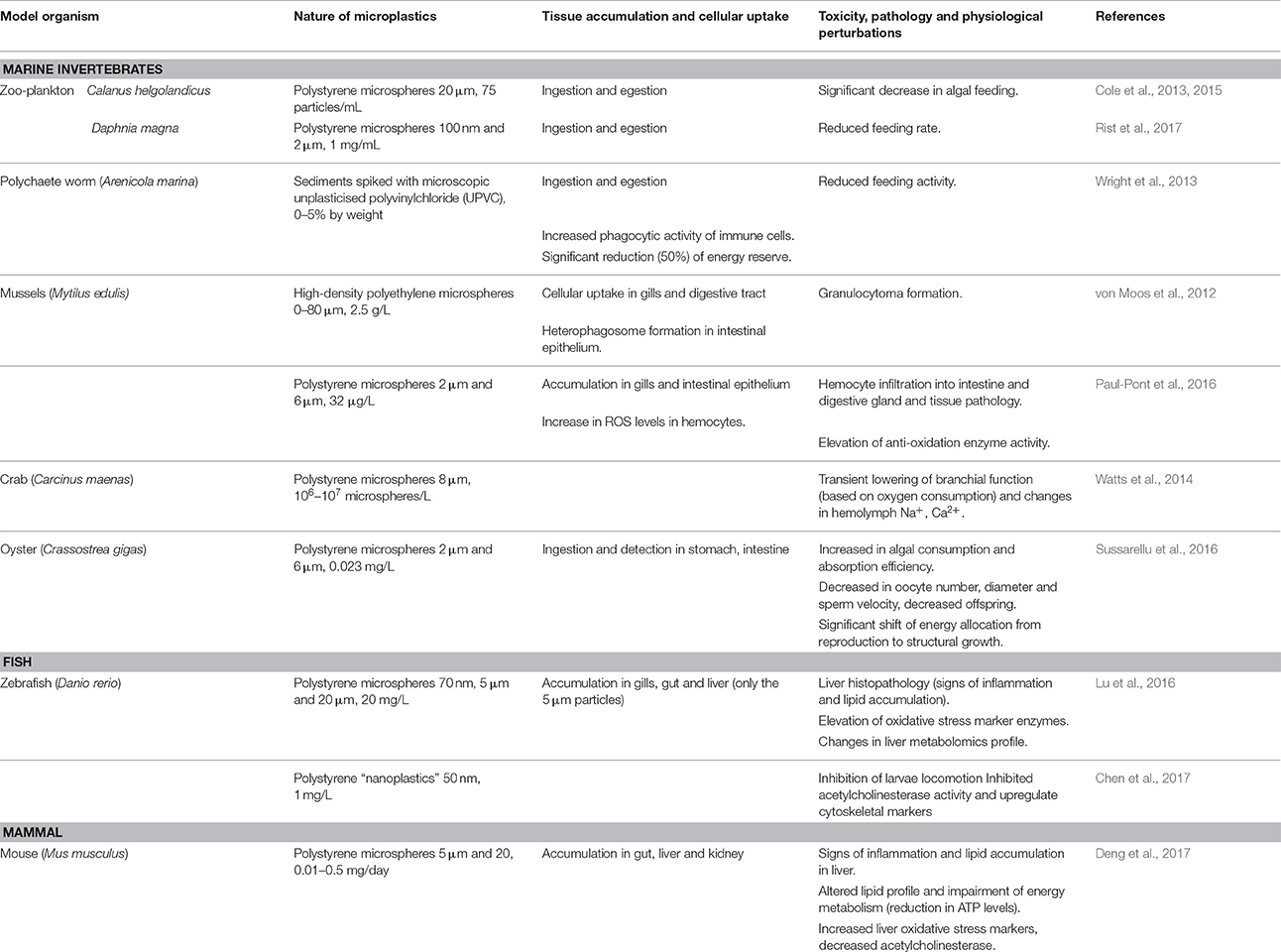
Table 1. A summary of histopathology and/or physiological perturbations observed in recent laboratory experiments with microplastics treatment of marine invertebrates, zebrafish and mouse.
Microplastics Ingestion and Adverse Effects in Mouse
The same group has now reported a similar analysis in a mammalian species (Deng et al., 2017). Ingested by the mice through drinking water, 5 and 20 μm polystyrene micro-particles elicited somewhat similar pathological and physiological changes in mice. These, particularly the 5 μm micro-particles, could be detected in histological sections of the gut, liver and kidney. Again, the authors reported that focused analysis on liver tissues revealed signs of inflammation, accumulation of lipid droplets, elevation of oxidative stress markers, defects in energy metabolism, and altered metabolomics. An inference on potential neurotoxicity of the micro-particles was also made based on changes in liver acetylcholinesterase (AChE) activity (Note: This point in the paper is confusing, as the authors stated in the text that AChE activity “decreased” after exposure to microplastics, but the data presented showed it to be elevated instead). On the whole, although the changes reported were moderate, they were statistically significant.
Implications and Perspectives
What is the significance of the findings of Deng et al.? On one hand, findings indicating that microplastics could accumulate in the tissues of marine vertebrates and even mammals at the apex of the trophic pyramid have important ecological implications, but these should be interpreted with some caution. For one, the relatively low abundance of microplastics in the ecological environment would likely not result in significant tissue accumulation via direct assimilation in larger organisms, but transfer across trophic levels and biomagnification along the food chain is a much touted possibility. There is no unequivocal evidence for the latter mechanisms pertaining to microplastics, largely because it is very difficult, given the heterogeneity and low abundance, to adequately assess microplastic distribution and bioaccumulation in the wild. One should bear in mind that the quantity of microplastics used in laboratory experiments are typically several magnitudes above environment abundance (Connors et al., 2017). In Deng et al.'s experimental setups, the lowest dose administered at 0.01 mg/day corresponded to ~105 of the 5 μm particles, a number that is likely 3–4 magnitudes above ecological abundance. Even the consumption of oysters which rather high accumulation of microplastics (Galloway and Lewis, 2016) would hardly approach the amounts used in the experiments.
How do microplastics elicit the adverse effects seen in mouse liver and the zebrafish liver in the earlier report (Lu et al., 2016)? To reach the liver, the ingested microplastics would need to somehow negotiate the gut-vascular barrier (Spadoni et al., 2015) or be taken up by intestinal enterocytes and transported across the epithelial mucosal lining in a manner akin to transcytosis of macromolecules. Thereafter, these have to enter the circulation via penetration of the vessel lining of endothelial cells and pericytes. Uptake of microplastics by gill and gut cells (von Moos et al., 2012) and translocation of ingested microplastic into the circulation (Browne et al., 2008) has been shown for the mussel Mytilus edulis, but not for the oyster (Sussarellu et al., 2016). How these microplastics negotiate the gut-vascular barrier and threshold concentrations at which this would effective occur are important questions that call for further investigation.
What about the toxicological/pathological perturbations measured in the liver? The data presented in Deng et al. are somewhat consistent with an acute injury-type inflammatory response, likely against circulating and liver tissue-accumulated microplastics, which would be accompanied by ROS elevation in adhering immune cells and a countering anti-oxidation response by the hepatocytes. This sort of acute inflammatory response is also evident in mussel tissues, as demonstrated by hemocyte infiltration and increase ROS (von Moos et al., 2012). The reduction in ATP and the formation of lipid droplets are indicative of changes in energy and lipid metabolism that accompanies mammalian liver injury and inflammation. The degree of injury was however not clear as common liver function tests were not performed. The metabolomics changes are difficult to interpret, and not particular useful except to support the notion that metabolic adaptations to injury and inflammation have likely occurred in liver tissues. Any claim of potential neurotoxicity based on changes in liver AChE is premature. If any, this is likely limited to the enteric nervous system as the blood-brain barrier presents a much more formidable obstacle for any plausible central nervous system accumulation of microplastics. The lack of any behavioral tests also precludes such a claim. The effect of ingested microplastics on longer term changes in feeding behavior and fecundity as previously observed for invertebrates (Wright et al., 2013; Sussarellu et al., 2016; Rist et al., 2017) were not investigated. These are more important parameters to assess pertaining to chronic exposure to any xenobiotic.
On the whole, the findings of Deng et al. (2017) were made under conditions that mimic a massive overdose of microplastics and the results seen are limited to what might resemble a tissue limited acute inflammatory response. Documenting the fact that such responses could be elicited by the presumably chemically inert pristine microplastics is not without value, but the suggestion of “widespread health risks of (microplastics) exposure” cannot be taken without some reservations.
Author Contributions
The author confirms being the sole contributor of this work and approved it for publication.
Conflict of Interest Statement
The author declares that the research was conducted in the absence of any commercial or financial relationships that could be construed as a potential conflict of interest.
Acknowledgments
The author is supported by the NUS Graduate School for Integrative Sciences and Engineering, and declares no conflict of interest.
References
Auta, H. S., Emenike, C. U., and Fauziah, S. H. (2017). Distribution and importance of microplastics in the marine environment: a review of the sources, fate, effects, and potential solutions. Environ. Int. 102, 165–176. doi: 10.1016/j.envint.2017.02.013
Browne, M. A., Dissanayake, A., Galloway, T. S., Lowe, D. M., and Thompson, R. C. (2008). Ingested microscopic plastic translocates to the circulatory system of the mussel, Mytilus edulis (L). Environ. Sci. Technol. 42, 5026–5031. doi: 10.1021/es800249a
Chen, Q., Gundlach, M., Yang, S., Jiang, J., Velki, M., Yin, D., et al. (2017). Quantitative investigation of the mechanisms of microplastics and nanoplastics toward zebrafish larvae locomotor activity. Sci. Total Environ. 584–585, 1022–1031. doi: 10.1016/j.scitotenv.2017.01.156
Cole, M., Lindeque, P., Fileman, E., Halsband, C., and Galloway, T. S. (2015). The impact of polystyrene microplastics on feeding, function and fecundity in the marine copepod Calanus helgolandicus. Environ. Sci. Technol. 49, 1130–1137. doi: 10.1021/es504525u
Cole, M., Lindeque, P., Fileman, E., Halsband, C., Goodhead, R., Moger, J., et al. (2013). Microplastic ingestion by zooplankton. Environ. Sci. Technol. 47, 6646–6655. doi: 10.1021/es400663f
Connors, K. A., Dyer, S. D., and Belanger, S. E. (2017). Advancing the quality of environmental microplastic research. Environ. Toxicol. Chem. 36, 1697–1703. doi: 10.1002/etc.3829
Deng, Y., Zhang, Y., Lemos, B., and Ren, H. (2017). Tissue accumulation of microplastics in mice and biomarker responses suggest widespread health risks of exposure. Sci. Rep. 7:46687. doi: 10.1038/srep46687
Galloway, T., Cole, M., and Lewis, C. (2017). Interactions of microplastic debris throughout the marine ecosystem. Nat. Ecol. Evol. 1:0116. doi: 10.1038/s41559-017-0116
Galloway, T. S., and Lewis, C. N. (2016). Marine microplastics spell big problems for future generations. Proc. Natl. Acad. Sci. U.S.A. 113, 2331–2333. doi: 10.1073/pnas.1600715113
Lu, Y., Zhang, Y., Deng, Y., Jiang, W., Zhao, Y., Geng, J., et al. (2016). Uptake and Accumulation of polystyrene microplastics in Zebrafish (Danio rerio) and toxic effects in liver. Environ. Sci. Technol. 50, 4054–4060. doi: 10.1021/acs.est.6b00183
Paul-Pont, I., Lacroix, C., González Fernández, C., Hégaret, H., Lambert, C., Le Goïc, N., et al. (2016). Exposure of marine mussels Mytilus spp. to polystyrene microplastics: Toxicity and influence on fluoranthene bioaccumulation. Environ Pollut. 216, 724–737. doi: 10.1016/j.envpol.2016.06.039
Rist, S., Baun, A., and Hartmann, N. B. (2017). Ingestion of micro- and nanoplastics in Daphnia magna-Quantification of body burdens and assessment of feeding rates and reproduction. Environ. Pollut. 228, 398–407. doi: 10.1016/j.envpol.2017.05.048
Spadoni, I., Zagato, E., Bertocchi, A., Paolinelli, R., Hot, E., Di Sabatino, A., et al. (2015). A gut-vascular barrier controls the systemic dissemination of bacteria. Science 350, 830–834. doi: 10.1126/science.aad0135
Sussarellu, R., Suquet, M., Thomas, Y., Lambert, C., Fabioux, C., Pernet, M. E. J., et al. (2016). Oyster reproduction is affected by exposure to polystyrene microplastics. Proc. Natl. Acad. Sci. U.S.A. 113, 2430–2435. doi: 10.1073/pnas.1519019113
Van Cauwenberghe, L., Claessens, M., Vandegehuchte, M. B., and Janssen, C. R. (2015). Microplastics are taken up by mussels (Mytilus edulis) and lugworms (Arenicola marina) living in natural habitats. Environ. Pollut. 199, 10–17. doi: 10.1016/j.envpol.2015.01.008
von Moos, N., Burkhardt-Holm, P., and Köhler, A. (2012). Uptake and effects of microplastics on cells and tissue of the blue mussel Mytilus edulis L. after an experimental exposure. Environ. Sci. Technol. 46, 11327–11335. doi: 10.1021/es302332w
Watts, A. J., Lewis, C., Goodhead, R. M., Beckett, S. J., Moger, J., Tyler, C. R., et al. (2014). Uptake and retention of microplastics by the shore crab Carcinus maenas. Environ. Sci. Technol. 48, 8823–8830 doi: 10.1021/es501090e
Keywords: microplastics, histopathology, ecotoxicity, Inflammation, health risk
Citation: Tang BL (2017) Commentary: Tissue accumulation of microplastics in mice and biomarker responses suggest widespread health risks of exposure. Front. Environ. Sci. 5:63. doi: 10.3389/fenvs.2017.00063
Received: 23 July 2017; Accepted: 27 September 2017;
Published: 11 October 2017.
Edited by:
Rajeev Pratap Singh, Banaras Hindu University, IndiaReviewed by:
Abhijit Sarkar, University of Gour Banga, IndiaYehia Eltemsah, Genøk, Norway
Denis Moledo De Souza Abessa, Universidade Estadual Paulista Júlio de Mesquita Filho (UNESP), Brazil
Copyright © 2017 Tang. This is an open-access article distributed under the terms of the Creative Commons Attribution License (CC BY). The use, distribution or reproduction in other forums is permitted, provided the original author(s) or licensor are credited and that the original publication in this journal is cited, in accordance with accepted academic practice. No use, distribution or reproduction is permitted which does not comply with these terms.
*Correspondence: Bor L. Tang, bchtbl@nus.edu.sg