einstein (São Paulo). 15/Dec/2021;19:eAI6347.
A T1-hypointense intracranial dermoid cyst
Marcos Gil da Veiga
![]() , Amets Sagarribay
, Amets Sagarribay
![]() , Carlos Marques Pontinha
, Carlos Marques Pontinha
![]() , Carla Conceição
, Carla Conceição
![]()
DOI: 10.31744/einstein_journal/2021AI6347
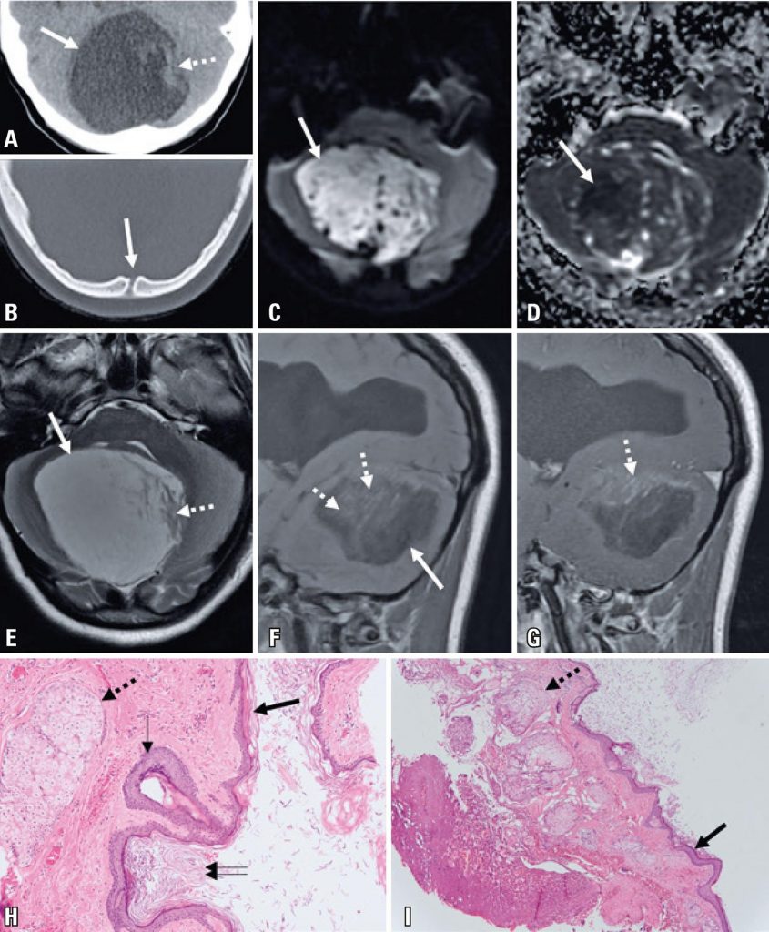
A 15-year old women presented with a 6-month history of progressive right-hand tremor with functional impairment, aggravated by a 1-month history of episodic confusion. The patient underwent a computed tomography scan, which revealed a posterior fossa expansive lesion with cerebrospinal fluid-like density and a midline bone discontinuity ( ; soft tissue window not shown). A magnetic resonance imaging (MRI) scan ( ) was performed after hospital admission, revealing a posterior fossa expansile lesion with predominant T2 hyperintensity, T1 hypointensity, linear and irregular areas of faint enhancement after gadolinium injection, and a large area with reduced water diffusion. Supratentorial images revealed signs of chronic hydrocephalus. The patient underwent surgery revealing a whitish extra-axial capsulated lesion containing dermal appendages, and histology confirmed the diagnosis of a dermoid cyst ( ).
[…]
777