einstein (São Paulo). 20/Oct/2020;18:eRC5876.
COVID-19 myocarditis: a case report
Patrícia Yokoo
![]() , Eduardo Kaiser Ururahy Nunes Fonseca
, Eduardo Kaiser Ururahy Nunes Fonseca
![]() , Roberto Sasdelli Neto
, Roberto Sasdelli Neto
![]() , Walther Yoshiharu Ishikawa
, Walther Yoshiharu Ishikawa
![]() , Murilo Marques Almeida Silva
, Murilo Marques Almeida Silva
![]() , Elaine Yanata
, Elaine Yanata
![]() , Rodrigo Caruso Chate
, Rodrigo Caruso Chate
![]() , Antonio Carlos Bacelar Nunes Filho
, Antonio Carlos Bacelar Nunes Filho
![]() , Marcelo Bettega
, Marcelo Bettega
![]() , João Ricardo Cordeiro Fernandes
, João Ricardo Cordeiro Fernandes
![]() , Flávio Tarasoutchi
, Flávio Tarasoutchi
![]() , Gilberto Szarf
, Gilberto Szarf
![]()
DOI: 10.31744/einstein_journal/2020RC5876
ABSTRACT
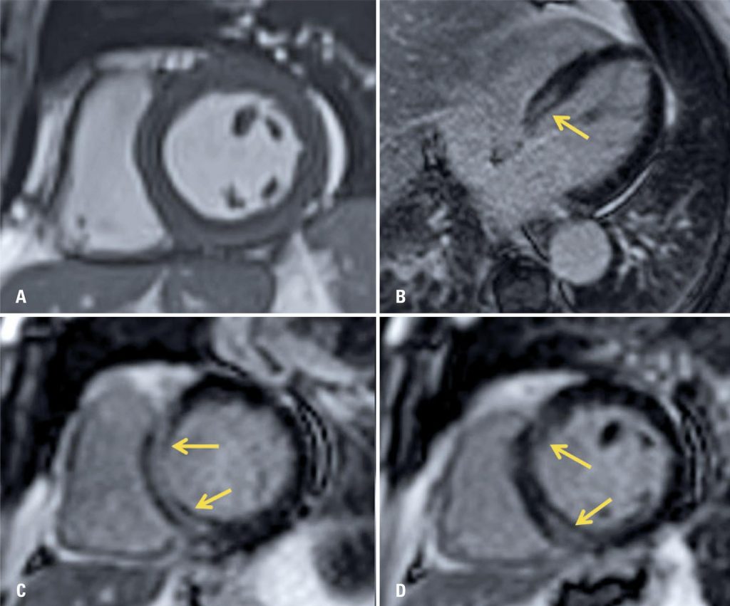
A male patient with flu-like symptoms and tomography and laboratory diagnosis of severe acute respiratory syndrome. He developed acute cardiac dysfunction during admission and was submitted to a cardiac magnetic resonance imaging examination, which confirmed acute myocarditis, indicating cardiac involvement by coronavirus disease 2019. A review and discussion about coronavirus disease 2019-related cardiac manifestations are reported, focusing on the imaging findings to make diagnosis.
5,856