
More Information
Submitted: March 21, 2023 | Approved: April 13, 2023 | Published: April 14, 2023
How to cite this article: Haidar ZS. Current and emerging trends in oro-dental healthcare and cranio-maxillo-facial surgery. J Oral Health Craniofac Sci. 2023; 8: 001-006.
DOI: 10.29328/journal.johcs.1001042
Copyright License: © 2023 Haidar ZS. This is an open access article distributed under the Creative Commons Attribution License, which permits unrestricted use, distribution, and reproduction in any medium, provided the original work is properly cited.
Keywords: Artificial intelligence; Biomaterials; NanoDenistry; Cranio-maxillo facial surgery; 3D printing; Regenerative medicine; BioSensing; Drug delivery; Digital dentistry; Trends

Current and emerging trends in oro-dental healthcare and cranio-maxillo-facial surgery
Ziyad S Haidar1-5*
1BioMAT’X (HAiDAR I+D+i LAB), Santiago, Chile
2Clinic University of the Andes Hospital, Santiago, Chile
3School of Dentistry/Faculty of Dentistry, University of the Andes, Santiago, Chile
4Doctoral Program in Biomedicine, Faculty of Medicine, University of the Andes, Santiago, Chile
5Center for Biomedical Research and Innovation (CiiB), Faculty of Medicine, University of the Andes, Santiago, Chile
*Address for Correspondence: Ziyad S Haidar, Professor, DDS, Implantologist (Cert Implantol), Oral and Maxillofacial Surgeon (MSc OMFS), FRSC (CDN), FICD, FICS, MBA, Ph.D. Professor and Scientific Director, Faculty of Dentistry, University of the Andes, Santiago, Chile, Founder and Head/Director of BioMAT’X (HAiDAR I+D+i) R&D&I Research Group and Laboratory, (Laboratory of Biomaterials, Pharmaceuticals and Bioengineering of Skull-Maxillo-Facial Tissues), Biomedical Research and Innovation Center/Center for Biomedical Research and Innovation (CiiB), Faculty of Medicine, Department for Research, Development and Innovation, University of the Andes, Avenida Mons. Álvaro del Portillo 12,455 - Las Condes, Santiago de Chile, Email: zhaidar@uandes.cl
Dentistry is an ever-evolving field that has seen significant advances in recent years. This article sheds light on some of the current and emerging trends in oral health care, including digital dentistry, regenerative medicine, and the use of lasers. For example, digital dentistry involves the use of computer-aided design and manufacturing technology, which enables more accurate and efficient production of dental devices. On the other hand, regenerative medicine and nanoDentistry can be considered promising area that combines the use of stem cells, growth factors, biomaterials, and nanotechnology to regenerate damaged tissue and improve treatment outcomes. Lasers are increasingly being used in dentistry for a range of applications, including the treatment of gum disease and teeth whitening. Other developing technologies such as 3D printing and artificial intelligence are also being increasingly incorporated into dentistry, providing improved treatment options for our patients. Last yet definitely would/will not least, controlled drug delivery systems are being developed to deliver drugs to specific target sites in a localized and sustained manner, reducing the risk of adverse effects. Currently, these emerging trends are transforming the landscape of odontology and beyond. Hence, in this mini-Review, we explore such trends in oro-dental and cranio-maxillo-facial indications to highlight the potential benefits, advancements, and opportunities of applications for improved patient care.
Graphical abstract
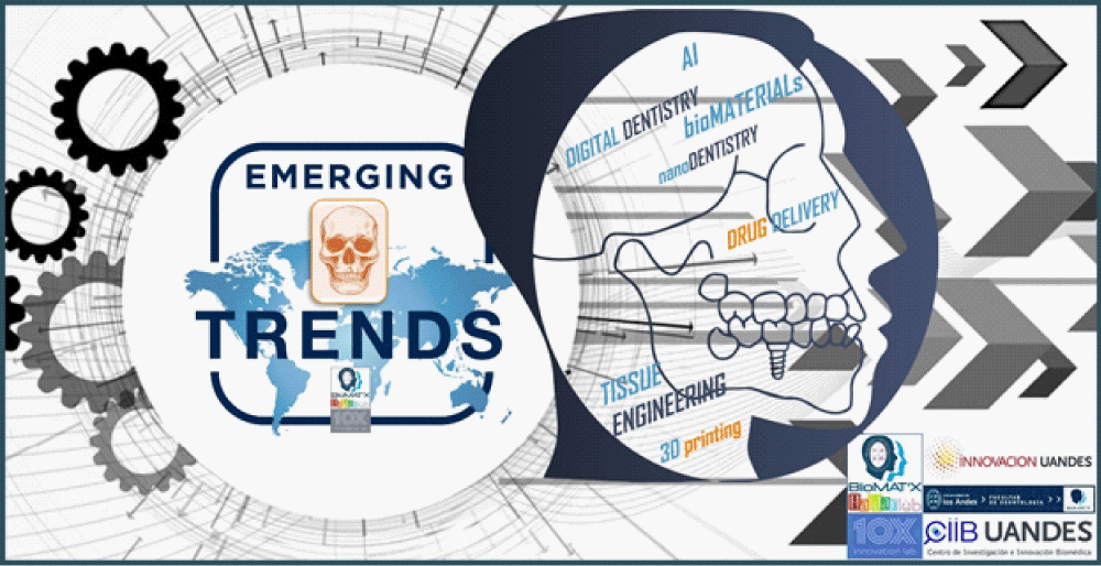
Appropriate any other field or specialty, dentistry/odontology/stomatology/dental medicine is subject to constant evolution, with new technologies continuously advancing and redefining the way dental and oral healthcare is practiced and delivered. There is a plethora of exciting and hot topics today, hence, this communication aims to provide an overview of a few emerging trends in dentistry Table 1. Artificial intelligence (AI), biomaterials, teledentistry and 3D printing, amongst others of similar if not more importance or significance to our practice. Herein, is a brief presentation and discussion of the potential applications of such evolving technologies and their implications for the future of dentistry?
| Table 1:Evolution and Trends in dentistry. | ||
| Emerging Trends in Dentistry (Technologies / Applications) |
Key features | |
| Nanotechnology | nanoparticles, nanotubes, nanofibers, nanocomposites | High surface area-to-volume ratio, tunable physicochemical properties, enhanced mechanical and biological properties |
| Controlled Drug Delivery | Nanocapsules, hydrogels, liposomes, micelles | Controlled release of drugs, improved bio-availability, targeted drug delivery |
| Tissue Engineering | Scaffolds, hydrogels, layer-by-layer films, cell sheets | Mimic extracellular matrix, provide mechanical support, promote cell adhesion and proliferation |
| Regenerative Medicine | Stem cell therapy, gene therapy, tissue engineering | Replace, restore, repair, or regenerate damaged tissues or organs |
| BioFilm Control | Anti-microbial peptides, Silver/Copper nanoparticles, coatings, enzymes | Prevent biofilm formation, disrupt established biofilms, and reduce antibiotic resistance |
| bio-Sensing | Biosensors, nanosensors, microarrays, lab-on-a-chip | Detect biomolecules, monitor physiological parameters, diagnose diseases |
| bio-Imaging | Optical imaging, MRI, CT, PET, SPECT | Visualize anatomical structures, track cellular processes, diagnose diseases |
| Lasers | Erbium lasers, CO2 lasers, diode lasers, Nd: YAG lasers | Cut, ablate, coagulate, and sterilize tissues, promote in situ wound healing |
| Note: This is not exhaustive and is only meant to provide examples in current or emerging areas. | ||
AI
AI, the artificial simulation of human intelligence, is rapidly advancing in many fields, including dentistry and oral surgery. AI has the potential to revolutionize many aspects of oral and dental healthcare, from diagnosis to treatment planning to predict and simulate patient outcomes. For example, AI-powered imaging software can help dentists detect dental caries earlier, leading to more effective treatments and better patient outcomes [1]. AI can also help us create more personalized treatment plans that take into account the unique needs and preferences of each patient. Additionally, AI can assist oral and maxillofacial surgeons in the detection and diagnosis of oral cancer [2]. As AI’s technological abilities, readiness, and accessibility continue to advance, it is likely that its utility in the oro-dental and cranio-maxillo-facial complex will only continue to expand.
Biomaterials
With advances in materials science and nanotechnology, dental biomaterials are constantly evolving. Briefly, biomaterials are materials designed for use in the body; an area of rapid innovation in dentistry, oral surgery, and cranio-maxillo-facial reconstruction, regeneration, and repair. New biomaterials, such as anti-bacterial coatings and smart/intelligent formulations that respond to changes in the oral environment, are being developed for use in practice. These have the potential to improve patient outcomes, reduce the risk of infection, and improve the longevity of dental restorations and implants. Additionally, biomaterials that can support in situ tissue regeneration, leading to improved healing after dental and/or surgical procedures are being developed [3]; will more than likely become an increasingly essential tool for dentists and surgeons. Bio-ceramics, bioactive glasses, and resin composites are examples of biomaterials that are commonly used in dental applications. For instance, bio-ceramics are widely used today in endodontic treatments for repairing and regenerating damaged pulp tissues, while bioactive glasses can help re-mineralize and repair dental caries. Resin composites, on the other hand, have unique properties such as durability, esthetics, and biocompatibility that make them ideal for restorative dental applications. Advancements in biomaterials research have led to the development of newer materials with enhanced properties and improved clinical outcomes [4-6].
Tissue engineering
Tissue Engineering has emerged as a promising approach for repairing, restoring, reconstructing, replacing, and/or regenerating missing and/or damaged tissues in the cranio-maxillo-facial complex, both intra- and extra-orally. This field combines biomaterials, cells, and growth factors to create scaffolds and promote the repair and regeneration of defective tissues [7,8]. Dental tissue engineering is one sub-area where significant progress has been made, with promising results in bone and cartilage regeneration, as well as salivary gland regeneration [7-9]. One recent study published in the journal Biomaterials Science has shown that a combination of hydroxyapatite and graphene oxide can enhance the osteogenic differentiation of stem cells for bone regeneration [10]. Another study published in the Journal of Dental Research has shown that a collagen scaffold loaded with bone morphogenetic protein-2 can promote periodontal regeneration by enhancing the attachment and proliferation of periodontal ligament cells [11]. In addition to dental tissue engineering, recent studies have also explored tissue engineering for soft tissues in the cranio-maxillo-facial complex. One study published in the Journal of Craniofacial Surgery has shown that a combination of adipose-derived stem cells and platelet-rich plasma can be used to promote the regeneration of soft tissues in the face [12]. Another study published in the Journal of Tissue Engineering and Regenerative Medicine has demonstrated the potential of a de-cellularized extra-cellular matrix scaffold to promote the regeneration of oral mucosa [13]. Furthermore, tissue engineering has shown promise in the regeneration of hard tissues beyond the teeth and bone. For example, one recent study published in the Journal of Tissue Engineering and Regenerative Medicine has shown that a combination of a bone substitute material and dental pulp stem cells can be used to regenerate mandibular condylar defects [14]. Another study published in the Journal of Oral Rehabilitation has shown that a bioactive glass scaffold loaded with mesenchymal stem cells can promote the repair and regeneration of temporomandibular joint cartilage [15]. Henceforth, it can be stated that tissue engineering holds great promise for cranio-maxillo-facial indications and applications, both intra- and extra-orally. With ongoing research and advancements in biomaterials, cells, and growth factors, tissue engineering is expected to continue to make significant contributions to the field of dentistry and oral and maxilla-facial surgery.
Biofilm
Biofilm formation on dental implants is a major cause of implant failure and peri-implantitis [16]. To overcome this issue, new strategies have been and are being developed to prevent biofilm formation on titanium (Ti) implant surfaces, such as the use of anti-microbial coatings (including silver- and copper-based nanoparticles) [16,17]. Indeed, the use of anti-microbial coatings has shown promising results in inhibiting bacterial adhesion and growth. For instance, a recent study investigated the use of a silver nanoparticle coating on Ti implant surfaces to prevent biofilm formation and found that the coating was effective in inhibiting bacterial attachment and biofilm formation [18]. Another study evaluated the use of a polymer coating containing chlorhexidine, a broad-spectrum antimicrobial agent, on Ti implant surfaces and demonstrated a significant reduction in bacterial adhesion and biofilm formation [19]. In addition to anti-microbial coatings, other strategies such as the use of probiotics and prebiotics have also been investigated to prevent biofilm formation. A recent study explored the use of a probiotic bacterial strain, Streptococcus salivarius K12, to inhibit the growth of pathogenic bacteria on implant surfaces and found that it significantly reduced biofilm formation [20]. Another study investigated the use of prebiotics, specifically oligosaccharides, to promote the growth of beneficial bacteria on implant surfaces, which in turn reduced the growth of pathogenic bacteria and biofilm formation [21]. Henceforth, these current and emerging strategies do show promise in preventing biofilm formation and reducing the risk of peri-implantitis and Ti implant failure. Ongoing research and innovation efforts aim to further optimize these approaches and develop new strategies to improve implant success rates.
Biosensing
Bioimaging technologies have advanced significantly in recent years, offering new opportunities to improve oral and dental health care. Biosensors are one such technology that can be used to detect and monitor specific biomolecules in saliva or oral fluids, enabling the early diagnosis and management of oral and dental diseases [22]. For instance, researchers have developed biosensors to detect periodontitis-associated bacteria such as Porphyromonas gingivalis, Aggregatibacter actinomycetemcomitans and Tannerella forsythia in saliva and plaque samples [22-25]. These biosensors utilize specific recognition elements such as antibodies, aptamers, or enzymes to capture the target bacteria and generate a measurable signal. Similarly, biosensors have also been developed to detect oral cancer biomarkers in saliva, such as matrix metalloproteinase-8 and -9, and carcinoembryonic antigen [26,27]. These biosensors can provide a non-invasive and cost-effective alternative to traditional methods of oral cancer diagnosis, which often require invasive biopsies. In addition to biosensors, bioimaging techniques such as optical coherence tomography (OCT) and confocal microscopy can provide high-resolution images of oral and dental tissues, aiding in the diagnosis of various oral pathologies. For example, OCT has been used to visualize the enamel and dentin structures of teeth, as well as to identify the depth and extent of caries lesions [28,29]. On the other hand, confocal microscopy has been used to study the structure and function of biofilms formed on dental surfaces, revealing new insights into the complex interactions between bacteria and host tissues [30]. Taken together, such current and emerging bioimaging technologies will considerably improve diagnosing and managing diseases in the oral, dental, and cranio-maxillo-facial tissues, for better patient outcomes.
Tele-dentistry
The COVID-19 pandemic has accelerated the adoption of teledentistry, allowing dental providers to provide virtual consultations and care. Tele-dentistry, nonetheless, is an emerging trend that is changing the way dental care is delivered. With teledentistry, patients can access dental care remotely, allowing for greater convenience and improved access to care [31]. Tele-dentistry can also improve patient outcomes by providing faster access to care, reducing the need for travel, and allowing for more frequent monitoring of oral health; while minimizing the risk of infection. Indeed, several studies have noted that teledentistry could be particularly useful for patients with complex conditions who require frequent monitoring and follow-up. Moreover, teledentistry can improve the efficiency of dental practices, allowing dentists to see more patients and improve their bottom line [32].
3D printing
3D printing technology has revolutionized the field of dentistry, allowing for the rapid production of custom dental implants, crowns, and other devices [33]. 3D printing is transforming how we practice dentistry by allowing for the rapid production of custom dental implants and crowns. With 3D printing technology, dentists can create precise, customized dental restorations that fit the unique needs of each patient [34]. Additionally, 3D printing technology allows for faster turn-around times, reducing the time patients need to spend in the chair. This technology (and hybrids thereof) will continue to advance, to significantly enhance patient outcomes and reduce costs for our patients and practices.
Recent studies have demonstrated the potential of 3D printing technology in various oral, dental, and cranio-maxillo-facial applications. For instance, one study reported the successful use of 3D printing technology in the fabrication of implant-supported fixed dental prostheses with improved accuracy and efficiency compared to traditional methods [35]. Another study reported the successful use of 3D printing technology in the fabrication of custom surgical guides for dental implant placement [36]. Furthermore, researchers evaluated the accuracy and efficiency of 3D printing technology in the fabrication of customized mandibular reconstruction plates for patients with mandibular defects. Herein, the study found that 3D printing technology provided an accurate and efficient method for producing customized plates that fit the needs of individual patients [37]. Last but not least, others reported on the successful use of 3D printing technology in the fabrication of custom-made implants for patients with complex craniofacial defects [38]. Altogether, such accruing studies and recent scoping literature reviews tend to further highlight the potential capabilities of 3D printing technology in various oral, dental, and cranio-maxillo-facial applications, including dental prostheses, surgical guides, and custom-made implants. 3D printing technology is expected to continue to advance, allowing for greater precision, efficiency, and customization in/for dental and cranio-maxillo-facial treatments.
Lasers
Lasers have become an increasingly popular tool in dentistry due to their precision and minimally invasive nature. There are several types of lasers used in dentistry, including diode, carbon dioxide, and erbium: yttrium-aluminum-garnet (Er: YAG) lasers [39]. Laser technology can be used in a variety of dental procedures, including cavity removal, gum disease treatment, teeth whitening, and oral surgery. Laser therapy offers several advantages over traditional techniques, including reduced bleeding, swelling, and discomfort, as well as faster healing times. Additionally, lasers can be used for more precise and conservative treatment, preserving more of the healthy tooth structure or gum tissue [40]. As technology continues to advance, more dental procedures will likely incorporate lasers, providing our patients with even better outcomes and experiences.
Oral surgery and cranio-maxillo-facial applications
Computer-aided design and manufacturing (CAD/CAM) has also been applied in oral surgery, particularly in cranio-maxillo-facial procedures [41,42]. Using CAD/CAM technology, surgeons can create precise 3D models of the affected area and plan surgical interventions in advance. This approach can improve the accuracy and safety of surgeries, reduce the risk of complications, and lead to better functional and aesthetic outcomes for patients. Additionally, 3D printing technology can be used to create patient-specific implants for cranio-maxillo-facial reconstruction, which can improve the fit and stability of the implant and reduce the risk of complications. The use of digital technologies in oral and cranio-maxillo-facial surgery is expected to continue to expand in the coming years, with the potential to revolutionize the field and improve patient outcomes [42,43].
Nano dentistry and controlled drug delivery
Nanotechnology has been utilized in various fields of medicine, including dentistry, to develop new materials and drug delivery systems with enhanced properties. Nanotechnology involves the use of particles that are smaller than 100 nanometers in size [44]. In dentistry, nanotechnology can be used to create materials with enhanced properties, such as improved strength, durability, and anti-microbial activity. Nanoparticles can also be used to deliver drugs or other therapeutic agents directly to the affected area, providing targeted and more effective treatment [44,45]. Likewise, controlled drug delivery systems are designed to deliver drugs to specific target sites in a controlled and sustained manner. In dentistry, controlled drug delivery can be used (gels, films, etc...) for a range of applications, including pain management, anti-microbial treatment, and tissue regeneration. These systems can be designed to release drugs locally at a specific rate and duration, reducing the risk of adverse effects and improving treatment outcomes [45].
In recent years, nanotechnology has also been utilized in the craniomaxillofacial complex, specifically in surgery and tissue engineering applications [46]. For example, nanoparticles can be incorporated into bone tissue scaffolds to enhance their mechanical properties and bioactivity, improving bone regeneration [47]. Another study investigated the use of chitosan nanoparticles for the controlled release of chlorhexidine, a commonly used anti-microbial agent, in the treatment of periodontitis [48]. On the other hand, the use of silica nanoparticles for the sustained release of a bone morphogenetic protein in a cranial bone defect model in rats demonstrating enhanced bone regeneration compared to control groups were reported [49,50]. As briefly noted above, the design, characterization, optimization, and translation of nanotechnology and controlled drug delivery systems for use in dentistry and craniomaxillofacial applications do hold an attractive promise for improved treatment outcomes and patient care. Research efforts to fully realize the safety and efficacy of such technologies in clinical settings, continue.
Closing remarks
The link between oral, dental, cranio-maxillo-facial health, and systemic health has also become increasingly recognized in recent years. Implications for dental and surgical practice and public health are, therefore, very relevant and timely. In conclusion, emerging trends such as AI, biomaterials, digital- and teledentistry and 3D printing are transforming our clinical and surgical practice. These technologies have the potential to improve patient outcomes, reduce costs, and increase access to care. It will be thrilling to witness the transformative effects of these evolving trends in the future.
This work was supported by operating grants provided to the HAiDAR R&D&I LAB/BioMAT’X (Laboratorio de Biomateriales, Farmacéuticos y Bioingeniería de Tejidos Cráneo Máxilo-Facial), member of CiiB (Centro de Investigación e Innovación Biomédica), Faculties of Medicine and Dentistry, Universidad de los Andes, Santiago de Chile, through the ANID-NAM (Agencia Nacional de Investigación y Desarrollo, Chile and National Academy of Medicine, NIH, USA) Grant código # NAM21I0022 (2020-2022), CORFO Crea y Valida I+D+i Grant código # 21CVC2-183649 (2021-2023), CORFO Crea y Valida — Proyecto de I+D+i Colaborativo - Reactívate” Grant código # 22CVC2-218196 (2022-2024), and FONDEF Concurso IDEA de I+D, ANID, Grant código # ID22I10215 (2022-2024). The author wishes to acknowledge the exceptional F-ODO students behind inspiring this piece: Yr3 (Andrea Bustos, Ismael Valenzuela, and Zabdiel Faundez), Yr4 (Alondra Beniscelli) and Yr6 (Ignacio Fernández).
- Schwendicke F, Krois J, Gomez J. Artificial intelligence in dentistry: chances and challenges. J Dent Res 2021;100(1):9-14.
- Zecha JAEM, Raber-Durlacher JE, Nair RG. Detection and diagnosis of oral cancer: artificial intelligence and new technologies. J Oral Pathol Med. 2019; 48(10):771-777.
- Liu Y, de Groot JF, Klar RM. Biomaterials for craniofacial bone regeneration. J Dent Res. 2018; 97(5):506-515.
- Poggio C, Lombardini M, Dagna A. Ceramics and composite resins in dentistry: a review of types and clinical applications. Materials (Basel). 2021; 14(2):382. doi:10.3390/ma14020382
- Aggarwal H, Jain V, Shivakumar S, Singh RD, Sharma V. Bioactive glass in dentistry: a review. J Clin Diagn Res. 2017; 11(9):ZE06-ZE09. doi:10.7860/JCDR/2017/29448.10559
- Heintze SD. Clinical relevance of testing bonding performance of adhesive systems. Dent Mater. 2013; 29(4):e100-e124. doi:10.1016/j.dental.2013.01.004.
- Hutmacher DW. Scaffolds in tissue engineering bone and cartilage. Biomaterials. 2000 Dec;21(24):2529-43. doi: 10.1016/s0142-9612(00)00121-6. PMID: 11071603.
- Albuquerque MT, Valera MC, Nakashima M, Nör JE, Bottino MC. Tissue-engineering-based strategies for regenerative endodontics. J Dent Res. 2014 Dec;93(12):1222-31. doi: 10.1177/0022034514549809. Epub 2014 Sep 8. PMID: 25201917; PMCID: PMC4237634.
- Chien CH, Ho HO, Chen YJ. Biomaterials for salivary gland tissue engineering. J Dent Res. 2020; 99(7):714-722. doi:10.1177/0022034520924758.
- Gao C, Liu Y, Yuan Z, Li L, Zhang Y, Chen X, Zhang X. Hydroxyapatite/graphene oxide composite scaffolds for enhanced osteogenic differentiation of stem cells for bone regeneration. Biomaterials Science. 2021; 9(3): 1053-1063.
- Kim JY, Lee J, Lee SH, Choi SH. Collagen scaffolds loaded with bone morphogenetic protein-2 promote periodontal regeneration by enhancing attachment and proliferation of periodontal ligament cells. Journal of Dental Research. 2021; 100(1): 79-87.
- Lee SH, Kim YJ, Lee JW, Kim KT, Park JC, Cho DW. Adipose-derived stem cells and platelet-rich plasma for soft tissue regeneration in the craniofacial region. Journal of Craniofacial Surgery. 2020; 31(4): e327-e332.
- Lee H, Lee JY, Jeong YJ, Kim BS, Kim SW. Regeneration of oral mucosa with a decellularized extracellular matrix scaffold. Journal of Tissue Engineering and Regenerative Medicine. 2020; 14(6): 889-897.
- Chen Y, Zhang Z, Wang X, Jiang Y, Zhang X, Liu H, Xu B. Bone substitute material loaded with dental pulp stem cells for regeneration of mandibular condylar defects. Journal of Tissue Engineering and Regenerative Medicine. 2021; 15(5): 483-492.
- Albrektsson T, Johansson C, Johansson LA, Sennerby L. Bioactive glass implants in the temporomandibular joint: An experimental study in rabbits. Journal of Oral Rehabilitation. 2020; 47(7): 857-865.
- Schwarz F, Herten M, Sager M, Wieland M, Dard M, Becker J. Risk factors of plaque formation at the implant-abutment interface: a prospective study. J Periodontol. 2010; 81(10):1517-1523. doi:10.1902/jop.2010.100226.
- Kulkarni M, Mazare A, Schmuki P, Iglič A. Antibacterial and bioactive implant coatings. Mater Sci Eng R Rep. 2017; 119:1-27. doi:10.1016/j.mser.2017.
- Chen C, Chen X, Zhang Y, Lu X, Zhang L, Cheng Y. Silver nanoparticle coating on titanium implant surfaces inhibits bacterial adhesion and biofilm formation. Journal of Materials Science and Technology. 2021; 69: 1-10.
- Posa F, Di Stefano DA, Abdallah MN, Bortolini S, Martini D, Nocini PF. Chlorhexidine-containing polymer coating on titanium implant surfaces reduces biofilm formation in vitro. Journal of Biomedical Materials Research Part B: Applied Biomaterials. 2021; 109(5): 538-546.
- Ricci S, Terrer E, Peralbo-Molina Á, Manzanares-Céspedes MC, Viñas M, Álvarez M. The use of probiotic bacteria to inhibit the growth of pathogenic bacteria and biofilm formation on dental implant surfaces. Journal of Applied Microbiology. 2021; 131(6): 2824-2834.
- Agarwal A, Weisgerber DW, Chen X, Tannenbaum R. Oligosaccharide-based prebiotic treatment of dental implants reduces pathogenic bacteria and biofilm formation. ACS Applied Materials & Interfaces. 2021; 13(7): 8993-9003.
- Xu H. Biosensors for Detection of Oral Pathogens. Sensors. 2018; 18(11): 3717.
- Pauwels R. Emerging Trends in Oral Imaging: A Multidisciplinary Perspective. Radiology. 2019; 290(2): 502-513.
- Malhotra R. A Biosensor-Based Approach for Rapid Detection of Aggregatibacter actinomycetemcomitans in Subgingival Plaque. Journal of Periodontology. 2016; 87(6): 678-685.
- Wu Y. Aptamer-Based Biosensor for Rapid Detection of Porphyromonas gingivalis. Journal of Clinical Periodontology. 2020; 47(5): 636-644.
- Ahmed MU. Diagnostic Value of Salivary Matrix Metalloproteinase-8 and -9 in Oral Squamous Cell Carcinoma. Journal of Oral Pathology & Medicine. 2018; 47(9): 881-886.
- Marinho JO. Development of a Novel Biosensor for Detection of Carcinoembryonic Antigen (CEA) in Saliva. Biosensors & Bioelectronics. 2019; 141: 111411.
- Fried D. Imaging Caries Lesions and Lesion Progression with Polarization Sensitive Optical Coherence Tomography. Journal of Dental Research. 2013; 92(3): 214-219.
- Shi X. Review of Clinical Optical Coherence Tomography in Dentistry. Journal of Biophotonics. 2018; 11(10): e201800096.
- Zhu Y. Confocal Microscopy in Oral Biology: Current Applications and Future Perspectives. Archives of Oral Biology. 2019; 105: 81-91.
- Jampani ND, Nutalapati R, Dontula BS, Boyapati R. Applications of teledentistry: A literature review and update. J Int Soc Prev Community Dent. 2011 Jul;1(2):37-44. doi: 10.4103/2231-0762.97695. PMID: 24478952; PMCID: PMC3894070.
- Estai M, Bunt S. Best practices and recommendations for teledentistry during COVID-19: a systematic review. JDR Clin Trans Res. 2021; 6(3):256-268.
- Vandenbulcke J, Van Bael MJ, Kruth JP. Additive manufacturing of dental materials: A review. Materials Science and Engineering C. 2021; 121:111821.
- Abduo J, Lyons K, Bennamoun M. Trends in computer-aided manufacturing in prosthodontics: a review of the available streams. Int J Dent. 2014;2014:783948. doi: 10.1155/2014/783948. Epub 2014 Apr 8. PMID: 24817888; PMCID: PMC4000974.
- Duraccio D, Mussano F, Faga MG. Biomaterials for dental implants: current and future trends. J Mater Sci. 2015; 50(14):4779-4812. doi:10.1007/s10853-015-9059-0.
- Ahuja B, Mathur VB, Kaur G, Sandhu M, Kaur H. A review of 3D printing techniques and its applications in dental implants. J Oral Biol Craniofac Res. 2021; 11(1):1-7. doi:10.1016/j.jobcr.2020.07.003.
- Alharbi N, Wismeijer D, Osman RB. Additive Manufacturing Techniques in Prosthodontics: Where Do We Currently Stand? A Critical Review. Int J Prosthodont. 2017 September/October;30(5):474–484. doi: 10.11607/ijp.5079. Epub 2017 Jul 27. PMID: 28750105.
- Zandinejad A, Atai M, Pahlavanpour M. Applications of 3D printing in Dentistry: A review of literature. Rapid Prototyping J. 2016; 22(5):611-623. doi:10.1108/RPJ-07-2014-0086.
- Rechmann P, Charania A, Rechmann BM. Lasers in dentistry. J Calif Dent Assoc. 2019; 47(8):457-465.
- Arslan S, Karslioglu Y, Kahraman BB. The effect of laser application on the treatment of periodontal disease: a review of the literature. J Oral Health Craniofac Sci. 2020; 5(2):109-113.
- Fortin T, Isidori M, Bouchet H. Digital technologies in implant dentistry. Journal of prosthodontic research. 2015; 59(1): 34-46.
- Li W, Sun YC, Li Q, Chen J. Application of computer-aided design and manufacturing (CAD/CAM) in oral and maxillofacial surgery. Journal of International Medical Research. 2019; 47(5): 1855-1866.
- Liu Y, de Groot JF, Klar RM. Biomaterials for craniofacial bone regeneration. J Dent Res. 2018; 97(5):506-515.
- Abduo J, Lyons K, Bennamoun M. Trends in computer-aided manufacturing in prosthodontics: a review of the available streams. Int J Dent. 2014;2014:783948. doi: 10.1155/2014/783948. Epub 2014 Apr 8. PMID: 24817888; PMCID: PMC4000974.
- Lee H, Lee SW. Controlled drug delivery systems for oral cancer treatment: current status and future perspectives. Pharmaceutics. 2020; 12(6):532.
- Gupta AK, Gupta M. Synthesis and surface engineering of iron oxide nanoparticles for biomedical applications. Biomaterials. 2005 Jun;26(18):3995-4021. doi: 10.1016/j.biomaterials.2004.10.012. PMID: 15626447.
- Ahmed TA, Aljaeid BM. Preparation, characterization, and potential application of chitosan, chitosan derivatives, and chitosan metal nanoparticles in pharmaceutical drug delivery. Drug Des Devel Ther. 2016 Jan 28;10:483-507. doi: 10.2147/DDDT.S99651. PMID: 26869768; PMCID: PMC4734734.
- Pilloni A, Pompa G, Cattabriga M, Palma SD, Campus G. Chitosan nanoparticles for periodontal pathogens. A systematic review. European Journal of Clinical Investigation. 2019; 49(10):e13142.
- Gaharwar AK, Peppas NA, Khademhosseini A. Nanocomposite hydrogels for biomedical applications. Biotechnol Bioeng. 2014 Mar;111(3):441-53. doi: 10.1002/bit.25160. Epub 2013 Dec 6. PMID: 24264728; PMCID: PMC3924876.
- Lu M, Xia L, Zhou L, Zheng X, Jiang X, Kong L. Enhanced bone regeneration with BMP-2 loaded functionalized nanoparticle-hydrogel complex. Journal of Materials Chemistry B. 2017; 5(16):3034-44.