
More Information
Submitted: October 04, 2021 | Approved: December 13, 2021 | Published: December 14, 2021
How to cite this article: Bello A, Wamakko HH. Comparative anatomy of selected bones of forelimb of local Mongrelian Dog (Canis lupus familiaris) in Sokoto, Nigeria. Insights Vet Sci. 2021; 5: 026-031.
DOI: 10.29328/journal.ivs.1001033
Copyright License: © 2021 Bello A, et al. This is an open access article distributed under the Creative Commons Attribution License, which permits unrestricted use, distribution, and reproduction in any medium, provided the original work is properly cited.
Keywords: Age related changes; Mongrelian dog; Scapula; Humerus; Radius and Ulna

Comparative anatomy of selected bones of forelimb of local Mongrelian Dog (Canis lupus familiaris) in Sokoto, Nigeria
Bello A1* and Wamakko HH2
1Department of Veterinary Anatomy, Faculty of Veterinary Sciences, Usmanu Danfodiyo University, Sokoto, Nigeria
2Sokoto State Veterinary Clinic, Sokoto State, Nigeria
*Address for Correspondence: Bello A, Department of Veterinary Anatomy, Faculty of Veterinary Sciences, Usmanu Danfodiyo University, Sokoto, Nigeria, Email: abccrcfge28@gmail.com
This research was conducted over period of 3 months with the aim of studying Age related changes of selected bones of forelimb (Scapula, Humerus, Radius and Ulna) in Local Mongrelian Dog (Canis lupus familiaris). The study entails biometrical and gross observations on the bones. The sample bones were acquired from the experimental animals of comparative anatomy in the department. The bones were categorized into various age groups for the research. The length, width, diameter and circumference of the samples (scapula, humerus, radius and ulna bones) were determined for all the groups. The shape, size, color, location, position and relation of each segment of the samples at various stages of development were determined. The differences across the age groups of different samples were observed and recorded. Based on the research result, it was concluded that, the biometric and morphometry data was found to be increasing with advancement of age. A baseline data was established with the view to enhance learning.
The domestic dog (Canis lupus familiaris when considered a subspecies of the wolf or Canis familiaris when considered a distinct species) [1] is a member of the genus Canis (canines), which forms part of the wolf-like canids [2] and is the most widely abundant terrestrial carnivore [3-5]. The dog and the extant gray are sister taxa [6,7], as modern wolves are not closely related to the wolves that were first domesticated [7] which implies that the direct ancestor of the dog is extinct [8]. The dog was the first species to be domesticated [9,10] and has been selectively bred over millennia for various behaviors, sensory capabilities, and physical attributes [11]. Their long association with humans has led dogs to be uniquely attuned to human behavior [12] and they are able to thrive on a starch-rich diet that would be inadequate for other canid species [13]. Dogs vary widely in shape, size and colors [14]. They perform many roles for humans, such as hunting, herding, pulling loads, protection, assisting police and military, companionship and, more recently, aiding disabled people and therapeutic roles. This influence on human society has given them the sobriquet of “man’s best friend”. The origin of the domestic Dog include the Dog’s evolutionary divergence from wolf, its domestication and its development into dog types and dog breed. The dogs is a member of the genus Canis, which forms part of the wolf-like canids, and was the first species and the only large carnivore to have been domesticated [9,15].
The dog and the extant gray wolf are sister taxa, as modem wolves are not closely related to the population of wolves that was first domesticated [16].
The term dog typically is applied both to the species (or subspecies) as a whole, and any adult male member of the same. An adult female is a bitch. An adult male capable of reproduction is a stud. An adult female capable of reproduction is a brood bitch, or brood mother. Immature males or females (that is, animals that are incapable of reproduction) are pups or puppies. A group of pups from the same gestation period is called a litter. The father of a litter is a sire. It is possible for one litter to have multiple sires. The mother of a litter is a dam. A group of any three or more adults is a pack.
There are different breeds of dogs (Exotic and Local breed), different dog breeds show a range of phenotypic variation. The domestic dog is the first species, and the only large carnivore, known to have been domesticated. Especially over the past 200 years, dogs have undergone rapid phenotypic change and were formed into today’s modern dog breeds due to artificial selection by humans. These breeds can vary in size and weight from a 0.46 kg (1.0 lb) teacup poodle to a 90 kg (200 lb) giant mastiff. Phenotypic variation can include height measured to the withers ranging from 15.2 centimeters (6.0 in) in the Chihuahua to 76 cm (30 in) in the Irish Wolfhound; color varies from white through grays (usually called “blue”) to black, and browns from light (tan) to dark (“red” or “chocolate”) in a wide variation of patterns; coats can be short or long, coarse-haired to wool-like, straight, curly, or smooth. The skull, body, and limb proportions vary significantly between breeds, with dogs displaying more phenotypic diversity than can be found within the entire order of carnivores. Some breeds demonstrate outstanding skills in herding, retrieving, scent detection, and guarding, which demonstrates the functional and behavioral diversity of dogs. The first dogs were domesticated from shared ancestors of modern wolves, however the phenotypic changes that coincided with the dog–wolf genetic divergence are not known [15].
Osteology is the study of bones; the skeleton provides the basic scaffolding for the body. The skeletal system includes the bones, and the cartilage, ligaments, and connective tissues that hold everything together. Skeleton is the rigid frame of the body that gives support and shape to the body. The skeleton can be divided into three parts; Axial Skeleton– runs from the skull to the tip of the tail and includes the skull, mandible, vertebrae and also the sternum. Appendicular skeleton; the pectoral (front) and pelvic (hind) limbs and the shoulder and pelvic girdles that attach (or append) them to the body. Splanchnic skeleton– in the dog and cat, this is represented by the os penis within the tissue of the penis. When studying the anatomy of the skeletal system it is helpful to understand the terms that are used to describe the various projections, passages and depressions that are found on and within bones (Anderson, et al. 1994). The bones of the skeletal system can be classified into five categories including long bones, short bones, flat bones, irregular bones, and sesamoid bones [17].
The functions of the skull are to house and protect the brain, to house the special sense organs – eye, ear, nose and tongue, to house and provide attachment for parts of the digestive system – teeth and tongue, etc., to provide attachment for the hyoid apparatus and the numerous muscles of mastication and facial expression. Others includes, to provide a bony cavity through which air can enter the body and to communicate – the muscles of facial expression are found on the region as a means of communication. The bones of the forelimb are; Clavicle, Humerus, Radius and ulna, Carpal bones, Metacarpal bone and Digits. There is little information and data about local domestic dogs which is the most predominant breed of dogs in my Nigeria, therefore, due to increase number of the breed, there is need for comprehensive data about anatomy, physiology and reproductive capacity of the breed. Considering the fact that research and documentation on morphometric data is this species of animal is scanty and researchers in the area are less concerned with dogs.
Dogs are important companion (pet) animals and there is paucity of on the morphometric data therefore research and documentation in this area is of paramount importance. Dogs are pets animals as many humans use it as pet, they perform many roles for humans, such as hunting, herding, pulling loads, protection, assisting police and military, companionship and, more recently, aiding disabled people and therapeutic roles hence the need for studying the developmental changes to ascertain the maturity of the species within the research area.
The aim of the study is to determine the age related changes in the skeleton of the forelimb of dogs using morphometric analysis. While the objectives are to determine the gross difference of scapula, humerus, radius and ulna bones across the various age group and to determine the biometrical differences of scapula, humerus, radius and ulna bone across the various age groups.
The knowledge of studying morphometric data in local Dogs base on shape, size and weight of the bone is scanty as such very little literature are available. This project will focus on the age-related changes on bones of forelimb of local domestic dog’s base on morphometric analysis.
Study area
The research was conducted in Sokoto metropolis, the capital of Sokoto state of Nigeria. Sokoto State is geographically located at north western part of Nigeria between longitude 110 30’ to 130 50’ East and latitude 40 to 60 40’ North. The State shares common borders with Niger Republic to the North, Kebbi State to the South and Zamfara State to the East [18].
Animal source
The dogs were sourced from anatomy laboratory of faculty of veterinary medicine Usmanu Danfodiyo University, Sokoto. The animals were purchased for the purpose of practical for the students. A total number of 14 dogs were obtained.
Materials and method
Gross anatomical study was conducted on some selected bones of forelimb of 14 Dogs of different ages namely; Scapula, Humerus, Radius and ulna bone respectively. The dogs were categorized into different age class as adopted by (Evans, et al. 2004). Viz;
A = 0-6 months
B = 6 months to 1 year
C = 1-2 years
D = 2-3years and
E = above 3 years
The dogs were obtained from anatomy laboratory of the faculty live and were then euthanized using formaldehyde intracardially. The dogs were flayed and muscles was removed using knives, scalpel blade; the joints were disarticulated and the remains was cooked using a large pot with potash and detergent as catalyst. After cooking, the bones were allowed to cool and was washed with detergent and allowed to air dry.
The morphometric data were recorded using a thread, tape rule in centimeters and electric weighing balance calibrated in gram.
The measurements include: elevations, depressions, curvatures, borders and surfaces present on the bones to understand the various developmental changes in dogs with respect to age.
The bones were weighed on electric weighing balance and recorded, the total length across the longest part were also measured and also recorded.
Statistical analysis
The data were analyzed using the General Linear Model (GLM) procedure of Statistical Analysis System (SAS) package version 9.2 software [19] and statistical significance was set at p < .05. Significant differences between means were detected using Duncan’s multiple range tests.
Gross observations
The samples were categorized into 5 classes according to age as follows:
A = 0-6 months
B = 6 months to 1 year
C = 1-2 years
D = 2-3years and
E = above 3 years.
The bone of the fore limb in dog is divided into shoulder blade otherwise known as scapular; arm otherwise known as Humerus; fore arm; Carpus and phalanges.
The observed parameters are:
- Shape; The shape can be rectangular, triangular, oval, round, straight, elongated.
- Size can be Big, Large, small, medium, light or heavy
- Color which can be white, brown, black, yellow, yellow, dark or light
- Region: This refers to the position or part which the bone is found example Axial, Appendicular or Splanchnic Skeleton
- Relation: This refers to the relationship of the bones with others bones in the area either proximally, medially, laterally or dorsally related to another bone (Figure 1).
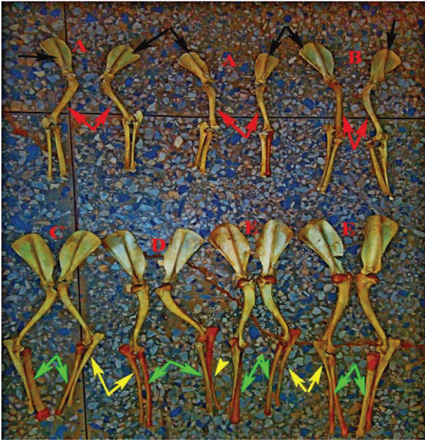
Figure 1: Photograph of selected bones of forelimb of Mangrelian Dog showing various identification marks and classification of the samples, 0-6 month (A), 6month-1 year (B), 1-2 years (C), 2-3years (D) and above 3 years (E), scapular (black arrow), humerus (red arrow), radius (green arrow) and ulna bone (yellow arrow).
The scapula
Is a rectangular bone with 2 surfaces, medial and lateral surface. The lateral surface is divided into two unequal halves by scapula spine and it ends as acromium. It has3 borders namely: cranial border, caudal border and dorsal border; the cranial border is sharp and the caudal border is broad. The distal end of the scapula forms the glenoid cavity for articulation with head of humerus as shown in Figure 2.
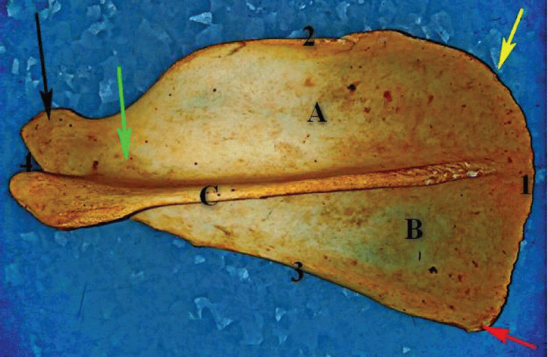
Figure 2: Photograph of the lateral view of Mangrelian Dog scapular bone above 3 years showing infraspinatus fossa (A), supraspinatus fossa(B), spinous process (C), cranial border (3), caudal border (2), dorsal border (1), basal border (4) cranial angle (Red arrow), caudal angle (yellow arrow), basal angle (black arrow)and neck of the scapular (green arrow).
The humerus
Belongs to long bones class, it has two ends that is proximal and distal end. The proximal end consist of the head which is located medially for articulation with scapula and lateral greater tuberosity separated intertrochanteric crest. Just below the neck, there is deltoid tuberosity and musculo-spiral groove. The shaft of the bone is slightly twisted. The distal end of the bone possess the tubercle for articulation with radio ulna bone (Figure 3).
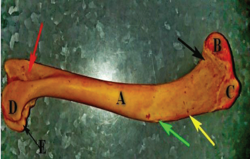
Figure 3: Photograph of the lateral view of Humerus of Mongrelian Dog of 2-3 years showing the shaft of the bone (A), head (B), tubercle (C), neck (black arrow) the capitulum (D) and the distal articular surface (red arrow).
Radio ulna bone
They occur as separate bones in dogs while in other animals they are fused with the ulna being the largest, the ulna bone is long with olecranon proximally which lodges into the radial fossa of the humerus bone and the distal end possess styloid process of ulna. The radius occurs as long bone which is anterior to the ulna bone and proximally supports the distal condyles of the humerus.
Biometrical observations
Scapula: Total length of the bone was taken from proximal border of the bone to the glenoid cavity; the dorsal length of the scapula was measured from cranial border to caudal border; the pine length was taken from dorsal border of the scapula to the acromium; the infraspinous and supraspinous spinous fossa measurements were taken proximal, midway and distal to the fossa.
Humerus: The total length was taken from proximal extremity to distal extremity of the bone. The circumference was taken using thread proximally below the head of humerus, midway below the musculospiral groove, deltoid tuberosity, the shaft circumference was taken and distal circumference just above the distal condyles.
Radius and ulna: Total length of the ulna was taken from proximal to distal and the shaft just the neck of olecranon process while that of the radius was taken proximally midway and distally (Figure 4).
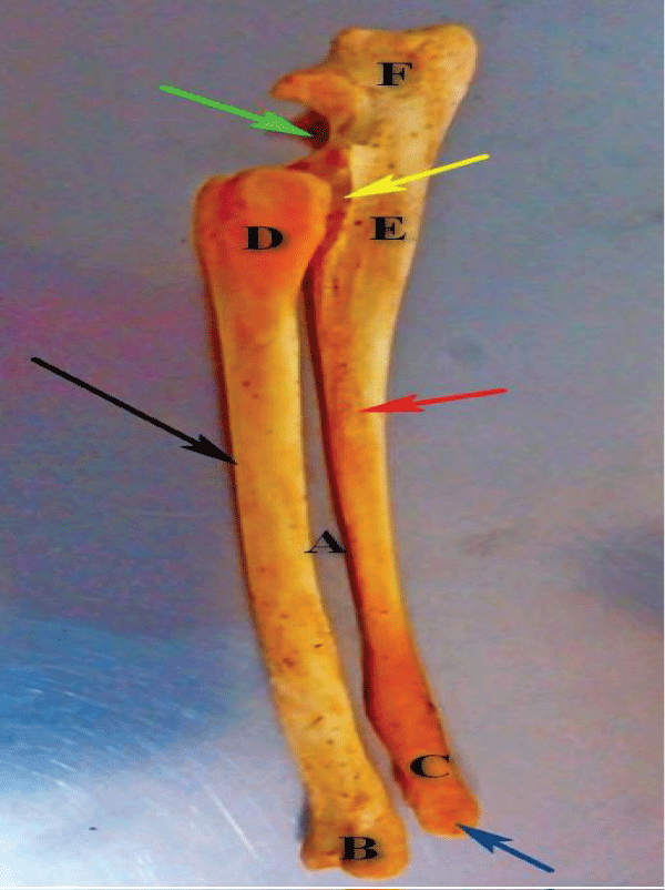
Figure 4: Photograph of the lateral view of radio ulnar bones of Mongrelian Dog at 2-3 years showing the shaft of the radial bone (black arrow), shaft of the ulna bone (red arrow), proximal extremity of the radius (D), proximal extremity of the ulna (E), the large interoceous space (A), the distal extremity of the radius (B), the distal extremity of the ulna bone (C), the ancornial process (green arrow) and the distal styloid process of the ulna bone (blue arrow).
The result of this study revealed the there is significant change in weight, length and diameter of some bones of the forelimb of local domestic dog. As the age increase there is change in parameters in some of the parameters increasing.
For scapula; there is no difference between the proximal aspects of the infraspinatous fossa across the age as it increases. For Group D and Group E, there is no difference statistically but difference exist from Group D to group A respectively as the age decreases. For Distal infraspinous fossa; there is no difference except for Group E which is greater than 3 years.
For humerus; there is no significant difference between the middle shaft of the humerus even as the age increases statistically. The deltoid tuberosity and teres tuberosity of the age group A = 0-6 months is not well developed thus not captured in this research work; while it is well developed as the age increases as in group (D = 2-3 years and E = above 3 years).
For ulna; the significant difference between the parameters as the age increases.
For radius; the weight increases as the age increase, but there is no significant difference but the proximal, middle and distal circumference statistically.
The average maximum length and breadth of scapula (Table 1) in Blue bull was 31.40 ± 0.616 cm and 15.62 ± 0.29 cm respectively, which was 13.94 ± 0.30 cm and 6.62 ± 0.11 cm in Black Bengal goat [20]; 14.07 ± 0.019 cm and 8.59 ± 0.016 cm in blackbuck [21], 20.46 ± 0.03 cm and 11.94 ± 0.03 cm, in chital [22] and 12.35 ± 0.12cm and 6.94 ± 0.06 cm in chinkara [24]. The scapular index in the present study was 49:74 for Blue bull which was 82.05 for tiger, 72.82 for leopard, 67.34 for Sambar, 65.83 for sheep, 62.43 for buffalo, 57.51 for deer, 55.74 for pig, 52.59 for ox, 45.86 for horse and 43.62 for goat as per calculations of Dalvi, et al. [23], 57.78 for Chinkara [24], 58.35 for Chital [22] and 61.05 for blackbuck [21]. The average maximum length of spines, breadth of necks of scapulae and breadth of glenoid cavities were 26.67 ± 0.33 cm, 6.833 ± 0.148 cm and 3.9 ± 0.217 cm, respectively Tables 1-4).
| Table 1: Morphometric data for Different Parameters of Scapula (Mean and Standard error measurement). | ||||||||||||||
| S/N | Age | Group A | Group B | Group C | Group D | Group E | SEM | |||||||
| Parameters | R | L | R | L | R | L | R | L | R | L | L | R | ||
| 1 | Weight(g) | 8.00b | 7.250c | 12.65a | 12.75b | 13.00a | 13.00b | 14.00a | 14.00b | 15.00a | 19.00a | 1.28 | 0.87 | |
| 2 | Total length(cm) | 7.45c | 7.45b | 8.15c | 8.15b | 9.70b | 9.35b | 12.15b | 12.20a | 13.00a | 14.00a | 0.87 | 0.77 | |
| 3 | Spine(cm) | 7.50d | 7.45d | 8.25d | 8.70d | 11.25b | 11.10b | 12.00b | 12.95b | 15.10a | 15.10a | 0.96 | 0.96 | |
| 4 | Dorsal Border (cm) | 4.70b | 4.85b | 5.35b | 5.75b | 5.55b | 6.00a | 6.30b | 6.35a | 6.70a | 6.70a | 0.23 | 0.28 | |
| 5 | Supraspinatus fossa (cm) | Proximal | 1.20d | 1.55c | 1.55c | 1.60c | 1.55c | 1.65c | 2.10b | 2.10b | 2.80a | 2.80a | 0.16 | 0.19 |
| Middle | 2.05a | 2.25a | 2.50a | 2.50a | 2.65a | 2.74a | 3.00a | 3.00a | 3.00a | 3.00a | 0.14 | 0.16 | ||
| Distal | 2.60a | 1.40b | 2.60a | 1.80b | 2.50a | 2.50a | 1.85b | 2.60a | 1.00c | 2.60a | 0.17 | 0.21 | ||
| 6 | Infraspinatous fossa (cm) | Proximal | 2.90a | 2.90a | 3.90a | 3.90a | 4.00a | 3.95a | 4.00a | 3.95a | 4.00a | 3.95a | 0.18 | 0.19 |
| Middle | 2.30b | 2.15b | 2.40b | 2.30b | 2.45b | 2.50b | 3.60a | 3.60a | 3.70a | 3.70a | 0.23 | 0.21 | ||
| Distal | 0.85b | 1.00a | 1.00b | 1.30a | 1.25b | 1.40a | 1.30b | 1.40a | 1.40a | 5.40a | 0.88 | 0.08 | ||
| Key: abcMeans bearing same letters within the same column are not statistically different (p < 0.05). Group A = 0-6 months; Group B = 6 Months to 1 year; Group C = 1-2 Years; Group D = 2-3 Years; Group E = Above 3 Years SEM = Standard Error of Mean; g = gram; L = Right; R = Right; Cm = Centimeters |
||||||||||||||
| Table 2: Morphometric data for Different Parameters of Humerus (Mean and Standard error measurement). | |||||||||||||
| S/N | Age | Group A | Group B | Group C | Group D | Group E | SEM | ||||||
| Parameters | R | L | R | L | R | L | R | L | R | L | R | L | |
| 1 | Weight (g) | 12.15d | 12.00d | 24.25c | 24.50c | 40.00b | 40.00b | 48.00a | 48.00a | 50.00a | 50.00a | 4.86 | 4.87 |
| 2 | Length (cm) | 11.00d | 11.15d | 14.90c | 15.10c | 16.70b | 16.70b | 16.70b | 16.70b | 20.00a | 20.00a | 0.98 | 0.96 |
| 3 | Circumference of proximal shaft (cm) | 5.15c | 5.15c | 5.35b | 5.50b | 7.00b | 7.00b | 8.60a | 8.60a | 8.600a | 8.60a | 0.52 | 0.51 |
| 4 | Circumference of middle Shaft (cm) | 4.35b | 4.35b | 4.45b | 4.45b | 5.30b | 5.30b | 5.50b | 5.50b | 5.90a | 5.90a | 0.22 | 0.23 |
| 5 | Circumference of distal shaft (cm) | 4.25c | 4.25d | 4.65c | 4.85c | 5.50b | 5.50b | 5.60b | 5.60b | 6.30a | 6.30a | 0.23 | 0.23 |
| 6 | Deltoid Tuberosity (cm) | 0.00d | 0.00d | 2.55c | 2.60c | 5.50b | 5.50b | 5.60b | 5.60b | 6.30a | 6.30a | 0.79 | 0.79 |
| 7 | Teres Tuberosity (cm) | 0.00c | 0.00b | 2.30b | 2.65a | 3.10b | 3.10a | 3.70a | 3.70a | 4.00a | 4.00a | 0.49 | 0.49 |
| Key: abcMeans bearing same letters within the same column are not statistically different (p < 0.05). Group A = 0-6 months; Group B = 6 Months to 1 year; Group C = 1-2 Years; Group D = 2-3 Years; Group E= Above 3 Years SEM = Standard Error of Mean; g = gram; L = Right; R = Right; Cm = Centimeters |
|||||||||||||
| Table 3: Morphometrical data for Different Parameters of Ulna bone (Mean and Standard error measurement) | |||||||||||||
| S/N | Age | Group A | Group B | Group C | Group D | Group E | SEM | ||||||
| Parameters | R | L | R | L | R | L | R | L | R | L | R | L | |
| 1 | Weight (G) | 8.00e | 9.00d | 13.25d | 13.75c | 19.50c | 20.00b | 23.50b | 23.50a | 28.50a | 26.50a | 2.43 | 2.15 |
| 2 | Length (cm) | 13.50d | 13.80d | 17.55c | 17.55c | 19.90c | 19.50b | 20.10c | 20.20b | 24.50a | 24.60a | 1.21 | 1.19 |
| 3 4 5 |
Proximal (cm) | 4.050d | 4.00d | 4.20c | 4.05d | 4.70b | 4.90c | 4.70b | 5.10b | 5.70a | 5.50a | 0.19 | 0.20 |
| Middle (cm) | 2.75c | 2.75b | 2.80c | 2.80b | 3.00b | 3.40a | 3.40b | 3.40a | 3.50a | 3.50a | 0.11 | 0.11 | |
| Distal (cm) | 2.10c | 2.20c | 2.55b | 2.60b | 2.60b | 2.65b | 2.70b | 2.80b | 3.40a | 3.20a | 0.14 | 0.11 | |
| Key: abcMeans bearing same letters within the same column are not statistically different (p < 0.05) Group A = 0-6 months; Group B = 6 Months to 1 year; Group C= 1-2 Years; Group D= 2-3 Years; Group E= Above 3 Years SEM = Standard Error of Mean; g = gram; L = Right; R = Right; Cm = Centimeters |
|||||||||||||
| Table 4: Morphometric data for Different Parameters of Radius (Mean and Standard error measurement). | |||||||||||||
| S/N | Age Parameters | Group A | Group B | Group C | Group D | Group E | SEM | ||||||
| R | L | R | L | R | L | R | L | R | L | R | L | ||
| 1 | Weight (g) | 10.00d | 10.25d | 21.50c | 21.50c | 23.50c | 23.50c | 26.50a | 26.50b | 48.00a | 48.00a | 4.15 | 4.12 |
| 2 | Length (cm) | 13.00c | 13.60c | 15.60c | 15.45c | 19.50b | 19.90c | 20.20b | 20.20b | 24.50a | 26.60a | 1.36 | 1.53 |
| 3 | Proximal (cm) | 3.45a | 3.55a | 4.70a | 4.70a | 4.70a | 4.90a | 4.90a | 5.10a | 5.70a | 5.50a | 0.35 | 0.34 |
| 4 | Middle (cm) | 2.90a | 2.95a | 3.00a | 3.00a | 3.40a | 3.40a | 3.50a | 3.60a | 4.00a | 4.00a | 0.20 | 0.20 |
| 5 | Distal (cm) | 2.55a | 2.70a | 2.80a | 2.70a | 3.00a | 2.80a | 3.05a | 3.00a | 3.90a | 4.00a | 0.22 | 0.23 |
| Key: abcMeans bearing same letters within the same column are not statistically different (p < 0.05) . Group A = 0-6 months; Group B = 6 Months to 1 year; Group C = 1-2 Years; Group D = 2-3 Years; Group E = Above 3 Years SEM = Standard Error of Mean; g = gram; L = Right; R = Right; Cm = Centimeters |
|||||||||||||
The age group D and E have the highest values of morphometry in all the parameters measured while A and B group falls under lower limit with C been the intermediate. However, some parameters do not change even as the age increases.
Recommendation
Based on this study, the morphometric data of forelimb of a local dog increases as the age increase and vice versa. Based on these data, the result of this can be referred for the following research work.
- Ageing of local dog base on morphometry for breeding
- Forensic studies
- Medication and vaccination administration
- Feeding system management
- Controlled breeding such as neutering and castration
- Xiaoming W, Mauricio A, Richard T, Dogs: Their Fossil Relatives and Evolutionary History. New York: Columbia University Press. 2008; 1.
- Lindblad-Toh K, Wade CM, Mikkelsen TS, Karlsson EK, Jaffe DB, et al. Genome sequence, comparative analysis & haplotype structure of the domestic dog. Nature. 2005; 438: 803–819.
- Young Julie K, Olson Kirk A, Reading Richard P, Sukh A, Joel B, et al. 2011.
- Fan Z, Silva P, Gronau I, Wang S, Armero AS, et al. Worldwide patterns of genomic variation and admixture in gray wolves. Genome Res. 2016; 26: 163–173. PubMed: https://pubmed.ncbi.nlm.nih.gov/26680994/
- Thalmann O, Shapiro B, Cui P, Schuenemann VJ, Sawyer SK, et al. Complete Mitochondrial Genomes of Ancient Canids Suggest a European Origin of Domestic Dogs. Science. 2013; 342: 871–874. PubMed: https://pubmed.ncbi.nlm.nih.gov/24233726/
- Vila C. Multiple and ancient origins of the domestic dog. Science. 1997; 276: 1687–1689. PubMed: https://pubmed.ncbi.nlm.nih.gov/9180076/
- Freedman AH, Gronau I, Schweizer RM, Del Vecchyo DO, Han E, et al. Genome Sequencing Highlights Genes Under Selection and the Dynamic Early History of Dogs. PLOS Genetics. 2014; 10: e1004016. PubMed: https://pubmed.ncbi.nlm.nih.gov/24453982/
- Vonholdt BM, Driscoll CA. 3-Origins of the dog: Genetic insights into dog domestication. In James Serpell (ed.) 2016.
- Larson G, Bradley DG. Perri Angela. A wolf in dog's clothing: Initial dog domestication and Pleistocene wolf variation. J Archaeological Sci. 2016; 68: 1–4.
- Perri A. A wolf in dog's clothing: Initial dog domestication and Pleistocene wolf variation. J Archaeological Sci. 68: 1–4.
- Dewey T, Bhagat s. Canis lupus familiaris, Animal Diversity Web. 2002.
- Berns GS, Brooks AM, Neuhauss SM, Stephan CF. Functional Magnetic Resonance Imaging in Awake Unrestrained Dogs. PLoS ONE. 2012; 7: e38027. PubMed: https://pubmed.ncbi.nlm.nih.gov/22606363/
- Axelsson E, Ratnakumar A, Arendt ML, Maqbool K, Webster MT, et al. The genomic signature of dog domestication reveals adaptation to a starch-rich diet. Nature. 2013; 495: 360–364. PubMed: https://pubmed.ncbi.nlm.nih.gov/23354050/
- Nikhil S. Why are different breeds of dogs all considered the same species? – Scientific AmericanArchived 10 October 2016 at the Wayback Machine. 2016.
- Freedman AH, Wayne RK. Deciphering the Origin of Dogs: From Fossils to Genomes. Annu Rev Anim Biosci. 2017; 5: 281–307. PubMed: https://pubmed.ncbi.nlm.nih.gov/27912242/
- Larson G, Bradley DG. How Much Is That in Dog Years? The Advent of Canine Population Genomics. PLOS Genetics. 2014; 10: e1004093.
- Dyce KM, Sack WO, Wensing CJG. Textbook of Veterinary Anatomy, 3rd edition. W.B sounders. 2002.
- National Population Commission (NPC), Census Data of 2006.
- Statistical Analysis System (SAS) package version 9.2 software (Statistical Analysis System, 2007, SAS Institute Inc., Cary, NC, USA).
- Siddiqui MSI, Khan MZI, Sarma M, Islam MN, Jahan MR. Macro-anatomy of the bones of the limb of Black Bengal Goat (Capra hircus). Bangladesh J Veterin Med. 2008; 6: 59-66.
- Choudhary OP, Singh I. Morphometrical studies on Scapula of Indian Blackbuck (Antelope cervicapra). Indian Veterin J. 2016; 93: 64-67.
- Choudhary OP, Mathur R, Joshi S, Beniwal G, Dangi A. Gross and biometrical studies on scapula of chital (Axis axis). Veterinary Practitioner. 2013; 14: 036- 039.
- Dalvi RS, Bhamburkar VR, Ladukar ON, Banubakode SB. Morphometric study on scapulae of some domestic and wild animals. Tech. Bul. XII Convention and National Symposium of IAVA. 1997; 43.
- Jangir DK. Thesis entitled “Gross studies on the bones of the Forelimb in Indian Gazelle (Gazella gazelle bennettii)” College of veterinary and animal sciences, RAJUVAS, Bikaner. 78-91.