

- Department of Pathology and Laboratory Medicine, Children’s Hospital of Philadelphia, University of Pennsylvania Perelman School of Medicine, Philadelphia, Pennsylvania, United States,
- Department of Neurosurgery, Hospital of the University of Pennsylvania, Philadelphia, Pennsylvania, United States.
Correspondence Address:
Angela N. Viaene
Department of Neurosurgery, Hospital of the University of Pennsylvania, Philadelphia, Pennsylvania, United States.
DOI:10.25259/SNI_501_2019
Copyright: © 2019 Surgical Neurology International This is an open-access article distributed under the terms of the Creative Commons Attribution-Non Commercial-Share Alike 4.0 License, which allows others to remix, tweak, and build upon the work non-commercially, as long as the author is credited and the new creations are licensed under the identical terms.How to cite this article: Angela N. Viaene, Steven Brem. Recurrent neurenteric cysts compressing the brainstem. 13-Dec-2019;10:245
How to cite this URL: Angela N. Viaene, Steven Brem. Recurrent neurenteric cysts compressing the brainstem. 13-Dec-2019;10:245. Available from: https://surgicalneurologyint.com/surgicalint-articles/9802/
Date of Submission
03-Oct-2019
Date of Acceptance
15-Nov-2019
Date of Web Publication
13-Dec-2019
Abstract
Background: Neurenteric cysts, also referred to as endodermal cysts and enterogenous cysts, are rare lesions of the neuroaxis occurring most frequently within the spinal cord and rarely intracranially. In the literature describing these lesions, examples of intraoperative imaging and cytology preparations are rare to non-existent.
Case Description: Here, we describe a case of a recurrent posterior fossa neurenteric cyst compressing the brainstem in a 47-year-old female and causing incontinence and progressive quadriparesis. Intraoperative findings and cytologic and histologic features are presented.
Conclusion: Neurenteric cysts are generally considered to be benign and slow-growing though recurrence is common. This case of a recurrent neurenteric cyst is illustrated by intraoperative macroscopic and cytologic images.
Keywords: Cytology, Endodermal cyst, Enterogenous cyst, Neurenteric cyst, Posterior fossa epithelial cyst
INTRODUCTION
Neurenteric cysts, also referred to as endodermal cysts and enterogenous cysts, are rare lesions of the neuroaxis occurring most frequently within the spinal cord and rarely intracranially. In the literature describing these lesions, examples of intraoperative imaging and cytology preparations are rare to non-existent.
CASE REPORT
A 47-year-old female with a history of posterior fossa neurenteric cysts presented with incontinence and progressive quadriparesis. In 1994, she experienced imbalance and falls, and head imaging revealed a 7 cm posterior fossa cyst which was resected. Another resection was performed in 1997. She developed hydrocephalus, resulting in a ventriculoperitoneal shunt placement with subsequent revisions due to infections and clotting. In the 2 months before presentation, she developed incontinence and progressive quadriparesis. Magnetic resonance imaging showed multiple, expanding, and recurrent neurenteric cysts in the left cerebellomedullary angle and left prepontine cistern compressing the medulla [
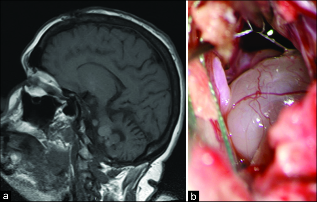
Figure 1:
Neurenteric cysts compressing the brainstem. Magnetic resonance imaging demonstrated multiple, nonenhancing, multiolobulated, expanding, and extra-axial recurrent neurenteric cysts compressing the medulla (a). An intraoperative image showing the cysts which were adherent to but separable from the medulla and upper cervical spinal cord (b).
Intraoperative squash preparations demonstrated sheets of epithelial cells with honeycomb architecture, distinct cell borders, and abundant cytoplasm within a background of mucin [
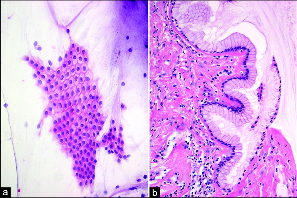
Figure 2:
Cytology and histology of the neurenteric cysts. Intraoperative squash preparation stained with hematoxylin and eosin revealed sheets of epithelial cells with honeycomb architecture, distinct cell borders, and abundant cytoplasm within a background of mucin, ×200 (a). Hematoxylin and eosin-stained permanent sections showing cyst wall lined by columnar epithelium with underlying basement membrane and connective tissue stroma, ×200 (b).
DISCUSSION
Neurenteric cysts, also referred to as endodermal cysts and enterogenous cysts, are rare lesions comprising 0.7–1.3% of all spinal cord tumors and occurring less frequently within the posterior fossa and cerebral hemispheres.[
CONCLUSION
Neurenteric cysts are generally considered to be benign and slow-growing though recurrence is common. This case of recurrent intracranial neurenteric cysts includes illustrations of intraoperative macroscopic and cytologic images.
Declaration of patient consent
The authors certify that they have obtained all appropriate patient consent forms.
Financial support and sponsorship
Nil.
Conflicts of interest
There are no conflicts of interest.
References
1. Bejjani GK, Wright DC, Schessel D, Sekhar LN. Endodermal cysts of the posterior fossa. Report of three cases and review of the literature. J Neurosurg. 1998. 89: 326-35
2. Priamo FA, Jimenez ED, Benardete EA. Posterior fossa neurenteric cysts can expand rapidly: Case report. Skull Base Rep. 2011. 1: 115-24
3. Rotondo M, D’Avanzo R, Natale M, Pasqualetto L, Bocchetti A, Agozzino L. Intramedullary neurenteric cysts of the spine. Report of three cases. J Neurosurg Spine. 2005. 2: 372-6
4. Vachhani JA, Fassett DR. Intramedullary neurenteric cyst associated with a tethered spinal cord: Case report and literature review. Surg Neurol Int. 2012. 3: 80-
5. Yang Y, Fang J, Li DA, Wang L, Ji N, Zhang J. Recurrent intracranial neurenteric cyst with malignant transformation: A case report and literature review. Oncol Lett. 2016. 11: 3395-402

