Abstract
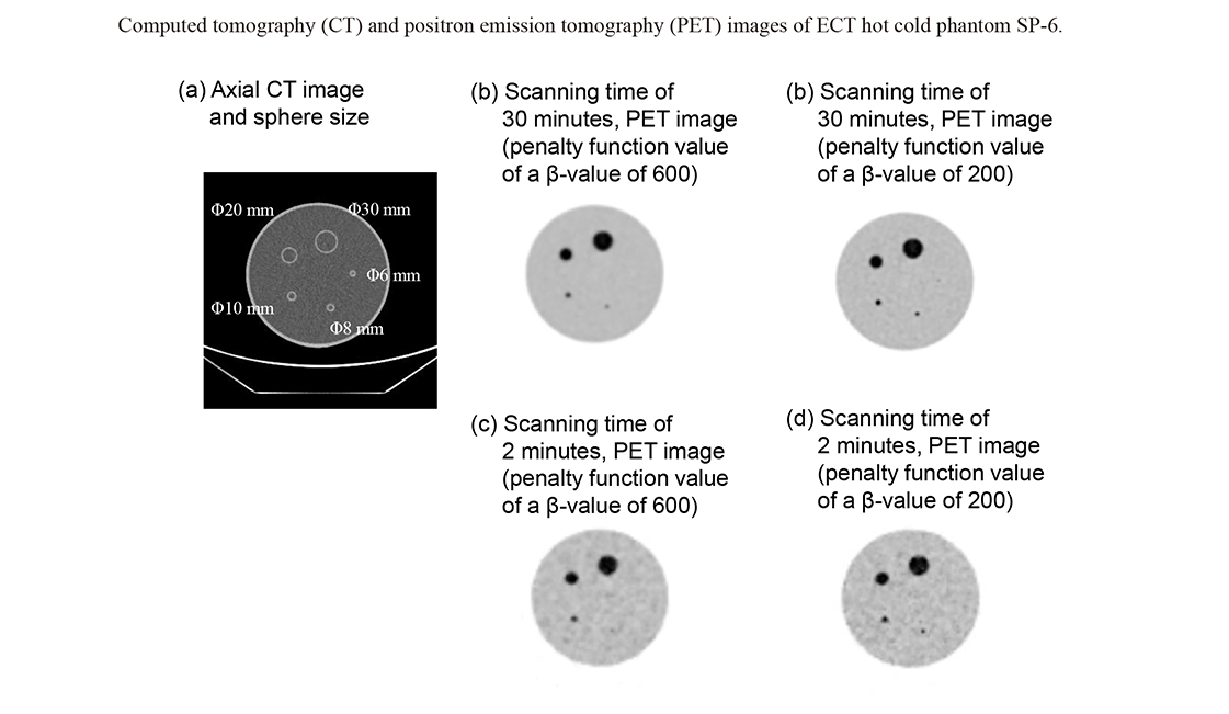
Positron emission tomography (PET)/computed tomography (CT) has improved sensitivity and resolution using silicon photomultiplier as a photosensor. Previously, only a fixed setting was available for the shooting time of 1 bed, but now, the shooting time can be changed for each bed. Time can be shortened or extended depending on the target area. A few studies reported on image reconstruction conditions for head and neck cancer in whole-body PET/CT examinations. Thus, this study aimed to optimize the imaging conditions of the head and neck region during whole-body imaging. A cylindrical acrylic container with a 200 mm diameter was used to simulate the head and neck area using a PET/CT system equipped with a semiconductor detector. Spheres of 6-30 mm in diameter were enclosed in the 200 mm diameter cylindrical acrylic vessel. Radioactivity in 18F solution (Hot:BG ratio 4:1) was enclosed in a phantom following the Japanese Society of Nuclear Medicine (JSNM) guidelines. Background radioactivity concentration was 2.53 kBq/mL. List mode acquisition of 1,800 s was collected at 60-1,800 s with the field of view of 700 mm and 350 mm. The image was reconstructed by resizing the matrix to 128 × 128, 192 × 192, 256 × 256, and 384 × 384, respectively. The imaging time per bed in the head and neck should be at least 180 s, and the reconstruction conditions should be a field of view (FOV) of 350 mm, matrix sizes of ≥ 192, and a Bayesian penalized likelihood (BPL) reconstruction with a β-value of 200. This allows detection of > 70% of the 8-mm spheres in the images.