Abstract
We report the case of a three-year-old child who, following long term treatment with topical corticosteroids and their associations for a case of ringworm on the face developed a form of folliculitis known as Majocchi's Granuloma. Treatment with oral Griseofulvin was successful.
Adrenal cortex hormones; Granuloma; Steroids; Tinea
INTRODUCTION
Majocchi's Granuloma was described in 1883 in Italy by Domenico Majocchi.11 Coelho WS, Diniz LM, Sousa Filho JB, Castro CM. Case for diagnosis. Granuloma trichophyticum (Majocchi's granuloma). An Bras Dermatol. 2009;84:85-6. It is a rare infection, possibly associated with depilation or with use of high potency topical corticosteroid therapy in areas of dermatophyte infection in immunocompetent patients.22 Bressan AL, Silva RS, Fonseca JC, Alves M de F. Majocchi's granuloma. An Bras Dermatol. 2011;86:797-8. The authors report a case of Majocchi's Granuloma during childhood.
CASE REPORT
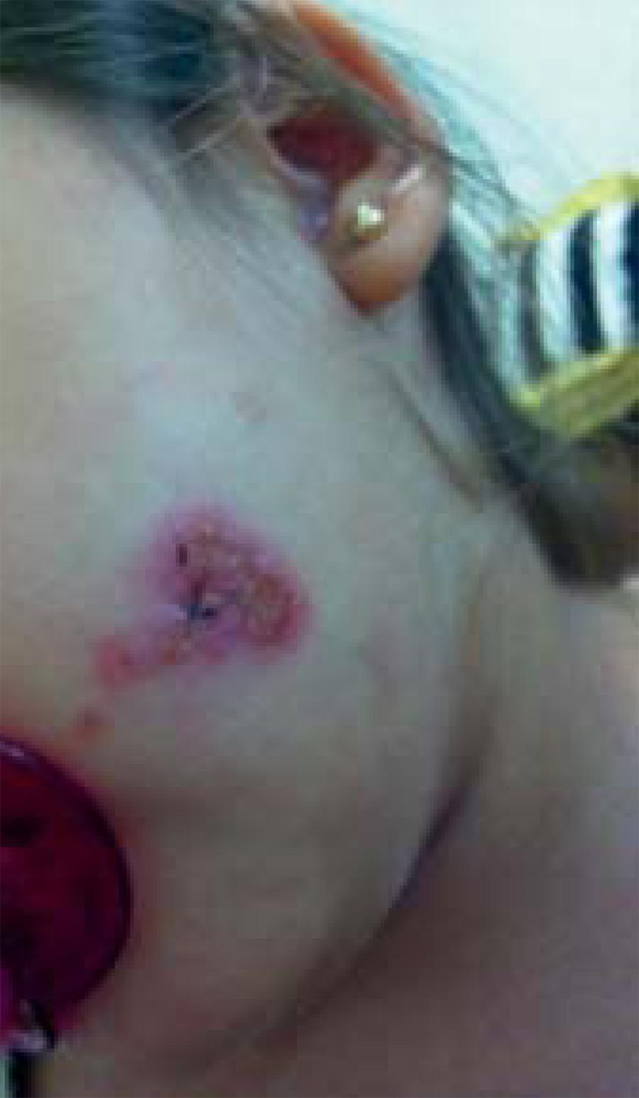
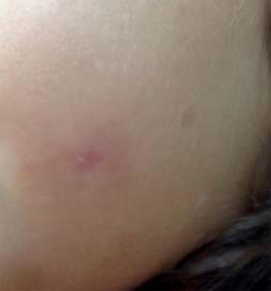
Female patient, three years old, white, from Rio de Janeiro, presenting an exulcerated lesion of desquamative edges in the left malar region with 8 months of evolution (Figure 1). It was reported that she had been using an association of topical corticoids, antifungals, antibiotics, antibiotic therapy and oral corticotherapy during this period with no improvement. She was also taking Pimecrolimus 0.03% twice a day for a period of 30 days with relative improvement during the use of the medications and worsening after ceasing their use. General state with no systemic changes. Drugs were suspended after 15 days and cutaneous biopsy was performed with histopathological examination. Direct mycological examination negative and culture for Microsporum gypseum positive. Histopathologics showed presence of hyphae and positive PAS spores in topography of follicular canal, abscess and outline of perifollicular granulomatous reaction compatible with fungal folliculitis, Majocchi's granuloma (Figures 2 and 3). Patient was treated with griseofulvin 250mg/day for 8 weeks presenting remission of the condition (Figure 4).
Exulcerated lesion, erythematous, with desquamative edges in the left malar region of the face
40X: Topography of follicular canal reveals presence of hyphae and positive PAS spores, abscess and outline of perifollicular granulomatous reaction
200X: Topography of follicular canal reveals presence of hyphae and positive PAS spores, abscess and outline of perifollicular granulomatous reaction compatible with fungal folliculitis, Majocchi's granuloma
DISCUSSION
Tinea corporis is a dermatophytic infection with greater incidence in the skin, mainly on the trunk and extremities, usually restricted to the stratum corneum. The atypical deep involvement is called Majocchi's granuloma, and it can be perifollicular secondary to traumas or the subcutaneous nodular type in the immunocompromised patient.33 Teixeira SP, Ruete LC, Yamashita JT. Dermatoses nos pacientes transplantados. Guia de Medicina ambulatorial e hospitalar da UNIFESP-EPM. 2008;(1):277-295.
This granuloma constitutes a nodular perifolliculitis with formation of foreign body granuloma, due to the infection of dermis and subcutaneous tissue by dermatophytes. Among the etiological agents described, Trichophyton rubrum is the most frequent one, followed by Trichophyton violaceum, Trichophyton mentagrophytes, Microsporum audouinii, Microsporum gypseum, Microsporum canis and Epidermophyton floccosum.11 Coelho WS, Diniz LM, Sousa Filho JB, Castro CM. Case for diagnosis. Granuloma trichophyticum (Majocchi's granuloma). An Bras Dermatol. 2009;84:85-6.,44 Azulay RD, Azulay DR, Abufalia LA. Micoses superficiais. In: Azulay DR. Dermatologia. 5 ed. Rio de Janeiro: Guanabara Koogan, 2008. p. 419-440.
In immunocompetent patients, clinical findings are typically characterized by a
localized area with erythematous papules, perifollicular or small nodules. Pustules
may also be present.55 Goldstein AO, Goldstein BG, Dellavale RP. Dermatophyte (tinea)
infections. Literature review current through: Jun 2013. | This topic last
updated: Fev 2, 2013. Immunocompromised
patients may present similar symptoms as immunocompetent patients or with
subcutaneous nodules and abscesses.66 Tse KC, Yeung CK, Tang S, Chan HH, Li FK, Chan TM, et al. Majocchi's
granuloma and posttransplant lymphoproliferative disease in a renal transplant
recipient. Am J Kidney Dis. 2001;38:E38.
7 Liao YH, Chu SH, Hsiao GH, Chou NK, Wang SS, Chiu HC. Majocchi's
granuloma caused by Trichophyton tonsurans in a cardiac transplant recipient. Br
J Dermatol. 1999;140:1194-6.-88 Kim ST, Baek JW, Kim TK, Lee JW, Roh HJ, Jeon YS, et al. Majocchi's
granuloma in a woman with iatrogenic Cushing's syndrome. J Dermatol.
2008;35:789-91. Trauma is also
considered an incitation factor in these cases. Cell-mediated immune depression and
inflammatory response, important for inhibition of infections by dermatophytes, may
contribute for the progression of the disease.99 Akiba H, Motoki Y, Satoh M, Iwatsuki K, Kaneko F. Recalcitrant
trichophytic granuloma associated with NK-cell deficiency in a SLE patient
treated with corticosteroid. Eur J Dermatol. 2001;11:58-62.,1010 Smith KJ, Neafie RC, Skelton HG 3rd, Barrett TL, Graham JH, Lupton
GP. Majocchi's granuloma. J Cutan Pathol. 1991;18:28-35. Systemic
dissemination seldom occurs.1010 Smith KJ, Neafie RC, Skelton HG 3rd, Barrett TL, Graham JH, Lupton
GP. Majocchi's granuloma. J Cutan Pathol. 1991;18:28-35.
In immunocompetent patients, the use of topic corticosteroids on a surface may lead to infection by dermatophytes by local immunosuppression, and promote the development of Majocchi's granuloma.44 Azulay RD, Azulay DR, Abufalia LA. Micoses superficiais. In: Azulay DR. Dermatologia. 5 ed. Rio de Janeiro: Guanabara Koogan, 2008. p. 419-440. The diagnosis is performed through direct mycological examination, culture and histopathology. In histopathology, in response to the agent or due to the releasing of follicular content with cellular immune reaction, there is formation of giant cell and foreign body granuloma containing the fungus. The histopathological as well as the mycological examination may not reveal fungal elements, and for this reason the best test for that is the culture of homogenate, and treatment guided by the result of culture with local anti-fungal. Surgical excision of lesion has also been reported with good results.55 Goldstein AO, Goldstein BG, Dellavale RP. Dermatophyte (tinea) infections. Literature review current through: Jun 2013. | This topic last updated: Fev 2, 2013.
A noteworthy fact is that tinea barbae is a fungal infection common to the beard and its surrounding area of teenage and adult males who shave, and is rare during infancy. Microsporum gypseum is a geophilic fungus. Deep reactions with high inflammatory lesions are common and respond well to therapy.44 Azulay RD, Azulay DR, Abufalia LA. Micoses superficiais. In: Azulay DR. Dermatologia. 5 ed. Rio de Janeiro: Guanabara Koogan, 2008. p. 419-440.
Indiscriminate use of topical corticoids, by diminishing local defense, may favor fungal infection and trigger Majocchi's granuloma, with penetration of hair follicle by the dermatophyte. Fungal infection diagnosis must be always remembered in the presence of lesions refractory to treatment with correct antibiotic therapy, elucidating the importance of tracking with direct mycological examination and culture of lesion, for they are low cost tests and of easy execution.
-
Financial Support: none
-
How to cite this article: Kanaan ICS, Santos TBP, Kac BK, Souza AMV, Cerqueira AMM. Majocchi's granuloma - Case report. An Bras Dermatol. 2015;90(2):251-3.
-
*
Work performed at Hospital Municipal Jesus - Rio de Janeiro (RJ), Brazil.
References
-
1Coelho WS, Diniz LM, Sousa Filho JB, Castro CM. Case for diagnosis. Granuloma trichophyticum (Majocchi's granuloma). An Bras Dermatol. 2009;84:85-6.
-
2Bressan AL, Silva RS, Fonseca JC, Alves M de F. Majocchi's granuloma. An Bras Dermatol. 2011;86:797-8.
-
3Teixeira SP, Ruete LC, Yamashita JT. Dermatoses nos pacientes transplantados. Guia de Medicina ambulatorial e hospitalar da UNIFESP-EPM. 2008;(1):277-295.
-
4Azulay RD, Azulay DR, Abufalia LA. Micoses superficiais. In: Azulay DR. Dermatologia. 5 ed. Rio de Janeiro: Guanabara Koogan, 2008. p. 419-440.
-
5Goldstein AO, Goldstein BG, Dellavale RP. Dermatophyte (tinea) infections. Literature review current through: Jun 2013. | This topic last updated: Fev 2, 2013.
-
6Tse KC, Yeung CK, Tang S, Chan HH, Li FK, Chan TM, et al. Majocchi's granuloma and posttransplant lymphoproliferative disease in a renal transplant recipient. Am J Kidney Dis. 2001;38:E38.
-
7Liao YH, Chu SH, Hsiao GH, Chou NK, Wang SS, Chiu HC. Majocchi's granuloma caused by Trichophyton tonsurans in a cardiac transplant recipient. Br J Dermatol. 1999;140:1194-6.
-
8Kim ST, Baek JW, Kim TK, Lee JW, Roh HJ, Jeon YS, et al. Majocchi's granuloma in a woman with iatrogenic Cushing's syndrome. J Dermatol. 2008;35:789-91.
-
9Akiba H, Motoki Y, Satoh M, Iwatsuki K, Kaneko F. Recalcitrant trichophytic granuloma associated with NK-cell deficiency in a SLE patient treated with corticosteroid. Eur J Dermatol. 2001;11:58-62.
-
10Smith KJ, Neafie RC, Skelton HG 3rd, Barrett TL, Graham JH, Lupton GP. Majocchi's granuloma. J Cutan Pathol. 1991;18:28-35.
Publication Dates
-
Publication in this collection
Mar-Apr 2015
History
-
Received
02 Sept 2013 -
Accepted
17 Oct 2013