Article

Hidden in Heart Failure

Register or Login to View PDF Permissions
Permissions× For commercial reprint enquiries please contact Springer Healthcare: ReprintsWarehouse@springernature.com.

For permissions and non-commercial reprint enquiries, please visit Copyright.com to start a request.

For author reprints, please email rob.barclay@radcliffe-group.com.
Information image
Average (ratings)
No ratings
Your rating

Abstract

Current diagnostic strategies fail to illuminate the presence of rare disease in the heart failure population. One-third of heart failure patients are categorised as suffering an idiopathic dilated cardiomyopathy, while others are labelled only as heart failure with preserved ejection fraction. Those affected frequently suffer from delays in diagnosis, which can have a significant impact on quality of life and prognosis. Traditional rhetoric argues that delineation of this patient population is superfluous to treatment, as elucidation of aetiology will not lead to a deviation from standard management protocols. This article emphasises the importance of identifying genetic, inflammatory and infiltrative causes of heart failure to enable patients to access tailored management strategies.

Disclosure:The authors have no conflicts of interest to declare.

Received:

Accepted:

Published online:

Correspondence Details:Perry Elliott, Paul O’Gorman Building, 72 Huntley Street, London WC1E 6BT, UK. E: perry.elliott@ucl.ac.uk

Open Access:

This work is open access under the CC-BY-NC 4.0 License which allows users to copy, redistribute and make derivative works for non-commercial purposes, provided the original work is cited correctly.

Heart failure is a global health challenge, affecting 1–2% of the population and up to an estimated 64 million people worldwide.1,2 In the UK, just under 1 million people have heart failure, with approximately 350 new diagnoses each year per 100,000 population.3 The lifetime risk of developing heart failure at 55 years of age is 33% for men and 28% for women.4 The heart failure population is heterogenous with 17 aetiologies defined in the Lancet Global Burden of Disease studies.5 Ischaemic heart disease predominates, accounting for 40–50% of cases, with hypertensive and valvular disease accounting for a further 15%.6,7 About one-third of heart failure patients are labelled as having idiopathic dilated cardiomyopathy (DCM). Global studies highlight a significantly higher burden of non-ischaemic heart failure outside of western Europe and the US.8–10

Patients attending a heart failure clinic undergo a clinical history and examination as per clinical practice guidelines. They will have a 12-lead ECG, echocardiography and serological tests to determine full blood count, renal function, thyroid function and selected cardiac biomarkers. Patients are classified using left ventricular ejection fraction (LVEF) into having heart failure with reduced ejection fraction (HFrEF; LVEF <40%), mid-range ejection fraction (HFmrEF; LVEF 40–49%) or preserved ejection fraction (HFpEF; LVEF ≥50%).11 For patients with a reduced or mid-range EF, further investigation aims to rule out ischaemic, valvular and hypertensive heart disease. Where these tests are negative, patients are frequently given a diagnosis of idiopathic DCM, with emphasis on prompt initiation and uptitration of evidence-based pharmacological treatment and consideration of device therapy.

In recent years, this approach has been augmented by improved community heart failure services and the introduction of newer pharmacological therapies.12–14 However, trends in survival rates have shown only a modest improvement, particularly when compared to fields such as oncology.15

Current diagnostic strategies fail to identify the presence of rare disease in the heart failure population. The EU defines rare disease as one that is present in fewer than 1 in 2,000 of the population. However, with as many as 8,000 recognised rare diseases, the cumulative burden of rare disease affects more than 30 million people in Europe alone. Those affected frequently suffer from delays in diagnosis with a significant impact on quality of life and, potentially, prognosis.16,17

Rarer causes of common presentations are seldom considered in everyday clinical practice. The diagnostic process often places too much emphasis on clinical traits such as LVEF, to the exclusion of a more nuanced approach. Traditional rhetoric argues that delineation of the idiopathic DCM population is superfluous to their treatment, as elucidation of aetiology will not lead to a deviation from standard management protocols. Increasingly, it is recognised that identification of an underlying genetic, inflammatory or infiltrative cause of heart failure can have profound implications for the patient and – where hereditary – their relatives.18–20

Genetic Cardiomyopathies

A significant proportion of people with idiopathic DCM have familial disease, with estimates typically in the region of 30–50%.21,22 A large scale meta-analysis, including 23 studies published from 1980 to 2010, resulted in a combined prevalence estimate of 23% with a large range (2–65%), highlighting significant heterogeneity across studies.21 The estimated prevalence of familial DCM increased with publication year, suggesting that greater awareness and access to screening has increased the number of recognised cases. A recent study identified familial disease in 26% of idiopathic DCM patients listed for cardiac transplantation.23

A Case of Breathlessness and Peripheral Oedema

Article image

Using these data, we can conservatively estimate that 10% of an undifferentiated heart failure population may have familial disease, emphasising the importance of a detailed family history.24 This has implications for patients and their families and should prompt consideration of family screening and genetic testing.25,26 Panel-based, next-generation sequencing has shown a pathogenic or likely pathogenic variant in almost 50% of patients with familial DCM and 25% in those with sporadic disease.27,28 The yield from genetic testing will change with a better understanding of variant pathogenicity and the discovery of new disease genes.

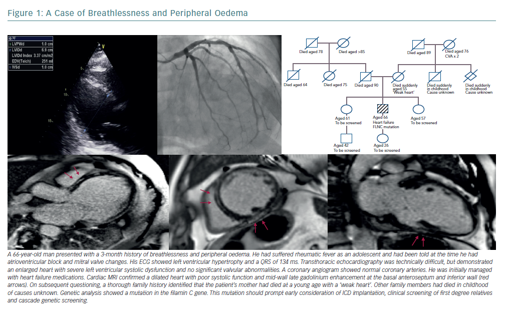
Identification of a definite genetic cause can have profound implications for the patient and their family (Figure 1). Mutations in the gene coding for lamins A and C (LMNA) are well recognised as predictors of a malignant clinical course with progressive conduction disease and arrhythmia, even where left ventricular systolic impairment is mild.29 Patients with an LMNA mutation must be considered for prophylactic ICD implantation at an early stage of disease.30 Filamin C (FLNC) mutations are similarly predictive of a high risk of arrhythmogenic complications. A recent study has found that 85% of DCM patients with truncating FLNC variants had either ventricular arrhythmias or sudden cardiac death.31,32 Pathogenic variants in SCN5A (encoding for the cardiac sodium channel), RMB20 (encoding for the RNA binding motif protein 20) and BAG3 (encoding for an antiapoptotic protein on the sarcomere Z-disc) have also been identified as carrying an increased risk of ventricular arrhythmia or progressive heart failure.33–37

Myocarditis

Regardless of aetiology, myocardial injury often prompts an inflammatory response and cardiac autoantibodies are found in high levels in patients with both ischaemic and non-ischaemic end-stage heart failure.38–41 However, care should be taken to differentiate between cardiac injury leading to inflammation and cardiac dysfunction driven by inflammation. The term inflammatory cardiomyopathy has been used to describe myocarditis that leads to impaired function. Causes of myocarditis are numerous, with viral infection being the most common and parvovirus B19, human herpes virus 6 and enteroviruses the most frequently implicated.42,43 Bacteria, parasites, protozoa and many toxic agents and autoimmune disorders also contribute.44

Histological evidence for myocarditis in patients with DCM and heart failure is frequently reported. In a study of 1,278 patients with congestive cardiac failure and DCM, 24% had an underlying diagnosis confirmed on endomyocardial biopsy that could be attributed to an inflammatory cause.45 Inflammatory disease may be particularly common in patients with idiopathic DCM and ventricular arrhythmia. A study of 103 non-ischaemic cardiomyopathy patients presenting with monomorphic or polymorphic ventricular tachycardia, or with premature ventricular complexes, found that just under 50% had ongoing focal myocardial inflammation on fluorodeoxyglucose PET (FDG-PET). Immunosuppressive therapy was associated with an improvement in ejection fraction, particularly in those identified at an early stage of the disease process. A third of those with a positive scan were subsequently diagnosed with sarcoidosis by tissue biopsy.46

The prevalence of myocarditis remains elusive due to the heterogeneity of aetiologies and modes of presentation. In approximately 50% of cases, acute myocarditis resolves in 2–4 weeks. However, up to 25% develop persistent cardiac dysfunction and 12–25% may acutely deteriorate or progress to end-stage DCM.47 Histological studies suggest that between 10–52% of patients with acute viral myocarditis progress to a DCM and biopsy-based analysis shows that viral persistence is associated with progressive LV dysfunction.48–51 Recent data suggest that more than half of patients presenting with acute lymphocytic myocarditis and an LVEF <50% suffer persistent left ventricular systolic dysfunction.52 Immunohistological analysis of biopsy material from patients with chronic DCM shows that 40% have evidence of myocardial inflammation.53 The subclinical nature of myocardial inflammation is well recognised in children and adults.54–56

That inflammatory cardiomyopathy exists within the heart failure population seems irrefutable. Identifying these patients offers the opportunity to significantly alter the course of the disease. For example, immunosuppression in patients with virus-negative, chronic inflammatory cardiomyopathy can result in a mean improvement in LVEF from 26% to 46%.57 It also improves rates of transplant-free survival.58 Independent of virus-status, DCM patients with increased human leukocyte antigen (HLA) system expression have also demonstrated significant improvements in LVEF with immunosuppression.53

Systemic Autoimmune Disorders

Systemic autoimmune disorders frequently affect the heart and autoimmunity plays a pivotal role in myocarditis and in DCM.59 Systemic lupus erythematosus (SLE) is associated with numerous cardiac disorders and carries a high risk of developing myocarditis, with clinical detection rates in the region of 3–15%.60 African-American people are particularly at risk.61 The inflammatory process is driven by immune complex and complement deposition in the myocardium leading to cardiac dysfunction, DCM and heart failure. SLE may present with myocarditis or with heart failure, which can rapidly progress.62,63 A low threshold of suspicion must be maintained, with awareness that cardiac biomarkers and ECG results may be normal.64 An echocardiogram may demonstrate regional or global ventricular dysfunction, with strain techniques offering earlier diagnosis.65 Cardiac MRI can detect subclinical inflammation in SLE.66,67 Recommended treatment is corticosteroids and immunosuppressive agents such as cyclophosphamide.68 Myocarditis in SLE is associated with lower survival rates.61

Rheumatoid arthritis and systemic sclerosis-associated myocarditis are diagnosed less frequently. However, cardiac MRI studies of these patient populations suggest a high prevalence of myocardial involvement, with half of patients demonstrating evidence of myocardial fibrosis with late gadolinium enhancement.69,70 Cardiac involvement is found in 27–47% of patients with eosinophilic granulomatosis with polyangiitis, previously called Churg-Strauss syndrome, with myocarditis one of the most common cardiac manifestations.71–73 Myocarditis is also seen in Behcet’s disease, Takayasu arteritis, dermatomyositis and polymyositis. It tends to be associated with poorer outcomes and treatment is with steroids and immunosuppression.44,68

Giant Cell Myocarditis

Giant cell myocarditis is a disease that predominantly affects young people. It typically presents with acute heart failure and ventricular arrhythmias. Approximately 20% of patients have a pre-existing autoimmune disorder and the severe inflammation appears to be mediated predominantly by a T-cell response. Early diagnosis is key as median survival without transplant or immunosuppression is less than 3 months.44

Sarcoidosis

Sarcoidosis is an inflammatory, non-caseating granulomatous disease affecting multiple organ systems including the skin, eyes, lungs, heart and the nervous system. The incidence rate in the US has been reported at 10.9 per 100,000 in white people and 35.5 per 100,000 in black people.74 Higher incidence rates are reported in people from Scandinavia and Japan.75 Cardiac manifestations are seen in only 2–5% of patients with systemic sarcoidosis but there is a significantly higher proportion of subclinical cardiac disease.76,77 At autopsy, cardiac involvement was observed in 14% of white people, 21% of black people and 68% of Japanese people with sarcoidosis.78 The aetiology is poorly understood. Environmental exposures and infectious organisms have been implicated and the involvement of genetic factors is supported by twin studies and familial clustering.79

Cardiac sarcoidosis is frequently asymptomatic but may present with conduction defects, arrhythmias or heart failure. Clinical clues include bilateral hilar lymphadenopathy on chest X-ray and elevated levels of serum angiotensin converting enzyme and serum calcium. The ECG may highlight conduction defects or repolarisation abnormalities, but is abnormal in less than 10% of people with clinically silent disease.74 An echocardiogram with longitudinal strain parameters is useful in identifying cardiac involvement and in predicting major cardiovascular events.80,81 Imaging techniques such as FDG-PET and cardiac MRI with gadolinium contrast offer a high degree of diagnostic accuracy, but diagnostic criteria either fail to recognise the benefits of advanced imaging or rely heavily on biopsy results.82–85

Corticosteroids remain the mainstay of treatment for cardiac sarcoidosis. Data is sparse and conflicting, but indicate a beneficial effect on left ventricular function, reduced heart failure admissions and improved long-term clinical outcomes.86,87 Early diagnosis and initiation of therapy is important and titration of immunosuppression can be guided by FDG-PET.87–89

Cardiac Infiltration

Heart failure with preserved ejection fraction remains poorly understood. However, recent evidence suggests that there is a subset of cardiomyopathies caused by cardiac amyloidosis in this patient group. Moreover, better understanding of these conditions is yielding disease-modifying treatments, placing a greater emphasis on early identification of these patients.

The term amyloidosis describes a condition in which structurally unstable proteins misfold and aggregate to form fibrils that are deposited in organs and tissues. The misfolding and deposition of two proteins results in the vast majority of cardiac amyloidosis: immunoglobulin light chain (AL) and transthyretin (ATTR). Transthyretin can be further classified into wild type (ATTRwt) and that due to a mutation in the gene encoding transthyretin (ATTRm).90

Systemic Light-chain Amyloidosis

Systemic AL amyloidosis is a rare disease with a yearly incidence estimated at 3 per million in the UK and 8.9 per million, or 3,000 new cases per year, in the US.91,92 Approximately 30–50% of sufferers have cardiac involvement with 5% having isolated cardiac involvement. The average time from initial symptoms to diagnosis is 2 years, with nearly a third of patients requiring review by five physicians before receiving a diagnosis of amyloidosis.90 Cardiologists were consulted more than haematologists, nephrologists and oncologists, but rarely made the diagnosis.93

Proteinuria or hepatomegaly in combination with heart failure should raise the suspicion of AL amyloidosis.94 Macroglossia and periorbital purpura are highly specific but present only in a minority of cases.90 Patients with significant cardiac involvement are unable to tolerate the preferred treatment of high-dose chemotherapy with autologous stem cell transplantation, emphasising the importance of early diagnosis.95 Mortality at 6 months remains high at 24%, but is improving as patients are diagnosed earlier with less severe cardiac involvement.96

A Case of Worsening Breathlessness

Article image

Transthyretin Amyloidosis

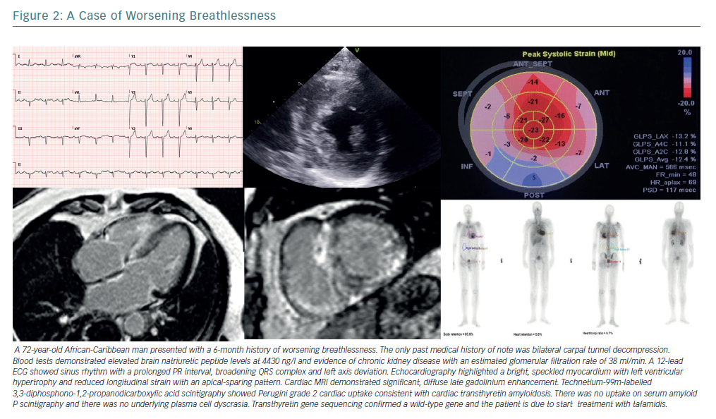
ATTR amyloidosis is a multisystem disorder caused by aggregation of TTR amyloid fibrils.97–100 TTR cardiomyopathy (TTR-CM) is usually a late-onset disease found in older men, with patients typically presenting aged 60 years or older.101 Symptoms include dyspnoea, fatigue and orthostatic hypotension. Signs of heart failure predominate in TTR-CM, often with a preserved ejection fraction. Clinical clues to its presence involve extra-cardiac amyloid deposition including carpal tunnel syndrome and peripheral neuropathy (Figure 2).102

TTR-CM can be hereditary or non-hereditary. The non-hereditary form of the disease is caused by aggregation of ATTRwt. The hereditary form of the disease can be caused by more than 100 mutations in the TTR gene, of which at least 22 are associated predominantly with TTR-CM.103–5 The point mutation valine 122 to isoleucine (Val122Ile) is the most common TTR-CM variant, with a prevalence of about 3–4% in the African-American population. The proportion that develop clinical disease is much smaller.106–108

In patients with ATTRwt-CM, median survival ranges from 26–67 months from diagnosis and 72 months from symptom onset. Patients with Val122Ile ATTRm-CM have a median survival time from diagnosis ranging from 36 months to 43 months. Death in most patients with TTR-CM is from sudden death or progressive heart failure.109,110

Recent data suggest that the prevalence of TTR-CM in older people has been under-appreciated. Autopsy data demonstrate that 25% of adults older than 80 years have TTR amyloid deposition in the myocardium, and 32% of patients aged 75 years or older with HFpEF demonstrate myocardial TTR amyloid deposits.111,112 Technetium-99m-labelled 3,3-diphosphono-1,2-propanodicarboxylic acid (99mTc-DPD) scintigraphy reliably detects TTR-CM. Its use highlights a prevalence of 13% of TTR-CM in patients admitted to hospital with HFpEF.113–116

Effective treatment may soon be available for TTR-CM. TTR is a 55-kD protein with a tetrameric structure that transports thyroxine and vitamin A complex. Amyloidogenesis occurs when the tetrameric structure dissociates resulting in the formation of intermediates that then deposit and polymerise as amyloid fibrils in the myocardium. The discovery of a stabilising polymorphism in the TTR gene has led to the discovery of tafamidis, a nonsteroidal anti-inflammatory drug benzoxazole derivative that adheres to the thyroxine binding sites and inhibits tetramer dissociation. Tafamadis has been shown to be superior to placebo in reducing the risk of death and cardiovascular-related hospitalisations and it reduces the decline in functional capacity and quality of life.117

Disorders of Metabolism

Anderson-Fabry Disease

Anderson-Fabry disease (AFD) is an X-linked recessive disorder caused by mutations in the GLA gene leading to reduced or absent activity of the enzyme alpha-galactosidase A. This deficiency leads to accumulation of glycosphingolipids in lysosomes in various tissues, including the heart. It results in myocardial remodelling and left ventricular hypertrophy and manifests as systolic and diastolic dysfunction, valvular abnormalities and conduction disease.

Presentation of AFD is typically in the first decade, with gastrointestinal symptoms, angiokeratomas or pain caused by a small-fibre neuropathy. However, up to 10% of patients can present with cardiac involvement, highlighting the importance of adopting a low-threshold of suspicion, particularly for patients with left ventricular hypertrophy.118,119 As many as 10% of patients with AFD develop severe heart failure (defined by New York Heart Association classification ≥3) and cardiac disease is the major cause of death, accounting for 38% of all-cause mortality.120,121 Enzyme replacement therapy is the standard treatment for AFD, aiming to compensate for reduced endogenous alpha-galactosidase activity with treatment leading to improved cardiac outcomes. It is readily apparent that enzyme replacement therapy started at a younger age leads to better outcomes illustrating the importance of identifying these patients at an early stage in the disease process.122–125

Flow Diagram Illustrating a Proposed Diagnostic Algorithm

Article image

Iron-overload Cardiomyopathy

Iron-overload cardiomyopathy (IOC) should be considered in any patient with unexplained heart failure. It can be caused by a primary haemochromatosis where iron accumulation occurs through increased gastrointestinal iron absorption, or by secondary iron-overload, most commonly due to a high frequency of blood transfusions.94 The pathophysiology is complex and not simply related to myocardial iron accumulation with immunoinflammatory, molecular and genetic factors implicated.126

The early stages of IOC are characterised by left ventricular diastolic dysfunction with restrictive physiology.127,128 Progression can lead to a dilated phenotype with biventricular dilatation and systolic dysfunction or to a restrictive phenotype with preservation of LVEF, pulmonary hypertension and right ventricular dilatation.129–131 It can also present with acute heart failure.132

Serum ferritin and transferrin saturation should be included in the initial investigation of any patient with newly diagnosed heart failure.11 Echocardiography may help with ventricular assessment and an estimation of pulmonary pressures, however cardiac MRI with T2* is able to quantitatively assess myocardial iron content and predict progression to heart failure within one year.133,134 Identification of patients with an IOC is imperative as both iron removal using phlebotomy and chelation therapy have been shown to improve heart function.135–138 With the introduction of chelation therapies and MRI with T2* there has been a significant improvement in mortality rates, predominantly driven by reductions in death due to cardiac iron overload.139 Early initiation of therapy is therefore crucial.

Improving the Detection of Actionable Diagnoses

The definition of heart failure rests on a constellation of typical symptoms and an underlying structural or functional cardiac abnormality leading to deranged cardiac physiology. This definition captures a patient cohort that, historically, have been managed with a simple treatment algorithm; a ‘one-size-fits-all’ approach. A third of these patients are categorised as suffering an idiopathic DCM. We contend that rare but readily identifiable aetiologies reside within this group and, more importantly, that they are actionable diagnoses leading to targeted therapies.

The diagnosis of rare disease among people with heart failure may be challenging, particularly where resources are constrained and access to specialist cardiac investigations limited. Diagnoses are often made following a conspicuous event such as unexplained syncope or sudden death in a relative. On other occasions, diagnoses are delayed and are reached only with retrospective recognition of atypical features.

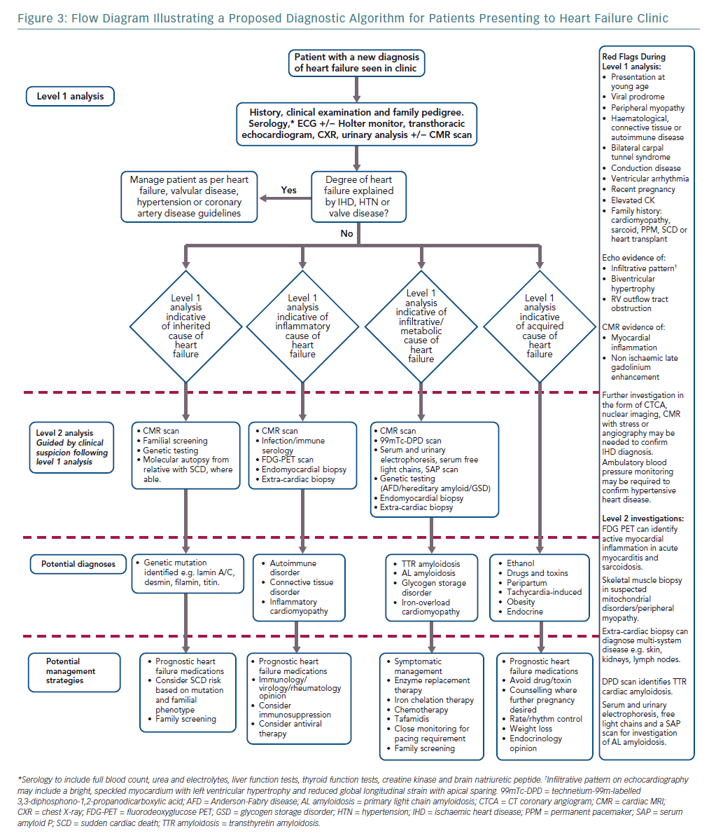
Elucidating the rarer causes of heart failure may need specialised knowledge or access to investigations such as genetic testing. We argue, however, that a diagnostic ‘red flag’ approach to clinical assessment in the heart failure clinic, combined with routinely available tests, can act as an initial filter to more readily identify uncommon disease. This approach begins with a thorough, hypothesis-driven clinical and family history and incorporates basic serology, an ECG and baseline imaging (Figure 3).18 Ischaemic, valvular and hypertensive heart disease should be managed as per guidelines.140–142 Acquired and reversible causes of heart failure such as alcohol excess and prolonged tachycardia are readily identifiable with thorough clinical assessment and routine investigations.11 Cardio-oncology is a growing sub-specialty, with increasing recognition of the cardiotoxic effects of both established chemotherapy agents and newer immunotherapies and targeted therapies. Left ventricular dysfunction and heart failure are relatively common and serious side-effects of cancer treatment.143

After preliminary assessment, patients without an obvious cause for heart failure can be directed to one of three investigative streams: genetic, inflammatory or infiltrative/metabolic.24,144,145 Awareness of dual pathologies is important as, for example, hypertension may coexist with infiltrative disease in a patient with left ventricular hypertrophy.146

Precision Medicine in Heart Failure

NHS England embraces the concept of personalised, precision medicine recognising that “complex diseases should no longer be considered a single entity” and that “different subtypes of patients within a given condition can be identified and treatment can be tailored to the underlying cause”. It urges more precise diagnoses and emphasises the usefulness of genomic medicine.147

The field of oncology offers a paradigm of the precision medicine approach. Where previously cytotoxic treatments with severe side-effects were the norm, we now see therapies designed to precisely target cancer cells. This is chiefly through two methods: pathway-targeted therapy and immunotherapy.148 These approaches seem particularly well-suited to future treatment of inflammatory and infiltrative heart disease.

The one-size-fits-all approach continues to prevail in the modern management of heart failure. This article highlights the diverse pathogeneses of the heart failure syndrome and how it could benefit from a precision medicine approach. The underlying biology of the conditions outlined is increasingly well described, resulting in the development of new therapies that offer significant clinical benefit, particularly when instituted at an early stage in the disease process. Furthermore, these conditions, traditionally thought of as rare, are more common than first thought and, in combination, account for a significant burden of disease.

We offer a diagnostic algorithm that aims to co-opt principles of precision medicine into heart failure clinics. If adopted, we hypothesise that the population prevalence of idiopathic DCM would decline as patients are given aetiology-specific diagnoses with accompanying disease-modifying treatments.

References

  1. Vos T, Alemu Abajobir A, Hassen Abate K, et al. Global, regional, and national incidence, prevalence, and years lived with disability for 328 diseases and injuries for 195 countries, 1990-2016: a systematic analysis for the Global Burden of Disease Study 2016. Lancet 2017: 390:1211–59.
    Crossref | PubMed
  2. Ziaeian B, Fonarow GC. Epidemiology and aetiology of heart failure. Nat Rev Cardiol 2016;13:368–78.
    Crossref | PubMed
  3. Conrad N, Judge A, Tran J, et al. Temporal trends and patterns in heart failure incidence: a population-based study of 4 million individuals. Lancet 2018;391:572–80.
    Crossref | PubMed
  4. Bleumink G, Knetsch A, Sturkenboom M, et al. Quantifying the heart failure epidemic: prevalence, incidence rate, lifetime risk and prognosis of heart failure. The Rotterdam Study. Eur Heart J 2004;25:1614–9.
    Crossref | PubMed
  5. Vos T, Flaxman AD, Naghavi M, et al. Years lived with disability (YLDs) for 1160 sequelae of 289 diseases and injuries 1990-2010: a systematic analysis for the Global Burden of Disease Study 2010. Lancet 2012;380:2163–96.
    Crossref | PubMed
  6. Baldasseroni S, Opasich C, Gorini M, et al. Left bundle-branch block is associated with increased 1-year sudden and total mortality rate in 5517 outpatients with congestive heart failure: a report from the Italian network on congestive heart failure. Am Heart J 2002;143:398–405.
    Crossref | PubMed
  7. Chioncel O, Lainscak M, Seferovic PM, et al. Epidemiology and one-year outcomes in patients with chronic heart failure and preserved, mid-range and reduced ejection fraction: an analysis of the ESC Heart Failure Long-Term Registry. Eur J Heart Fail 2017;19:1574–85.
    Crossref | PubMed
  8. Malamba-Lez D, Ngoy-Nkulu D, Steels P, et al. Heart failure etiologies and challenges to care in the developing world: an observational study in the Democratic Republic of Congo. J Card Fail 2018;854–9.
    Crossref | PubMed
  9. Sliwa K, Damasceno A, Mayosi BM. Epidemiology and etiology of cardiomyopathy in Africa. Circulation 2005;112:3577–83.
    Crossref | PubMed
  10. Dokainish H, Teo K, Zhu J, et al. Global mortality variations in patients with heart failure: results from the International Congestive Heart Failure (INTER-CHF) prospective cohort study. Lancet Glob Heal 2017;5:665–72.
    Crossref | PubMed
  11. Ponikowski P, Voors AA, Anker SD, et al. 2016 ESC Guidelines for the diagnosis and treatment of acute and chronic heart failure. Eur J Heart Fail 2016;18:891–975.
    Crossref | PubMed
  12. Swedberg K, Komajda M, Böhm M, et al. Ivabradine and outcomes in chronic heart failure (SHIFT): a randomised placebo-controlled study. Lancet 2010;376:875–85.
    Crossref | PubMed
  13. McMurray JJV, Packer M, Desai AS, et al. Angiotensin-neprilysin inhibition versus enalapril in heart failure. N Engl J Med 2014;371:993–1004.
    Crossref | PubMed
  14. Blue L, Lang E, McMurray JJ, et al. Randomised controlled trial of specialist nurse intervention in heart failure. BMJ 2001;323:715–8.
    Crossref | PubMed
  15. Taylor CJ, Ordóñez-Mena JM, Roalfe AK, et al. Trends in survival after a diagnosis of heart failure in the United Kingdom 2000-2017: population based cohort study. BMJ 2019;364:l223.
    Crossref | PubMed
  16. Orphanet. About rare diseases. Available at: https://www.orpha.net/consor/cgi-bin/Education_AboutRareDiseases.php?lng=EN (accessed 19 June 2019).
  17. EURORDIS. Rare Diseases Europe. Available at: https://www.eurordis.org (accessed 19 June 2019).
  18. Rapezzi C, Arbustini E, Caforio ALP, et al. Diagnostic work-up in cardiomyopathies: bridging the gap between clinical phenotypes and final diagnosis. A position statement from the ESC Working Group on Myocardial and Pericardial Diseases. Eur Heart J 2013;34:1448–58.
    Crossref | PubMed
  19. Majid Akhtar M, Elliott PM. Rare disease in cardiovascular medicine I. Eur Heart J 2017;38:1625–1628.
    Crossref | PubMed
  20. Majid Akhtar M, Elliott PM. Rare diseases in cardiovascular medicine II. Eur Heart J 2017;38:1629–31.
    Crossref | PubMed
  21. Grünig E, Tasman JA, Kücherer H, et al. Frequency and phenotypes of familial dilated cardiomyopathy. J Am Coll Cardiol 1998;31:186–94.
    Crossref | PubMed
  22. Burkett EL, Hershberger RE. Clinical and genetic issues in familial dilated cardiomyopathy. J Am Coll Cardiol 2005;45:969–81.
    Crossref | PubMed
  23. Seidelmann SB, Laur O, Hwa J, et al. Familial dilated cardiomyopathy diagnosis is commonly overlooked at the time of transplant listing. J Heart Lung Transpl 2016;35:474–80.
    Crossref | PubMed
  24. Ruiter JS, Berkenbosch-Nieuwhof K, van den Berg MP, et al. The importance of the family history in caring for families with long QT syndrome and dilated cardiomyopathy. Am J Med Genet A 2010;152A:607–12.
    Crossref | PubMed
  25. Merlo M, Cannatà A, Gobbo M, et al. Evolving concepts in dilated cardiomyopathy. Eur J Heart Fail 2018;20:228–39.
    Crossref | PubMed
  26. Moretti M, Merlo M, Barbati G, et al. Prognostic impact of familial screening in dilated cardiomyopathy. Eur J Heart Fail 2010;12:922–7.
    Crossref | PubMed
  27. Akinrinade O, Ollila L, Vattulainen S, et al. Genetics and genotype-phenotype correlations in Finnish patients with dilated cardiomyopathy. Eur Heart J 2015;36:2327–37.
    Crossref | PubMed
  28. Dellefave L, McNally EM. The genetics of dilated cardiomyopathy. Curr Opin Cardiol 2010;25:198–204.
    Crossref | PubMed
  29. Captur G, Arbustini E, Bonne G, et al. Lamin and the heart. Heart 2018;104:468–79.
    Crossref | PubMed
  30. Anselme F, Moubarak G, Savouré A, et al. Implantable cardioverter-defibrillators in lamin A/C mutation carriers with cardiac conduction disorders. Heart Rhythm 2013;10:1492–8.
    Crossref | PubMed
  31. Begay RL, Tharp CA, Martin A, et al. FLNC gene splice mutations cause dilated cardiomyopathy. JACC Basic to Transl Sci 2016;1:344–59.
    Crossref | PubMed
  32. Begay RL, Graw SL, Sinagra G, et al. Filamin c truncation mutations are associated with arrhythmogenic dilated cardiomyopathy and changes in the cell-cell adhesion structures. JACC Clin Electrophysiol 2018;4:504–14.
    Crossref | PubMed
  33. Domínguez F, Cuenca S, Bilinska Z, et al. Dilated cardiomyopathy due to BLC2-associated athanogene 3 (BAG3) mutations. J Am Coll Cardiol 2018;72:2471–81.
    Crossref | PubMed
  34. Peters S, Kumar S, Elliott P, et al. Arrhythmic genotypes in familial dilated cardiomyopathy: implications for genetic testing and clinical management. Heart Lung Circ 2019;28:31–8.
    Crossref | PubMed
  35. Hey TM, Rasmussen TB, Madsen T, et al. Pathogenic RBM20-variants are associated with a severe disease expression in male patients with dilated cardiomyopathy. Circ Heart Fail 2019;12 :e005700.
    Crossref | PubMed
  36. van den Hoogenhof MMG, Beqqali A, Amin AS, et al. RBM20 mutations induce an arrhythmogenic dilated cardiomyopathy related to disturbed calcium handling. Circulation 2018;138:1330–42.
    Crossref | PubMed
  37. McNair WP, Sinagra G, Taylor MRG, et al. SCN5A mutations associate with arrhythmic dilated cardiomyopathy and commonly localize to the voltage-sensing mechanism. J Am Coll Cardiol 2011;57:2160–8.
    Crossref | PubMed
  38. Kalogeropoulos AP, Georgiopoulou VV, Butler J. From risk factors to structural heart disease: the role of inflammation. Heart Fail Clin 2012;8:113–23.
    Crossref | PubMed
  39. Frangogiannis NG. The inflammatory response in myocardial injury, repair and remodelling. Nat Rev Cardiol 2014;11:255–65.
    Crossref | PubMed
  40. Smith JG. Molecular epidemiology of heart failure. JACC Basic to Transl Sci 2017;2:757–69.
    Crossref | PubMed
  41. Youker KA, Assad-Kottner C, Cordero-Reyes AM, et al. High proportion of patients with end-stage heart failure regardless of aetiology demonstrates anti-cardiac antibody deposition in failing myocardium: humoral activation, a potential contributor of disease progression. Eur Heart J 2014;35:1061–8.
    Crossref | PubMed
  42. Sagar S, Liu PP, Cooper LT. Myocarditis. Lancet 2012;379:738–47.
    Crossref | PubMed
  43. Sotiriou E, Heiner S, Jansen T, et al. Therapeutic implications of a combined diagnostic workup including endomyocardial biopsy in an all-comer population of patients with heart failure: a retrospective analysis. ESC Heart Fail 2018;5:630–41.
    Crossref | PubMed
  44. Trachtenberg BH, Hare JM. Inflammatory cardiomyopathic syndromes. Circ Res 2017;121:803–18.
    Crossref | PubMed
  45. Felker GM, Hu W, Hare JM, et al. The spectrum of dilated cardiomyopathy. The Johns Hopkins experience with 1,278 patients. Medicine (Baltimore) 1999;78:270–83.
    Crossref | PubMed
  46. Tung R, Bauer B, Schelbert H, et al. Incidence of abnormal positron emission tomography in patients with unexplained cardiomyopathy and ventricular arrhythmias: The potential role of occult inflammation in arrhythmogenesis. Heart Rhythm 2015;12:2488–98.
    Crossref | PubMed
  47. Caforio ALP, Pankuweit S, Arbustini E, et al. Current state of knowledge on aetiology, diagnosis, management, and therapy of myocarditis: a position statement of the European Society of Cardiology Working Group on Myocardial and Pericardial Diseases. Eur Heart J 2013;34:2636–48.
    Crossref | PubMed
  48. Billingham ME, Tazelaar HD. The morphological progression of viral myocarditis. Postgrad Med J 1986;62:581–4.
    Crossref | PubMed
  49. Quigley PJ, Richardson PJ, Meany BT, et al. Long-term follow-up of acute myocarditis. Correlation of ventricular function and outcome. European Heart Journal 1987;8(suppl J):39–42.
    Crossref
  50. D’Ambrosio A, Patti G, Manzoli A, et al. The fate of acute myocarditis between spontaneous improvement and evolution to dilated cardiomyopathy: a review. Heart 2001;85:499–504.
    Crossref | PubMed
  51. Kühl U, Pauschinger M, Seeberg B, et al. Viral persistence in the myocardium is associated with progressive cardiac dysfunction. Circulation 2005;112:1965–70.
    Crossref | PubMed
  52. Merlo M, Ammirati E, Gentile P, et al. Persistent left ventricular dysfunction after acute lymphocytic myocarditis: Frequency and predictors. PLoS One 2019;14:e0214616.
    Crossref | PubMed
  53. Wojnicz R, Nowalany-Kozielska E, Wojciechowska C, et al. Randomized, placebo-controlled study for immunosuppressive treatment of inflammatory dilated cardiomyopathy: two-year follow-up results. Circulation 2001;104:39–45.
    Crossref | PubMed
  54. Talmon G, Fink DL, Horowitz Y, et al. The prevalence of subclinical myocarditis among young children with acute viral infection. Harefuah 2015;154:641–5,676,675 [in Hebrew].
    PubMed
  55. Rady HI, Zekri H. Prevalence of myocarditis in pediatric intensive care unit cases presenting with other system involvement. J Pediatr (Rio J) 2015;91:93–7.
    Crossref | PubMed
  56. Peters NS, Poole-Wilson PA. Myocarditis – continuing clinical and pathologic confusion. Am Heart J 1991;121:942–7.
    Crossref | PubMed
  57. Frustaci A, Russo MA, Chimenti C. Randomized study on the efficacy of immunosuppressive therapy in patients with virus-negative inflammatory cardiomyopathy: the TIMIC study. Eur Heart J 2009;30:1995–2002.
    Crossref | PubMed
  58. Merken J, Hazebroek M, Van Paassen P, et al. Immunosuppressive therapy improves both short- and long-term prognosis in patients with virus-negative nonfulminant inflammatory cardiomyopathy. Circ Hear Fail 2018;11:e004228.
    Crossref | PubMed
  59. Caforio ALP, Vinci A, Iliceto S. Anti-heart autoantibodies in familial dilated cardiomyopathy. Autoimmunity 2008;41:462–9.
    Crossref | PubMed
  60. Tincani A, Rebaioli CB, Taglietti M, et al. Heart involvement in systemic lupus erythematosus, anti-phospholipid syndrome and neonatal lupus. Rheumatology 2006;45(suppl_4):8–13.
    Crossref | PubMed
  61. Apte M, McGwin G, Vilá LM, et al. Associated factors and impact of myocarditis in patients with SLE from LUMINA, a multiethnic US cohort. Rheumatology 2008;47:362–7.
    Crossref | PubMed
  62. Ashrafi R, Garg P, McKay E, et al. Aggressive cardiac involvement in systemic lupus erythematosus: a case report and a comprehensive literature review. Cardiol Res Pract 2011;2011:578390.
    Crossref | PubMed
  63. Berg G, Bodet J, Webb K, et al. Systemic lupus erythematosis presenting as isolated congestive heart failure. J Rheumatol 1985;12:1182–5.
    PubMed
  64. Thomas G, Cohen Aubart F, Chiche L, et al. Lupus myocarditis: initial presentation and longterm outcomes in a multicentric series of 29 patients. J Rheumatol 2017;44:24–32.
    Crossref | PubMed
  65. Nikdoust F, Bolouri E, Tabatabaei SA, et al. Early diagnosis of cardiac involvement in systemic lupus erythematosus via global longitudinal strain (GLS) by speckle tracking echocardiography. J Cardiovasc Thorac Res 2018;10:231–5.
    Crossref | PubMed
  66. Puntmann VO, D’Cruz D, Smith Z, et al. Native myocardial T1 mapping by cardiovascular magnetic resonance imaging in subclinical cardiomyopathy in patients with systemic lupus erythematosus. Circ Cardiovasc Imaging 2013;6:295–301.
    Crossref | PubMed
  67. Mavrogeni S, Koutsogeorgopoulou L, Markousis-Mavrogenis G, et al. Cardiovascular magnetic resonance detects silent heart disease missed by echocardiography in systemic lupus erythematosus. Lupus. 2018;27:564–71.
    Crossref | PubMed
  68. Comarmond C, Cacoub P. Myocarditis in auto-immune or auto-inflammatory diseases. Autoimmun Rev 2017;16:811–6.
    Crossref | PubMed
  69. Ntusi NAB, Piechnik SK, Francis JM, et al. Diffuse myocardial fibrosis and inflammation in rheumatoid arthritis. JACC Cardiovasc Imaging 2015;8:526–36.
    Crossref | PubMed
  70. Ntusi NAB, Piechnik SK, Francis JM, et al. Subclinical myocardial inflammation and diffuse fibrosis are common in systemic sclerosis – a clinical study using myocardial T1-mapping and extracellular volume quantification. J Cardiovasc Magn Reson 2014;16:21.
    Crossref | PubMed
  71. Courand P-Y, Croisille P, Khouatra C, et al. Churg-Strauss syndrome presenting with acute myocarditis and cardiogenic shock. Heart Lung Circ 2012;21:178–81.
    Crossref | PubMed
  72. Qiao L, Gao D. A case report and literature review of Churg-Strauss syndrome presenting with myocarditis. Medicine (Baltimore) 2016;95:e5080.
    Crossref | PubMed
  73. Guillevin L, Pagnoux C, Seror R, et al. The Five-Factor Score revisited: assessment of prognoses of systemic necrotizing vasculitides based on the French Vasculitis Study Group (FVSG) cohort. Medicine (Baltimore) 2011;90:19–27.
    Crossref | PubMed
  74. Muchtar E, Blauwet LA, Gertz MA. Restrictive cardiomyopathy: genetics, pathogenesis, clinical manifestations, diagnosis, and therapy. Circ Res 2017;121:819–37.
    Crossref | PubMed
  75. Bresnitz EA, Strom BL. Epidemiology of sarcoidosis. Epidemiol Rev 1983;5:124–56.
    Crossref | PubMed
  76. Baughman RP, Teirstein AS, Judson MA, et al. Clinical characteristics of patients in a case control study of sarcoidosis. Am J Respir Crit Care Med 2001;164:1885–9.
    Crossref | PubMed
  77. Martusewicz-Boros MM, Boros PW, Wiatr E, et al. Cardiac sarcoidosis: is it more common in men? Lung 2016;194:61–6.
    Crossref | PubMed
  78. Iwai K, Sekiguti M, Hosoda Y, et al. Racial difference in cardiac sarcoidosis incidence observed at autopsy. Sarcoidosis 1994;11:26–31.
    PubMed
  79. Newman LS, Rose CS, Bresnitz EA, et al. A case control etiologic study of sarcoidosis. Am J Respir Crit Care Med 2004;170:1324–30.
    Crossref | PubMed
  80. Murtagh G, Laffin LJ, Patel KV, et al. Improved detection of myocardial damage in sarcoidosis using longitudinal strain in patients with preserved left ventricular ejection fraction. Echocardiography 2016;33:1344–52.
    Crossref | PubMed
  81. Schouver ED, Moceri P, Doyen D, et al. Early detection of cardiac involvement in sarcoidosis with 2-dimensional speckle-tracking echocardiography. Int J Cardiol 2017;227:711–6.
    Crossref | PubMed
  82. Youssef G, Leung E, Mylonas I, et al. The use of 18F-FDG PET in the diagnosis of cardiac sarcoidosis: a systematic review and metaanalysis including the Ontario experience. J Nucl Med 2012;53:241–8.
    Crossref | PubMed
  83. Smedema JP, Snoep G, van Kroonenburgh MPG, et al. Evaluation of the accuracy of gadolinium-enhanced cardiovascular magnetic resonance in the diagnosis of cardiac sarcoidosis. J Am Coll Cardiol 2005;45:1683–90.
    Crossref | PubMed
  84. Hiraga H, Yuwai K, Hiroe M. Diagnostic standard and guidelines for sarcoidosis. Jpn J Sarcoidosis Granulomatous Disord 2007;27:89–102.
  85. Birnie DH, Sauer WH, Bogun F, et al. HRS expert consensus statement on the diagnosis and management of arrhythmias associated with cardiac sarcoidosis. Heart Rhythm 2014;11:1305–23.
    Crossref | PubMed
  86. Nagai T, Nagano N, Sugano Y, et al. Effect of corticosteroid therapy on long-term clinical outcome and left ventricular function in patients with cardiac sarcoidosis. Circ J 2015;79:1593–600.
    Crossref | PubMed
  87. Osborne MT, Hulten EA, Singh A, et al. Reduction in 18F-fluorodeoxyglucose uptake on serial cardiac positron emission tomography is associated with improved left ventricular ejection fraction in patients with cardiac sarcoidosis. J Nucl Cardiol 2014;21:166–74.
    Crossref | PubMed
  88. Kandolin R, Lehtonen J, Airaksinen J, et al. Cardiac sarcoidosis. Circulation 2015;131:624–32.
    Crossref | PubMed
  89. Kusano KF, Satomi K. Diagnosis and treatment of cardiac sarcoidosis. Heart 2016;102:184–90.
    Crossref | PubMed
  90. Maurer MS, Elliott P, Comenzo R, et al. Addressing common questions encountered in the diagnosis and management of cardiac amyloidosis. Circulation 2017;135:1357–77.
    Crossref | PubMed
  91. Pinney JH, Smith CJ, Taube JB, et al. Systemic amyloidosis in England: an epidemiological study. Br J Haematol 2013;161:525–32.
    Crossref | PubMed
  92. Kyle R, Linos A, Beard C, et al. Incidence and natural history of primary systemic amyloidosis in Olmsted County, Minnesota, 1950 through 1989. Blood 1992;79:1817–22.
    Crossref | PubMed
  93. Lousada I, Comenzo RL, Landau H, et al. Light chain amyloidosis: patient experience survey from the Amyloidosis Research Consortium. Adv Ther 2015;32:920–8.
    Crossref | PubMed
  94. Pereira NL, Grogan M, Dec GW. Spectrum of restrictive and infiltrative cardiomyopathies: part 2 of a 2-part series. J Am Coll Cardiol 2018;71:1149–66.
    Crossref | PubMed
  95. Gutiérrez-García G, Cibeira MT, Rovira M, et al. Improving security of autologous hematopoietic stem cell transplant in patients with light-chain amyloidosis. Bone Marrow Transplant 2019; Jan 21 [Epub ahead of print]
    Crossref | PubMed
  96. Muchtar E, Gertz MA, Kumar SK, et al. Improved outcomes for newly diagnosed AL amyloidosis between 2000 and 2014: cracking the glass ceiling of early death. Blood 2017;129:2111–9.
    Crossref | PubMed
  97. Ando Y, Coelho T, Berk JL, et al. Guideline of transthyretin-related hereditary amyloidosis for clinicians. Orphanet J Rare Dis 2013;8:31.
    Crossref | PubMed
  98. Sekijima Y. Transthyretin (ATTR) amyloidosis: clinical spectrum, molecular pathogenesis and disease-modifying treatments. J Neurol Neurosurg Psychiatry 2015;86:1036–43.
    Crossref | PubMed
  99. Wechalekar AD, Gillmore JD, Hawkins PN. Systemic amyloidosis. Lancet 2016;387:2641–54.
    Crossref | PubMed
  100. Ruberg FL, Berk JL. Transthyretin (TTR) cardiac amyloidosis. Circulation 2012;126:1286–300.
    Crossref | PubMed
  101. Dharmarajan K, Maurer MS. Transthyretin cardiac amyloidoses in older North Americans. J Am Geriatr Soc 2012;60:765–74.
    Crossref | PubMed
  102. Gertz MA, Benson MD, Dyck PJ, et al. Diagnosis, prognosis, and therapy of transthyretin amyloidosis. J Am Coll Cardiol 2015:66:2451–66.
    Crossref | PubMed
  103. Connors LH, Lim A, Prokaeva T, et al. Tabulation of human transthyretin (TTR) variants, 2003. Amyloid 2003;10:160–84.
    Crossref | PubMed
  104. Saraiva MJM. Transthyretin mutations in hyperthyroxinemia and amyloid diseases. Hum Mutat 2001;17:493–503.
    Crossref | PubMed
  105. Rowczenio DM, Noor I, Gillmore JD, et al. Online registry for mutations in hereditary amyloidosis including nomenclature recommendations. Hum Mutat 2014;35:E2403–12.
    Crossref | PubMed
  106. Coelho T, Maurer MS, Suhr OB. THAOS – The Transthyretin Amyloidosis Outcomes Survey: initial report on clinical manifestations in patients with hereditary and wild-type transthyretin amyloidosis. Curr Med Res Opin 2013;29:63–76.
    Crossref | PubMed
  107. Jacobson DR, Alexander AA, Tagoe C, et al. Prevalence of the amyloidogenic transthyretin (TTR) V122I allele in 14 333 African-Americans. Amyloid 2015;22:171–4.
    Crossref | PubMed
  108. Connors LH, Prokaeva T, Lim A, et al. Cardiac amyloidosis in African Americans: Comparison of clinical and laboratory features of transthyretin V122I amyloidosis and immunoglobulin light chain amyloidosis. Am Heart J 2009;158:607–14.
    Crossref | PubMed
  109. Ruberg FL, Maurer MS, Judge DP, et al. Prospective evaluation of the morbidity and mortality of wild-type and V122I mutant transthyretin amyloid cardiomyopathy: The Transthyretin Amyloidosis Cardiac Study (TRACS). Am Heart J 2012;164:222–8.
    Crossref | PubMed
  110. Givens RC, Russo C, Green P, et al. Comparison of cardiac amyloidosis due to wild-type and V122I transthyretin in older adults referred to an academic medical center. Aging Health 2013;9:229–35.
    Crossref | PubMed
  111. Tanskanen M, Peuralinna T, Polvikoski T, et al. Senile systemic amyloidosis affects 25% of the very aged and associates with genetic variation in alpha2-macroglobulin and tau: a population-based autopsy study. Ann Med 2008;40:232–9.
    Crossref | PubMed
  112. Mohammed SF, Mirzoyev SA, Edwards WD, et al. Left ventricular amyloid deposition in patients with heart failure and preserved ejection fraction. JACC Heart Fail 2014;2:113–22.
    Crossref | PubMed
  113. Castano A, Haq M, Narotsky DL, et al. Multicenter study of planar technetium 99m pyrophosphate cardiac imaging. JAMA Cardiol 2016;1:880–9.
    Crossref | PubMed
  114. Gillmore JD, Maurer MS, Falk RH, et al. Nonbiopsy diagnosis of cardiac transthyretin amyloidosis clinical perspective. Circulation 2016;133:2404–12.
    Crossref | PubMed
  115. Haq M, Pawar S, Berk JL, et al. Can 99m Tc-pyrophosphate aid in early detection of cardiac involvement in asymptomatic variant TTR amyloidosis? JACC Cardiovasc Imaging 2017;10:713–4.
    Crossref | PubMed
  116. González-López E, Gallego-Delgado M, Guzzo-Merello G, et al. Wild-type transthyretin amyloidosis as a cause of heart failure with preserved ejection fraction. Eur Heart J 2015;36:2585–94.
    Crossref | PubMed
  117. Maurer MS, Schwartz JH, Gundapaneni B, et al. Tafamidis treatment for patients with transthyretin amyloid cardiomyopathy. N Engl J Med 2018;379:1007–16.
    Crossref | PubMed
  118. Nakao S, Takenaka T, Maeda M, et al. An atypical variant of Fabry’s disease in men with left ventricular hypertrophy. N Engl J Med 1995;333:288–93.
    Crossref | PubMed
  119. Mehta A, Ricci R, Widmer U, et al. Fabry disease defined: baseline clinical manifestations of 366 patients in the Fabry Outcome Survey. Eur J Clin Invest 2004;34:236–42.
    Crossref | PubMed
  120. Patel V, O’Mahony C, Hughes D, et al. Clinical and genetic predictors of major cardiac events in patients with Anderson-Fabry Disease. Heart 2015;101:961–6.
    Crossref | PubMed
  121. Mehta A, Clarke JTR, Giugliani R, et al. Natural course of Fabry disease: changing pattern of causes of death in FOS – Fabry Outcome Survey. J Med Genet 2009;46:548–52.
    Crossref | PubMed
  122. Germain DP, Elliott PM, Falissard B, et al. The effect of enzyme replacement therapy on clinical outcomes in male patients with Fabry disease: A systematic literature review by a European panel of experts. Mol Genet Metab Reports 2019;19:100454.
    Crossref | PubMed
  123. Germain DP, Arad M, Burlina A, et al. The effect of enzyme replacement therapy on clinical outcomes in female patients with Fabry disease – a systematic literature review by a European panel of experts. Mol Genet Metab 2018;126:224–35.
    Crossref | PubMed
  124. Warnock DG, Ortiz A, Mauer M, et al. Renal outcomes of agalsidase beta treatment for Fabry disease: role of proteinuria and timing of treatment initiation. Nephrol Dial Transplant 2012;27:1042–9.
    Crossref | PubMed
  125. Weidemann F, Niemann M, Breunig F, et al. Long-term effects of enzyme replacement therapy on Fabry cardiomyopathy. Circulation 2009;119:524–9.
    Crossref | PubMed
  126. Kremastinos DT, Farmakis D. Iron overload cardiomyopathy in clinical practice. Circulation. 2011;124:2253–63.
    Crossref | PubMed
  127. Spirito P, Lupi G, Melevendi C, et al. Restrictive diastolic abnormalities identified by Doppler echocardiography in patients with thalassemia major. Circulation 1990;82:88–94.
    Crossref | PubMed
  128. Mancuso L, Vitrano A, Mancuso A, et al. Left ventricular diastolic dysfunction in β-thalassemia major with heart failure. Hemoglobin 2018;42:68–71.
    Crossref | PubMed
  129. Kremastinos DT, Farmakis D, Aessopos A, et al. β-Thalassemia Cardiomyopathy. Circ Hear Fail 2010;3:451–8.
    Crossref | PubMed
  130. Kremastinos DT, Tsiapras DP, Tsetsos GA, et al. Left ventricular diastolic Doppler characteristics in beta-thalassemia major. Circulation 1993;88:1127–35.
    Crossref | PubMed
  131. Hahalis G, Manolis AS, Apostolopoulos D, et al. Right ventricular cardiomyopathy in β-thalassaemia major. Eur Heart J 2002;23:147–56.
    Crossref | PubMed
  132. Cooray SD, Heerasing NM, Selkrig LA, et al. Reversal of end-stage heart failure in juvenile hemochromatosis with iron chelation therapy: a case report. J Med Case Rep 2018;12:18.
    Crossref | PubMed
  133. Anderson LJ, Holden S, Davis B, et al. Cardiovascular T2-star (T2*) magnetic resonance for the early diagnosis of myocardial iron overload. Eur Heart J 2001;22:2171–9.
    Crossref | PubMed
  134. Kirk P, Roughton M, Porter JB, et al. Cardiac T2* magnetic resonance for prediction of cardiac complications in thalassemia major. Circulation 2009;120:1961–8.
    Crossref | PubMed
  135. Candell-Riera J, Lu L, Serés L, et al. Cardiac hemochromatosis: beneficial effects of iron removal therapy. An echocardiographic study. Am J Cardiol 1983;52:824–9.
    Crossref | PubMed
  136. Rivers J, Garrahy P, Robinson W, et al. Reversible cardiac dysfunction in hemochromatosis. Am Heart J 1987;113:216–7.
    Crossref | PubMed
  137. Anderson LJ, Westwood MA, Holden S, et al. Myocardial iron clearance during reversal of siderotic cardiomyopathy with intravenous desferrioxamine: a prospective study using T2* cardiovascular magnetic resonance. Br J Haematol 2004;127:348–55.
    Crossref | PubMed
  138. Tanner MA, Galanello R, Dessi C, et al. A randomized, placebo-controlled, double-blind trial of the effect of combined therapy with deferoxamine and deferiprone on myocardial iron in thalassemia major using cardiovascular magnetic resonance. Circulation 2007;115:1876–84.
    Crossref | PubMed
  139. Modell B, Khan M, Darlison M, et al. Improved survival of thalassaemia major in the UK and relation to T2* cardiovascular magnetic resonance. J Cardiovasc Magn Reson 2008;10:42.
    Crossref | PubMed
  140. Baumgartner H, Falk V, Bax JJ, et al. 2017 ESC/EACTS Guidelines for the management of valvular heart disease. Eur Heart J 2017;38:2739–91.
    Crossref | PubMed
  141. Task Force Members, Montalescot G, Sechtem U, et al. 2013 ESC guidelines on the management of stable coronary artery disease. Eur Heart J 2013;34:2949–3003.
    Crossref | PubMed
  142. Williams B, Mancia G, Spiering W, et al. 2018 ESC/ESH Guidelines for the management of arterial hypertension. Eur Heart J 2018;39:3021–104.
    Crossref | PubMed
  143. Zamorano JL, Lancellotti P, Rodriguez Muñoz D, et al. 2016 ESC position paper on cancer treatments and cardiovascular toxicity developed under the auspices of the ESC Committee for Practice Guidelines. Eur Heart J 2016;37:2768–801.
    Crossref | PubMed
  144. Milandri A, Longhi S, Gagliardi C, et al. Prevalence, risk factors and correlation with cardiac involvement of carpal tunnel syndrome in amyloidosis. Orphanet J Rare Dis 2015;10:P32.
    Crossref
  145. Nakagawa M, Sekijima Y, Yazaki M, et al. Carpal tunnel syndrome: a common initial symptom of systemic wild-type ATTR (ATTRwt) amyloidosis. Amyloid 2016;23:58–63.
    Crossref | PubMed
  146. Terryn W, Deschoenmakere G, De Keyser J, et al. Prevalence of Fabry disease in a predominantly hypertensive population with left ventricular hypertrophy. Int J Cardiol 2013;167:2555–60.
    Crossref | PubMed
  147. NHS England. Improving outcomes through personalised medicine: working at the cutting edge of science to improve patients’ lives. Leeds: NHS England, 2016. Available at: https://www.england.nhs.uk/wp-content/uploads/2016/09/improving-outcomes-personalised-medicine.pdf (accessed 19 June 2019).
  148. Dugger SA, Platt A, Goldstein DB. Drug development in the era of precision medicine. Nat Rev Drug Discov 2018;17:183–96.
    Crossref | PubMed