Article

Women with Stable Angina Pectoris and No Obstructive Coronary Artery Disease: Closer to a Diagnosis

Register or Login to View PDF Permissions
Permissions× For commercial reprint enquiries please contact Springer Healthcare: ReprintsWarehouse@springernature.com.

For permissions and non-commercial reprint enquiries, please visit Copyright.com to start a request.

For author reprints, please email rob.barclay@radcliffe-group.com.
Average (ratings)
No ratings
Your rating

Abstract

A large proportion of women with chest pain have no obstructive coronary artery disease. Recent studies have demonstrated that these women continue to have symptoms and are at increased risk of cardiovascular morbidity and mortality. Coronary microvascular dysfunction (CMD) leads to an impairment of blood flow regulation to the myocardium and possible transient ischaemia. CMD is a disease entity with several pathophysiologic aspects and diagnostic modalities continue to be developed. However, due to the complexity of the disease, it remains elusive whether CMD is the explanation for the symptoms and the poor prognosis in women with angina and no obstructive coronary artery disease.

Disclosure:The authors have no conflicts of interest to declare.

Received:

Accepted:

Correspondence Details:Marie Mide Michelsen, Department of Cardiology, Building 67, Bispebjerg University Hospital, Bispebjerg Bakke 23, 2400 Copenhagen NV, Denmark. E: marie.m.michelsen@gmail.com

Copyright Statement:

The copyright in this work belongs to Radcliffe Medical Media. Only articles clearly marked with the CC BY-NC logo are published with the Creative Commons by Attribution Licence. The CC BY-NC option was not available for Radcliffe journals before 1 January 2019. Articles marked ‘Open Access’ but not marked ‘CC BY-NC’ are made freely accessible at the time of publication but are subject to standard copyright law regarding reproduction and distribution. Permission is required for reuse of this content.

Women have for many years been regarded as being at relatively low risk for the development of ischaemic heart disease (IHD).1,2 Recently this pattern has changed and cardiovascular disease (CVD) is now the leading cause of death for women in Europe.3,4 Under the age of 60 years acute coronary syndrome occurs 3–4 times more often in men; however, after the age of 75 years women represent the majority of the patients. Although coronary artery disease (CAD) develops on average 10 years later in women, CAD is responsible for approximately 20 % of deaths in both men and women.5,6 In regard to mortality, due to CVD women surpass men at 51 versus 42 %7,8 and over the last decades CVD mortality has declined rapidly for men, while the decline has been significantly lower for women.9,10

Chest Pain in Women

The typical symptom of CAD is oppressive chest pain radiating to the left arm. Nevertheless, the literature often notes that women tend to present with ’atypical’ symptoms: pain occurring at rest, during sleep or atypically located in the neck and shoulder regions, or fatigue, emotional stress, dyspnoea and general weakness.10,11 Studies have shown that women with characteristic symptoms have a higher likelihood of significant CAD.11,12 This suggests that the main difference between men and women is not so much the presentation of symptoms, but the high proportion of women with angina who have no obstructive CAD. In some studies this proportion has reached 70 % or more.13 Overall, treatment of stable angina pectoris is guided by the presence of obstructive CAD, which leaves patients with angina and no obstructive CAD under-diagnosed with few or no treatment options.1

Prognosis for Women with Angina and No Obstructive Coronary Artery Disease

Women with chest pain and no obstructive CAD were thought to have a benign prognosis, but contrary to previous beliefs, more women than expected may have a cardiac origin to their symptoms. Recent studies have found the condition to be associated with increased cardiovascular (CV) morbidity and mortality, and with a prognosis almost as poor as for subjects with obstructive CAD.13–15 In a study of 4,711 women with stable angina pectoris referred for coronary angiography (CAG) and 3,326 asymptomatic women, the hazard ratio for major CV events (MACE) in women with angina and normal coronary arteries and diffuse non-obstructive stenosis was 1.52 and 1.85, respectively, compared with asymptomatic women. The increased risk was comparable to patients with obstructive CAD and primarily due to CV death, heart failure or myocardial infarction.13 Similarly, the Women’s Ischemia Syndrome Evaluation (WISE) trial demonstrated higher prevalence of CVD events in 540 symptomatic women without CAD compared with 1,000 asymptomatic age- and race-matched women.15 Furthermore, the prognosis might be poorer for women compared with men. A large study (n=13,695) investigated sex differences in outcome after CAG in patients with angina and diffuse non-obstructive CAD and found that women were three-times more likely to experience a cardiac event within the first year compared with men.16

Coronary Microvascular Dysfunction

The pathophysiology behind angina with no obstructive CAD remains unknown. Coronary microvascular dysfunction (CMD) has been proposed as a possible explanation for the continued symptoms and higher CV morbidity and mortality. CMD is an impaired coronary microcirculatory function. Whereas epicardial coronary arteries are conduit vessels, the microcirculation consists of resistance vessels that regulate myocardial blood flow by dilation or contraction according to cardiac demand.17 A dysfunction of the resistance vessels reduces the capability to regulate myocardial perfusion leading to a decreased blood flow, stress-induced ischaemia and altered myocardial metabolism.18–20 The regulation of myocardial perfusion is complex and can be controlled via endothelial-dependent pathways by shear stress and endothelial-independent pathways, including regulation by metabolic substances, auto-regulation, myogenic control in response to increased intraluminal pressure and sympathetic nerve responses.21,22

CMD has multiple pathophysiologic pathways. Vascular causes include functional causes such as endothelial and smooth muscle dysfunction as well as structural causes such as microvascular atherosclerosis, intraluminal microembolism and vascular wall hypertrophy creating stiffness. Extravascular causes might also be a part of the pathogenesis, such as inflammation, structural changes such as ventricular hypertrophy causing vascular compression and impairment of the pathways regulating blood flow, i.e. imbalance of autonomic system with an inappropriate high sympathetic tone.23–25 Presumably, CMD occurs due to a synergy of many causes and it is difficult to isolate and identify the sole responsible cause.

Whether CMD explains both symptoms and the increased CVD risk in patients with angina and no obstructive CAD remains elusive due to diagnostic difficulties and lack of evidence-based intervention.

Diagnostic Methods to Assess Coronary Microvascular Dysfunction

A single method may not be sufficient to rule out CMD since each method may assess different aspects of the pathophysiology.26

Vasodilator Stimuli

Myocardial perfusion can be examined by using various vasodilator stimuli. Administration of adenosine, A1 –A3 receptor agonists, leads to coronary microvascular dilatation by acting directly on the vascular smooth muscle cells. Dipyridamole inhibits reuptake of adenosine and has a similar effect.27,28 Like adenosine, the selective A2a receptor agonist, regadenoson, dilates the coronary and peripheral arterial microvessels by acting on the smooth muscle cells.28 These vasodilators assess endothelial-independent coronary microvascular function27 in the absence of epicardial coronary artery stenosis.29 Microvascular function is expressed as a flow reserve calculated as the ratio between flow during stress and rest. Although CMD should be seen as a continuum,30 European guidelines suggest a flow reserve <2 as a strong indicator of CMD,31 but studies have used both higher and lower cut-off values depending on the method of assessment.19,20,32–34

Endothelium-dependent dysfunction and both macrovascular and microvascular spasm can be assessed by an intracoronary acetylcholine provocation test. 35 Vascular response to acetylcholine is predominantly caused by the release of nitric oxide (NO) from endothelial cells causing vasodilation.36 In patients with vascular dysfunction, examination with infusion of acetylcholine shows a diminished vasodilation, a paradoxical vasoconstriction or in larger doses coronary artery spasms may be elicited. This reaction has been suggested as part of the definition of vasospastic angina.37 The cold pressor test, the submersion of the patient’s hand in ice water, is also used to assess endothelial-dependent function. This procedure induces sympathetic stimulation, thereby increased heart rate, blood pressure and blood flow. The proposed mechanism is through adrenergic receptors releasing NO.38 However, for both acetylcholine and cold pressor tests, a direct effect on smooth muscle cells cannot be excluded and the response is not solely endothelial dependent.39

Invasive Assessment by Angiography

Epicardial blood flow can be measured by intracoronary Doppler flow where the tip of the guidewire has ultrasound capabilities measuring resting and peak velocities at maximal hyperaemia. To investigate resistance changes in the distribution of the epicardial coronary arteries, coronary vascular resistance can be calculated as mean arterial pressure divided by coronary blood flow.40 Additionally, quantitative vasomotor response in epicardial coronary arteries can be evaluated as a change in vessel diameter. Measurements are usually repeated using increasing concentrations of acetylcholine. Endothelial dysfunction is defined as vasoconstriction, lack of coronary blood flow increase and/or vasospasm as a result of direct activation of muscarinic receptors on vascular smooth muscle cells.37,41

Endothelial-independent function can also be assessed during vasodilator infusion with adenosine, dipyridamole or regadenoson with calculation of the coronary flow velocity reserve (CFVR).29 Flow velocity changes in the coronary arteries are assumed to be representative of changes in flow volume due to dilation of the smaller resistance vessels, based on the assumption that coronary artery diameters remain constant during hyperaemia. However, dilation of the epicardial coronary arteries during hyperaemia may occur. Therefore, intracoronary NO to achieve maximum dilation of the epicardial coronary arteries before CFVR measurement can be used as well as quantification of the cross-sectional area of the measuring point in the coronary artery calculating volumetric blood flow.42 Invasive CFVR measurement has been extensively used and validated and is at present considered the gold standard for assessment of endothelial-independent CMD.

Corrected thrombolysis in myocardial infarction (TIMI) frame counts can also be used as an invasive measure of CMD. The ratio between number of angiographic frames for the contrast to reach a distal landmark at maximal hyperaemia and rest reflects coronary microvascular function and has shown good correlation with invasive CFVR.29,43,44 CMD may also be assessed through thermodilution, although this method is less widely used.45

Positron Emission Tomography

Positron emission tomography (PET) can assess absolute myocardial blood flow (ml/min/g) by a dynamic imaging protocol calculating the rate of tracer uptake into the myocardium at rest and during stress.46 Rubidium (Rb)-82, oxygen (O)-water-15 and nitrogen (N)-13-ammonia are the most widely used tracers. Focusing on endothelial-dependent function, cold pressor testing can be used to induce stress by sympathetic stimulation calculating the absolute flow difference between rest and stress.34,47 The use of endothelial-independent vasodilators allows for calculation of the myocardial blood flow reserve (MBFR) for the assessment of coronary microvascular function.34,48 The few studies investigating the repeatability of MBFR have not been able to produce convincing results.49–51 However, one study found PET MBFR valid against invasively measured CFVR calculated by the volumetric blood flow method, where the cross-sectional area of the coronary artery was quantified to calculate blood flow volume.42

Echocardiographic Assessment

Transthoracic Doppler echocardiography (TTDE) can visualise the left anterior descending artery and peak coronary flow velocities at rest and maximum hyperaemia during vasodilator infusion with adenosine, dipyridamole or regadenoson can be measured by pulsed wave Doppler technique. The resulting CFVR reflects coronary microvascular function but may be affected by the possible dilation of the epicardial coronary arteries. The method can be technically challenging, but is highly reproducible in experienced hands with good agreement with invasively measured CFVR.52–58 However, agreement with PET measured MBFR is only modest in both women with no obstructive CAD (n=107) and patients with CAD (n=35).26,57 Advantages of the TTDE assessment is that it is easily accessible, non-invasive and radiation-free.59 Another method to assess CMD is myocardial perfusion echocardiography.60

Cardiac Magnetic Resonance

The microvasculature can be assessed by cardiac magnetic resonance (CMR) perfusion with images acquired immediately after the injection of gadolinium contrast.61 Contrast signal intensity gives an estimate of myocardial perfusion. The myocardial perfusion reserve index is the ratio of contrast signal intensity/upslope during pharmacologically induced hyperaemia and at rest. Use of CMR perfusion imaging in diagnosing CMD has not yet been standardised but having in mind that perfusion defects in CAD are visible even before angina, it is likely that women with CMD in the absence of obstructive CAD could have inducible perfusion abnormalities detectable by CMR. The reproducibility of perfusion CMR has been assessed in healthy volunteers with a coefficient of variation of 13.7 % for resting values and 12.9 % for stress values. Furthermore, inter- and intra-observer agreement was good.62 In patients with obstructive CAD, perfusion CMR correlated significantly with CFVR by Doppler flow guidewire.63

Computed Tomography Angiography

Computer tomography (CT) perfusion is a rapidly developing noninvasive diagnostic test evaluating the haemodynamic significance of coronary artery stenosis.64 Studies are currently establishing whether semi-quantitative measures, including the myocardial perfusion reserve (hyperaemic versus resting myocardial blood flow) and transmural perfusion ratio (subendocardial to subepicardial attenuation density) can identify patients with CMD.

The Coronary Microvascular Dysfunction Diagnosis

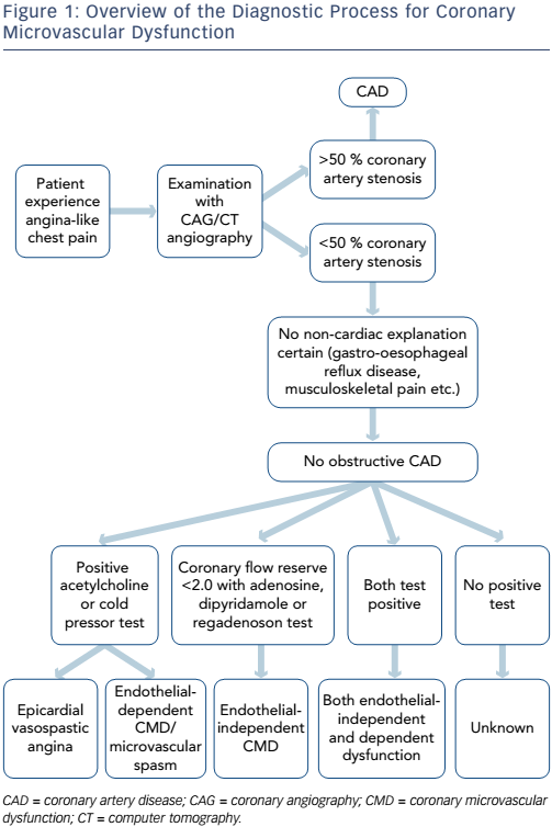
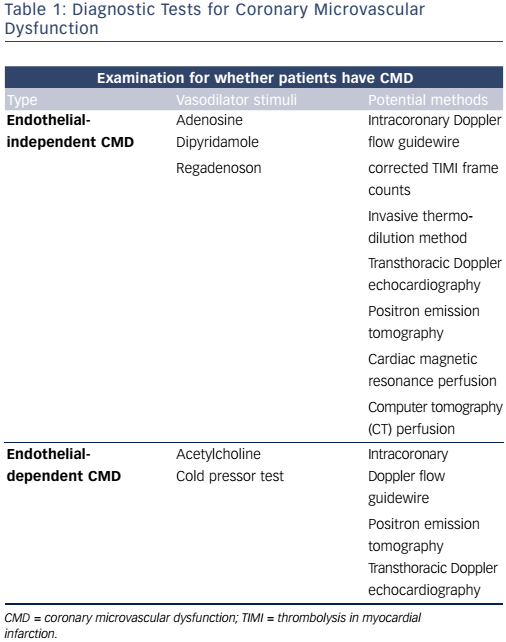
In summary, patients with no obstructive CAD can be diagnosed with either endothelial-dependent and/or endothelial-independent CMD depending on the choice of vasodilator using several methods (see Figure 1 and Table 1). However, there are multiple methods assessing coronary microvascular function and some seem to diverge and may be assessing different aspects of the complex pathophysiological mechanisms of both the endothelial independent and dependent part of CMD.26

Is Coronary Microvascular Dysfunction the Cause of Angina in Women with No Obstructive Coronary Artery Disease?

CMD is highly prevalent in a population of women and men with angina and no obstructive CAD. In a cross-sectional study of 159 women with no obstructive CAD examined invasively with CFVR, 47 % had CMD (CFVR <2.5).65 Another cross-sectional study of 1,439 patients with angina and no obstructive CAD found that 1,171 (81 %) had CMD with angiographic testing and infusion of both acetylcholine and adenosine: 19 % had both endothelial dependent and independent CMD; 12 % only endothelial-independent CMD and 33 % only endothelial-dependent CMD.66 In prospective and cross-sectional TTDE studies, CMD (CFVR <2.0) prevalence has been reported lower ranging from 22 to 40 %.33,67,68 In a prospective study of 1,218 patients assessed by PET more than 50 % of women were diagnosed as having endothelial-independent CMD (MBFR <2.0).69 Focusing on endothelial-dependent function, a prospective study of 470 patients with angina and no obstructive CAD found CMD defined as ≤50 % increase in myocardial blood flow to acetylcholine measured angiographically to be present in 56 %.70 Similarly 95 out of 147 patients (66 %) had CMD in a prospective study using >20 % diameter reduction in response to acetylcholine measured angiographically as criterion for CMD.71

Figure 1: Overview of the Diagnostic Process for Coronary Microvascular Dysfunction

Article image

Table 1: Diagnostic Tests for Coronary Microvascular Dysfunction

Article image

Classification of angina symptoms, frequency, stability and treatment satisfaction were not associated with CMD assessed by TTDE CFVR in a cohort study of 919 women with angina. Burden of symptoms in terms of physical limitation and self-perception of disease was related to CMD, although differences between CFVR level were small.68 In accordance, PET MBFR (study n=1,218) was not different for patients with typical or atypical angina, compared with symptoms classified as non-cardiac or no symptoms.69 The WISE study compared CMR perfusion in women with angina and no obstructive CAD (n=53) to healthy controls (n=12) and found that the symptomatic women had a significantly lower myocardial perfusion reserve index but the burden of risk factors was also lower in the controls.72 Furthermore, trials (n=51 and n=46) have failed to show that improvement of symptoms are related to changes in coronary microvascular function.73,74 This questions whether CMD and the burden of symptoms are actually related; although symptoms are generally hard to study being indeed subjective.

Cardiovascular Risk Factors in Women with No Obstructive Coronary Artery Disease

A proportion of patients with angina and no obstructive CAD have IHD caused by insignificant atherosclerosis, vasospastic angina or CMD. Prevalence of CV risk factors in women with angina and no obstructive CAD is relatively high compared with the general population of women at similar ages and comparable to patients with coronary heart disease13,65,68,75 and independently predicts cardiovascular mortality.76,77 In recent large studies from the US and Denmark, approximately 50 % of women with angina and no obstructive CAD had hypertension, hyperlipidaemia and a family history of CAD, one-fifth had diabetes mellitus and generally these women had a high body mass index (BMI). However, active smoking varied from 16 %, 20 % and 30 % probably due to a general decline in smoking in the recent years.13,65,68 In comparison, around 35 % women in a Danish general population had hypertension, 8 % had diabetes and 20 % were actively smoking in the year 2013.75 CMD has been found to be associated with CV risk factors such as age,32,33,65,67–69,78,79 hypertension,33,68,69,79,80 diabetes mellitus,68,69,79,80 smoking,68,78 heart rate,68,81 HDL cholesterol,68,78 BMI,69,81 menopause and absence of hormone therapy post-menopause.65 However, not all studies identify the same CV risk factors and the contribution of risk factors explaining variation in CFR assessing CMD is low.66,68,82

The fact that prevalence of CV risk factors is high in women with angina and no obstructive CAD may play a role in the increased risk of CV events and possible CMD in this population.13,65,68,76

Signs of Myocardial Affection/Ischaemia

CMD is thought to be global or to involve random patchy areas of hypoxia, which might lead to transient myocardial ischaemia.83

Cardiac Exercise Stress Testing

Even in the absence of CAD, a large percentage of patients with persistent chest pain present with transient ischaemia on electrocardiogram due to regional myocardial hypoperfusion.18 However, ST-segment depression during an exercise test is thought to be less accurate in identifying CAD in women compared with men.84 Presence of impaired CFVR and endothelial dysfunction consistent with CMD is a plausible explanation for perfusion abnormalities sufficient to produce electrocardiogram changes and angina during stress testing. However, two large studies found no association between presence of CMD and results from diagnostic stress testing prior to CAG in women with angina and no obstructive CAD.66,68 The explanation may lie in the size and the possible patchy distribution of the affected, ischaemic myocardium, undetectable by present techniques in cardiac stress testing. Decreased exercise tolerance, vasospastic angina and oestrogen levels have also been suggested as causes for ST-depression in women with no obstructive CAD.84,85

Markers of Inflammation

Chronic inflammation, expressed as elevated C-reactive protein levels, has been found associated with endothelial dysfunction and CMD in patients with no obstructive CAD.86,87 Patients with angina and a normal CAG were in one study (n=137) found to have elevated C-reactive protein levels and reduced PET measured MBFR compared with controls.88

Prognosis for Patients with Coronary Microvascular Dysfunction

Epicardial and microvascular coronary endothelial dysfunction assessed invasively by acetylcholine predict long-term risk of CV events in patients with no obstructive CAD (study n between 147 and 470), independently of traditional risk factors.70,71,89,90 In a study of72, cold pressor testing in PET imaging can be used to predict CV outcome.47

Endothelial-independent CMD assessed both invasively (n=152) by PET (n=1,218) and by Doppler echocardiography (n=394) has been shown to be strongly associated with adverse CV outcome in several prospective studies of patients with no obstructive CAD.32,33,69

Large prognostic studies of perfusion CMR in patients without obstructive CAD are lacking, but in one study of 103 patients with angina and normal stress perfusion CMR no patient with a normal CMR experienced events, whereas 16 events occurred in 14 patients with abnormal CMR during follow-up.91 In the WISE study, global perfusion abnormalities reflecting CMD could also predict adverse events.92

Continued Symptoms

Patients with angina pectoris and no obstructive CAD are costly for society due to continued symptoms resulting in multiple hospital re-admissions and re-assessments for obstructive CAD.93 Angina is persistent in 45–61 % of patients after 1–3 years.94–97 Persistent angina has been reported to be associated with non-fatal myocardial infarction and stroke.95 Frequent angina was furthermore found to be associated with lower physical functioning, angina stability, treatment satisfaction and quality of life.94 Patients with angina and no obstructive CAD have increased risk of hospitalisation primarily due to stable and unstable angina, repeat CAG, heart failure and stroke compared with healthy controls. Furthermore, higher rates of followup consultations at the general practitioners have been reported.93,98

Besides high healthcare costs, patients with angina and no obstructive CAD often exit the workforce prematurely requiring disability pension. Surprisingly, costs are comparable to patients with obstructive CAD.97,99 Cumulative costs over a five-year period has, in an American study, been estimated to be lower, though still very high, for women with no obstructive CAD (US$32,239; 95 % confidence interval [CI], US$29,496–US$34,982) than for patients with obstructive CAD (increasing to US$53,398; 95 % CI, US$43,854–US$62,942 for patients with three-vessel disease).93

Treatment Strategies

Currently, there are no evidence-based treatment strategies for women with no obstructive CAD. However, guideline-based management of the relative high burden of risk factors in all patients with IHD, including women with no obstructive CAD, is important. Further approaches are lifestyle modification (smoking cessation, exercise and a healthy diet).100 Moreover, large clinical trials are lacking for those diagnosed with any aspect of CMD. Small clinical trials indicate that anti-ischaemic and antianginal medication (such as treatment with angiotensin-converting enzyme inhibitor, statins and NO modulators) could have a role in improving CMD. However, the trials conducted have been of moderate or poor quality and/or with too few subjects (n from 6 to 13) to have an impact on guidelines.24

Closer to a Diagnosis?

Women with angina pectoris and no obstructive CAD have continued symptoms and a poor CV prognosis. CMD is an entity comprising several pathologic pathways. New invasive and non-invasive diagnostic modalities for identifying CMD are being developed and accumulating research points to CMD as a cause of poor prognosis in women with angina and no obstructive CAD. Whether CMD directly causes angina cannot be decisively concluded at present, for this we must await intervention studies showing simultaneous improvement in CMD and angina symptoms. Furthermore, evidence-based treatment strategies for women with no obstructive CAD are lacking.

References

  1. Task Force Members, Montalescot G, Sechtem U, Achenbach S, et al. 2013 ESC guidelines on the management of stable coronary artery disease: the Task Force on the management of stable coronary artery disease of the European Society of Cardiology. Eur Heart J 2013;34:2949–3003.
    Crossref | PubMed
  2. Wilson PW, D’Agostino RB, Levy D, et al. C/W Reading Prediction of Coronary Heart Disease Using Risk Factor Categories. Circulation 1998;97:1837–47.
    Crossref | PubMed
  3. Mosca L, Hammond G, Mochari-Greenberger H, et al. Fifteen-year trends in awareness of heart disease in women: results of a 2012 American Heart Association national survey. Circulation 2013;127(11):1254–63, e1-29. PMCID: PMC3684065
    Crossref | PubMed
  4. Maas AHEM, van der Schouw YT, Regitz-Zagrosek V, et al. Red alert for women’s heart: the urgent need for more research and knowledge on cardiovascular disease in women: proceedings of the workshop held in Brussels on gender differences in cardiovascular disease, 29 September 2010. Eur Heart J 2011;32(11):1362–8.
    Crossref | PubMed
  5. Safdar B, Nagurney JT, Anise A, et al. Gender-specific research for emergency diagnosis and management of ischemic heart disease: proceedings from the 2014 Academic Emergency Medicine Consensus Conference Cardiovascular Research Workgroup. Acad Emerg Med 2014;21 (12):1350–60.
    Crossref | PubMed
  6. Anand SS, Islam S, Rosengren A, et al. Risk factors for myocardial infarction in women and men: Insights from the INTERHEART study. Eur Heart J 2008;29(7):932–40.
    Crossref | PubMed
  7. Townsend NM, Luengo-Fernandez R, Leal J, et al. European cardiovascular disease statistics 2012. European Heart Network, Brussels, European Society of Cardiology, Sophia Antipolis: 2012. Available at: www.escardio.org/ static_file/Escardio/Press-media/press-releases/2013/ EU-cardiovascular-disease-statistics-2012.pdf (Accessed 13 January 2017).
  8. Nichols M, Townsend N, Scarborough P, Rayner M. Cardiovascular disease in Europe 2014: epidemiological update. Eur Heart J 2014;35(42):2950–9.
    Crossref | PubMed
  9. Ford ES, Capewell S. Coronary heart disease mortality among young adults in the U.S. from 1980 through 2002: concealed leveling of mortality rates. J Am Coll Cardiol 2007;50(22):2128–32.
    Crossref | PubMed
  10. Park SM, Merz CN. Women and Ischemic Heart Disease: Recognition, Diagnosis and Management. Korean Circ J 2016;46(4):433–42. PMCID: PMC4965421
    Crossref | PubMed
  11. Douglas PS, Ginsburg GS. The evaluation of chest pain in women. N Engl J Med 1996;334(20):1311–5.
    Crossref | PubMed
  12. Sharaf BL, Pepine CJ, Kerensky RA, et al. Detailed angiographic analysis of women with suspected ischemic chest pain (pilot phase data from the NHLBI-sponsored Women’s Ischemia Syndrome Evaluation [WISE] Study Angiographic Core Laboratory). Am J Cardiol 2001;87(8): 937–41, A3.
    Crossref | PubMed
  13. Jespersen L, Hvelplund A, Abildstrøm SZ, et al. Stable angina pectoris with no obstructive coronary artery disease is associated with increased risks of major adverse cardiovascular events. Eur Heart J 2012;33(6):734–44.
    Crossref | PubMed
  14. Sharaf B, Wood T, Shaw L, et al. Adverse outcomes among women presenting with signs and symptoms of ischemia and no obstructive coronary artery disease: findings from the National Heart, Lung, and Blood Institute-sponsored Women’s Ischemia Syndrome Evaluation (WISE) angiographic core laboratory. Am Heart J 2013;166(1):134–41. PMCID: PMC3703586
    Crossref | PubMed
  15. Gulati M, Cooper-DeHoff RM, McClure C, et al. Adverse cardiovascular outcomes in women with nonobstructive coronary artery disease: a report from the Women’s Ischemia Syndrome Evaluation Study and the St James Women Take Heart Project. Arch Intern Med 2009;169(9): 843–50. PMCID:PMC2782882
    Crossref | PubMed
  16. Sedlak TL, Lee M, Izadnegahdar M, et al. Sex differences in clinical outcomes in patients with stable angina and no obstructive coronary artery disease. Am Heart J 2013;166(1):38–44.
    Crossref | PubMed
  17. Panza JA. Coronary atherosclerosis: extending to the microcirculation? Eur Heart J 2010;31 (8):905–7.
    Crossref | PubMed
  18. Crea F, Camici PG, Bairey Merz CN. Coronary microvascular dysfunction: an update. Eur Heart J 2014;35(17):1101–11.PMCID:PMC4006091
    Crossref | PubMed
  19. Graf S, Khorsand A, Gwechenberger M, et al. Typical chest pain and normal coronary angiogram: cardiac risk factor analysis versus PET for detection of microvascular disease. J Nucl Med 2007;48(2):175–81.
    PubMed
  20. Reis SE, Holubkov R, Lee JS, et al. Coronary flow velocity response to adenosine characterizes coronary microvascular function in women with chest pain and no obstructive coronary disease. Results from the pilot phase of the Women’s Ischemia Syndrome Evaluation (WISE) study. J Am Coll Cardiol 1999;33(6):1469–75.
    Crossref | PubMed
  21. Marcus ML, Chilian WM, Kanatsuka H, et al. Understanding the coronary circulation through studies at the microvascular level. Circulation 1990;82(1):1–7.
    Crossref | PubMed
  22. Muller JM, Davis MJ, Chilian WM. Integrated regulation of pressure and flow in the coronary microcirculation. Cardiovasc Res 1996;32(4):668–78.
    Crossref | PubMed
  23. Mosseri M, Yarom R, Gotsman MS, Hasin Y. Histologic evidence for small-vessel coronary artery disease in patients with angina pectoris and patent large coronary arteries. Circulation 1986;74(5):964–72.
    Crossref | PubMed
  24. Marinescu MA, Löffler AI, Ouellette M, et al. Coronary microvascular dysfunction, microvascular angina, and treatment strategies. JACC Cardiovasc Imaging 2015;8(2): 210–20.PMCID:PMC4384521
    Crossref | PubMed
  25. Beltrame JF, Crea F, Camici P. Advances in coronary microvascular dysfunction. Heart Lung Circ 2009;18(1):19–27.
    Crossref | PubMed
  26. Michelsen MM, Mygind ND, Pena A, et al. Transthoracic Doppler echocardiography compared with positron emission tomography for assessment of coronary microvascular dysfunction: The iPOWER study. Int J Cardiol 2017;228: 435–43.
    Crossref | PubMed
  27. Youn HJ, Foster E. Demonstration of coronary artery flow using transthoracic Doppler echocardiography. J Am Soc Echocardiogr 2004;17(2):178–85.
    Crossref | PubMed
  28. Al Jaroudi W, Iskandrian AE. Regadenoson: a new myocardial stress agent. J Am Coll Cardiol 2009;54:1123–30.
    Crossref | PubMed
  29. Layland J, Nerlekar N, Palmer S, et al. Invasive assessment of the coronary microcirculation in the catheter laboratory. Int J Cardiol 2015;199:141–9.
    Crossref | PubMed
  30. Murthy VL, Naya M, Foster CR, et al. Improved cardiac risk assessment with noninvasive measures of coronary flow reserve. Circulation 2011;124(20):2215–24.PMCID:PMC3495106
    Crossref | PubMed
  31. Montalescot G, Sechtem U, Achenbach S, et al. 2013 ESC guidelines on the management of stable coronary artery disease—addenda. Available at: www.escardio.org/static_file/ Escardio/Guidelines/publications/ANGINA2013_Stable_ Coronary_Artery_Disease_web_addenda.pdf (Accessed 23 October 2015).
  32. Pepine CJ, Anderson RD, Sharaf BL, et al. Coronary microvascular reactivity to adenosine predicts adverse outcome in women evaluated for suspected ischemia results from the National Heart, Lung and Blood Institute WISE (Women’s Ischemia Syndrome Evaluation) study. J Am Coll Cardiol 2010;55(25):2825–32.PMCID:PMC2898523
    Crossref | PubMed
  33. Sicari R, Rigo F, Cortigiani L, et al. Additive prognostic value of coronary flow reserve in patients with chest pain syndrome and normal or near-normal coronary arteries. Am J Cardiol 2009;103(5):626–31.
    Crossref | PubMed
  34. Schindler TH, Schelbert HR, Quercioli A, Dilsizian V. Cardiac PET imaging for the detection and monitoring of coronary artery disease and microvascular health. JACC Cardiovasc Imaging 2010;3:623–40.
    Crossref | PubMed
  35. Sun H, Mohri M, Shimokawa H, et al. Coronary microvascular spasm causes myocardial ischemia in patients with vasospastic angina. J Am Coll Cardiol 2002;39(5):847–51.
    Crossref | PubMed
  36. Quyyumi AA, Dakak N, Mulcahy D, et al. Nitric Oxide Activity in the Atherosclerotic Human Coronary Circulation. J Am Coll Cardiol 1997;29(2):308–17.
    Crossref | PubMed
  37. Beltrame JF, Crea F, Kaski JC, et al. International standardization of diagnostic criteria for vasospastic angina. Eur Heart J 2015;epub ahead of press.
    Crossref | PubMed
  38. Hwang HJ, Chung WB, Park JH, et al. Estimation of coronary flow velocity reserve using transthoracic Doppler echocardiography and cold pressor test might be useful for detecting of patients with variant angina. Echocardiography 2010;27(4):435–41.
    Crossref | PubMed
  39. Lanza GA. Angina Pectoris and Myocardial Ischemia in the Absence of Obstructive Coronary Artery Disease: Role of Diagnostic Tests. Curr Cardiol Rep 2016;18(2):15.
    Crossref | PubMed
  40. Bellamy R. Calculation of coronary vascular resistance. Cardiovasc Res 1980;14(5):261–9.
    Crossref | PubMed
  41. Matsuzawa Y, Lerman A. Endothelial dysfunction and coronary artery disease: assessment, prognosis, and treatment. Coron Artery Dis 2014;25(8):713–24. PMCID:PMC4220301
    Crossref | PubMed
  42. Kaufmann PA, Namdar M, Matthew F, et al. Novel doppler assessment of intracoronary volumetric flow reserve: validation against PET in patients with or without flowdependent vasodilation. J Nucl Med 2005;46(8):1272–7. PMID:
    PubMed
  43. Manginas A, Gatzov P, Chasikidis C, et al. Estimation of coronary flow reserve using the Thrombolysis In Myocardial Infarction (TIMI) frame count method. Am J Cardiol 1999;83(11):1562–5, A7.
    Crossref | PubMed
  44. Stoel MG, Zijlstra F, Visser CA. Frame count reserve. Circulation 2003;107(24):3034–9.
    PubMed
  45. Fearon WF, Balsam LB, Farouque HM, et al. Novel index for invasively assessing the coronary microcirculation. Circulation 2003;107(25):3129–32.
    Crossref | PubMed
  46. Nakazato R, Berman DS, Alexanderson E, Slomka P. Myocardial perfusion imaging with PET. Imaging Med 2013;5(1):35–46. PMCID: PMC3650901
    Crossref | PubMed
  47. Schindler TH, Nitzsche EU, Schelbert HR, et al. Positron emission tomography-measured abnormal responses of myocardial blood flow to sympathetic stimulation are associated with the risk of developing cardiovascular events. J Am Coll Cardiol 2005;45(9):1505–12.
    Crossref | PubMed
  48. Camici PG, Crea F. Coronary Microvascular Dysfunction. N Engl J Med 2007;356(8):830–40.
    Crossref | PubMed
  49. Manabe O, Yoshinaga K, Katoh C, et al. Repeatability of rest and hyperemic myocardial blood flow measurements with 82Rb dynamic PET. J Nucl Med 2009;50(1):68–71.
    Crossref | PubMed
  50. Prior JO, Allenbach G, Valenta I, et al. Quantification of myocardial blood flow with 82Rb positron emission tomography: clinical validation with 15O-water. Eur J Nucl Med Mol Imaging 2012;39(6):1037–47.PMCID:PMC3342496
    Crossref | PubMed
  51. Sdringola S, Johnson NP, Kirkeeide RL, et al. Impact of unexpected factors on quantitative myocardial perfusion and coronary flow reserve in young, asymptomatic volunteers. JACC Cardiovasc Imaging 2011;4(4):402–12.
    Crossref | PubMed
  52. Lethen H, Tries HP, Brechtken J, et al. Comparison of transthoracic Doppler echocardiography to intracoronary Doppler guidewire measurements for assessment of coronary flow reserve in the left anterior descending artery for detection of restenosis after coronary angioplasty. Am J Cardiol 2003;91 (4):412–7.
    Crossref | PubMed
  53. Caiati C, Montaldo C, Zedda N, et al. Validation of a new noninvasive method (contrast-enhanced transthoracic second harmonic echo Doppler) for the evaluation of coronary flow reserve: comparison with intracoronary Doppler flow wire. J Am Coll Cardiol 1999;34(4):1193–200.
    Crossref | PubMed
  54. Hozumi T, Yoshida K, Akasaka T, et al. Noninvasive assessment of coronary flow velocity and coronary flow velocity reserve in the left anterior descending coronary artery by Doppler echocardiography: comparison with invasive technique. J Am Coll Cardiol 1998;32(5):1251–9.
    Crossref | PubMed
  55. Hildick-Smith DJR, Maryan R, Shapiro LM. Assessment of coronary flow reserve by adenosine transthoracic echocardiography: validation with intracoronary Doppler. J Am Soc Echocardiogr 2002;15(9):984–90.
    Crossref | PubMed
  56. Saraste M, Koskenvuo J, Knuuti J, et al. Coronary flow reserve: measurement with transthoracic Doppler echocardiography is reproducible and comparable with positron emission tomography. Clin Physiol 2001;21 (1):114–22.
    Crossref | PubMed
  57. Olsen RH, Pedersen LR, Snoer M, et al. Coronary flow velocity reserve by echocardiography: feasibility, reproducibility and agreement with PET in overweight and obese patients with stable and revascularized coronary artery disease. Cardiovasc Ultrasound 2016;14(1):22;PMC: PMC4897868
    Crossref | PubMed
  58. Michelsen MM, Pena A, Mygind ND, et al. Coronary Flow Velocity Reserve Assessed by Transthoracic Doppler: The iPOWER Study: Factors Influencing Feasibility and Quality. J Am Soc Echocardiogr 2016;29(7):709–16.
    Crossref | PubMed
  59. Meimoun P, Tribouilloy C. Non-invasive assessment of coronary flow and coronary flow reserve by transthoracic Doppler echocardiography: a magic tool for the real world. Eur J Echocardiogr 2008;9(4):449–57.
    Crossref | PubMed
  60. Barletta G, Del Bene MR. Myocardial perfusion echocardiography and coronary microvascular dysfunction. World J Cardiol 2015;7(12):861–74. PMCID:PMC4691812
    Crossref | PubMed
  61. Shehata ML, Basha TA, Hayeri MR, et al. MR myocardial perfusion imaging: insights on techniques, analysis, interpretation, and findings. Radiographics 2014;34(6):1636–57.
    Crossref | PubMed
  62. Larghat AM, Maredia N, Biglands J, et al. Reproducibility of first-pass cardiovascular magnetic resonance myocardial perfusion. J Magn Reson Imaging 2013;37(4):865–74.
    Crossref | PubMed
  63. Kurita T, Sakuma H, Onishi K, et al. Regional myocardial perfusion reserve determined using myocardial perfusion magnetic resonance imaging showed a direct correlation with coronary flow velocity reserve by Doppler flow wire. Eur Heart J 2009;30(4):444–52.
    Crossref | PubMed
  64. Rochitte CE, George RT, Chen MY, et al. Computed tomography angiography and perfusion to assess coronary artery stenosis causing perfusion defects by single photon emission computed tomography: the CORE320 study. Eur Heart J 2014;35:1120–30.
    Crossref | PubMed
  65. Reis SE, Holubkov R, Conrad Smith AJ, et al. Coronary microvascular dysfunction is highly prevalent in women with chest pain in the absence of coronary artery disease: results from the NHLBI WISE study. Am Heart J 2001;141(5):735–41.
    Crossref | PubMed
  66. Sara JD, Widmer RJ, Matsuzawa Y, et al. Prevalence of Coronary Microvascular Dysfunction Among Patients With Chest Pain and Nonobstructive Coronary Artery Disease. JACC Cardiovasc Interv 2015;8(11):1445–53.
    Crossref | PubMed
  67. Sade LE, Eroglu S, Bozbas¸ H, et al. Relation between epicardial fat thickness and coronary flow reserve in women with chest pain and angiographically normal coronary arteries. Atherosclerosis 2009;204(2):580–5.
    Crossref | PubMed
  68. Mygind ND, Michelsen MM, Pena A, et al. Coronary Microvascular Function and Cardiovascular Risk Factors in Women With Angina Pectoris and No Obstructive Coronary Artery Disease: The iPOWER Study. J Am Heart Assoc 2016;5(3):e003064. PMCID:PMC4943278
    Crossref | PubMed
  69. Murthy VL, Naya M, Taqueti VR, et al. Effects of sex on coronary microvascular dysfunction and cardiac outcomes. Circulation 2014;129(24):2518–27.PMCID:PMC4076200
    Crossref | PubMed
  70. Reriani M, Sara JD, Flammer AJ, et al. Coronary endothelial function testing provides superior discrimination compared with standard clinical risk scoring in prediction of cardiovascular events. Coron Artery Dis 2016;27(3):213–20. PMCID:PMC4823151
    Crossref | PubMed
  71. Sánchez-Recalde A, Galeote G, Moreno R, et al. Long-term prognostic value of endothelial dysfunction in patients with chest pain and angiographically normal coronary arteries. Rev Port Cardiol 2009;28(7–8):785–91.
    PubMed
  72. Shufelt CL, Thomson LEJ, Goykhman P, et al. Cardiac magnetic resonance imaging myocardial perfusion reserve index assessment in women with microvascular coronary dysfunction and reference controls. Cardiovasc Diagn Ther 2013;3(3):153–60. PMCID: PMC3839208
    Crossref | PubMed
  73. Di Franco A, Villano A, Di Monaco A, et al. Correlation between coronary microvascular function and angina status in patients with stable microvascular angina. Eur Rev Med Pharmacol Sci 2014;18(3):374–9.
    PubMed
  74. Villano A, Di Franco A, Nerla R, et al. Effects of ivabradine and ranolazine in patients with microvascular angina pectoris. Am J Cardiol 2013;112(1):8–13.
    Crossref | PubMed
  75. The National Institute of Public Health. The National Health Interview Surveys. 2013. Available at: www. sundhedsprofil2010.dk (Accessed 13 January 2016).
  76. Conroy RM, Pyörälä K, Fitzgerald AP, et al. Estimation of tenyear risk of fatal cardiovascular disease in Europe: the SCORE project. Eur Heart J 2003;24(11):987–1003.
    Crossref | PubMed
  77. Khot UN, Khot MB, Bajzer CT, et al. Prevalence of conventional risk factors in patients with coronary heart disease. JAMA 2003;290(7):898–904.
    Crossref | PubMed
  78. Lee DH, Youn HJ, Choi YS, et al. Coronary flow reserve is a comprehensive indicator of cardiovascular risk factors in subjects with chest pain and normal coronary angiogram. Circ J 2010;74(7):1405–14.
    Crossref | PubMed
  79. Tuccillo B, Accadia M, Rumolo S, et al. Factors predicting coronary flow reserve impairment in patients evaluated for chest pain: an ultrasound study. J Cardiovasc Med (Hagerstown).2008;9(3):251–5.
    Crossref | PubMed
  80. Ahmari SA, Bunch TJ, Modesto K, et al. Impact of individual and cumulative coronary risk factors on coronary flow reserve assessed by dobutamine stress echocardiography. Am J Cardiol 2008;101 (12):1694–9.
    Crossref | PubMed
  81. Tona F, Serra R, Di Ascenzo L, et al. Systemic inflammation is related to coronary microvascular dysfunction in obese patients without obstructive coronary disease. Nutr Metab Cardiovasc Dis 2014;24(4):447–53.
    Crossref | PubMed
  82. Wessel TR, Arant CB, McGorray SP, et al. Coronary microvascular reactivity is only partially predicted by atherosclerosis risk factors or coronary artery disease in women evaluated for suspected ischemia: results from the NHLBI Women’s Ischemia Syndrome Evaluation (WISE). Clin Cardiol 2007;30(2):69–74.
    Crossref | PubMed
  83. Pries AR, Reglin B. Coronary microcirculatory pathophysiology: can we afford it to remain a black box? Eur Heart J 2016; epub ahead of press.
    Crossref | PubMed
  84. Kohli P, Gulati M. Exercise Stress Testing in Women: going back to the basics. Circulation 2010;122(24):2570–80.
    Crossref | PubMed
  85. Yasue H, Omote S, Takizawa A, et al. Circadian variation of exercise capacity in patients with Prinzmetal’s variant angina: role of exercise-induced coronary arterial spasm. Circulation 1979;59(5):938–48.
    Crossref | PubMed
  86. Arroyo-Espliguero R, Mollichelli N, Avanzas P, et al. Chronic inflammation and increased arterial stiffness in patients with cardiac syndrome X. Eur Heart J 2003;24(22):2006–11.
    Crossref | PubMed
  87. Cosín-Sales J, Pizzi C, Brown S, Kaski JC. C-reactive protein, clinical presentation, and ischemic activity in patients with chest pain and normal coronary angiograms. J Am Coll Cardiol 2003;41 (9):1468–74.
    Crossref | PubMed
  88. Recio-Mayoral A, Rimoldi OE, Camici PG, Kaski JC. Inflammation and microvascular dysfunction in cardiac syndrome X patients without conventional risk factors for coronary artery disease. JACC Cardiovasc Imaging 2013;6(6):660–7.
    Crossref | PubMed
  89. Halcox JPJ, Schenke WH, Zalos G, et al. Prognostic Value of Coronary Vascular Endothelial Dysfunction. Circulation 2002;106(6):653–8.
    Crossref | PubMed
  90. von Mering GO, Arant CB, Wessel TR, et al. Abnormal coronary vasomotion as a prognostic indicator of cardiovascular events in women: results from the National Heart, Lung, and Blood Institute-Sponsored Women’s Ischemia Syndrome Evaluation (WISE). Circulation 2004;109(6):722–5.
    Crossref | PubMed
  91. Lerakis S, McLean DS, Anadiotis A V, et al. Prognostic value of adenosine stress cardiovascular magnetic resonance in patients with low-risk chest pain. J Cardiovasc Magn Reson 2009;11 :37. PMCID:PMC2758876
    Crossref | PubMed
  92. Doyle M, Weinberg N, Pohost GM, et al. Prognostic value of global MR myocardial perfusion imaging in women with suspected myocardial ischemia and no obstructive coronary disease: results from the NHLBI-sponsored WISE (Women’s Ischemia Syndrome Evaluation) study. JACC Cardiovasc Imaging 2010;3(10):1030–6.PMCID:PMC3278772
    Crossref | PubMed
  93. Shaw LJ, Merz CNB, Pepine CJ, et al. The economic burden of angina in women with suspected ischemic heart disease: results from the National Institutes of Health--National Heart, Lung, and Blood Institute--sponsored Women’s Ischemia Syndrome Evaluation. Circulation 2006;114(9): 894–904.
    Crossref | PubMed
  94. Jespersen L, Abildstrøm SZ, Hvelplund A, Prescott E. Persistent angina: highly prevalent and associated with long-term anxiety, depression, low physical functioning, and quality of life in stable angina pectoris. Clin Res Cardiol 2013;102(8):571–81.
    Crossref | PubMed
  95. Johnson BD, Shaw LJ, Pepine CJ, et al. Persistent chest pain predicts cardiovascular events in women without obstructive coronary artery disease: results from the NIH-NHLBI-sponsored Women’s Ischaemia Syndrome Evaluation (WISE) study. Eur Heart J 2006;27(12):1408–15.
    Crossref | PubMed
  96. Pasternak RC, Thibault GE, Savoia M, et al. Chest pain with angiographically insignificant coronary arterial obstruction. Clinical presentation and long-term follow-up. Am J Med 1980;68(6):813–7.
    Crossref | PubMed
  97. Juelsgaard P, Rønnow Sand NP. Somatic and social prognosis of patients with angina pectoris and normal coronary arteriography: a follow-up study. Int J Cardiol 1993;39(1):49–57.
    Crossref | PubMed
  98. Jespersen L, Abildstrom SZ, Hvelplund A, et al. Burden of hospital admission and repeat angiography in angina pectoris patients with and without coronary artery disease: a registry-based cohort study. PLoS One 2014;9(4):e93170. PMCID:PMC3976412
    Crossref | PubMed
  99. Jespersen L, Abildstrøm SZ, Hvelplund A, et al. Symptoms of angina pectoris increase the probability of disability pension and premature exit from the workforce even in the absence of obstructive coronary artery disease. Eur Heart J 2013;34(42):3294–303.
    Crossref | PubMed
  100. Dean J, Cruz SD, Mehta PK, Merz CNB. Coronary microvascular dysfunction: sex-specific risk, diagnosis, and therapy. Nat Rev Cardiol 2015;12(7):406–14. DOI:10.1038/ nrcardio.2015.72;
    Crossref | PubMed