Published online Jul 6, 2019. doi: 10.12998/wjcc.v7.i13.1686
Peer-review started: February 15, 2019
First decision: March 9, 2019
Revised: March 27, 2019
Accepted: May 1, 2019
Article in press: May 1, 2019
Published online: July 6, 2019
Orthokeratinized odontogenic cyst (OOC) is a benign odontogenic cyst. It is a variant of the common odontogenic keratocyst (OKC). This case report describes a rare malignant transformation of OOC, with the aim of raising awareness of the malignant potential of OOC and distinguishing it from OKC.
In August 2018, a 52-year-old man was referred to the Department of Oral Maxillofacial and Head–Neck Oncology of Wuhan University. The patient presented with severe pain in the left mandible for 2 mo, and had a 5-year history of osteomyelitis and mandibular cyst with three recurrences. His latest diagnosis by pathological examination was OOC of the left mandible with mild-to-moderate local proliferation. However, the cyst showed malignant potential by radiographic examination. We performed partial mandibulectomy and sent the lesion tissue for pathological examination. As expected, the cyst had deteriorated to moderately differentiated squamous cell carcinoma. During postoperative follow-up, the patient went for chemotherapy in September 2018 and successfully completed four cycles.
Surgeons should be more aware of OOC, which is usually benign but can become malignant.
Core tip: Orthokeratinized odontogenic cyst (OOC) is a benign odontogenic cyst that rarely undergoes malignant transformation. In order to highlight the malignant potential of OOC, especially in patients with a history of recurrence and inflammation, we describe a rare case of recurrent OOC that transformed into squamous cell carcinoma after the patient underwent curettage twice.
- Citation: Wu RY, Shao Z, Wu TF. Chronic progression of recurrent orthokeratinized odontogenic cyst into squamous cell carcinoma: A case report. World J Clin Cases 2019; 7(13): 1686-1695
- URL: https://www.wjgnet.com/2307-8960/full/v7/i13/1686.htm
- DOI: https://dx.doi.org/10.12998/wjcc.v7.i13.1686
Orthokeratinized odontogenic cyst (OOC) has recently been described as a new odontogenic cyst, distinct from odontogenic keratocyst (OKC), by the World Health Organization (WHO). OKC is a benign odontogenic cyst that has the possibility of recurrence and malignant transformation. OOC is milder than OKC, with lower recurrence and more favorable prognosis. A variety of studies have shown that OOC is different from OKC when it comes to clinical features, histological features and immunohistochemistry. After obtaining informed consent from the patient, we reported a rare case of recurrent OOC that transformed into squamous cell carcinoma (SCC) after undergoing curettage twice, and distinguished OOC from OKC.
OOC commonly affects men aged 30–40 years[1], and affects the mandible more often than the maxilla[2], especially the mandible angle and ramus. Sometimes OOC is accompanied by impacted teeth; similar to a dentigerous cyst. If the lesion is large and extensive, the bone involved can expand[3]. OKC is believed to be the first clinical manifestation of nevoid basal cell carcinoma syndrome (NBCCS)[4], but OOC does not seems not to be associated with NBCCS[5]. OKC can occur at several sites simulta-neously, but multiple OOC is rarely reported[5].
In imaging examinations, the lesion often shows well-defined boundaries, with or without unerupted teeth. The teeth may or may not be absorbed. Only if the cyst un-dergoes malignant transformation, the boundaries can be unclear.
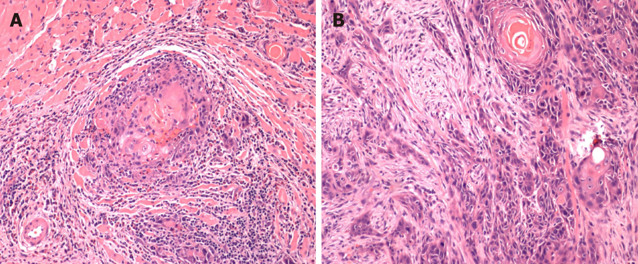
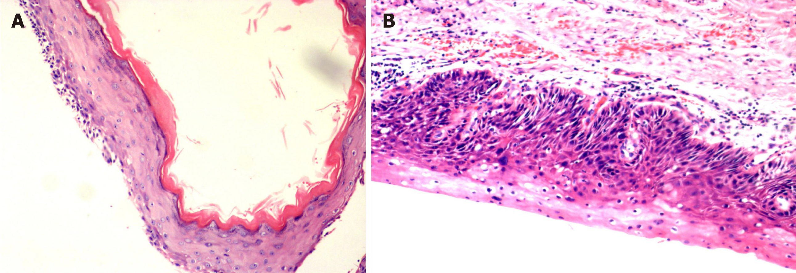
OOC has a thick “onion skin-like” uniform orthokeratinized squamous epithelial lining of 4–8 cell layers, while OKC has a thin parakeratotic epithelial lining. The basal cells of OOC lack polarity and nuclear hyperchromatism, while OKC commonly displays prominent palisaded and hyperchromatic basal cells with polarity. In addition, OKC often has a corrugated surface layer of parakeratin[6]. Light microscopy shows small daughter cysts in OKC but not in OOC, which might be related to the high recurrence rate of OKC.
Immunohistochemically, there are many differences between OOC and OKC. The most prominent is that OOC has less proliferative potential, which might be due to lower p63 expression. According to Sarvaiya et al[3], p63 is a p53 tumor suppressor gene family member, and it is essential for terminal differentiation of epithelial stem cells and their maintenance. Varsha et al[7] also found that the high level of p63 can be correlated with the aggressive behavior of OKC. In consequence, treatment of OKC might also be more aggressive. OOC has been shown to have fewer Ki67-positive proliferating cells compared with OKC, which reveals that OOC has less proliferative and malignant potential. Modi et al[8] found that compared with radicular and denti-gerous cysts, Ki67-positive cells were more common. Moreover, Aragaki et al[9] revealed that GLI2 (a downstream effector of Hedgehog signaling) is significantly expressed in OKC and basal cell carcinoma. Expression of keratin 17, mammalian target of rapamycin, and BCL2 is more evident in OKC than OOC. These findings support the view that OOC and OKC are separate entities.
There are two widely accepted methods to treat OKC and OOC: Resection or enuclea-tion with peripheral ostectomy and decompression.
OOC used to be considered a benign variant of OKC that was unlikely to recur, and MacDonald-Jankowski found that the recurrence rate of OOC was 4%, which was less than that of OKC. It is widely believed that OKC has more malignant potential than OOC.
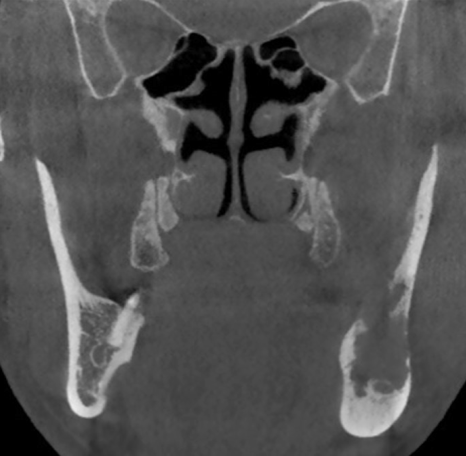
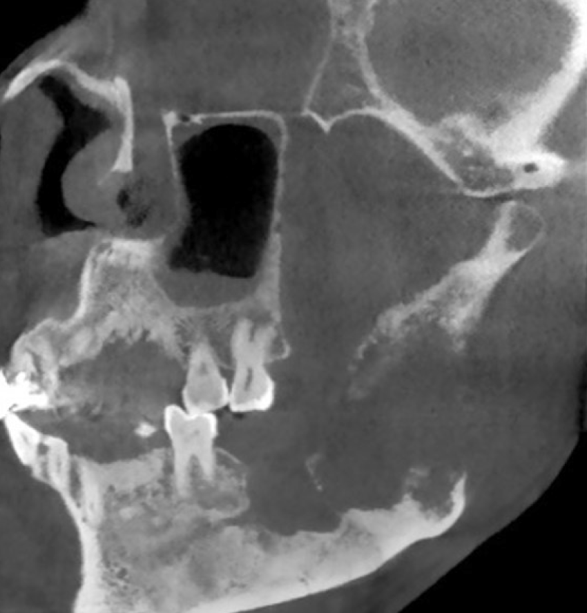
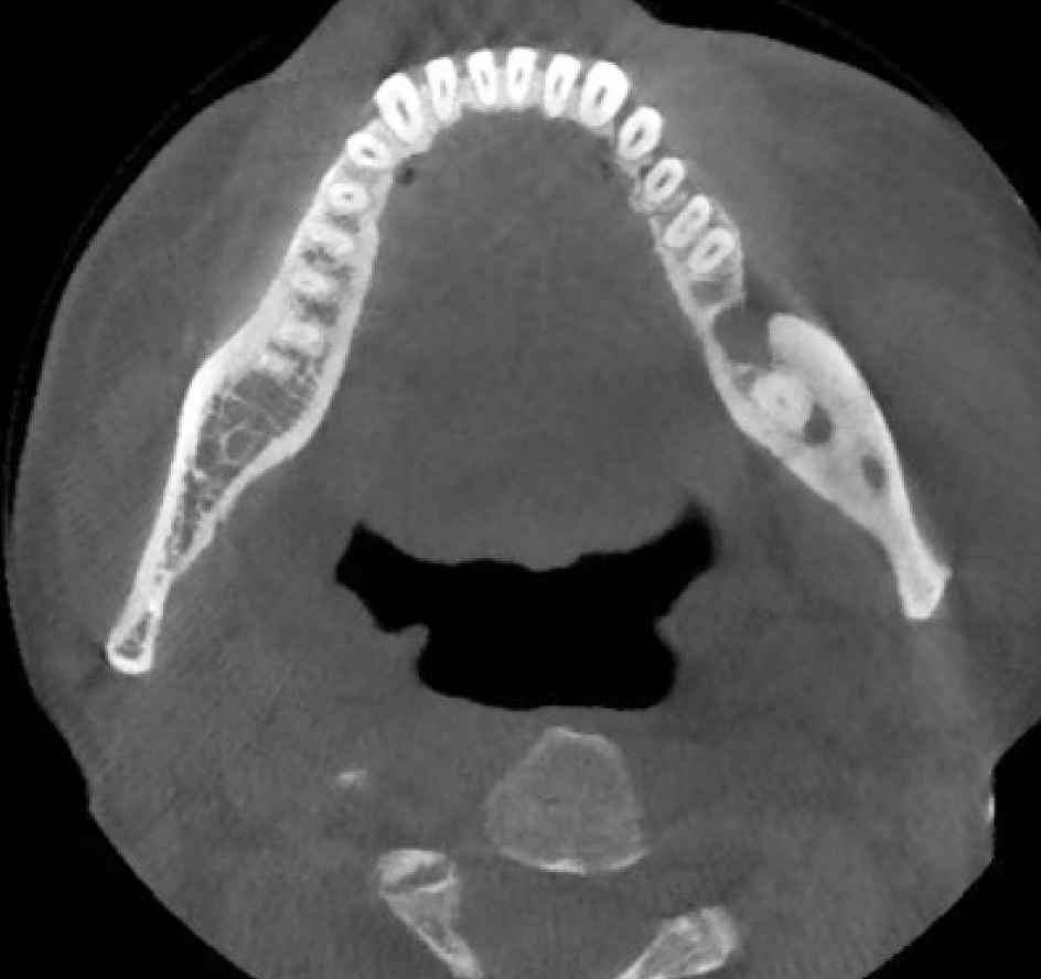
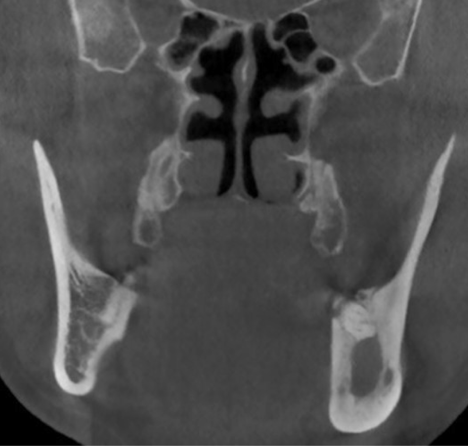
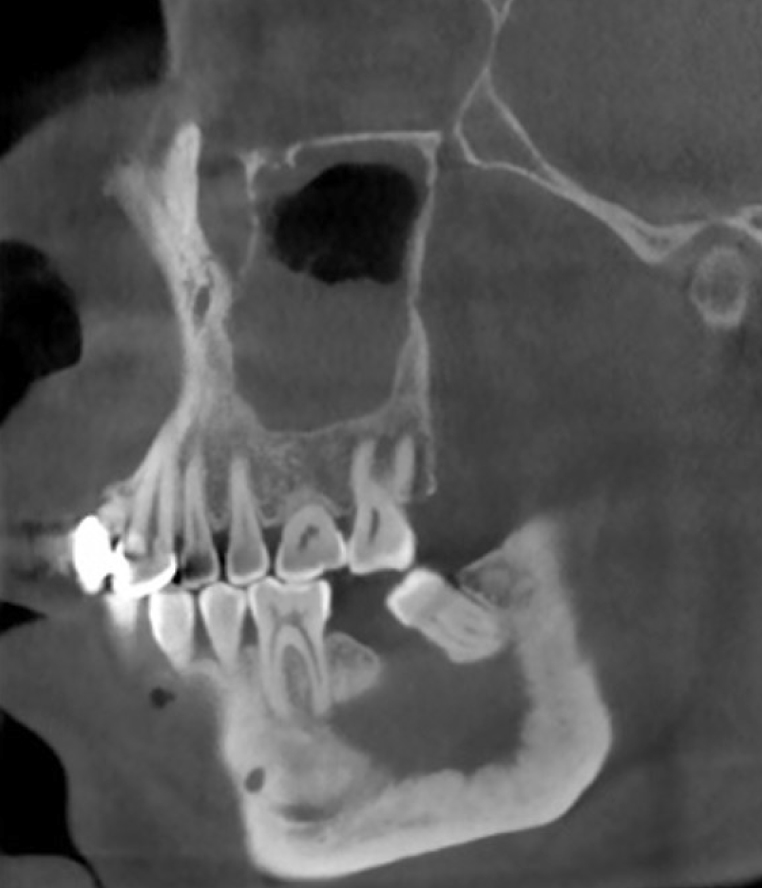
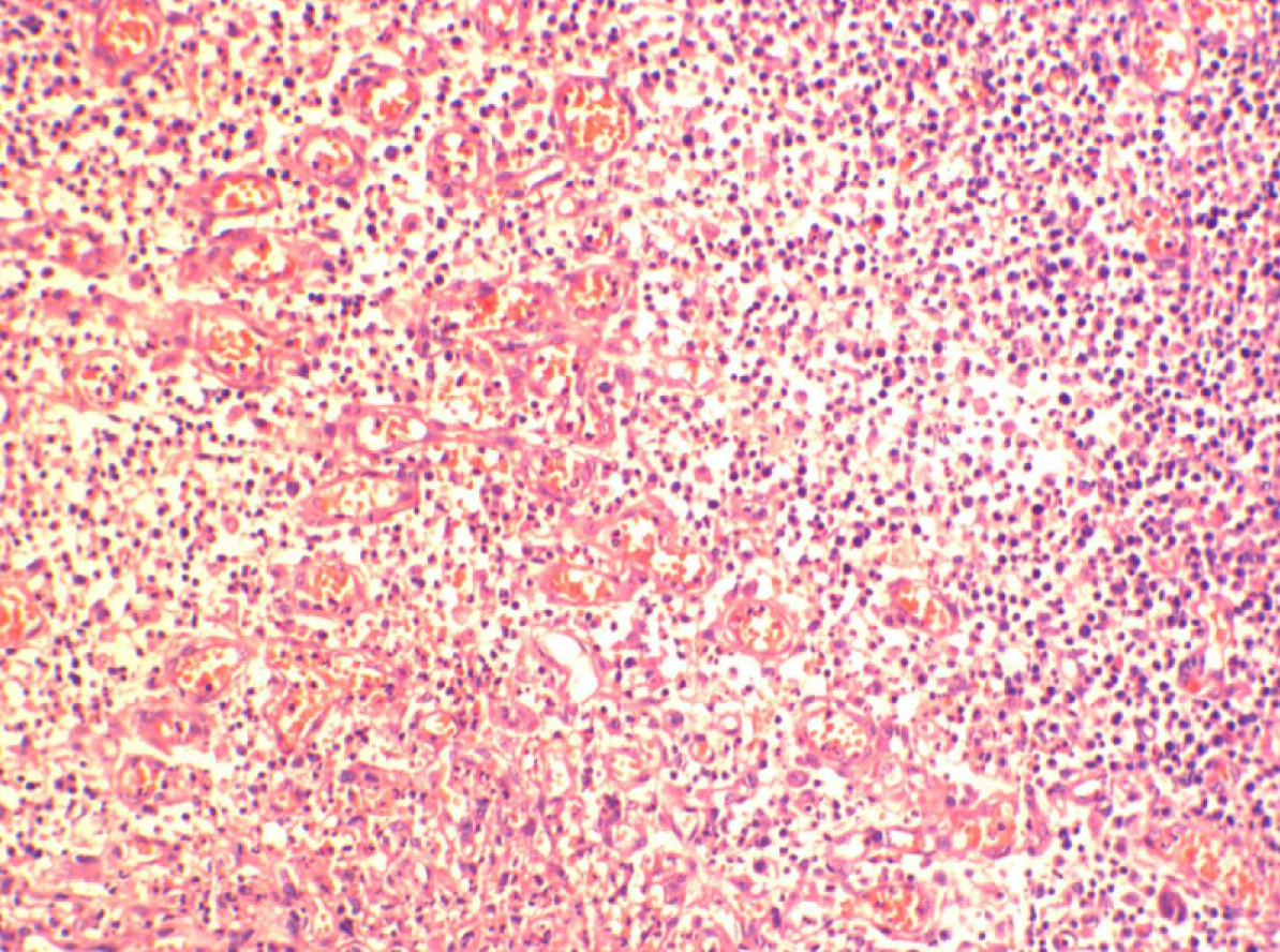
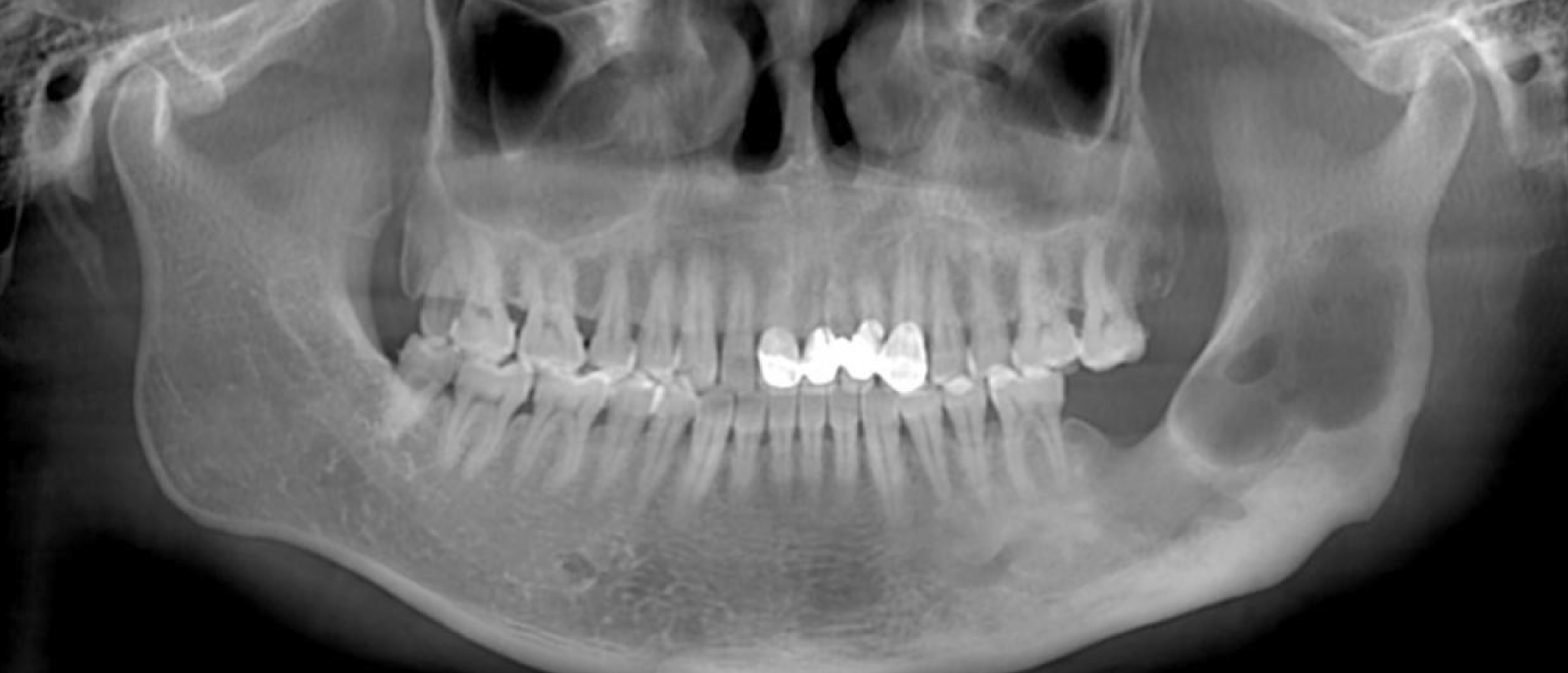
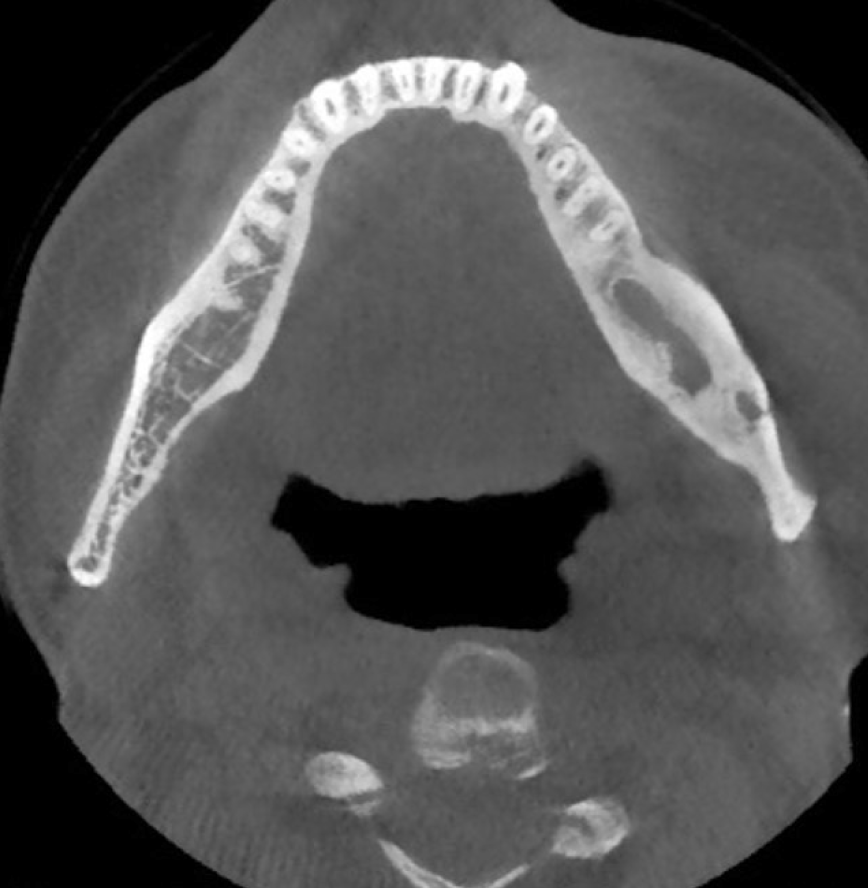
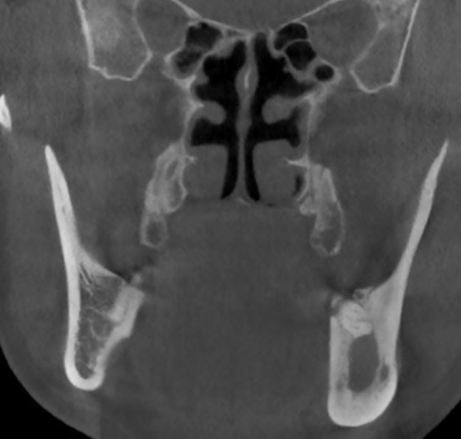
In August 2018, a 52-year-old man was referred to the Department of Oral Maxillo-facial and Head–Neck Oncology at our hospital. The patient presented with severe pain in the left mandible for 2 mo. His left mandible gradually became swollen with severe pain. He denied a family history. Oral examination revealed a 6 cm × 3 cm firm and ill-defined mass on the left mandible angle and ramus. Other physical exami-nation results were normal. Laboratory examinations such as routine blood tests, routine urine tests and urinary sediment examination, routine fecal tests, serum tumor marker measurement, and blood biochemistry revealed no obvious anomalies. There was a cystic lesion in cone beam computed tomography (CBCT); the left mandibular angle and ramus bone density decreased irregularly; and the lesion boundaries were unclear (Figure 1-4). Combined with history and CBCT images, we considered that the OOC could be cancerous. Therefore, we performed partial mandibulectomy. The diseased tissue was obtained for pathological examination and showed pleomorphic nests of squamous cells infiltrating muscles and nerves. The results revealed moderately differentiated squamous cell carcinoma (SCC) (Figure 5). We recommen-ded the patient for subsequent chemotherapy. Central type oral SCC can sometimes be mistaken for odontogenic SCC. However, we believe that the SCC came from OOC for the following reasons: (1) The SCC arose from the primary lesion site; (2) The central type oral SCC was rarely keratinized; and (3) Combined with clinical and radiographic features, this case conformed more to odontogenic SCC. Therefore, we were sure that the SCC arose from OOC. Besides, the patient had a 5-year history of osteomyelitis and mandibular cyst with three recurrences.
On the first visit in January 2013, his left tempus and parotid region were painful and swollen for about 1 year, and pus was leaking out of the fistula. CBCT showed that tooth 37 was lost and the root zone of 37 and the left mandible ramus had a lower density, and the mesial tissue of the lesion area formed new bone (Figure 6-9). Abscess drainage and curettage of mandibular osteomyelitis was performed, and we diagnosed osteomyelitis and mandibular cyst of the left mandible by pathological examination (Figure 10). After surgery, a fistula appeared in the left cheek with exudation of pale yellow pus, but the patient refused further treatment.
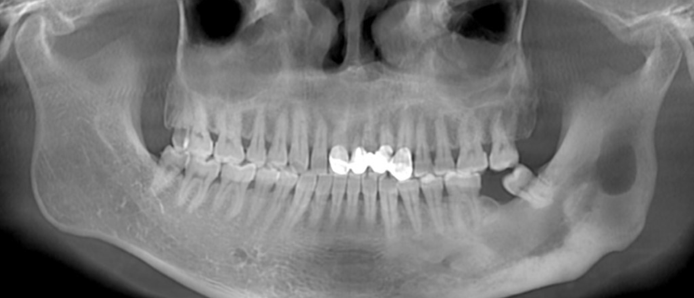
The second visit (first recurrence) was in January 2015, and the patient was diagnosed with osteomyelitis of the left mandible and fistula of the parotideoma-sseterica region by clinical examination. Further treatment was recommended, but the patient refused. After this recurrence, the disease deteriorated. In February 2017, the patient was admitted to our hospital again (second recurrence), but was sent to Wuhan General Hospital of Guangzhou Military Region because of sudden precordial pain with pus leaking into the thorax. After the pain alleviated and other compli-cations were cured, he was admitted to our hospital in June 2017. CBCT showed that the left mandibular angle and ramus density decreased and the cyst had clear boundaries (Figure 11-14). Cyst curettage was performed and the pathological diagnosis was OOC of the left mandible. The cyst component had an orthokeratinized lining (Figure 15A), and the cystic epithelium showed local mild-to-moderate proli-feration (Figure 15B).
The final diagnosis was moderately differentiated SCC.
The patient was treated twice with curettage, partial mandibulectomy, and chemo-therapy.
After surgery, the patient experienced pain in the surgical area. Therefore, he attended another hospital for chemotherapy in September 2018. Positron emission tomography–CT showed recurrence of the left mandibular SCC. The patient success-fully underwent four cycles of chemotherapy.
This case showed low patient compliance, which might lead to missed diagnosis of cancerization. Multiple molecular factors and inflammation-cancer chain involved mechanism, and more unknown factors might play crucial roles in malignant transformation. Examination of known factors, such as p63 and Ki67 expression, although not performed in this case, might provide meaningful indications for early intervention.
Surgeons should be alert to recurrent OOC, which might change with time. Furth-ermore, this patient was diagnosed by pathological examination in 2017 with OOC of the left mandible with mild-to-moderate local proliferation, which was a sign of risk. On the whole, we should be more vigilant about OOC, especially recurrent and proliferative OOC.
The naming and classification of OKC and OOC have been controversial. In the 1950s, odontogenic cyst with keratin formation was designated as OKC for the first time. Afterwards, researchers found that many other cysts also form keratin; there-fore, they were called keratocysts. OKC was used to describe a specific cyst in the 1992 classification. In 2005, there was a controversy between cyst and neoplasm, which involved OKC. The WHO working group recommended that OKC should be replaced by keratocystic odontogenic tumor (KCOT) for the following reasons: (1) Aggressive behavior; (2) Occurrence of a solid variant; (3) Possibility of recurrence; and (4) Mutations of the PTCH gene[10]. However, some researchers debated whether gene mutations could be found in other non-neoplastic diseases such as fibrous dysplasia. In other words, the genetic variation that influences OKC can also influence other types of cysts that are not defined as neoplasms[10]. There is also controversy about the aggressive behavior of OKC. Therefore, in 2017, the WHO working group renamed KCOT as OKC[6]. As for OOC, it was found and described as a type of OKC in 1981[11]. However, because of its different clinical and histological behavior, OOC was acce-pted as a separate entity in the 2017 classification.
OOC used to be considered as a variant of OKC and unlikely to recur, but there are rare reports about its malignant transformation[3]. The systematic review by MacDonald-Jankowski[12] in 2010 showed that only 4% of OOC recurred and the average age at first presentation was 35 years. Although OOC was generally believed to have benign clinical behavior, we have found two case reports of malignant OOCs. The first one was a report of OOC with 8 mo of chronic inflammation, which turned into SCC[13]. Kamarthi et al[14] reported a case of OOC that turned into verrucous carcinoma after 1 year of painful swelling of the left maxillary alveolus. Beyond that, OOC is not associated with NBCCS, and differs in many aspects from other odontogenic cysts, especially dentigerous cyst and OKC. Therefore, oral and maxillo-facial surgeons should distinguish OOC from other types of odontogenic cysts and be more alert to the malignant potential of OOC with a history of recurrence and inflammation and encourage patients to have frequent visits after treatment.
This case report discussed the chronic progression of recurrent OOC into SCC. It revealed that OOC can also be cancerous. Surgeons should perform long-term follow-up and examination of OOC cases.
Manuscript source: Unsolicited Manuscript
Specialty type:
Country of origin:
Peer-review report classification
Grade A (Excellent):
Grade B (Very good):
Grade C (Good):
Grade D (Fair):
Grade E (Poor):
P-Reviewer: Chen YK, Gupta C, Vynios D S-Editor: Ma YJ L-Editor: Filipodia E-Editor: Wang J
| 1. | Dong Q, Pan S, Sun LS, Li TJ. Orthokeratinized odontogenic cyst: a clinicopathologic study of 61 cases. Arch Pathol Lab Med. 2010;134:271-275. [PubMed] [DOI] [Cited in This Article: ] [Cited by in F6Publishing: 4] [Reference Citation Analysis (0)] |
| 2. | Mahdavi N, Taghavi N. Orthokeratinized Odontogenic Cyst of the Maxilla: Report of a Case and Review of the Literature. Turk Patoloji Derg. 2017;33:81-85. [PubMed] [DOI] [Cited in This Article: ] [Cited by in Crossref: 3] [Cited by in F6Publishing: 7] [Article Influence: 1.0] [Reference Citation Analysis (0)] |
| 3. | Sarvaiya B, Vadera H, Sharma V, Bhad K, Patel Z, Thakkar M. Orthokeratinized odontogenic cyst of the mandible: A rare case report with a systematic review. J Int Soc Prev Community Dent. 2014;4:71-76. [PubMed] [DOI] [Cited in This Article: ] [Cited by in Crossref: 14] [Cited by in F6Publishing: 16] [Article Influence: 1.6] [Reference Citation Analysis (0)] |
| 4. | Pazdera J, Kolar Z, Zboril V, Tvrdy P, Pink R. Odontogenic keratocysts/keratocystic odontogenic tumours: biological characteristics, clinical manifestation and treatment. Biomed Pap Med Fac Univ Palacky Olomouc Czech Repub. 2014;158:170-174. [PubMed] [DOI] [Cited in This Article: ] [Cited by in Crossref: 11] [Cited by in F6Publishing: 12] [Article Influence: 1.0] [Reference Citation Analysis (0)] |
| 5. | Cheng YS, Liang H, Wright J, Teenier T. Multiple orthokeratinized odontogenic cysts: a case report. Head Neck Pathol. 2015;9:153-157. [PubMed] [DOI] [Cited in This Article: ] [Cited by in Crossref: 16] [Cited by in F6Publishing: 15] [Article Influence: 1.5] [Reference Citation Analysis (0)] |
| 6. | Soluk-Tekkeşin M, Wright JM. The World Health Organization Classification of Odontogenic Lesions: A Summary of the Changes of the 2017 (4th) Edition. Turk Patoloji Derg. 2018;34. [PubMed] [DOI] [Cited in This Article: ] [Cited by in Crossref: 28] [Cited by in F6Publishing: 48] [Article Influence: 4.4] [Reference Citation Analysis (0)] |
| 7. | Varsha B, Gharat AL, Nagamalini B, Jyothsna M, Mothkur ST, Swaminathan U. Evaluation and comparison of expression of p63 in odontogenic keratocyst, solid ameloblastoma and unicystic ameloblastoma. J Oral Maxillofac Pathol. 2014;18:223-228. [PubMed] [DOI] [Cited in This Article: ] [Cited by in Crossref: 13] [Cited by in F6Publishing: 15] [Article Influence: 1.5] [Reference Citation Analysis (0)] |
| 8. | Modi TG, Chalishazar M, Kumar M. Expression of Ki-67 in odontogenic cysts: A comparative study between odontogenic keratocysts, radicular cysts and dentigerous cysts. J Oral Maxillofac Pathol. 2018;22:146. [PubMed] [DOI] [Cited in This Article: ] [Cited by in Crossref: 5] [Cited by in F6Publishing: 8] [Article Influence: 1.3] [Reference Citation Analysis (0)] |
| 9. | Aragaki T, Michi Y, Katsube K, Uzawa N, Okada N, Akashi T, Amagasa T, Yamaguchi A, Sakamoto K. Comprehensive keratin profiling reveals different histopathogenesis of keratocystic odontogenic tumor and orthokeratinized odontogenic cyst. Hum Pathol. 2010;41:1718-1725. [PubMed] [DOI] [Cited in This Article: ] [Cited by in Crossref: 38] [Cited by in F6Publishing: 38] [Article Influence: 2.7] [Reference Citation Analysis (0)] |
| 10. | Wright JM, Odell EW, Speight PM, Takata T. Odontogenic tumors, WHO 2005: where do we go from here? Head Neck Pathol. 2014;8:373-382. [PubMed] [DOI] [Cited in This Article: ] [Cited by in Crossref: 64] [Cited by in F6Publishing: 62] [Article Influence: 6.2] [Reference Citation Analysis (0)] |
| 11. | Wright JM. The odontogenic keratocyst: orthokeratinized variant. Oral Surg Oral Med Oral Pathol. 1981;51:609-618. [PubMed] [DOI] [Cited in This Article: ] [Cited by in Crossref: 142] [Cited by in F6Publishing: 153] [Article Influence: 3.6] [Reference Citation Analysis (0)] |
| 12. | Macdonald-Jankowski DS. Orthokeratinized odontogenic cyst: a systematic review. Dentomaxillofac Radiol. 2010;39:455-467. [PubMed] [DOI] [Cited in This Article: ] [Cited by in Crossref: 49] [Cited by in F6Publishing: 58] [Article Influence: 4.5] [Reference Citation Analysis (0)] |
| 13. | Acharya S, Tayaar AS, Hallkeri K, Adirajaiah S, Gopalkrishnan K. Squamous cell carcinoma emerging in an orthokeratinized odontogenic cyst: A case report and brief review. J Oral Maxillofac Surg Med Pathol. 2014;26:563-568. [DOI] [Cited in This Article: ] [Cited by in Crossref: 9] [Cited by in F6Publishing: 9] [Article Influence: 0.9] [Reference Citation Analysis (0)] |
| 14. | Kamarthi N, Palakshappa SG, Wadhwan V, Mohan RS. Intraosseous verrucous carcinoma arising from an orthokeratinized odontogenic keratocyst: A report of a rarest entity. Contemp Clin Dent. 2016;7:416-419. [PubMed] [DOI] [Cited in This Article: ] [Cited by in Crossref: 10] [Cited by in F6Publishing: 10] [Article Influence: 1.3] [Reference Citation Analysis (0)] |


