Article Text

Abstract
Background Enlargement of the major salivary glands (SGs) is a major risk factor for B-cell lymphoma among patients with primary Sjögren’s syndrome (pSS). Ultrasound-guided core needle biopsy (US-guided CNB) could be a novel technique to manage SG enlargement among patients with pSS.
Objective Accordingly, this study’s main aim was to evaluate the safety, patient tolerance and diagnostic accuracy of US-guided CNB procedure for patients with pSS with major SG enlargement.
Methods Patients with clinical diagnosis of pSS and a clinical indication for SG biopsy consecutively underwent US-guided CNB between September 2019 and June 2021. These patients were evaluated clinically 1, 2 and 12 weeks after US-guided CNB. Patients were asked to complete a questionnaire about postprocedural complications as well as periprocedural pain, using the Visual Analogue Scale. Complications were categorised as transient (<12 weeks) or persistent (≥12 weeks).
Results US-guided CNB was performed on 30 major salivary glands (22 parotid glands and 8 submandibular glands). The procedure was well tolerated. Transient complications—such as haematoma, swelling—were observed among 43% of patients, and mean periprocedural pain was low. However, no persistent complications were reported during the study’s follow-up period.
Conclusion US-guided CNB represents a novel approach for the management of patients with pSS with SG enlargement. The procedure showed remarkable patient safety and tolerance, allowing adequate glandular sampling and a definite diagnosis for almost all participating patients without long-term complications.
- Sjogren's syndrome
- ultrasonography
- autoimmune diseases
Data availability statement
Data are available upon reasonable request.
This is an open access article distributed in accordance with the Creative Commons Attribution Non Commercial (CC BY-NC 4.0) license, which permits others to distribute, remix, adapt, build upon this work non-commercially, and license their derivative works on different terms, provided the original work is properly cited, appropriate credit is given, any changes made indicated, and the use is non-commercial. See: http://creativecommons.org/licenses/by-nc/4.0/.
Statistics from Altmetric.com
Key messages
What is already known about this subject?
Salivary gland enlargement in primary Sjögren’s syndrome (pSS) is a main risk of lymphoma development, whose diagnosis must be pathologically proven through histological analysis. In this scenario, ultrasound-guided core needle biopsy (US-guided CNB) has been proposed as a diagnostic tool, but evidence for its safety, tolerance and procedural standardisation remains limited.
What does this study add?
This study provides insights into the technical aspects and anatomical issues of the US-guided CNB procedure, the safety and patients’ tolerability through objective measures (ie, pain visual analogue scale).
How might this impact on clinical practice or further developments?
US-guided CNB could be a valuable procedure for salivary gland lymphoma diagnosis in patients with pSS, and in the next future, it might obtain a growing prognostic value, empowering disease activity and tissue damage monitoring as well as shedding light on patients’ response to treatment.
Introduction
Primary Sjögren’s syndrome (pSS) is a connective tissue disease characterised by a wide spectrum of glandular and extraglandular features.1 The hallmark of pSS is considered to be the immune-mediated destruction of exocrine glands and B-cell hyperactivation. This condition exposes patients with pSS to an increased risk of lymphoproliferative disease,2 the highest risk among various autoimmune diseases,3 usually affecting the mucosa-associated lymphoid tissue (MALT).4 5
Persistent major salivary gland (SG) enlargement is a major clinical risk factor for B-cell lymphoma, and it may signify an established lymphoma stage, a prelymphomatous stage or even a completely different disease.6 7 The parotid glands are the main site of B-cell lymphoma development among patients with pSS, and biopsies of the major SGs are reserved for patients with pSS suspected of having glandular lymphoma, mainly based on SG swelling.
An ultrasonographic examination of the major SGs could assist clinicians in assessing the gland’s typical structural abnormalities and detecting parenchyma lesions,8–10 suggesting that SG ultrasonography (SGUS) may come to be used as a stratification (scoring system) and prognostic tool.11–13
The presence of a focal lesion within altered glandular parenchyma (figure 1A) or severe glandular inhomogeneity (eg, diffuse, large-confluent, hypoechoic areas; figure 1B) or both are the typical sonographic patterns of SG lymphoma among patients with pSS.14–16 Nevertheless, currently, lymphoma is mainly suspected among patients with pSS due to clinical characteristics, rather than imaging features, and the diagnosis of B-cell lymphoma must be pathologically proven.17 18

Sonographic images of a focal lesion (the white arrows) in the salivary gland parenchyma (A); a peculiar appearance of the salivary gland, showing confluent hypoechoic areas (B).
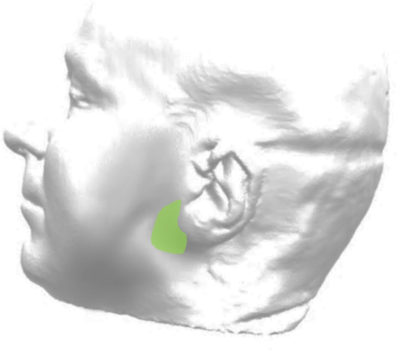
An open surgical biopsy of the parotid gland is the recommended procedure in case of SG enlargement;19 20 however, the need of a skilled surgeon and the risk of potential complications limit its generalisability.20 21 Recently, ultrasound (US)-guided core needle biopsy (US-guided CNB) has been proposed as a diagnostic procedure for patients with pSS with suspected glandular lymphoma.17 22 However, evidence of this procedure’s safety, patient tolerance and standardisation remains limited.23 The most feared complication is the injury of the facial nerve, which presents a strict anatomical relation with the parotid gland. In interventional procedures on the parotid glands, a ‘safety zone’ or ‘safety area’ has been identified. This region is located between 1 cm anterior and 1 cm below the ear lobe, where the facial nerve runs in the glandular parenchyma protected by the superficial lobe of the parotid for a length of approximately 2 cm (figure 2).

The ‘safety area’ or ‘safety zone’ (green area) for a parotid biopsy.
To overcome these limitations, the current prospective study aimed to (1) strengthen our preliminary experience,15 (2) describe the US-guided CNB procedure in detail and (3) evaluate the procedure’s safety, patient tolerance and the diagnostic accuracy for lymphoproliferative disease of US-guided CNB among a cohort of definite patients with pSS or patients suspected to have pSS with persistent parotid or submandibular gland swelling.
Methods
Patients
Our study included consecutive patients referring to the Institute of Rheumatology, University Hospital of Udine (Italy), from September 2019 to June 2021, with a clinical diagnosis of pSS, who underwent US-guided CNB due to suspected major SG lymphoma. The suspicion of lymphoma development was clinically based on parotid and/or submandibular gland swelling, defined as either (1) chronic (>12 months), unilateral or bilateral parotid or submandibular gland swelling or (2) recurrent glandular swelling of at least 2 months of duration in one episode.24 25
Written informed consent was obtained from each patient in accordance with the Declaration of Helsinki and with local guidelines for good clinical practice.26
Demographic, clinical and laboratory data
Patients’ clinical data were collected from their medical charts, including their age, gender, disease duration, previous unstimulated sialometry and Schirmer’s tests and evidence of serum antibodies, as well as antinuclear, anti-Ro/SSA and anti-La/SSB antibodies. Furthermore, the presence of lymphoma development risk factors for patients with pSS was noted at the time of participants’ biopsy procedures (glandular swelling, lymphadenopathy, cryoglobulinemia, a serum monoclonal component, rheumatoid factor, low serum C4 and leucopenia).6
The US-guided CNB procedure for the major SGs
In this study, US-guided CNB was performed by a radiologist (ML) with extensive experience (ie, 10 years of practice) in ultrasound-guided biopsies. The procedures were conducted under real-time US guidance with linear high-frequency transducers (RS85, probe LM4-15B (Samsung, Seoul, South Korea) or Affiniti 70G, probe L18-5 (Philips, Amsterdam, the Netherlands)). The procedures were performed with an aseptic, free-hand technique under real-time US guidance in a non-operating room.
Biopsies were performed with a 14-gauge, semiautomatic CNB system (Precisa 14G, HS Hospital Service, Aprilia, Italy), with a sampling length set on 10 mm or, more frequently, 20 mm, depending on the size of the target area. US-guided CNB was performed on participants’ clinically most swollen gland. The biopsy was performed on the area with the most suspicious ultrasonographic appearance (severe inhomogeneity of the glandular parenchyma or focal lesion, whenever present). For deep lesions, the procedure’s risks and benefits were weighed by the radiologist and the clinicians. The optimal procedure technique was adapted to participants’ SG type (ie, parotid vs submandibular glands) and lesion type (ie, localised vs diffuse).
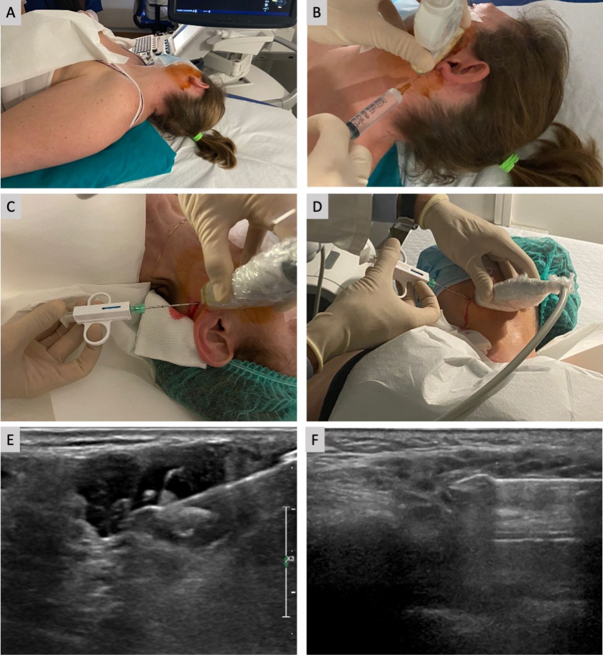
During their biopsies, patients were positioned in a supine position.27 Their shoulders were slightly lifted (usually with a pillow below their upper back), and their necks were slightly hyperextended, turned towards the contralateral side of the target gland (figure 3A).

(A): A patient’s supine positioning for a parotid gland CNB, with their shoulders slightly lifted (and a pillow below their upper back), slight hyperextension of their neck and facing towards the contralateral side of the target gland. (B): Local anaesthetic injected under ultrasound guidance into the subcutaneous tissue and the posterior, superficial part of the parotid gland while moving the needle in a caudocranial direction. (C): A semi-automatic needle inserted into the ‘safety area’ of the left parotid gland. (D): A semi-automatic needle inserted into the left submandibular gland, (E): The needle’s sonographic appearance in a focal lesion of the parotid gland. (F): The needle’s sonographic appearance in a peculiar appearance of the salivary gland, in the absence of focal lesion. CNB, core needle biopsy.
For patients with parotid focal lesions, the spatial relationship between the ‘safety area’ and the lesions themselves was primarily assessed to determine the procedure’s technical feasibility and safety. After the radiologist analysed the safety and feasibility, the procedure targeted lesions posteriorly through the ‘safety area’, maintaining the shortest path and the most superficial needle position possible within a depth of 1–1.5 cm from the glandular surface.
For patients with diffusely inhomogeneous parenchyma without focal lesions, parotid biopsies targeted the posterior-caudal part of the gland in a caudocranial direction. The needle entered within the ‘safety area’, maintaining the most superficial needle direction possible within 1–1.5 cm of the glandular surface.
For US-guided submandibular gland biopsies, patients were positioned like patients receiving a US-guided biopsy of the parotid gland, with their heads fully turned to the opposite side. Patients’ submandibular glands were accessed anteriorly or posteriorly, depending on patients’ cooperation, operator preferences and the eventual US detection of a focal lesion, as described above, in this case, nerve injury was not a concern. Attention was paid also on localising and then avoiding the path of the facial artery, which represents the major noble anatomical structure in strict contact with the gland.
The asepsis of the procedure was guaranteed by accurate disinfection of the skin with gluconate chlorhexidine or povidone–iodine solution and the use of single use prove covers. A local anaesthetic was injected under US guidance with a fine needle (23 G) through the skin and subcutaneous fat (figure 3B). Shortly after this local anaesthetic injection, a small incision of the anesthetised skin was made with a scalpel. The biopsy was then performed with a 14 G semiautomatic needle under US guidance (figure 3B,C). One to five needle passes were performed through the same skin incision (figure 3E,F). The biopsy samples were fixed in formalin and sent for histological analysis. After this procedure, patients compressed their puncture sites and remained under observation for at least 30 min.
Evaluation of postbiopsy complications and peri-procedural pain
This study’s participants were evaluated clinically 1, 2 and 12 weeks after their US-guided CNB and beyond the 12 weeks in case of persistent complications. All patients were asked to complete a questionnaire, reporting any postprocedural complications (online supplemental figure 1) as well as assessing their intraprocedural and postprocedural pain using the Visual Analogue Scale (VAS 0–10). Complications were categorised as transient (lasting <12 weeks) or persistent (lasting ≥12 weeks). Any complication that had developed during the follow-up period was assessed and recorded, including swelling, haematoma, bleeding, pain, local infection, anaesthesia or paraesthesia, sialocele and fistulae. Potential damage of the facial nerve was assessed by clinically monitoring the eventual development of sensorimotor sign or symptoms.
Supplemental material
Results
Patients’ demographic and clinical characteristics and laboratory findings
Thirty patients who had undergone US-guided CNB of the major SGs were evaluated. Of these patients, 27 (90%) were women. Patients’ mean age at the time of the biopsies was 59.8 years (SD: 13.2 years). Moreover, 23 of the study’s 30 patients (76.6%) met the American College of Rheumatology (ACR) and EULAR classification criteria for pSS,28 and 24 (80%) had anti-Ro/SSA antibodies. Among the seven patients who did not fulfil the 2016 ACR/EULAR criteria, pSS was highly suspected based on clinical grounds in five patients, while in two patients, the clinical picture did not allow one particular disease as much more likely. They all refused a minor SG biopsy performed concomitantly with US-guided CNB of the major SGs.
Additionally, 22 patients (73.3%) had parotid gland swelling. Among this group, 11 patients showed chronic (≥12 months) parotid gland enlargement, 8 patients presented with episodical parotid enlargement of long duration (2–11 months) and 3 patients showed episodical parotid gland enlargement of short duration (<2 months). Eight patients (26.7%) reported submandibular gland swelling. Particularly, five patients presented chronic submandibular gland enlargement, while three patients presented episodical submandibular enlargement of long duration. Patients’ characteristics are presented in table 1.
Patients’ clinical and laboratory features
Sonographic characteristics
SGUS examinations were performed in all studied patients before US-guided CNB. SGUS detected a focal lesion among 19 of 30 (63.3%) patients, whereas 11 patients (36.7%) showed inhomogeneous glandular parenchyma, suggesting lymphoproliferative disease in the absence of glandular focal lesions. All biopsied glands showed a sonographic OMERACT (Outcome Measures in Rheumatology) score ≥2 (grade 2 in 4 out of 30 patients and grade 3 in 26 out of 30 patients). Overall, 22 parotid glands (73.33%) and 8 submandibular glands (26.67%) were biopsied.
Out of 22 US-guided CNBs of the parotid glands, 14 (63.6%) were performed on focal lesions, and in all cases, the procedure was technically and safely feasible through the ‘safety area’. For biopsies of the submandibular glands, five of eight (62.5%) procedures were performed on focal lesions. At least two samples were taken in all suspected cases. During the US-guided parotid gland CNB, the facial nerve was not clearly identifiable by SGUS in none of 22 patients (100%).
Safety and patient acceptance
US-guided CNB was well tolerated, and patients reported no long-term complications during the follow-up period of mean±SD: (10.6±7.3 months). Only transient complications (lasting <12 weeks) were reported by 13 patients (43%). Specifically, five cases of local swelling at the biopsy site were reported, lasting no more than 6 days, along with two cases of local bleeding and subsequent haematoma of the submandibular area, one case of transient facial paresis (lasting less than 1 hour, due to the anaesthetic) and four cases of local paraesthesia lasting less than 2 hours. More details about these complications are presented in figure 4A.

(A) Complications of US-guided CNB; all complications were transient (<12 weeks), and no persistent complications were reported during follow-up. (B): Peri-procedural pain: patients’ reported intra- and post-operative pain. CNB, core needle biopsy; US, ultrasound.
Intraprocedural pain, evaluated using the VAS, was assessed via a questionnaire and found to be low (mean VAS: 1.67±2.47), like postprocedural pain (mean VAS: 1.23±2.3). Specifically, 17 of 30 patients (56.67%) reported no intraprocedural pain, eight patients (26.67%) reported mild pain, two patients (6.67%) reported moderate pain and three patients (10%) reported severe pain (maximum VAS pain: 8/10). Postprocedural pain did not occur for 21 of 30 patients (70%), while 5 patients (16.67%) experienced mild postprocedural pain, two patients (6.67%) experienced moderate postprocedural pain and two patients (6.67%) experienced severe postprocedural pain. Figure 4B presents more details about participants’ procedure-related pain.
Overall, for the 9 of 30 patients (30%) who reported postprocedural pain, their median postprocedural pain duration was only 2 days (Q25–75: 1–3 days). No statistically significant difference was found between parotid and submandibular US-guided CNB in pain assessment. Importantly, patients did not report any surgical wounds or scar formation after the procedures’ skin incisions. The entire duration of the US-guided CNB procedure, from patients’ entrance into the examination room to their exit, took a maximum of 40 min.
Histopathological diagnoses
The biopsy samples were diagnostic in 28 of 30 patients (93.3%), in two cases (one parotid, one submandibular gland biopsy), a tissue sample of sufficient quality for histopathological diagnosis was not obtained. Patients’ pathological diagnoses included MALT lymphoma for 14 of 30 patients (46.6%), focal lymphoepithelial sialadenitis (LESA) with initial acquisition of the MALT tissue for two patients (6.7%), LESA for three patients (10%). Other histopathological diagnoses were reported in online supplemental table 1. In 3 of 30 patients, the gland biopsies’ histological results allowed respective differential diagnoses of sarcoidosis, IgG4-related disease and chronic sclerosing sialadenitis without pSS.
Of note, in 11 out of 14 cases (78.6%), MALT lymphoma was identified in patients satisfying pSS ACR/EULAR criteria. Moreover, in 10 out of 14 cases (71.42%), MALT lymphoma was detected in a focal lesion. All patients diagnosed with MALT lymphoma underwent further investigations (ie, PET/CT imaging, haematologic consult).
Among the seven patients who did not fulfil the 2016 ACR/EULAR criteria, subsequent US-guided CNB of the major SGs was consistent with pSS (focal lesions ≥1/4 mm2, LESA or MALT lymphoma) in six patients.
Discussion
Sampling quality tissue for histopathological analysis is crucial for diagnostic, prognostic and research purposes. In pSS cases, quality samples are even more valuable since this autoimmune disease presents the highest risk of lymphoproliferative disorders and, moreover, the exact lymphomagenesis is not yet clearly defined.6 Currently, an effective treatment of pSS is lacking; however, the recent availability of biologic target therapies might offer new treatment options.29
SGUS is frequently used to assess the structural abnormalities typical of pSS.8 Furthermore, recent reports have suggested that the sonographic detection of focal lesions or a peculiar sonographic appearance of SGs (eg, large, confluent, hypoechoic areas spread over the gland) could indicate glandular B-cell lymphoma,17 18 which has yet to be proven by histology.
For patients without pSS who present SGUS-detected focal lesions of the major SGs, fine-needle aspiration cytology (FNAC) is performed to differentiate between benign and malignant lesions. FNAC is a safe technique, but it frequently fails to provide material sufficient for a diagnosis.30 In patients with pSS at a high risk of lymphoma development, a histological, rather than cytological, sampling is usually needed. Therefore, such other procedures as open surgical biopsy or US-guided CNB may play a role.18
Open surgical biopsy of the major SGs is a safe technique when performed by expert surgeons but presents several disadvantages, such as the need for an operating room and possible adverse effects (such as facial nerve damage), which could limit its accessibility. Importantly, the focal lesions identified by SGUS may not be precisely identified during surgical biopsy procedures; therefore, this approach cannot accurately perform biopsies of SGUS-identified focal lesions. Furthermore, as reported in the preliminary experience by Zabotti et al,15 the open surgical biopsy approach showed a higher number of persistent complication, compared with US-guided CNB approach.
US-guided CNB could overcome both FNAC and surgical limitations. Recent evidence suggests that, for patients with pSS with major SG enlargement—a significant risk factor for B-cell lymphoma6—US-guided CNB can provide sufficient sampling for pathological examination.31
The current study completed a preliminary study15 demonstrating that US-guided CNB of the SGs is accurate and safe for patients with pSS suspected to have lymphoma, providing sufficient material for a histopathological diagnosis in most cases. The safety of US-guided CNB of the parotid glands was recently assessed in a meta-analysis,19 which reported high diagnostic accuracy for both sensitivity and specificity as well as a very low complication rate and facial nerve paralysis as the most severe complication. The identification of the facial nerve via ultrasound is challenging. Currently, few data are available on ultrasonographic scanning of the facial nerve. High-resolution ultrasonography might play a role in the assessment and scanning of the facial nerve.32 To identify the facial nerve, Tawfik et al described the facial nerve’s sonographic appearance, providing reference values from 50 healthy volunteers.33 Meanwhile, other authors34–36 have tried to scan the facial nerve in order to manage Bell’s palsy. However, the extracranial part of the facial nerve after its emergence from the stylomastoid foramen is only partly assessable via ultrasound.37 To the best of our knowledge, none of the studies was able to assess by US of the facial nerve on its extracranial emergence when it enters the parotid parenchyma from a longitudinal or transverse view of way.
In the current authors’ experience, the sonographic assessment of the facial nerve as it exits the stylomastoid foramen has not been feasible, particularly in cases where the disease affects the parotid gland, leading to ultrasonographic glandular impairment. Therefore, we performed US-guided parotid gland biopsies in the ‘safety zone’ for our patients.
In the current study, we described our US-guided approach and the technique used to safely perform a US-guided CNB in SGs of patients with pSS in detail, focusing on parotid gland biopsies in the ‘safety zone’. In this area, the facial nerve runs deeply in the glandular parenchyma, and the parotid gland’s superficial lobe protects the facial nerve at a length of approximately 2 cm (with some variability between individuals), minimising the risk of nerve damage.
The few reports available for submandibular US-guided CNB among patients with pSS17 38 have described the procedure’s efficacy in providing sufficient material for diagnosis and a good safety profile. Our patient cohort reported no long-term complications, confirming US-guided CNB’s safety profile for both parotid and submandibular glands.
Overall, in our opinion, US-guided CNB offers several advantages, the main being its safety and feasibility: in fact, it provides relatively easy access to both the parotid gland and the submandibular gland without a need for surgery, being less invasive and more patient friendly. In the future, the US-guided CNB approach might be performed directly by rheumatologists with expertise in sonography and bioptic procedures, as is now common practice for synovial biopsies. Second, this procedure allows for the targeting of glandular areas with different sonographic patterns in both the parotid and submandibular glands. According to our study, major SG biopsies may provide adequate samples for the early detection of suspected SG lymphoma, thus playing a role in the follow-up of suspected SG lymphoma in pSS.24 39 Moreover, the procedure allowed us to improve differential diagnoses for our patients initially suspected to have pSS (such as granulomatous sialadenitis consistent with sarcoidosis and IgG4-related disease). These findings emphasise the importance of SG biopsies—especially in patients with pSS with glandular swelling—in order to detect not only lymphoproliferative disease but also less common diseases that would otherwise be underdiagnosed.40–42
Furthermore, this new sampling technique might provide histological material from the major SGs that offers possible future research implications for tissue sampling.21 It may also provide superior diagnostic accuracy while improving prognostic value,39 allowing for the monitoring of disease activity and tissue damage10 43 as well as patients’ response to treatment.44 45 Further studies should better assess US-guided CNB’s diagnostic accuracy and safety for patients with pSS, as well as more researches should focus on the relationship between major SG and minor SG histology, and between salivary histology and sonographic appearances in pSS.
Conclusion
US-guided CNB represents a novel approach for the management of patients with pSS. This procedure has shown remarkable patient safety and tolerance, allowing for adequate tissue sampling and definite diagnoses for almost all patients who participated in this study.
Data availability statement
Data are available upon reasonable request.
Ethics statements
Patient consent for publication
Ethics approval
The study was conducted according to a protocol approved by the Regional Ethical Committee (CEUR-2017-Os-027- ASUIUD). This study involves human participants and was approved by Ethics Committee of Friuli Venezia Giulia. Participants gave informed consent to participate in the study before taking part.
References
Supplementary materials
Supplementary Data
This web only file has been produced by the BMJ Publishing Group from an electronic file supplied by the author(s) and has not been edited for content.
Supplementary Data
This web only file has been produced by the BMJ Publishing Group from an electronic file supplied by the author(s) and has not been edited for content.
Footnotes
Correction notice This article has been amended since it was first published online. Luca Quartuccio was spelt Quartuccio Luca.
Contributors Conceptualisation: AZ, IG, SZC, CD, AH, ML, EP. Methodology: AZ, ML, EP. Investigation: IG, SZC, VM. Data curation: IG, SZC, VM, AZ, EP. Writing—original draft preparation: IG, AZ. Writing—review and editing: IG, VM, AZ, SDV, ML. Supervision: SDV, LQ, CD, CZ, RG, AH. Guarantors: AZ. All authors have read and agreed to the published version of the manuscript.
Funding The authors have not declared a specific grant for this research from any funding agency in the public, commercial or not-for-profit sectors.
Competing interests None declared.
Provenance and peer review Not commissioned; externally peer reviewed.
Supplemental material This content has been supplied by the author(s). It has not been vetted by BMJ Publishing Group Limited (BMJ) and may not have been peer-reviewed. Any opinions or recommendations discussed are solely those of the author(s) and are not endorsed by BMJ. BMJ disclaims all liability and responsibility arising from any reliance placed on the content. Where the content includes any translated material, BMJ does not warrant the accuracy and reliability of the translations (including but not limited to local regulations, clinical guidelines, terminology, drug names and drug dosages), and is not responsible for any error and/or omissions arising from translation and adaptation or otherwise.