Article Text

Abstract
Objectives To investigate if inflammation detected by MRI or ultrasound at rheumatoid arthritis (RA) onset is predictive of erosive progression or poor response to methotrexate monotherapy, and to investigate if subclinical inflammation in remission is predictive of future treatment escalation or erosive progression.
Methods In a 2-year study, 218 patients with disease-modifying antirheumatic drug-naïve early RA were treated by a tight-control treat-to-target strategy corresponding to current recommendations. MRI and ultrasound were performed at regular intervals. Baseline imaging-based inflammation measures were analysed as predictors for early methotrexate failure and erosive progression using univariate and multivariate regression adjusted for clinical, laboratory and radiographic measures. In patients in remission after 1 year, imaging measures were analysed as predictors of treatment escalation and erosive progression during the second year. The added value of imaging in prediction models was assessed using receiver operating characteristic analyses.
Results Baseline MRI inflammation was associated with MRI erosive progression and ultrasound with radiographic erosive progression. No imaging inflammation measure was associated with early methotrexate failure. Imaging inflammation was present in a majority of patients in clinical remission. Tenosynovitis was associated with treatment escalation, and synovitis and tenosynovitis with MRI/radiographic erosive progression during the second year. Imaging information did not improve prediction models for any of the outcomes.
Conclusions Imaging-detected inflammation, both at diagnosis and in remission, is associated with elements of future disease development. However, the lack of a significant effect on prediction models indicates limited value of systematic MRI and ultrasound in management of early RA.
- arthritis
- rheumatoid
- magnetic resonance imaging
- ultrasonography
- synovitis
This is an open access article distributed in accordance with the Creative Commons Attribution Non Commercial (CC BY-NC 4.0) license, which permits others to distribute, remix, adapt, build upon this work non-commercially, and license their derivative works on different terms, provided the original work is properly cited, appropriate credit is given, any changes made indicated, and the use is non-commercial. See: http://creativecommons.org/licenses/by-nc/4.0/.
Statistics from Altmetric.com
Key messages
What is already known about this subject?
MRI and ultrasound are sensitive methods to monitor inflammatory disease activity in rheumatoid arthritis (RA). Associations between imaging inflammation measures and structural damage progression have been documented in previous longitudinal studies, both in early active disease and in remission.
What does this study add?
This study provides knowledge on the use of imaging in prediction of future disease course of patients with RA in a modern treat-to-target strategy. Our results suggest that although inflammatory imaging findings are associated with future outcomes, prediction models incorporating imaging information do not perform significantly better than models based only on routine clinical and laboratory measures.
How might this impact on clinical practice?
Based on our results, systematic use of MRI or ultrasound does not substantially improve precision of current individualised treatment strategies.
Introduction
Rheumatoid arthritis (RA) management has improved greatly over the last two decades, and many patients achieve remission by modern treatment.1 However, a considerable portion of patients still fail to adequately respond to the first-line methotrexate or have an initial good response, but eventually experience relapse of disease activity.2–6 Several studies have shown that progression of structural joint damage may occur even in patients who maintain stringent clinical remission criteria.7–12 To increase the precision of individualised treatment algorithms, it is necessary to improve methods for prediction of the patient’s disease course.
MRI and ultrasound are sensitive methods to monitor disease activity in RA.13–19 High disease activity at the time of treatment initiation is associated with a higher risk of insufficient methotrexate response,20 and imaging-detected synovitis and bone marrow edema (BME) have been shown to be predictive of progression of joint damage.11 21–25 Studies have also found that a majority of patients who have been successfully treated to remission by current clinical index-based criteria have residual joint inflammation when examined by MRI or ultrasound.10 11 Such subclinical inflammation has been proposed as the underlying pathology of continued radiographic progression in patients in remission, and it has been debated whether RA treatment should target abrogation of subclinical disease activity.9 26 However, three randomised trials investigating MRI and ultrasound did not find additional benefit from their use in conventional treat-to-target strategies.27–29 The value of imaging-detected inflammation for treatment decision-making in modern RA treatment is thus uncertain. Imaging, particularly MRI, is highly resource demanding, and updated knowledge about its value in risk-stratification and outcome prediction is essential.
We aimed to investigate if MRI or ultrasound examination at the time of diagnosis is useful in anticipating poor response to methotrexate monotherapy, or future structural damage progression, in patients treated by modern recommendations. Second, we investigated the presence of imaging-detected residual inflammation in patients who have achieved clinical remission after 1 year of treat-to-target therapy, and if such subclinical inflammation is predictive of treatment escalation or continued erosive progression during the following year.
Materials and methods
Study design
The ARCTIC trial (clinicaltrials.gov ID: NCT01205854) was a 24-month multicentre, randomised clinical strategy study, designed to compare outcomes of an ultrasound guided and a conventionally guided treat-to-target strategy in early RA. All included patients (n=230) were treated according to the same disease-modifying antirheumatic drug (DMARD) escalation strategy,28 which is consistent with current EULAR recommendations.1 Starting treatment was methotrexate 15 mg/week increased to 20 mg/week by week 5, with bridging prednisolone. The treatment algorithm further included increased methotrexate dose to 25 mg/week, triple conventional synthetic DMARD (methotrexate/sulfasalazine/hydroxychloroquine) and biological DMARD treatment. In the conventional treatment arm, the decision to adjust therapy was based on level of and change in Disease Activity Score (DAS).30 The treatment target was DAS <1.6 and no swollen joints, and treatment was escalated in case of insufficient response (defined as a decrease in DAS from the previous visit of less than 0.6 if DAS ≤2.4, or less than 1.2 if DAS >2.4) until the target was reached. In the ultrasound treatment arm, therapy was additionally escalated if the ultrasound examination indicated an unacceptable disease activity or unsatisfactory decrease of disease activity from the previous visit (defined as <10% change in ultrasound score if DAS ≤2.4, or <20% change in ultrasound score if DAS >2.4); in such cases the ultrasound assessment would thus over-rule the clinical assessment. According to the protocol, inflamed joints were treated with intra-articular corticosteroids; in the conventional arm, swollen joints were injected using a landmark-based approach; and in the ultrasound arm, joints with clinical swelling or power Doppler signal were injected with ultrasound guidance.28 As the primary analyses did not show significant differences in treatment outcomes between the study arms,28 all patients were regarded as one cohort in the current analyses. Sensitivity analyses were performed on the study arms separately.
Participants
The main inclusion criteria were age 18–75 years, fulfilment of the 2010 American College of Rheumatology/EULAR classification criteria for RA, DMARD-naivety, time from first patient-reported swollen joint less than 2 years and indication for DMARD treatment.
Clinical, laboratory and radiographic assessments
The study included 13 visits during the 2-year follow-up period.28 Clinical, laboratory data and patient-reported outcomes were recorded at every visit. Further assessments included age, gender, body mass index, smoking status, duration of symptoms (days since first patient-recorded symptom), swollen joint count (SJC, 44 joints), Ritchie Articular Index (RAI),31 anti-citrullinated protein antibody (ACPA) and rheumatoid factor status, patient global assessment (PGA), physician global assessment, joint pain (Visual Analogue Scale), calprotectin, erythrocyte sedimentation rate (ESR) and C reactive protein (CRP). The DAS was calculated as a composite of RAI, SJC, ESR and PGA.30 Remission was defined as DAS <1.6, low disease activity as DAS ≥1.6 ≤2.4, and moderate–high disease activity as DAS >2.4. Investigators and patients were aware of the allocated treatment group, and clinical assessments were performed by unblinded study personnel. Radiographs of hands, wrists and feet were obtained at baseline, after 12, 16 and 24 months for all patients, and scored in chronological order by two readers blinded for clinical data according to the van der Heijde modified Sharp score (vdHSS).32 The average of the two scores was used for the analyses.
Magnetic resonance imaging
MRI of dominant wrist and hand was performed at 0, 3, 6, 12, 16 and 24 months. Acquisitions were done according to the Outcome Measures in Rheumatology RA MRI scoring system (RAMRIS) recommendations,33 34 with coronal and axial T1 pulse sequences without contrast, transversal T1 turbo spin echo with contrast and coronal short tau inversion recovery. Images were scored for the RAMRIS features synovitis, tenosynovitis, BME, bone erosions and joint space narrowing in known chronological order by one reader (US) blinded for clinical data. A combined inflammation score was computed by normalised summation of the synovitis, tenosynovitis and BME scores.35 Reliability of MRI readings was performed by calculating intraclass correlation coefficients (ICCs) for scores of 12 patients scored at all six timepoints separately by DG and US. Inter-reader comparisons were estimated by a two-way mixed-effects model, individual measure and consistency of agreement. Intrareader comparisons were estimated using readings by US using a two-way mixed-effects model, individual measure and absolute agreement.35 Of the 230 patients from the ARCTIC primary analyses, 218 had an MRI assessment at baseline and at least one of the follow-up visits, and were included in the current analyses.
Ultrasound
The protocol for ultrasound assessments has been described previously.28 All patients were assessed by ultrasound at baseline and after 12 and 24 months. Joints were scored according to a validated scoring system with semiquantitative scoring of grey scale and power Doppler synovial inflammation in 32 joints of the hands, wrists, elbows, knees, ankles and feet bilaterally, with a range of 0–96 units for both scores.36 In the analyses of ultrasound data, 5 of the 218 patients were excluded from the baseline analyses, and two patients were excluded from the 1-year analyses, due to missing data.
Missing data
Less than 10% of observations of clinical, laboratory, radiographic, ultrasound and MRI parameters were missing. Missing clinical, laboratory and ultrasound data were imputed using multiple imputation using 10 imputations drawn from the observed distribution. Missing radiographic scores were imputed using linear intrapolation or extrapolation. Missing MRI scores were imputed with 50 imputations using a linear mixed-effects model using available MRI data, with random intercepts for patients and joints nested within patient.
Baseline MRI and ultrasound as predictors for treatment escalation and joint damage
We defined three dichotomous outcomes for assessment of the predictive value of MRI and ultrasound at time of diagnosis: (a) early methotrexate failure, (b) radiographic erosive progression and (c) MRI erosive progression. Early methotrexate failure was defined as escalation from the initial treatment methotrexate monotherapy to triple therapy, or biological DMARD/tumour necrosis factor inhibitor, within 6 months after initiation of treatment. Radiographic erosive progression was defined as an increase of ≥2 units in vdHSS erosion score during the 2-year course of the study. MRI erosive progression was defined as an increase of ≥2 units MRI erosion score during the course of the study. To assess the value of adding MRI or ultrasound information to clinical information in predicting the outcomes, we compared a clinical prediction model including variables routinely used for treatment decision-making, with prediction models including MRI or ultrasound variables. Predictors used in the clinical model were age, gender, SJC, RAI, PGA, CRP, ACPA status and radiographic erosions; the MRI model additionally included the RAMRIS scores for MRI synovitis, tenosynovitis and BME, and the ultrasound model additionally included the ultrasound grey scale and power Doppler scores as predictors.
MRI and ultrasound as predictors for treatment escalation and joint damage in patients in remission
For the analyses of the predictive value of MRI and ultrasound in patients achieving remission, we used data of patients in clinical remission (DAS <1.6) after 1 year of treatment. We defined three outcomes: (a) treatment escalation, (b) radiographic erosive progression and (c) MRI erosive progression. Treatment escalation was defined as change of therapy to a higher level of the treatment regimen during the second year. The decision to change treatment was taken by the treating physician in response to inadequate disease control, based on the ARCTIC trial’s treatment decision rules.28 Patients who escalated treatment on the 1-year visit were excluded from the analysis of this outcome. Radiographic erosive progression was defined as increase ≥1 units for the vdHSS erosion score during the second year. MRI erosive progression was defined as increase ≥1 units for the MRI erosion score during the second year. Similar to the baseline analyses, we compared prediction of the outcomes between three models: a clinical model with predictors age, gender, SJC, RAI, PGA, CRP, ACPA status and radiographic erosions; a clinical+MRI model and a clinical+ultrasound model. The assessment at the 1-year timepoint was used for all included predictor variables. In the analyses of radiographic/MRI erosive progression, only patients with at least two radiographic/MRI examinations in the second year were included. In exploratory analyses, we also assessed prediction of sustained DAS remission between 12 and 24 months.
Statistical analyses
Baseline and 1-year patient characteristics, clinical variables, radiographic and ultrasound scores are presented as means (SD) or frequencies (%) as appropriate. MRI and ultrasound scores are described as mean (SD) values, median (minimum–maximum) values, and number (%) of patients with positive findings (score >0) for all patients at baseline and for patients in remission (DAS <1.6) after 1 year. For each of the outcomes described in the previous sections, the number of positive outcomes is presented as n (%). For the MRI and radiographic erosive progression outcomes, the mean score changes and 95% CIs are reported. We used univariate and multivariate logistic regression to estimate the associations of the continuous MRI and ultrasound scores to the binary outcomes. In the univariate analyses, the linearity of the associations was assessed by visual and likelihood-ratio comparison of linear and cubic spline models. In the multivariate analyses, each of the scores (MRI synovitis, tenosynovitis, BME, ultrasound grey scale and power Doppler) was tested separately in multivariate models with the clinical, laboratory and radiographic variables. To account for an association between MRI inflammation and MRI erosions at baseline or at 1 year, the multivariate models were additionally adjusted for the MRI erosion scores in analyses of MRI erosive progression. In supplementary analyses, calprotectin was included as a covariate, see the online supplemental file. In the 1-year analyses, duration of remission was evaluated in sensitivity analyses. To assess the added value of including MRI or ultrasound data in prediction of the outcomes, we used receiver operating characteristic (ROC) analysis to compare the ROC area under the curve (AUC) of the clinical model to the AUC of the clinical+MRI and the clinical+ultrasound models.37 Additionally, we performed supplementary ROC analyses of models which only included MRI or ultrasound data. Supplementary analyses were also performed using the vdHSS total score and the combined MRI damage score (calculated by summation of the RAMRIS erosion and joint space narrowing scores) to define radiographic/MRI damage progression. All analyses were performed in STATA V.16 (StataCorp, USA).
Supplemental material
Results
At baseline, key clinical and radiographic variables were consistent with a typical population with early RA. Mean age (SD) was 51.7 years, 62.8% of the patients were women and 82.6% were ACPA-positive (table 1).
Patient characteristics of all patients at baseline and 1-year values in patients in clinical remission after 1 year; dichotomous variables presented as N (%), continuous variables presented as mean (SD)
The overall reliability of the MRI scores was very good when tested in intrareader and inter-reader comparisons (synovitis/tenosynovitis/BME/erosions mean inter-reader ICC (baseline) 0.96 (range 0.92–0.98), mean intrareader ICC 0.94 (range 0.89–0.98) (online supplemental table S1). Inflammation could be detected by MRI or ultrasound in a majority of the patients at baseline. The most frequent inflammatory imaging feature was ultrasound grey scale synovitis, which was present to some degree in all patients examined by ultrasound (n=213). The least frequent imaging feature was MRI BME, which was present in about two out of three patients. Imaging scores generally had right-tailed skewed distributions (table 2). The development of the MRI and ultrasound parameters over the study period has been described in previous publications.28 38
MRI and ultrasound (US) findings in all patients at baseline (MRI n=218, US n=213)
Baseline MRI and ultrasound as predictors for treatment escalation and joint damage
Of the 218 patients, 53 (24%) escalated treatment to triple therapy or biological DMARD within 6 months of initiation of treatment. There was no indication of an association between baseline MRI or ultrasound inflammation scores and early treatment escalation in either univariate or multivariate analyses (table 3). Radiographic erosive progression occurred in 63 (29%) patients during the 2-year follow-up, with a mean (95% CI) change in vdHSS erosion score of 2.7 (2.5–3.0) in these patients. The ultrasound, but not the MRI inflammation scores, were associated with radiographic progression in univariate and multivariate analyses (table 3). Thirty-three (15%) patients had MRI erosive progression during follow-up, and the mean (95% CI) change in MRI erosion score in these patients was 1.9 (1.7–2.2). All of the MRI and ultrasound inflammatory scores were strongly associated with MRI erosive progression in univariate analyses. The MRI synovitis, BME, combined MRI inflammation and ultrasound grey scale scores were independently associated with MRI erosive progression in the multivariate analyses (table 3). Sensitivity analyses performed on the randomised study arms separately showed overall similar results as the present analysis, although MRI BME was only independently associated to MRI erosive progression in the patients treated by a conventional treat-to-target strategy. Inclusion of calprotectin in the multivariate baseline prediction models did not significantly affect the coefficients or significance levels of the imaging variables (online supplemental table S2).
Predictive association of baseline MRI and ultrasound scores to early methotrexate failure, radiographic erosive progression and MRI erosive progression, ORs (95% CI)
When comparing the AUC of ROC analysis of the prediction model using only routine measures with the model using MRI data, and the model using ultrasound data, there was no significant contribution of MRI or ultrasound for prediction of either early methotrexate failure, radiographic erosive progression or MRI erosive progression. Analyses of outcomes based on the vdHSS total score and the combined MRI damage score provided comparable results (online supplemental table S4 and online supplemental figure S1A,B). The AUC for the model including MRI data was similar to the AUC of the model including ultrasound data (figure 1A–C). Pure imaging models showed low-to-moderate predictive performance (online supplemental figure S2A–C).
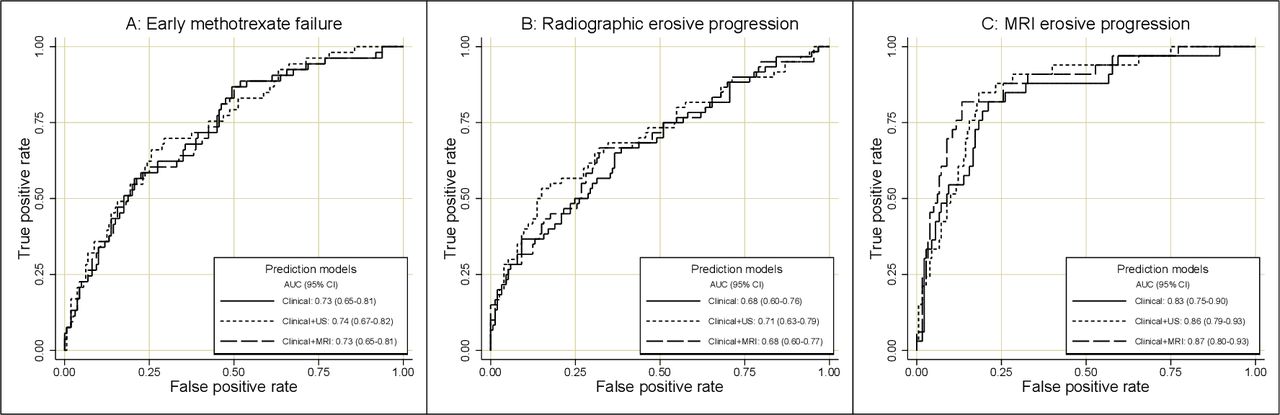
(A–C) ROC curves of the clinical, clinical+ultrasound and the clinical+MRI prediction models for (A) early methotrexate failure, (B) radiographic erosive progression and (C) MRI erosive progression. Early methotrexate failure: escalation from initial treatment methotrexate monotherapy to triple therapy or bDMARD within the first 6 months of treatment. Radiographic erosive progression: increase ≥2 vdHSS erosion score during follow-up. MRI erosive progression: increase ≥2 RAMRIS erosion score during follow-up. AUC, area under the curve; bDMARD, biological disease-modifying antirheumatic drug; RAMRIS, rheumatoid arthritis MRI scoring system; ROC, receiver operating characteristics; US, ultrasound; vdHSS, van der Heijde modified Sharp score.
MRI and ultrasound features after 1 year
After 1 year of tight-control targeted therapy, 164 of the 218 patients (75%) were in clinical remission (DAS <1.6). Signs of inflammation were seen in 159 patients (97%) on MRI and in 126 patients (78%) on ultrasound. Joints with ultrasound power Doppler signal were detected in 22% of patients (table 4).
MRI and ultrasound (US) scores in patients in clinical remission after 1 year (MRI n=164, US n=162)
MRI and ultrasound as predictors for treatment escalation and joint damage in patients in remission
Of 156 patients who were in remission and did not escalate treatment at the 1-year visit, 36 patients (23%) escalated treatment during the second year of follow-up. MRI tenosynovitis was associated with treatment escalation in the multivariate analysis. No associations were found for MRI synovitis, BME or ultrasound scores (table 5). Similar results were found when assessing predictors for not sustaining DAS remission between 12 and 24 months. Of 151 patients who had at least two conventional radiographs during the second year, radiographic erosive progression occurred in 50 (33%) cases, and the mean (95% CI) increase in vdHSS erosion score in these patients was 1.7 (1.4–1.9). All of the MRI inflammation scores and the ultrasound grey scale score were associated with radiographic erosive progression in univariate analyses. The MRI synovitis, tenosynovitis and combined inflammation scores were significantly associated with radiographic erosive progression also in multivariate analyses (table 5). Of 149 patients who had at least two MRI examinations during the second year, MRI erosive progression occurred in 18 (12%), with a mean (95% CI) score increase in these patients of 1.4 (1.2–1.7). All of the MRI inflammatory scores, but not the ultrasound scores, were associated with MRI erosive progression in univariate and multivariate analyses (BME borderline significant in multivariate analysis) (table 5). In analyses performed in the respective study arms separately, similar associations were found between the imaging features and the outcomes. Adjustment for duration of remission did not alter the estimates (data not shown).
Predictive association of inflammatory MRI and ultrasound scores of patients in clinical remission after 1 year to treatment escalation, radiographic erosive progression and MRI erosive progression during the second year of follow-up, ORs (95% CI)
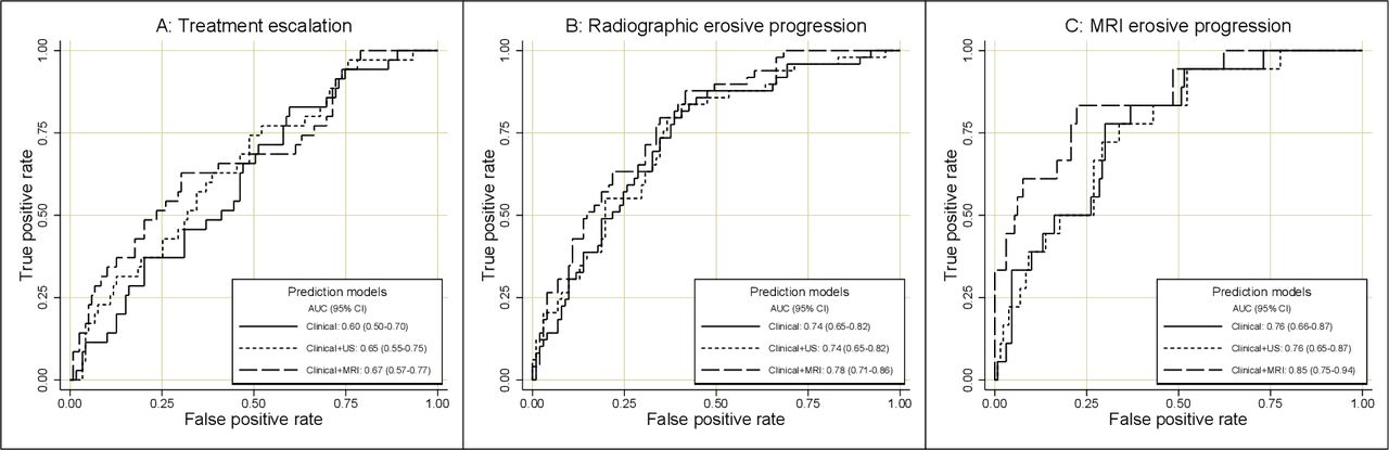
Comparison of the ROC curves of the prediction models showed a non-significant trend towards higher AUC values for the model including MRI data, in comparison with the clinical model and the model including ultrasound data (figure 2A–C). Analyses of outcomes based on the vdHSS total score and the combined MRI damage score showed no significant differences between the prediction models (online supplemental table S6 and online supplemental figure S3A,B). Pure imaging models had low-to-moderate predictive performance, with the exception that the MRI model showed good performance in prediction of MRI erosive progression (online supplemental figure S4A–C).
(A–C) ROC curves of the clinical, clinical+ultrasound and the clinical+MRI prediction models for (A) treatment escalation, (B) radiographic erosive progression and (C) MRI erosive progression during the second year in patients in clinical remission after 1 year. Treatment escalation: change to a higher level of therapy during the second year. Radiographic erosive progression: increase ≥1 vdHSS erosion score during the second year. MRI erosive progression: increase ≥1 RAMRIS erosion score during the second year. AUC, area under the curve; RAMRIS, rheumatoid arthritis MRI scoring system; ROC, receiver operating characteristics; US, ultrasound; vdHSS, van der Heijde modified Sharp score.
Discussion
In this study, we found that a majority of patients with RA in clinical remission after 1 year of treat-to-target therapy had some degree of joint inflammation on MRI and ultrasound, and that the level of MRI inflammation was associated with treatment escalation and radiographic progression in the following year. MRI inflammation at the time of treatment initiation was associated with subsequent MRI erosive progression and ultrasound inflammation with radiographic erosive progression. Nevertheless, addition of MRI or ultrasound information did not substantially improve prediction models based on routine clinical and demographic measures.
Current first-line treatment with methotrexate monotherapy in combination with short-term glucocorticoids is sufficient to achieve clinical remission in approximately 50%–60% of early RA cases,3 39–43 while further escalation of therapy will be necessary to reach the treatment target in remaining patients. In some patients, disease relapse and joint damage continue to occur, despite absence of clinically active disease.5 7–9 We have investigated whether supplementing routinely used clinical, laboratory and radiographic assessments with ultrasound or MRI examinations would improve the ability to predict such adverse developments. In our ‘clinical’ prediction models, we chose to include variables age, gender, ACPA-positivity, radiographic erosions and the components of the DAS (SJC, RAI, PGA and CRP). The variables in the clinical models were selected not based on statistical significance testing, but rather based on clinical reasoning and experience to represent key factors in treatment decision-making and prognosis assessment in current practice. For patients in clinical remission, the duration of remission may be a predictive factor for future outcomes,44 though in our data, adjustment for remission duration did not significantly affect the results of the analyses. To ensure comparability with previously published results from the ARCTIC trial, where radiographic progression was defined as an increase of ≥1 unit of the radiographic score per year, we used a cut-off of ≥1 unit increase of the MRI/radiographic erosion scores per year to define progression. MRI images were read in known chronological order, as this has been shown to increase sensitivity to change without introducing significant bias.45–47 In our analyses, imaging-detected inflammation at baseline was independently predictive of subsequent erosive progression. In patients in remission, MRI subclinical tenosynovitis was independently predictive of future treatment escalation, and MRI tenosynovitis and synovitis were predictive of continued erosive progression, which is consistent with secondary analyses of the IMAGINE-RA trial.48 However, despite statistically significant associations to elements of the future disease course, prediction models incorporating MRI or ultrasound information were not superior to models using only routine measures. Our interpretation of these results is that MRI and ultrasound imaging, by the protocols we applied, have limited value as an addition to routine examinations for improving treatment decision-making in current treat-to-target strategies. This is in line with the main conclusions of the ARCTIC, TaSER and IMAGINE-RA trials.27–29 In future studies, it would be of interest to perform comprehensive cost–benefit analyses, examining the cost of MRI and ultrasound examinations in relation to long-term medical treatment expenses, clinical and functional outcomes.
A limitation of this study is that a rescue option in the ARCTIC trial treatment regimen allowed for patients with high levels of MRI BME at baseline to be directly escalated from methotrexate to biological DMARD after 4 or 6 months, passing over triple therapy, in case of insufficient response to methotrexate. Potentially, this might have weakened the association between baseline BME and subsequent erosive progression. Apart from the baseline assessment of BME, no MRI data were available to the treating physicians during the course of the study. It should also be noted that for 121 of the 218 patients, treatment was partly guided by ultrasound findings, and joints with power Doppler signal were treated with intra-articular corticosteroid injections, which may have negatively biased the associations of ultrasound scores to future outcomes such as erosive progression. Nonetheless, sensitivity analyses of our data did not indicate stronger predictive associations in the patient group managed without ultrasound guidance, and earlier analyses of the same trial have not shown significant differences in treatment outcomes between the study arms.28 35 Another limitation is that in the ARCTIC trial, joint tenderness was assessed using the RAI, which is an infrequently used measure in clinical practice and therefore may be unfamiliar to some practitioners. The strengths of this study include the comprehensive clinical, laboratory and multimodality imaging examinations at different phases of the disease course, and at different levels of disease activity, in a study sample representative of a general population with early RA treated according to current recommendations.
In conclusion, we found that MRI and ultrasound inflammation at the time of RA diagnosis is associated with subsequent erosive progression, but not with poor methotrexate response. In patients in remission after 1 year of treat-to-target therapy, imaging-detected subclinical inflammation is frequently present, and is associated with future treatment escalation and continued erosive progression. We were however unable to establish a significant benefit of including imaging information in prediction models, supporting that systematic use of MRI and ultrasound examinations has limited value in routine follow-up of patients with early RA.
Acknowledgments
We would like to thank all investigators, study personnel and patients who have contributed to the ARCTIC trial. We thank Øyvind Skare for help and advice on statistical calculations and support on using statistical software, and to Daniel Glinatsi for guidance in MRI assessments using the RAMRIS system. We also thank all contributing radiology personnel, in particular Dag Sjølie, for help on MRI technical issues, and Karwan Faraj for help with MRI assessment.
References
Supplementary materials
Supplementary Data
This web only file has been produced by the BMJ Publishing Group from an electronic file supplied by the author(s) and has not been edited for content.
Footnotes
EAH and SL are joint last authors.
Twitter @UlfSundin
Collaborators The ARCTIC Study Group: Hallvard Fremstad; Tor Magne Madland; Åse Stavland Lexberg; Hilde Haukeland; Erik Rødevand; Christian Høili; Hilde Stray; Anne Noraas Bendvold; Inger Johanne W Hansen; Gunnstein Bakland.
Contributors All authors were involved in the analysis and/or interpretation of data, and in drafting the article or revising it critically for important intellectual content. All authors approved the final manuscript to be submitted and agreed to be accountable for all aspects of the work. Conception and design of the study—US, A-BA, NPS, JS, TKK, EAH and SL. Acquisition of data—US, A-BA, NPS, LBN, DvdH, HBH, EAH and SL. Members of the ARCTIC Study Group, HF, TMM, ÅSL, HH, ER, CH, HS, ANB, IWH and GB, recruited and enrolled patients, and collected and interpreted data.
Funding The ARCTIC Study has received grants from the Norwegian Research Council, the South-East Health Region in Norway, the Norwegian Rheumatism Association, the Norwegian Women’s Public Health Association, and unrestricted grant support from AbbVie, Pfizer, MSD, Roche and UCB.
Disclaimer The funders of the study had no role in study design, data collection, data analysis, data interpretation or writing of the report. The corresponding author had full access to all the data in the study and had final responsibility for the decision to submit for publication.
Competing interests A-BA reports grants from Pfizer, UCB, Roche, MSD, AbbVie, the Norwegian Research Council, the Norwegian South-Eastern Health Region, the Norwegian Women’s Public Health Association and the Norwegian Rheumatism Association, and personal fees from AbbVie, Eli Lilly, Novartis, Pfizer and UCB Pharma. HBH reports personal fees from AbbVie, Lilly and Novartis. DvdH reports personal fees from AbbVie, Astellas, AstraZeneca, Boehringer Ingeleheim, BMS, Celgene, Cyxone, Daiichi, Eisa, Eli-Lilly, Galapagos, Gilead, Glaxo-Smith-Kline, Janssen, Merck, Novartis, Pfizer, Regeneron, Roche, Sanofi, Takeda, UCB Pharma, and is Director of Imaging Rheumatology BV. TKK reports consulting fees, speaking fees, and/or honoraria from AbbVie, Amgen, Biogen, BMS, Celltrion, Egis, Eli Lilly, Evapharma, Ewopharma, Janssen, MSD, Mylan, Oktal Pharma, Orion Pharma, Pfizer, Roche, Sandoz, Sanofi and UCB Pharma. EAH reports grants from Pfizer, UCB, Roche, MSD, AbbVie, the Norwegian Research Council and the Norwegian South-Eastern Health Region, and speaker/consultant honorariums from Eli Lilly, Pfizer, UCB, Janssen, Celgene, and AbbVie. SL reports grants from Pfizer, UCB, Roche, MSD, AbbVie, the Norwegian Research Council and the Norwegian South-Eastern Health Region.
Patient and public involvement statement No patients were involved in the development of the study design, conduct of the study or reporting of these data.
Patient consent for publication Not required.
Ethics approval The study was approved by an independent ethics committee (the Regional Committee for Medical and Health Research Ethics South-East; reference number 2010/744), and performed in compliance with the Helsinki Declaration and guidelines for good clinical practice.
Provenance and peer review Not commissioned; externally peer reviewed.
Data availability statement The authors commit to making the relevant anonymised patient-level data available on reasonable request. Requests should be directed to the corresponding author, uffe.sundin@gmail.com.