Article Text

Abstract
Glioblastoma is the most common form of primary brain cancer and remains one of the most aggressive forms of human cancer. Current standard of care involves maximal surgical resection followed by concurrent therapy with radiation and the DNA alkylating agent temozolomide. Despite this aggressive regimen, the median survival remains approximately 14 months. Meaningful strategies for therapeutic intervention are desperately needed. Development of such strategies will require an understanding of the therapeutic concepts that have evolved over the past three decades. This article reviews the key principles that drive the formulation of therapeutic strategies in glioblastoma. Specifically, the concepts of tumour heterogeneity, oncogene addiction, non-oncogene addiction, tumour initiating cells, tumour microenvironment, non-coding sequences and DNA damage response will be reviewed.
- Glioblastoma
- cancer
- targeted therapy
- oncogene addiction
- non-oncogene addiction
- tumour-initiating cells
- microenvironment
- non-coding sequences
- DNA damage response
- neurosurgery
- epidemiology
- neuroanatomy
- oncology
- molecular biology
Statistics from Altmetric.com
- Glioblastoma
- cancer
- targeted therapy
- oncogene addiction
- non-oncogene addiction
- tumour-initiating cells
- microenvironment
- non-coding sequences
- DNA damage response
- neurosurgery
- epidemiology
- neuroanatomy
- oncology
- molecular biology
Introduction
Glioblastoma is the most common form of primary brain tumour. The incidence of this tumour is fairly low, with two to three cases per 100 000 people in Europe and North America.1 It is one of the most aggressive forms of human cancer.2 Without treatment, the median survival is approximately 3 months.3 The current standard of treatment involves maximal surgical resection followed by concurrent radiation therapy and chemotherapy with the DNA alkylating agent temozolomide (TMZ).4 5 With this regimen, the median survival is approximately 14 months. For nearly all affected, the treatment remains palliative.
The best available evidence suggests that glioblastomas originate from cells that give rise to glial cells.6 7 These glial-derived tumours are graded by WHO into four categories, termed WHO grade 1–4. The higher grade denotes histological features of increased malignancy. WHO grade 4 glioma is essentially synonymous with glioblastoma.8
Studies carried out over the past three decades suggest that glioblastomas, like other cancers, arise secondary to the accumulation of genetic alterations. These alterations can take the form of epigenetic modifications, point mutations, translocations, amplifications or deletions and modify gene functions in ways that deregulate cellular signalling pathways leading to the cancer phenotype.9 The exact number and nature of genetic alterations and deregulated signalling pathways required for tumorigenesis remains an issue of debate,10 although it is now clear that central nervous system (CNS) carcinogenesis requires multiple disruptions to the normal cellular circuitry. These genetic alterations result in either activation or inactivation of specific gene functions that contribute to the process of carcinogenesis.10 Genes that, when activated, contribute to the carcinogenesis are generally termed proto-oncogenes. The mutated forms of these genes are referred to as oncogenes. Genes that, when inactivated, contribute to the carcinogenesis are termed tumour suppressor genes.
Recent research in the area of experimental and clinical oncology has identified the key signalling pathways, critical regulatory nodes, genes and their protein products, and their mutual cross-talks, thereby providing a solid molecular basis for selection of candidate therapeutic targets and drug discovery programmes. These lines of investigation complement the recent efforts to sequence entire genomes of a growing number of human tumours including glioblastoma. The efforts have led to the formulation of new concepts and principles in tumour cell biology. Exploitation of these major advances has begun to provide exciting leads that may afford innovative therapeutic strategies. This article will aim to review these critical concepts and their relevance for glioblastoma therapeutic development.
Concept 1: glioblastoma subtypes
There is an old adage that cancer is a hundred diseases masquerading in one. While this adage is based on clinical and pathologic observations, systemic genomic characterisation of a large number of glioblastoma specimens confirms the notion that subtypes with distinct pathological molecular events and therapeutic responses exist.
The Cancer Genome Atlas project (TCGA) is a major National Institutes of Health initiative involving institutions spanning the continental USA with the goal of tumour specimen collection and molecular characterisation.11 Glioblastoma was one of the first tumour types characterised in this effort. This vast wealth of data is unprecedented, and despite the enormous challenge to process and analyse this incoming information, correlations of such emerging ‘genetic and expression profiles’ or ‘tumour landscapes’ with tumour biology and clinico-pathological features of the patients (including therapeutic responses) are beginning to impact oncology.
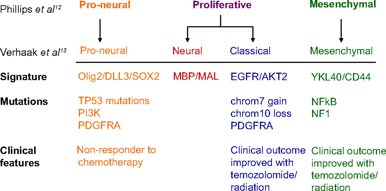
These studies12 have led to the understanding of glioblastoma as an umbrella term that encapsulates subtypes characterised by distinct molecular properties. Based on global transcript profiling, glioblastoma can be divided into three to four distinct subtypes.12 13 Interestingly, each subtype harbours distinct genetic aberrations13 and proteomic profiles.14 The recognition that glioblastoma consists of subtypes varying in molecular circuitry and biological behaviour suggests that no therapy can be universally efficacious. The major importance of this concept of heterogeneity is that meaningful therapeutic gain can only be attained by customising the therapy to the underlying molecular circuit. One subtype (termed classical by the TCGA and proliferative by Phillips et al) is characterised by frequent amplification or mutations in the epidermal growth factor receptor (EGFR) gene.11 12 In contrast, another subtype, termed proneural by both groups, harbours frequent mutations in p53, platelet-derived growth factor receptor A and isocitrate dehydrogenase 1.13 A third type, termed mesenchymal, is characterised by frequent mutations in the neurofibromatosis type 1 gene (NF-1).
Importantly, these transcriptomal subtypes appear to differ in their clinical courses and therapeutic responses. In terms of prognosis, studies by Phillips et al12 and Verhaak et al13 demonstrated increased overall survival in patients with proneural glioblastoma relative to other molecular subtypes. In terms of therapeutic response, Verhaak et al.13 explored this issue by stratifying the patients with various molecular subtypes into two groups: those that received concurrent chemo-radiation therapy or received more than three cycles of chemotherapy; and those that did not receive concurrent chemo-radiation therapy or received less than four cycles of chemotherapy. When stratified this way, the authors found that the two groups exhibited comparable survival in the proneural group. In contrast, for other molecular subtypes, patients in the first group exhibited improved survival relative to the second group.13 Since the analysis combined the survival effect of concurrent chemo-radiation therapy and prolonged chemotherapy, it is difficult to assess whether the effect is due to the former or the latter. Taken as a whole, these datasets suggest that the patients with the proneural glioblastomas tend to survive longer but are less responsive to conventional chemotherapy or chemo-radiation therapy (figure 1).

Transcriptomal subtypes of glioblastoma.
Concept 2: oncogene addiction
The term ‘oncogene addiction’ was initially coined by Dr Bernard Weinstein to describe the phenomenon that some tumours exhibit exquisite dependence on a single oncogenic protein (or pathway) for sustaining growth and proliferation.15 Such dependence has been convincingly demonstrated in both tissue culture and transgenic mice systems for oncogenic versions of MYC16–18 and RAS.19 Application of this concept to the clinical setting has achieved variable success in some various cancer types, including chronic myelogeneous leukaemia harbouring the BCR-ABL translocation, Erb2 overexpressing breast cancer, and non-small cell lung cancer harbouring a subset of EGFR mutations.20 21 A simplistic application of this concept in glioblastoma would involve identification of the critical ‘addicted’ oncogene followed by the inhibition of such oncogenes. Unfortunately, the actual biology of glioblastoma is far more complex.
To understand this complexity, a careful analysis of the fundamental notion of oncogenic addiction is needed. In some ways, the observation that tumours exhibit dependence on a particular oncogenic pathway at some point in its history is not surprising. However, considering the plethora of dynamic genetic changes that accumulated during cancer progression,22 it is somewhat counter-intuitive to suspect that any particular pathway would play a prominent role in maintaining cell viability. Moreover, inactivation of the normal counterpart of the addicted oncogenic protein is often tolerated in normal tissue. These observations suggest that the genetic circuitry of the cancer cell have been extensively reprogrammed to result in this ‘addicted’ state.15
The molecular nature of this reprogramming remains poorly understood. Several hypotheses have been put forward. One hypothesis involves the notion of ‘genetic streamlining’, where genetic instability in cancer cells is thought to mutationally or epigenetically inactivate certain signalling pathways that are operational in a normal cell but not required for growth in the cancer cell. In this ‘streamlined’ state, the tumour cell becomes hyper-dependent on the oncogene-driven processes.23 A more generalised form of this explanation involves the notion of synthetic lethality. Two genes are considered synthetically lethal if cells remain viable with inactivation of either gene. Simultaneous inactivation of both genes, on the other hand, results in cell death.24 It is thought that the cancer cells have accumulated mutations that are synthetically lethal with the absence of critical oncogenes. The main difference between this hypothesis and the ‘streamline’ hypothesis is that the mutation in the former can result in a gain or loss of function, whereas the later specifically proposes a loss of function. A third hypothesis suggests that oncogenes reprogramme the tumour cell by both pro-survival and pro-apoptotic signalling.23 25 26 With acute inactivation, the pro-survival signalling decayed faster than the pro-apoptotic signalling, resulting in tumour death. This thesis has been coined the ‘oncogene shock’ hypothesis.23 25 26
The main reason for revisiting the framework of oncogene addiction is to discuss the mechanism by which the cells can evolve to avoid such addiction. For instance, in the context of synthetic lethality, EGFR inhibition may be cytotoxic to glioblastoma cells only in the appropriate genetic context. Indeed, therapeutic effects of EGFR inhibition were observed only in patients with tumours expressing an oncogenic form of EGFR and an intact phosphatase and tensin homolog (PTEN) tumour suppressor gene.27 To complicate the matter, recent studies demonstrate that glioblastomas harbour activation of multiple oncogenic receptor tyrosine kinases, such that inactivation of any single oncogene merely diverts signalling through other active oncogenes.28 In these contexts, it is evident that meaningful therapy will require simultaneous inhibition of multiple oncogenes or identification of the fitting genetic context (figure 2).

Oncogene and non-oncogene addiction.
Concept 3: non-oncogene addiction
Emerging literature suggests an alternative strategy to the multi-target approach. These studies reveal that oncogene activation introduces secondary physiological changes that stress cellular capacity for survival. Consequently, tumour cells becomes more dependent (or hyper-dependent) on processes required to compensate for these stressful conditions.29 30 This phenomenon is termed ‘non-oncogene addiction’ since the compensatory processes required for tumour survival do not directly contribute to the cancer formation. In other words, even the genes that are not themselves targeted by tumorigenic mutations may well become essential for the tumour to survive the stressful environment and fuel the demanding process of tumour progression. Consequently, interfering with the function of such genes could cause tumour kill while sparing the normal counterpart (figure 2).29 30
There are several examples of such critical non-oncogenic pro-survival functions required for the maintenance of the tumorigenic state in glioblastoma. EGFR is a critical proto-oncogene in glioblastoma pathogenesis.11 31 Our laboratory has demonstrated that EGFR hyperactivation results in an increased accumulation of reactive oxygen species (ROS), which in turn cause cytotoxic DNA damage. To compensate for the deleterious effect of ROS, EGFR hyperactive glioblastomas exhibit increased reliance on the DNA repair process required for the repair of ROS-related DNA damage.32 Selective targeting of EGFR hyperactive glioblastomas can, thus, be achieved by inhibition of these repair processes. Other groups have demonstrated that EGFR hyperactivation in glioblastoma cell lines heightens requirement for lipogenesis.33 34 Additional examples of such critical non-oncogenic pro-survival functions required for maintenance of the tumorigenic state include dependency on mechanism for compensating mitotic and proteotoxic stress and interplay with the tumour microenvironment including the immune system.29
The principle of non-oncogene addiction suggests that there is a wider spectrum of therapeutic options than afforded under the paradigm of ‘oncogene addiction’. In many cases, compensatory processes involved in ‘non-oncogene addiction’ are the same as those that basic scientists have studied for years (for instance, DNA repair). Mechanistic investigations into these biological processes by the basic scientists have yielded a rich database of inhibitors. Thus, identifying gene functions that compensate for oncogene-induced cellular stress should afford opportunities to tap into this rich database and expand the denominator of drugs available for combinatorial therapy. Targeting genes that are synthetically lethal with oncogenes constitutes an attractive means to this end.
It is important to note that the effects of therapies designed based on the principles of ‘oncogene addiction’ and ‘non-oncogene addiction’ are inherently antagonistic. For instance, EGFR inhibition leads to a reduction in ROS, obviating the need for DNA repair.32 In this context, the combination of DNA repair inhibition and EGFR inhibition would not be desirable. Rational strategies for synthesising the two therapeutic paradigms remains a major intellectual challenge.
Concept 4: tumour-initiating cells
Another advance that may profoundly change our thinking about solid tumours including glioblastoma involves the concept of tumour-initiating cells. The experimental observation is that within a total population of glioblastoma cells, there appears to be a small subpopulation of cells that are highly tumorigenic (hence the term ‘tumour-initiating cells’ or ‘TICs’), with capacity for self-renewal.35 36 In some studies in which severely immune-compromised mice are used as assay for melanoma xenograft formation, the proportion of TICs within a tumour has been reported to be as high as 27%.37 Because glioblastoma TICs share many common properties with neural stem cells, it is proposed that TICs originated from stem cells. While there are some data supporting this hypothesis,6 the universality of this hypothesis remains controversial.
Protein markers to prospectively identify and isolate these putative TICs have been reported, such as the transmembrane glycoprotein CD133 (prominin-1) in glioblastomas.6 However, the value of CD133 as a single marker of glioblastoma TICs remains controversial, partly because CD133-negative glioblastoma cells could also give rise to tumours in an intracranial mouse xenograft model.38–40 These uncertainties motivate an ongoing search for additional candidate TIC markers. Candidate cell surface molecules suggested in this context include the adhesion glycoprotein L1CAM,41 surface carbohydrate antigen CD15 (SSEA-1),42 surface marker A2B543 and integrin α6.44 Currently, there are no generally accepted cell surface markers for defining TICs. The definition of TICs remains a functional one as defined by the ability of a tumour cell to sustain self-renewal and initiate glioblastoma formation in immunocompromised xenograft models (figure 3).

Hierarchy of tumour initiating cells.
Arguably, the most important aspect of the concept of TICs is that this population appeared particularly resistant to conventional radiation and chemotherapy.35 In this context, TICs may be responsible for glioblastoma recurrence after conventional therapy. Given such properties, it is understandable that glioblastoma research has recently focused on identification and development of potential anti-TIC therapies. Two of these strategies, namely targeting the TICs as part of a vascular niche, and attempts to overcome their therapeutic resistance, are discussed in the following sections on glioblastoma angiogenesis and the role of DNA damage response pathways, respectively. Here, we briefly consider other strategies that are emerging as potentially fruitful approaches to treat glioblastoma through targeting TICs.
The first strategy reflects the efforts to identify suitable cell surface markers to reliably identify glioblastoma TICs—with the hope of conjugating the corresponding antibody to cytotoxic compounds as therapeutic agents. The second strategy is based on observations that some TICs, like neural stem cells, can be induced into a differentiated state in which the self-renewal properties are lost. Among the suggested agents to induce such TIC differentiation, the bone morphogenetic proteins appear promising.45 The third strategy involves modulating specific signalling pathways required for maintaining the TIC state. Pathways targeted include those mediated by EGFR, Wnt-beta catenin, signal transducer and activator of transcription 3, Sonic Hedgehog-Gli and Notch pathways.46 Finally, normal neural stem cells have been shown to migrate towards and track TICs. Based on this principle, neural stem cells have been used as delivery vehicles to increase local concentration of therapeutic agents in the vicinity of TICs.47
Concept 5: tumour microenvironment
Over the past two decades, conceptualisation of glioblastomas has evolved from a collection of relatively homogenous cells to the recognition of distinct subpopulations of tumour cells to that of a complex organ, with constant interactions between tumour cells and aberrant stromal elements. Analogous to the distinct functions of different tissues in an organ, genomic characterisation using cells derived from distinct regions of the tumour revealed genetic heterogeneity.48 A major concept in oncology has emerged that reciprocal signalling between the distinct subpopulations of neoplastic cells and the aberrant stromal elements serve to sustain progressive neoplastic transformation and possibly functional specialisation.49 Understanding of these interactions has afforded novel therapeutic targets. For the purpose of this review, distinct subpopulation of neoplastic cells (tumour heterogeneity) and aberrant stromal interactions will both be considered as components of the microenvironment.
Studies of EGFR revealed a beautiful illustration of signalling between the subpopulations of genetically distinct neoplastic cells in glioblastoma. EGFRvIII is a variant of EGFR that arose from spontaneous deletion of exons 2–7.50 This variant is present in about 20% of glioblastomas51 and results in constitutive hyper-activation of EGFR.52 Clinical studies suggest that patients with glioblastoma harbouring this variant tend to have a worse prognosis.53 54 Interestingly, the vIII variant is rarely found in the absence of EGFR overexpression.54 55 Further, when found, the variant is typically present in only a subset of the total tumour mass.54 55 Investigations into the molecular mechanism underlying these observations revealed that EGFRvIII overexpression increased the secretion of interleukin 6 (IL-6) and leukaemia inhibitory factor, two soluble cytokines. These cytokines trigger phosphorylation of gp130 in the non-EGFRvIII expressing cells, which in turn activate EGFR of these cells.56 This activation increases the tumorigenicity and aggressiveness of the cancer. Such signalling may serve to actively maintain tumour cell heterogeneity (figure 4).

Complexity of the tumor microenvironment.
In addition to the signalling between distinct and genetically defined subpopulations of tumour cells, normal cells without genetic alterations associated with carcinogenesis are often recruited to the foci of tumour cells.49 In the process, these normal cells undergo phenotypic changes in response to direct physical interaction with cell surface proteins on the tumour cells or through interaction with secreted soluble factors. These changes result in the release of growth factors that further enable and sustain neoplastic transformation57 or lead to new blood vessel formation.58 Cycles of such reciprocal interaction facilitate stepwise progression in neoplastic progression.49
In terms of the non-neoplastic cell types shown to facilitate neoplastic information, they can generally be divided into three categories. The first category involves endothelial cells or endothelial cell precursors. These cells are critical for tumour growth since there are inherent limitations on the distance that oxygen and macromolecules can travel. In xenograft models, solid tumours can only proliferate up to a size of 1–2 mm without the development of a new blood supply.59 Quiescent endothelial cells in proximity of the neoplastic foci may be induced to initiate biological programmes that lead to blood vessel formation by secreted factors such as vascular endothelial growth factor (VEGR, see below).60 Alternatively, endothelial cell precursors in the bloodstream may be recruited into the tumour foci.58 61 A final mechanism involves the trans-differentiation of TICs to become endothelial cells.62
The second class of non-neoplastic cells that actively participate in tumour progression is fibroblasts. There is good evidence that these otherwise genetically normal fibroblasts, when in proximity of tumour cells, can become ‘re-programmed’ to promote/sustain neoplastic transformation. Transplantation experiments mixing cancer-associated fibroblast with cancer cells lead to a more aggressive tumour phenotype than ‘normal’ tumour cells. This tumour-promoting activity is largely thought to be the combined effect of cell-to-cell interaction and cytokine release.57 Non-neoplastic astrocytes perform many of the functions associated with fibroblasts. Thus, the interactions between glioblastoma cells and non-neoplastic astrocytes warrant further investigations.
The final class of non-neoplastic cells recruited are cells that mediate immune function. In general, these cells may possess tumour-antagonising activity or tumour-promoting activity.49 These divergent properties may be rationalised by understanding that the immune system is required for both the destruction of foreign cells and facilitating wound healing. Properties associated with the former will likely lead to tumour ablation.63 However, cytokines and growth factors associated with wound healing may promote tumour growth.64
The glioblastoma cells have evolved a large number of mechanisms that allow escape from immune detection and ablation, including release of immunosuppressive cytokines, such as IL-10, cytotoxic T-lymphocyte antigen 4 and transforming growth factor ß65 66 or expression of cell surface molecules that facilitate immunosuppression, such as B7-H1.67 These events, in turn, lead to the induction of regulatory T cells (Treg), downmodulation of antigen-presenting cells, with concomitant loss of T-cell effector function68 or loss of functional major histocompatibility class I receptors.69
These factors contribute to a ‘hostile’ microenvironment that compromises the immune cells' ability to achieve tumour eradication. For instance, primed CD8+ cytotoxic T cells can penetrate the blood–brain barrier and access the CNS.70 However, they are incapable of tumour eradication. Indeed, in patients with glioblastoma, tumour progression is seen despite the presence of tumour-infiltrating lymphocytes.71
Interestingly, glioblastoma's capacity to suppress immune response appears intimately associated with the process of neoplastic transformation. PTEN encodes a tumour-suppressing phosphatase that is frequently mutated during glioblastoma pathogenesis. The translation of many immune-suppressive cytokines and molecules, including IL10 and B7-H1, are under the regulation of PTEN. Thus, PTEN loss during neoplastic transformation leads to increased expression of immune-suppressive cytokines and cell surface molecules.67 This expression, in turn, creates a hostile environment for immune cells that otherwise target tumour for ablation.
Understanding the interaction between the genetically distinct subpopulation of glioblastoma cells and their microenvironment has yielded novel therapeutic developments. The endeavour most frequently cited in this regard involves angiogenesis inhibitors. Realising that VEGF is critical in angiogenesis, bevacizumab, a humanised antibody against VEGF, was developed.72 While there has not been a randomised control trial to assess the efficacy of bevacizumab, a phase II clinical trial demonstrated improved progression-free survival in recurrent glioblastomas (after concurrent TMZ/radiation treatment) relative to historical data of patients who received TMZ at recurrence.73–75 However, no overall survival benefit has been demonstrated with bevacizumab treatment. Clearly, angiogenesis inhibition is but one of the many strategies that can be developed based on the concept of tumour microenvironment.
Concept 6: non-coding DNA sequences
Classically, coding sequences are defined as the strand of DNA that has the same base sequence as the RNA transcript produced (with the caveat that thymines are replaced by uracil) that are ultimately translated into proteins. While the identification of nucleotide alterations within the coding sequences of proto-oncogene or tumour suppressor genes has significantly contributed to our understanding of carcinogenesis, there is an emerging appreciation that alterations in non-coding sequences similarly contribute to carcinogenesis.76 A notable example involves the regulation of gene transcription by reversible modification of gene promoter regions—a phenomenon sometimes referred to as ‘epigenetic regulation’.77 Similarly, we are beginning to appreciate the importance of transcripts that do not encode for proteins but are transcribed, such as microRNAs (miRNAs)78 and long non-coding RNAs (or LincRNAs),79 80 in terms of transcriptional and post-transcriptional modifications. The concept that non-coding DNA sequences regulate gene function and impact carcinogenesis has significantly expanded the repertoire of strategies available for glioblastoma therapeutics. To review this concept, we will discuss illustrative examples of epigenetic regulation, miRNAs and LincRNAs (figure 5).

Gene regulation by non-coding RNAs.
The term ‘epigenetic regulation’ has been coined to describe the phenomenon that heritable changes in gene expression can occur in the absence of changes in the DNA sequences encoding for gene function.77 The mechanism underlying this regulation involves cytosine methylation81 or histone modifications that, in turn, modulate the accessibility of gene promoter regions to transcriptional factors.82 Cytosine methylation typically occurs in the context of CpG di-nucleotide repeats, or CpG islands.81 Promoters harbouring heavily methylated CpG islands are typically transcriptionally silenced. There are two types of promoter methylation that are particularly pertinent to glioblastoma therapy: methylation in the promoter region of the DNA repair gene, methyl-guanine methyl transferase (MGMT) and the glioma-CpG island methylator (G-CIMP) phenotype.
MGMT encodes an enzyme that removes alkyl adducts at the O6 position of guanine.83 Because alkyl modification at this position is highly toxic and constitutes the primary mechanism for the tumoricidal activity of the chemotherapeutic agent TMZ, MGMT expression level correlates well with TMZ response in patients with glioblastoma.84 The human MGMT gene possesses a CpG island that spans approximately 1000 bases around the transcriptional start site. Detailed analysis of this region revealed 108 CpG sites85 that are methylated. Methylation of a subset of these CpGs has been associated with transcriptional silencing of MGMT86 87 and is associated with improved clinical outcome in patients with glioblastoma receiving TMZ therapy. Interestingly, MGMT promoter methylation is also associated with improved survival in patients who did not receive TMZ therapy.88 89 While the mechanism underlying this observation remains unclear, it seems likely that MGMT may participate in detoxifying the accumulation of endogenous DNA damage that is typically associated with the oncogenic state.32 As discussed in concept 7, glioblastoma cells accumulate endogenous DNA damage in the absence of DNA damaging agents.32 These endogenous DNA damages are not unlike those induce by temozolomide or radiation in that they could trigger cell death if unrepaired. Thus, tumours with high levels of MGMT may grow more robustly since MGMT is capable of detoxifying these endogenous DNA damages. If the tumour cells grow more robustly, the patient will survive for a shorter duration. In contrast, the glioblastoma cells with low MGMT may be more susceptible to the deleterious effects of the endogenous DNA damages. These tumours may grow less robustly, resulting in longer patient survival.
The G-CIMP phenotype refers to the observation that a subset of glioblastomas exhibits concerted CpG island methylation at a large number of loci.90 Since genes required for tumour growth are located at many of these loci, glioblastomas harbouring the G-CIMP phenotype tend to be more benign. Correspondingly, patients with G-CIMP glioblastomas experienced significantly improved outcome. Understanding the concept that the patterns of CpG island methylation directly impact outcomes in patients with glioblastoma open the door to therapeutic strategies aimed at enhancing promoter methylation at select promoter loci. Importantly, recent studies suggest that promoter methylation at distinct loci may be affected by specific chromatin-modulating factors.91
miRNAs are small non-coding RNAs of 20–22 nucleotides that, through imperfect pairing, bind to the 3′ untranslated regions of protein-coding mRNAs. Typically, this binding leads to mRNA degradation or inhibition of protein translation to suppress the expression of the target proteins.78 Bioinformatic analysis predicts that a single miRNA can potentially regulate hundreds of target oncogenes or tumour suppressor proteins. Expectedly, miRNAs have been implicated in carcinogenesis and resistance to chemotherapy.78 As one illustrative example, our laboratory recently demonstrated that the protein MGMT is under the regulation of miR-181d.92 Cell biological studies revealed that binding of miR-181d to the 3′ untranslated regions of MGMT caused decreased MGMT expression. This inverse relationship was validated in glioblastoma specimens. Importantly, patients with high miR-181d expression (hence low MGMT) are more likely to respond to TMZ chemotherapy.
LincRNAs are transcripts >5 kb that are evolutionarily conserved across mammalian genomes. These RNAs are transcribed by polymerase II but do not encode proteins. The LincRNAs serve to suppress transcription by targeting chromatin-modifying complexes to specific genomic loci.79 80 While the role of LincRNA in glioblastoma awaits careful scrutiny, LincRNA have been shown to mediate the function of tumour suppressor genes pertinent to glioblastoma pathogenesis. As one example, TP53 encodes a transcription factor that regulates gene sets critical for cell cycle progression and apoptosis. Under normal conditions, p53 is a short-lived protein.93 In response to cellular stress (for instance, DNA damage or oncogene expression), p53 undergoes post-translational modifications and protein–protein interactions that enhance its stability and transcriptional activity.93 One of the downstream effectors of p53 is a LincRNA. This LincRNA serves as a key mediator to suppress transcription of other p53 effectors.94 Such mechanisms may be operational in glioblastomas.
Understanding the concept that non-coding sequences play critical roles in glioblastoma pathogenesis and resistance to chemotherapy offers novel strategies for biomarker development and therapy. For instance, direct introduction of select miRNAs into glioblastoma has been shown to inhibit growth and proliferation.95 Similarly, incorporation of miR-181d expression level may further augment the predictive value of MGMT promoter methylation. Importantly, the concept predicts certain situations where the effects of an oncogenic mutation can be voided by the effects of non-coding sequences. Integrating the biology of non-coding sequences in the context of mutational profile will be critical in understanding tumour physiology and meaningful therapeutic development.
Concept 7: DNA damage response
From a broader perspective, the status of the molecular machinery that detects, signals and repairs DNA damage, and overall orchestrates the multifaceted cellular response to genotoxic insults (here referred to as the DNA damage response: ‘DDR’96) critically impacts tumour development and clinical outcome. While this is arguably relevant for any type of tumour to some extent, the DDR concept is particularly important for glioblastomas for the following reasons. First, the standard-of-care non-surgical modalities used to treat glioblastomas, namely ionising radiation and TMZ-based chemotherapy, operate through their genotoxic effects by causing mainly DNA double strand breaks (DSBs) and alkylated DNA lesions, respectively. Therefore, each individual patient's germ-line disposition of the DDR-related genes, along with any somatic alterations within the DDR machinery that have been selectively acquired by the tumour dictate (along with other factors such as the tumour microenvironment discussed above) their response to therapy. Second, among the hallmarks of glioblastomas is their resistance to radiotherapy and chemotherapy.97 These phenomena highlight the intimate involvement of the cellular DDR network, particularly DNA damage signalling, cell-cycle checkpoints and DNA repair pathways, in the pathobiology of glioblastomas. Third, the harmful side effects of the standard therapies, including brain damage and consequently cognitive changes, are also attributable to DNA damage and the cellular and tissue responses to such treatments. Fourth, genetic and/or epigenetic aberrations of a range of DDR factors, including the above mentioned p53 tumour suppressor or DNA repair genes such as MGMT, occur commonly during glioblastoma pathogenesis and/or upon treatment. This aspect of gliomagenesis has been suspected and partly known for years, however it has only been validated by the recent insights gained through comprehensive analyses by complete tumour genome sequencing within the framework of the TCGA initiative.11 Finally, the TICs (see concept 4), appear to be particularly resistant to DNA-damaging therapies. This resistance is, at least in part, due to enhanced DNA damage signalling and checkpoint machinery.35
Conceptually very relevant for such DDR-related features of gliomas is the recently described strong, constitutive activation of the DDR signalling pathways, observed from the early stages (grade II gliomas) of gliomagenesis up to glioblastomas.98 This spontaneous DDR activation precedes any genotoxic treatment, and it appears to be even more pronounced in gliomagenesis than in early lesions of major epithelial tumour types, in which this phenomenon represents a candidate intrinsic barrier against activated oncogenes and tumour progression.98–102 A major source of such DDR activation in early lesions including low-grade gliomas appears to be oncogene-induced replication stress, while in later stages of tumour progression, particularly in glioblastomas, the constitutive DNA damage signalling is fuelled by continued replication stress and by enhanced oxidative stress.32 98 100 103 Biologically, such oncogene-evoked DDR activation often leads to cell death or permanent proliferation arrest known as cellular senescence. This activation eliminates nascent tumour cells from the proliferative pool, thereby delaying or preventing tumour progression.99 102 Those lesions that do progress in the face of such constitutively activated DDR often do so by selection of various defects along the DDR signalling or effector pathways, such as mutations in the ATM-Chk2-p53 DDR pathway.99 102 104 Importantly, while such selected DDR aberrations facilitate tumour progression by allowing escape from DDR-induced senescence or apoptosis, the very same defects may create tumour-specific vulnerabilities that can be exploited by therapeutic strategies based on the synthetic lethality principle (see concept 2 above).29 32 96
In terms of exploiting the status of the DDR machinery for glioblastoma therapies, two major avenues are under intensive research and validation. First, there are promising attempts to sensitise glioblastoma cells (including the more resistant TICs) to conventional genotoxic therapy, such as ionising radiation, by concomitantly inhibiting the DNA damage signalling to downstream checkpoint and repair effectors. This strategy relies mainly on small molecule inhibitors of DDR kinases ATM, ATR, Chk1 and Chk2. This strategy appears particularly suitable for tumours with mutant p53. Such cancer cells lack the major p53-dependent G1/S checkpoint, and upon inhibition of the DDR kinases (whose activity underlies the still operational G2/M checkpoint) enter mitosis with an overload of unrepaired DNA damage, both endogenous and therapy induced, followed by cell death.29 105 An analogous strategy to overload glioblastoma cells with unrepaired DNA damage involves TMZ treatment with concurrent inhibition of MGMT in those cases where the MGMT gene promoter is not methylated.106
An emerging alternative treatment strategy takes advantage of the synthetic lethality and the accumulated knowledge about the DDR mechanisms.29 107 This strategy exploits tumour-selective defects in certain DNA repair pathways, such as DSB repair, by homologous recombination (HR). HR is a mechanism to copy a DNA sequence from an intact DNA molecule (mainly from the newly synthesised sister chromatid) to bypass or repair replication-associated DNA lesions. This promising strategy exploits HR defects that are found in some tumours. These HR-deficient tumours are particularly dependent on other repair processes to avoid the generation of DSBs. These tumour cells are therefore particularly sensitive to inhibition of these other repair processes. Such a strategy has shown promise in preclinical studies in which breast tumour cells defective to HR appear hypersensitive to inhibition of base excision repair by small molecule inhibitors of poly(ADP-ribose) polymerase (PARP).96 108 Of note, PARP inhibition has shown promise in glioblastoma treatment in cell culture models.109 and several PARP inhibitors are under investigation in clinical glioblastoma trials.110
Summary
In this review, we have discussed key principles underlying the current development of glioblastoma therapeutics. Emphasis was placed on conceptual frameworks rather than specific drugs or targets. These frameworks should serve as the basis for translating fundamental biological tenets into clinically useful therapeutic strategies.
References
Supplementary materials
Supplementary Data
This web only file has been produced by the BMJ Publishing Group from an electronic file supplied by the author(s) and has not been edited for content.
Files in this Data Supplement:
- Download Supplementary Data (PDF) - Manuscript file of format pdf
- Download Supplementary Data (PDF) - Manuscript file of format pdf
Footnotes
Funding This work was supported by the Doris Duke Charitable Foundation Clinical Scientist Development Award, the Sontag Foundation Distinguished Scientist Award, the Burroughs Wellcome Fund Career Awards for Medical Sciences, the Kimmel Scholar award, a Discovery Grant from the American Brain Tumour Association, a National Cancer Institute K12 award, the Danish National Research Foundation, the Czech Ministry of Health (NT/11065-5/2010), and the European Commission (projects DDResponse, CZ.1.05/2.1.00/01.0030, and Infla-Care).
Competing interests None.
Provenance and peer review Commissioned; externally peer reviewed.