Article Text

Abstract
Background Tumor cells modulate host immunity by secreting extracellular vesicles (EV) and soluble factors. Their interactions with myeloid cells lead to the generation of myeloid-derived suppressor cells (MDSC), which inhibit the antitumor function of T and NK cells. We demonstrated previously that EV derived from mouse and human melanoma cells induced immunosuppressive activity via increased expression of programmed cell death ligand 1 (PD-L1) on myeloid cells that was dependent on the heat-shock protein 90α (HSP90α) in EV. Here, we investigated whether soluble HSP90α could convert monocytes into MDSC.
Methods CD14 monocytes were isolated from the peripheral blood of healthy donors, incubated with human recombinant HSP90α (rHSP90α) alone or in the presence of inhibitors of TLR4 signaling and analyzed by flow cytometry. Inhibition of T cell proliferation assay was applied to assess the immunosuppressive function of rHSP90α-treated monocytes. HSP90α levels were measured by ELISA in plasma of patients with advanced melanoma and correlated with clinical outcome.
Results We found that the incubation of monocytes with rHSP90α resulted in a strong upregulation of PD-L1 expression, whereas reactive oxygen species (ROS) and nitric oxide (NO) production as well as the expression of arginase-1, ectoenzymes CD39 and CD73 remained unchanged. The PD-L1 upregulation was blocked by anti-TLR4 antibodies and a nuclear factor-κB inhibitor. rHSP90α-treated monocytes displayed the downregulation of HLA-DR expression and acquired the resistance to apoptosis. Moreover, these monocytes were converted into MDSC as indicated by their capacity to inhibit T cell proliferation, which was mediated by TLR4 signaling as well as PD-L1 and indoleamine 2,3-dioxygenase (IDO) 1 expression. Higher levels of HSP90α in plasma of patients with melanoma correlated with augmented PD-L1 expression on circulating monocytic (M)-MDSC. Patients with melanoma with high levels of HSP90α displayed shorter progression-free survival (PFS) on the treatment with immune checkpoint inhibitors (ICIs).
Conclusion Our findings demonstrated that soluble rHSP90α increased the resistance of normal human monocytes to apoptosis and converted them into immunosuppressive MDSC via TLR4 signaling that stimulated PD-L1 and IDO-1 expression. Furthermore, patients with melanoma with high concentrations of HSP90α displayed increased PD-L1 expression on M-MDSC and reduced PFS after ICI therapy, suggesting HSP90α as a promising therapeutic target for overcoming immunosuppression in melanoma.
- Melanoma
- Tumor Microenvironment
- Myeloid-Derived Suppressor Cells
- Tumor Escape
Data availability statement
Data are available upon reasonable request. Sets of raw data generated, used and analyzed during the current study are available from the corresponding author upon reasonable request. The microarray data generated during the study is made available on the GEO repository under the number GSE207075.
This is an open access article distributed in accordance with the Creative Commons Attribution Non Commercial (CC BY-NC 4.0) license, which permits others to distribute, remix, adapt, build upon this work non-commercially, and license their derivative works on different terms, provided the original work is properly cited, appropriate credit is given, any changes made indicated, and the use is non-commercial. See http://creativecommons.org/licenses/by-nc/4.0/.
Statistics from Altmetric.com
WHAT IS ALREADY KNOWN ON THIS TOPIC
Tumor-derived extracellular vesicles (TEV) can convert normal myeloid cells into myeloid-derived suppressor cells (MDSC). Heat shock protein (HSP) 90α is known to be present on TEV and is needed for the conversion of myeloid cells by TEV. Soluble HSP90α was shown to modulate tumor microenvironment and promote tumor invasiveness and metastasis formation. However, its role in the acquisition of immunosuppressive properties by myeloid cells in cancer was not investigated.
WHAT THIS STUDY ADDS
We demonstrated that soluble HSP90α induces upregulation of programmed cell death ligand 1 (PD-L1) and indoleamine 2,3-dioxygenase expression on human monocytes that acquire the capacity to inhibit T cells. In patients with melanoma, higher plasma levels of HSP90α correlated with higher PD-L1 expression on monocytic (M)-MDSC and with shorter progression-free survival.
HOW THIS STUDY MIGHT AFFECT RESEARCH, PRACTICE OR POLICY
Inhibition of soluble HSP90α could be used to hamper M-MDSC generation and thereby improve melanoma immunotherapy.
Background
Melanoma belongs to tumors with a high mutational burden that could induce a rigorous antitumor immune response.1 However, advanced melanoma can escape from immune control and inhibit the antitumor activity of effector immune cells due to generation, recruitment and activation of various immunosuppressive cells and factors.2 Among them, myeloid-derived suppressor cells (MDSC), which represent a heterogeneous population of myeloid cells with a strong ability to inhibit tumor-reactive T and NK cells, play a major role.3 Increased MDSC frequency was shown to be associated with a poor survival of patients with melanoma.4 5 Furthermore, higher blood levels of MDSC with elevated immunosuppressive capacity were found in patients with melanoma who failed to respond to immune checkpoint inhibitor (ICI) treatment than in those who responded.6 7 MDSC can be divided into two main subpopulations: monocytic and polymorphonuclear MDSC.3 In cancer, these cells could be generated as a result of impaired differentiation of myeloid cells or conversion of circulating monocytes.8 9 Immunosuppressive activity of MDSC is conducted via multiple mechanisms, including the upregulation of programmed cell death ligand 1 (PD-L1), increased production of reactive oxygen species (ROS), nitric oxide (NO) and adenosine as well as the depletion of L-arginine via augmented activity of arginase (ARG)−1.10
It has been demonstrated that myeloid cells can acquire an immunosuppressive phenotype by taking up tumor-derived EV, which can trigger surface signaling or deliver molecular cargo.11 We reported previously that the generation of immunosuppressive myeloid cells was induced by EV derived from mouse and human melanoma cells carrying heat shock protein 90α (HSP90α)9 that represents an inducible isoform of the vital cellular chaperone HSP90 in addition to the HSP90β constitutive isoform.12 Beyond its role in protein folding, HSP90 takes part in various cellular processes like DNA repair, neuronal signaling, immune response and by chaperoning oncogenes in cancer development and progression.13 HSP90 is widely studied as a therapeutic target since it is overexpressed by all tumor cells,12 including melanoma cells.12 14 15 In regard to the antitumor immunity, inhibition of HSP90 was shown to result in increased T cell recognition of melanoma cells,16 prevention of MDSC induction17 and enhanced effectiveness of immunotherapy in a mouse melanoma model.18 In the extracellular space, HSP90α was demonstrated to enhance cancer cell invasiveness and migration,19 modulate TME20 21 and promote metastasis formation.22 23 Plasma HSP90α is suggested as a pan-cancer biomarker24 and a prognostic indicator for immunotherapy.25 26
In the present study, we investigated molecular mechanisms of conversion of human monocytes into MDSC mediated by soluble HSP90α that involved the upregulation of PD-L1 and indoleamine 2,3-dioxygenase (IDO) 1 via TLR4 signaling. Since patients with malignant melanoma with high concentrations of HSP90α showed increased PD-L1 expression on MDSC and reduced progression-free survival (PFS) after ICI therapy, blocking this mechanism could improve melanoma immunotherapy by reducing the accumulation of immunosuppressive MDSC.
Methods
Expression and purification of rHSP90α
The plasmids encoding human HSP90α and subfragments F-5 and F-6 covering the central M domain of HSP90α were described before.19 The plasmid was transfected into E. coli KRX cells (Promega), and protein expression was induced by 1 mM isopropyl β-d-1-thiogalactopyranoside and 0,1% rhamnose (both Carl Roth). After overnight culture, cells were harvested and lysed mechanically with a French Press. Lysate was centrifuged at 10,000 g and 2–8°C for 30 min. The supernatant was filtered through a 0.45 µm syringe filter (ROTILABO), loaded onto the spin column (Biozym) filled with Ni-NTA superflow resin (Qiagen) and incubated at 4°C overnight. Then, the supernatant was centrifuged for 5 min at 400 g, 4°C, and the flow-through was discarded. The lipopolysaccharide (LPS) contamination was minimized as previously described27 with our modifications. Briefly, the Ni-NTA beads were washed twice with buffer containing 1.25% Triton X-114, 50 mM NaH2PO4, 300 mM NaCl and 20 mM imidazole (pH 8.0) followed by two wash steps with buffer without Triton-X 114. His-tagged proteins were eluted from the Ni-NTA beads five times using elution buffer (50 mM NaH2PO4, 300 mM NaCl and 500 mM imidazole, pH 8.0). Purity of recombinant protein was confirmed by staining with Coomassie (0.1% Coomassie brilliant blue R250 (Sigma-Aldrich), 10% acetic acid and 40% ethanol) for 10 min followed by destaining (10% acetic acid and 30% ethanol). Peak fractions were pooled, concentrated and put into the storage buffer (50 mM NaH2PO4, 50 mM NaCl, 50 mM 2-mercaptoethanol and 20% glycerol) with Amicon Ultra-15 Centrifugal Filter Devices (Millipore) according to manufacturer’s instructions. Protein concentration was determined with the Pierce bicinchoninic acid Protein Assay Kit (Thermo Fisher Scientific) according to manufacturer’s instructions. Protein solution was passed through a 0.45 µm syringe filter (ROTILABO), aliquoted, snap-frozen in liquid nitrogen and stored at −80°C. Expression of rHSP90α was confirmed by mass spectrometry performed at DKFZ Proteomics core facility as described28 29 and Western Blot analysis. Residual LPS contamination was measured by Limulus Amebocyte Lysate (LAL) (Pierce LAL Chromogenic Endotoxin Quantification Kit, Thermo Scientific) according to manufacturer’s instructions. In addition, trypsin on-beads digestion (Immobilized TPCK Trypsin, Thermo Scientific) of the rHSP90α was performed according to manufacturer’s instructions.
Isolation and culture of CD14 monocytes and MDSC
Peripheral blood mononuclear cells (PBMC) were isolated from buffy coats of healthy donors by density gradient centrifugation using Biocoll (Biochrom). CD14 monocytes were isolated by MACS (Miltenyi Biotec) according to the manufacturer’s instructions. Monocytes were cultured at a concentration of 1×10⁶/mL in RPMI-1640 medium supplemented with 10 mM 4-(2-hydroxyethyl)-1-piperazinee thanesulfonic acid (HEPES), 1 mM sodium pyruvate, 1 mM non-essential amino acids, 50 µM β-mercaptoethanol, 10% fetal bovine serum (FBS) and 1% penicillin/streptomycin (all Gibco). Monocytes were treated with rHSP90α at a concentration of 2 µg/mL/106 cells or phosphate-buffered saline (PBS) (control) for 16, 24 and 48 hours.
For isolation of patient-derived monocytes and M-MDSC, PBMC were stained with anti-human CD33-PE (Clone WM53, BD Biosciences) and HLA-DR-V500 (Clone G46-6, BD Biosciences) monoclonal antibodies (mAbs). Monocytes (CD33+HLA-DR+) and M-MDSC (CD33+HLA-DR−) were purified by FACS (FACSAria, BD Biosciences).
Flow cytometry
Cells (1×10⁵ cells/well) were treated with FcR Blocking Reagent (Miltenyi Biotec) for 15 min at 4°C. Then they were stained with the following antibodies and reagents for 20 min at 4°C: fixable viability dye 700 (BD Biosciences), CD14-FITC (clone MФP9, BD Biosciences), PD-L1-BV421 (clone MIH1, BD Horizon), isotype IgG1 BV421 (BD Horizon), HLA-DR-V500 (clone G46-6, Biolegend), HLA-DR-APC-H7 (clone G46-6 BD Biosciences), CD33-PE (clone WM53 BD Biosciences), anti-CD39 APC (clone TU66, BD Biosciences), anti-CD73 BV605 (clone AD2, Biolegend), ARG-1-PE (clone A1exF5, Invitrogen), 7-AAD (Miltenyi Biotec), Annexin V-APC (Biolegend), ROS Detection Kit (Cell Technology), NO detection reagent (diaminofluorescein FM diacetate, Cayman Chemical), cell proliferation dye eFluor 450 (BD Biosciences). In some experiments, the following blocking mAbs and reagents were added: mouse IgG2a kappa Isotype Control (10 µg/mL, clone eBM2a, Invitrogen), anti-human TLR2 (10 µg/mL, clone 6C2, Invitrogen), anti-human TLR4 (10 µg/mL, clone HTA125, Invitrogen),resatorvid (5 µM, TAK-242—CAS, Sigma-Aldrich), the inhibitor of nuclear factor (NF)-κB (2 µM, Bay 11-7082, Invivogen), anti-human PD-L1 mAb (0.5 µg/mL, clone 29E.2A3, Biolegend), IDO-1 inhibitor 1-methyl-L-tryptophan (1 DM-T) (0.5 mM, Sigma-Aldrich). Acquisition was performed using the 10-color flow cytometer BD FACSLyric and FACSuite software (BD Biosciences). Data were analyzed by FlowJo V.10 software (BD Biosciences).
Apoptosis assay
Cells were stained for the monocyte markers, washed and resuspended in 100 µL Ca2+-containing binding buffer at a concentration of 1×10⁵ cells/well. Annexin V and 7-AAD were added according to the manufacturer’s instructions followed by the incubation for 15 min at room temperature (RT). Then, cells were resuspended in a Ca2+-containing binding buffer and analyzed by flow cytometry.
Inhibition of T cell proliferation assay
The assay was performed according to the standardized Mye-EUNITER protocol as described.30 Briefly, allogenic to monocytes CD3 T cells were isolated from healthy donor PBMC by MACS (Milteny Biotec) and labeled with 10 µM cell proliferation dye eFluor 450 (eBioscience) at RT for 20 min followed by the co-incubation for 4 days with monocytes prestimulated for 24 hours with rHSP90a (2 µg/mL). Cells were co-cultured in 96-well round-bottom plates (Sarstedt) precoated for 2 hours with anti-CD3 (1 µg/mL, clone OKT-3, eBioscience) and anti-CD28 antibodies (2 µg/mL, clone CD28.2, Beckman Coulter). T cell proliferation was measured by assessing proliferation dye eFluor 450 dilution by FACS-Lyric (BD Biosciences) flow cytometer.
Microarray analysis
Total RNA was isolated from purified CD14 human monocytes treated with rHSP90α or non-treated (control) by RNeasy Mini Kit (Qiagen) according to the manufacturer’s instructions. RNA concentration was determined by the microplate reader Tecan Infinite M200 using a Nanoquant plate. For gene expression analysis, Affymetrix Clariom S human assay (Thermo Fisher Scientific) was applied according to the manufacturer’s instructions. Affymetrix CEL files were RMA normalized and expression values log2-transformed. Differentially expressed genes in experimental groups were identified using the empirical Bayes approach31 based on moderated t-statistics as implemented in the Bioconductor package limma32 accounting for batch effects. Gene set enrichment analysis was performed using the camera test.33 KEGG,34 Reactome35 and Gene Ontology36 databases were used in pathway analysis. An enrichment map was used to visualize results from pathway tests. The analysis was performed with statistical software R Statistical Software (V.4.0; R Core Team 2020) using the add-on package EnhancedVolcano, and p values were adjusted for multiple testing using the Benjamini-Hochberg correction.
Western blot analysis
Monocytes were resuspended in radioimmunoprecipitation assay buffer containing protease inhibitor (Promega), incubated on ice followed by the centrifugation at 10,000 g for 15 min at 4°C. Supernatant was mixed with 4× NuPage lithium dodecyl sulfate (LDS)-Sample Buffer (Laemmli), heated 5 min at 95°C, loaded onto SDS-PAGE gels and blotted onto a polyvinylidene fluoride (PVDF) membrane for 90 min. PVDF membrane was then blocked with 3% BSA in TBS-Tween-20 buffer and incubated with primary antibodies such as anti-human IDO-1 (D5J4E, Cell Signaling), anti-human HSP90α (Abcam) or GAPDH (FF26A, Invitrogen) for 1 hour at RT followed by the incubation with secondary Horseradish peroxidase (HRP)-conjugated goat anti-mouse antibody (Jackson ImmunoResearch) for 1 hour RT. Membrane was washed three times with TBS-Tween-20 buffer, and the chemiluminescence was measured using HRP substrate (Thermo Fisher Scientific) at FUSION-SL-Advance Chemiluminescence reader (PEQLAB Biotechnologie).
HSP90α assay in patients with melanoma
We selected 44 patients with stage III–IV melanoma who did not receive any systemic therapy at least for 3 months before their enrollment. The patients were treated with ICI (nivolumab or pembrolizumab as a monotherapy or nivolumab in combination with ipilimumab) or received targeted therapy (dabrafenib and trametinib or binimetinib). Peripheral blood samples were collected after written informed consent. For the patients who received ICIs, response to the treatment was assessed according to the Immunotherapy Response Evaluation Criteria in Solid Tumours (iRECIST) 12 weeks after the first administration of ICI. Based on the response, patients were divided into responders showing complete response, partial response or mixed response and non-responders (progressive disease). PBMC were isolated using density gradient centrifugation with Biocoll (Biochrom). After the PBMC removal, plasma was collected and stored at −80°C. Then the concentration of HSP90α was measured by ELISA kit (Invitrogen, Cat # BMS2090) or Bio-Techne (Cat # NBP2-29914) according to the manufacturer’s protocols.
Statistical analysis
Data were analyzed using the GraphPad Prism software. The two-tailed Student’s t-test was used for the normally distributed data and the Mann-Whitney test for not normally distributed data. Generation of the survival curves was made by the Kaplan-Meier method, and the statistical comparison was performed by the log-rank (Mantel-Cox) test. Data are presented as mean±SD. Differences were considered significant if *p<0.05, **p<0.01 and ***p<0.001.
Results
Recombinant HSP90α induces PD-L1 expression in human monocytes via TLR4-NF-κB signaling
We have earlier described that EV-associated HSP90α induced an increase of PD-L1 expression on normal human monocytes.9 Here, we stimulated freshly isolated human monocytes with soluble rHSP90α for 16 hours and observed a strong upregulation of PD-L1 (figure 1A). The gating strategy is shown in online supplemental figure S2A. Since LPS contamination of bacterially expressed rHSP90α proteins might also upregulate PD-L1 on monocytes, we measured the LPS concentration and found that it was only 0.01 EU/mL in the final dilution of rHSP90α solution. Notably, LPS up to 0.2 UE/mL was not sufficient to augment the expression of PD-L1 on monocytes (figure 1B). To further prove that PD-L1 upregulation was mediated by rHSP90α and not contaminating LPS, we performed digestion with bead-immobilized trypsin. Such treatment led to the complete disappearance of rHSP90α (online supplemental figure S1) that was associated with the abrogated PD-L1 upregulation (figure 1C).
Supplemental material

Induction of PD-L1 on human monocytes by rHSP90α is dependent on the TLR4-NF-κB signaling. (A) Human monocytes were treated with 2 µg/mL rHSP90α or PBS (control) for 16 hours in vitro. The expression of PD-L1 was analyzed by flow cytometry. (B) PD-L1 expression on human monocytes on treatment with lipopolysaccharide (LPS). (C) PD-L1 expression on human monocytes stimulated with trypsin-digested rHSP90α. (D) PD-L1 expression on monocytes stimulated with F-5 and F-6 subfragments of HSP90α for 16 hours. (E) Monocytes were treated with rHSP90α in the presence of anti-TLR2 (10 µg/mL) or anti-TLR4 mAbs (10 µg/mL) or the TLR4 inhibitor resatorvid (5 µM). (F) Monocytes were stimulated with rHSP90α in the presence of the NF-κB inhibitor Bay (2 µM). Results (mean±SD) are presented as the percentage of PD-L1+ monocytes among total monocytes. *p<0.05, **p<0.01, ***p<0.001. mAbs, monoclonal antibodies; NF, nuclear factor; PD-L1, programmed cell death ligand 1; rHSP90α, recombinant heat-shock protein 90α.
It was shown that the central M domain of HSP90α was responsible for protumoral effects of soluble HSP90α and its interaction with low-density lipoprotein receptor-related protein-1 (LRP1 or CD91).19 Therefore, we purified the F-5 and F-6 subfragments of HSP90α that contain a key amino acid sequence of central M domain of HSP90α.19 Both fragments failed to induce PD-L1 expression in human monocytes when tested at the same concentrations as rHSP90α (figure 1D). This finding indicated that the HSP90α−CD91 interaction was not responsible for PD-L1 induction.
Instead, we found that blocking anti-TLR4 mAbs and a TLR4 inhibitor resatorvid could block the upregulation of PD-L1 expression (figure 1E). In contrast, blocking anti-TLR2 mAbs or isotype control mAbs failed to mediate such blockade, indicating that rHSP90α indeed binds to TLR4. Furthermore, the addition of the nuclear factor (NF)-κB inhibitor Bay abrogated the upregulation of PD-L1, suggesting an involvement of the downstream signaling pathway (figure 1F). Collectively, our data suggested that soluble rHSP90α, similar to exosomal HSP90α, can trigger PD-L1 upregulation in human monocytes.
Recombinant HSP90α rescues monocytes from apoptosis and converts them into immunosuppressive cells via TLR4 signaling
To study the effect of rHSP90α on the monocyte viability, we performed an apoptosis assay with Annexing V and 7AAD staining (online supplemental figure S2B) and found that it could rescue monocytes from apoptosis (figure 2A,B). Importantly, this antiapoptotic action of rHSP90α was blocked by the anti-TLR4 mAbs (figure 2A) and by Bay (figure 2B).

rHSP90α converts human monocytes into immunosuppressive MDSC. (A) Human monocytes were treated for 24 hours with PBS (control) or 2 µg/mL rHSP90α alone or in the presence of anti-TLR2 (10 µg/mL) or anti-TLR4 mAbs (10 µg/mL). Apoptosis of monocytes was measured by flow cytometry. Data are presented as the percentage of live (Annexin V−7AAD−) cells among total monocytes (mean±SD; n=3–4). (B) Monocytes were treated with rHSP90α alone or together with the NF-κB inhibitor Bay (2 µM). Results are presented as the percentage of live (Annexin V−7AAD−) cells within total monocytes (mean±SD; n=3). (C) Representative histograms for proliferated T cells cultured for 96 hours alone or together with non-treated (control) monocytes or cells treated with 2 µg/mL rHSP90α. (D) Cumulative data for T cell proliferation are presented as the percentage of divided T cells normalized (norm) to the respective control of stimulated T cells alone (mean±SD; n=11). (E) HLA-DR expression on rHSP90α-treated monocytes was shown as the percentage of HLA-DR+ cells among total monocytes (mean±SD; n=5). (F) Representative dot plot showing HLA-DR expression on CD14 monocytes treated with rHSP90α for 24 hours. Data are presented as mean fluorescence intensity (MFI). *p<0.05, **p<0.01, ***p<0.001. mAbs, monoclonal antibodies; rHSP90α, recombinant heat-shock protein 90α; PBS, phosphate-buffered saline; AAD, aminoactinomycin D; HLA-DR, human leukocyte antigen-DR isotype; SSC-A, side scatter-area.
Next, we investigated the capacity of rHSP90α-treated monocytes to inhibit T cell proliferation. Human monocytes were prestimulated for 24 hours with rHSP90α and co-cultured for 96 hours with activated CD3 T cells labeled with cell proliferation dye. The cells with diluted cell proliferation dye were considered as proliferated (Online supplemental figure S2C). We observed that monocytes treated with rHSP90α could significantly reduce the proliferation of co-cultured T cells compared with control (non-treated) monocytes or activated T cells cultured alone (figure 2C,D). This indicates that rHSP90α-treated monocytes could acquire MDSC-like properties. It is known that human M-MDSC are characterized by the loss or significant downregulation of the HLA-DR expression.3 Therefore, we studied the modulation of such expression on monocytes induced by rHSP90α and observed a significant decrease in the frequency of HLA-DR+ monocytes on 24 hours of culture compared with the control group (figure 2E,F).
rHSP90α-treated monocytes acquired immunosuppressive capacity via TLR4-dependent upregulation of PD-L1
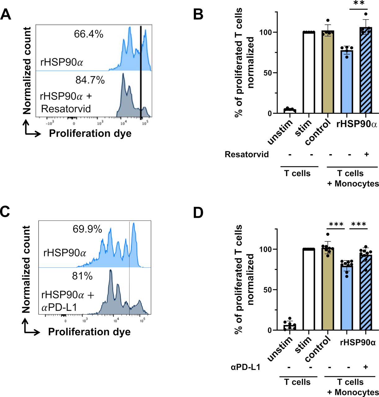
To study the role of TLR4 on monocytes in their acquisition of immunosuppressive activity, we added the TLR4 inhibitor resatorvid to the culture together with rHSP90α. Resatorvid completely abrogated the induction of the immunosuppressive activity of monocytes by rHSP90α (figure 3A,B). When adding blocking anti-PD-L1 antibodies to the co-culture of rHSP90α-treated monocytes and T cells, we found a significant reduction (but not a complete abrogation) of monocyte immunosuppressive activity, suggesting possible additional mechanisms of this immunosuppression (figure 3C,D). Therefore, we tested other immunosuppressive markers on human monocytes induced by the treatment with rHSP90α, including increased production of ROS and NO as well as the expression of ARG-1 and ectoenzymes CD39 and CD73.10 However, no significant changes between treated and non-treated monocytes were detected (online supplemental figure S3A-G).

Role of TLR4 signaling and PD-L1 expression in immunosuppressive capacity of rHSP90α-treated monocytes. (A) Representative histograms for proliferated T cells co-cultured with rHSP90α-treated monocytes with or without the TLR-4 inhibitor resatorvid (5 µM). (B) Cumulative data for T cell proliferation co-cultured with rHSP90α-treated monocytes and resatorvid. Results are presented as the percentage of divided T cells normalized (norm) to the respective control of stimulated T cells alone (mean±SD; n=5). (C) Representative histograms for proliferated T cells co-cultured with rHSP90α-treated monocytes with or without blocking anti-PD-L1 mAbs (0.5 µg/mL). (D) Cumulative data for T cell proliferation co-cultured with rHSP90α-treated monocytes and anti-PD-L1 mAbs. Data are shown as the percentage of divided T cells normalized (norm) to the respective control of stimulated T cells alone (mean±SD; n=7–9). **p<0.01, ***p<0.001. mAbs, monoclonal antibodies; PD-L1, programmed cell death ligand 1; rHSP90α, recombinant heat-shock protein 90α.
Expression of MDSC-related genes in rHSP90α-stimulated monocytes
To obtain a more comprehensive view on the effect of rHSP90α on human monocytes, we performed a microarray analysis, comparing rHSP90α-treated and control monocytes. Gene expression profiling confirmed the elevated expression of PD-L1 (CD274) (figure 4A). In addition, the expression of several MDSC-related genes including IDO1, C-C motif ligand 2 (CCL2) and C-X-C motif chemokine 5 (CXCL5), interleukin (IL)-6 was found to be elevated (figure 4A). In addition, we observed a slight upregulation of CD80, whereas CD86 expression was downregulated (figure 4A). Pathway analysis revealed upregulated expression of genes related to oxidative phosphorylation, IL-1 signaling and antigen processing in rHSP90α-treated monocytes (figure 4B). All differentially regulated genes in rHSP90α-treated versus non-treated monocytes are provided in the online supplemental table S1.
Supplemental material

Microarray analysis of rHSP90α-treated monocytes. Transcriptome of monocytes treated for 24 hours with 2 µg/mL rHSP90α versus control (untreated) monocytes (n=4). (A) Volcano plot representing differentially expressed genes. Arrows indicate selected differentially regulated genes. Horizontal dashed line indicates the significance threshold (p<0.05). Vertical dashed line indicates twofold change. (B) Enrichment map representing selected upregulated pathways in rHSP90α-treated versus control monocytes. Intensity of the red color indicates significance, and the size of the circle indicates the number of genes. The line thickness indicates the number of overlapping genes. FC, fold change; NF-κB, nuclear factor κB; rHSP90α, recombinant heat-shock protein 90α; ROS, reactive oxygen species;TNF, tumor necrosis factor; TNIK, Traf2 and Nck interacting kinase.
TLR4-NF-κB-dependent upregulation of IDO-1 endows rHSP90α-treated monocytes with immunosuppressive capacity
Upregulation of IDO-1 expression in rHSP90α-treated monocytes raised the question whether the immunosuppressive capacity is also regulated by IDO-1. We demonstrated an upregulation of IDO-1 expression at the protein level in human monocytes on the treatment with rHSP90α for 16 hours that was found to be NF-κB and TLR4-dependent (figure 5A). Importantly, the IDO-1 inhibitor 1-methyl-D-tryptophan (1-D-MT) was able to reduce significantly the immunosuppressive activity of rHSP90α-treated monocytes in the co-culture with T cells (figure 5B,C).

Impact of IDO-1 on immunosuppressive activity of rHSP90α-treated monocytes. (A) Expression of IDO-1 in monocytes treated with 2 µg/mL rHSP90α was measured by Western blot. The representative experiment out of three is shown. (B) Representative histograms for proliferated T cells co-cultured with rHSP90α-treated monocytes with or without the IDO-1 inhibitor 1-methyl-D-tryptophan (1-D-MT, 0.5 mM). (C) Cumulative data for T cell proliferation co-cultured with rHSP90α-treated monocytes and 1-D-MT. Data are shown as the percentage of divided T cells normalized (norm) to the respective control of stimulated T cells alone (mean±SD; n=5–8). (D) Representative histograms for proliferated T cells co-cultured with rHSP90α-treated monocytes alone or together with anti-PD-L1 mAbs (0.5 µg/mL) and/or 1-D-MT (0.5 mM). (E) Cumulative data for T cell proliferation co-cultured with rHSP90α-treated monocytes together with anti-PD-L1 mAb and/or 1-D-MT 1-D-MT. Results are presented as the percentage of divided T cells normalized (norm) to the respective control of stimulated T cells alone (mean±SD; n=4). *p<0.05, **p<0.01, ***p<0.001. IDO, indoleamine 2,3-dioxygenase; mAbs, monoclonal antibodies; PD-L1, programmed cell death ligand 1; rHSP90α, recombinant heat-shock protein 90α.
In a separate set of experiments, we co-cultured rHSP90α-treated monocytes and T cells together with anti-PD-L1 antibodies and the IDO-1 inhibitor 1-D-MT. It was found that both blocking agents induced no additional blocking effect compared with anti-PD-L1 or anti-IDO-1 monotherapy (figure 5D,E).
HSP90α in plasma of patients with melanoma
To investigate an impact of soluble HSP90α on myeloid cells in patients with advanced melanoma, we measured the frequency of circulating M-MDSC and monocytes and their PD-L1 expression in correlation with the HSP90α concentration in plasma from the same patients by ELISA. Gating strategy for the surface markers analysis and cell sorting is shown in online supplemental figure S4. Since EV can also contain HSP90α,9 we first tested if the ELISA kit could detect only soluble or also EV-derived HSP90α. For this, we depleted EV by the ultracentrifugation of plasma samples from patients with melanoma at 100,000 g for 16 hours and found no significant differences in HSP90α concentrations between whole and EV-depleted plasma samples, indicating that the ELISA kit detects only soluble HSP90α (online supplemental figure S5). We observed a tendency for the correlation between decreased monocyte frequencies and increased HSP90α plasma levels (online supplemental figure S6A). An elevated PD-L1 expression on circulating M-MDSC showed a significant correlation with an increased concentration of HSP90α (online supplemental figure S6B).

Association between concentration of HSP90α and clinical outcome of patients with metastatic melanoma. The concentration of HSP90α was measured by ELISA in plasma taken before the treatment starts. (A) Progression-free and (B) overall survival of patients with melanoma with high (>12.42 ng/mL; n=16) and low (<12.42 ng/mL; n=16) HSP90α levels at the baseline are shown as a Kaplan-Meier curve. (C) The level of HSP90α in patients with melanoma responding (n=13) and non-responding (n=14) to ICI treatment was expressed in ng/mL (mean±SD). HSP90α, heat-shock protein 90α; ICI, immune checkpoint inhibitors.
Finally, we investigated the impact of soluble HSP90α levels on the clinical outcome of patients with melanoma treated with ICI. The concentration of HSP90α was measured in plasma before the treatment initiation. We found a strong tendency for prolonged PFS in patients with lower concentrations of HSP90α (figure 6A), whereas overall survival was similar in patients with higher and lower levels of HSP90α (figure 6B). Furthermore, patients responding to the ICI treatment tended to have lower plasma levels of HSP90α before the therapy begin (figure 6C).
DISCUSSION
Tumor-derived EV were reported to convert myeloid cells into MDSC.11 The complexity of signals carried by these EV demands dissecting their single components and involved signaling pathways in recipient cells to improve cancer immunotherapy. We have previously shown that melanoma-derived EV can convert myeloid cells into MDSC via TLR4-dependent PD-L1 upregulation.9 However, other TLRs were also involved in the PD-L1 upregulation revealing the complexity of EV signals. Importantly, EV isolated from melanoma cell lines deficient for HSP90α or from melanoma cells treated with HSP90α inhibitor (KNK 437) were shown to lose the capacity to induce immunosuppressive activity in normal myeloid cells.9 Here, we addressed the question whether a component of melanoma-derived EV, HSP90α could induce the conversion of normal human monocytes into MDSC-like cells. We demonstrated that rHSP90α can upregulate PD-L1 on monocytes in a TLR4-NF-κB-dependent manner that was similar to the effect of melanoma-derived EV. It was previously reported that HSP90α located on the surface of autophagosomes released by tumor cells could promote an immunosuppressive phenotype of CD4 T cells via TLR2 signaling.37 However, in our experiments, anti-TLR2 blocking antibodies did not affect the rHSP90α-mediated upregulation of PD-L1.
It is known that HSP90α interacts also with LRP1.21 38 Zou et al19 found that two evolutionarily conserved lysine residues, Lys-270 and Lys-277, of HSP90α contained in the subfragments F-5 and F-6 were responsible for the binding of HSP90α to LRP1 on human cancer cells. In contrast, we demonstrated that these subfragments failed to upregulate PD-L1 on human monocytes. This could suggest that HSP90α may induce variable effects in different cell types.
It has been recently published that rHSP90α can trigger both LRP1 and TLR4 on THP1-derived macrophages, RAW264.7 macrophage cell line and bone marrow-derived macrophages, resulting in the upregulation of CD163, CD204, IL-10 and downregulation of TNF-α, IL-1β as well as in the depletion of CD4 T cells from the pancreatic tumor.21 It is possible that rHSP90α in myeloid cells forms a complex with TLR4 and LRP1, in which TLR4 plays a pivotal role. Moreover, HSP90α and HSP90β were reported to be expressed on the surface of monocytes and macrophages and to participate in cytokine response to TLR4 ligands by building a signaling complex with TLR4.39 40 Since we found that TLR4 blocking abrogated the upregulation of PD-L1 completely, we conclude that TLR4 could be a key receptor for interaction with soluble rHSP90α, leading to the acquisition of an immunosuppressive phenotype by monocytes.
We also showed that rHSP90α downregulated HLA-DR expression on monocytes. Low expression of this marker feature is considered to be typical for human M-MDSC.3 Frequencies of such HLA-DRlo/neg M-MDSC were reported to be increased in patients with melanoma compared with their counterparts in healthy donors.7 Moreover, M-MDSC prevailed in responders to the ICI treatment compared with non-responders.6 7 41
In addition to HLA-DR downregulation, we have shown TLR4-NF-κB-dependent antiapoptotic effect of rHSP90α on monocytes, indicating that HSP90α does not only play a role in the conversion of monocytes but also promotes longer persistence of converted cells. This finding is in agreement with the results of Franco et al42 who also found an antiapoptotic effect of extracellular HSP90 on the monocytic cell line U937.
A critical feature for defining myeloid cell as MDSC is the immunosuppressive function.3 We demonstrated that rHSP90α-treated monocytes inhibited proliferation of CD3 T cells and that their immunosuppressive capacity is abrogated by the inhibitor of TLR4. These data are in agreement with the previous study showing that TLR4 signaling enhanced the immunosuppressive function of already established MDSC.43 However, other reports demonstrated that the TLR4 inhibitor resatorvid (TAK-242) induces MDSC accumulation44 or that stimulation of TLR4 by cinnamaldehyde leads to MDSC apoptosis.45 It is plausible that the magnitude, timing and molecular context determine the outcome of TLR4 signaling. Since we showed that blocking PD-1/PD-L1 interactions also significantly diminishes the immunosuppression mediated by rHSP90α-treated monocytes, this approach could be an alternative to the inhibition of TLR4 signaling. However, the presence of patients with melanoma with increased frequencies of M-MDSC who are resistant to the nivolumab therapy41 suggests the role of additional suppressive molecules.10
Indeed, we found in rHSP90α-treated monocytes an upregulation of genes relevant for MDSC recruitment and expansion such as CCL2, CXCL5 and IL-646 47 as well as downregulation of a co-stimulatory molecule CD86. Importantly, we observed also an upregulation of the IDO-1 gene, which was confirmed at the protein level. Furthermore, the inhibitor of IDO-1 was able to reduce the immunosuppressive capability of HSP90α-treated monocytes significantly. In another study, IDO-1+ mononuclear cells were found in melanoma microenvironment, and the expression of IDO-1 was positively correlated with PD-L1 expression on melanoma cells.48 Moreover, higher frequencies of IDO-1+ M-MDSC were found to be correlated with melanoma progression.49 When exploring the synergistic effect of anti-PD-L1 blocking antibody and IDO-1 inhibition in rHSP90α monocytes, we observed no significant differences compared with the monotherapy. This could be due to the fact that blocking either PD-L1 or IDO-1 alone almost completely restored the proliferation of T cells, leaving only a narrow window for further improvement.
To verify the clinical significance of our findings on rHSP90α in patients with advanced melanoma, we measured the plasma concentration of HSP90α as well as the frequency of M-MDSC and their PD-L1 expression. It was shown that HSP90α was elevated in plasma of patients with various cancers and some non-cancer diseases such as psoriasis, chronic glomerulonephritis and idiopathic pulmonary fibrosis.50 We found that an increased PD-L1 expression on circulating M-MDSC tended to correlate with higher HSP90α concentrations in plasma. Furthermore, patients with higher plasma levels of HSP90α showed a tendency for worse clinical outcome that goes in line with another publication.26
Conclusion
Taken together, our study demonstrates a TLR4-NF-κB-dependent mechanism of the conversion of normal human monocytes into PD-L1+IDO-1+ M-MDSC mediated by HSP90α. Inhibiting intracellular HSP90 was reported previously to deplete MDSC,17 51 52 but none of the available inhibitors of intracellular HSP90 have demonstrated sufficient benefits in clinical trials so far. Our study highlights the role of extracellular soluble HSP90α, which could be a promising target to inhibit MDSC functions and to improve current immunotherapy of patients with melanoma.
Data availability statement
Data are available upon reasonable request. Sets of raw data generated, used and analyzed during the current study are available from the corresponding author upon reasonable request. The microarray data generated during the study is made available on the GEO repository under the number GSE207075.
Ethics statements
Patient consent for publication
Ethics approval
This study involves human participants and was approved by the ethics committee of University Medical Center Mannheim (2010-318N-MA). Participants gave informed consent to participate in the study before taking part.
Acknowledgments
We thank S Uhlig (Flow Core Team, University Medical Center Mannheim, Germany) for the assistance with FACS sorting of the MDSC subpopulations. We thank the Microarray Unit of the Genomics and Proteomics core facility, German Cancer Research Center (DKFZ), for providing excellent Expression Profiling services. We thank Yvonne Nowak, Sayran Arif-Said (both Skin Cancer Unit, German Cancer Research Center (DKFZ) and University Medical Center Mannheim) for the preparation of plasma samples.
References
Supplementary materials
Supplementary Data
This web only file has been produced by the BMJ Publishing Group from an electronic file supplied by the author(s) and has not been edited for content.
Footnotes
IA and FGÖK contributed equally.
Contributors VU, PA, JU, IA and FGOK designed the study. IA, FGOK, RB, DN, VP, SL, CG, AL and XH performed the experiments and the data analysis. WL provided the HSP90α plasmids. TH performed the microarray data analysis. IA, FGOK, PA and VU wrote the manuscript. All authors read and approved the manuscript. VU is responsible for the overall content as a principal investigator and guarantor.
Funding This work was supported by the German Federal Ministry of Education and Research– SERPENTINE project in the ERA PerMed network (01KU2017 to VU), the German Research Foundation (project number 259332240/RTG 2099 (to IA, JU and VU)) and the German Academic Exchange Service (DAAD to FGOK).
Competing interests None declared.
Provenance and peer review Not commissioned; externally peer reviewed.
Supplemental material This content has been supplied by the author(s). It has not been vetted by BMJ Publishing Group Limited (BMJ) and may not have been peer-reviewed. Any opinions or recommendations discussed are solely those of the author(s) and are not endorsed by BMJ. BMJ disclaims all liability and responsibility arising from any reliance placed on the content. Where the content includes any translated material, BMJ does not warrant the accuracy and reliability of the translations (including but not limited to local regulations, clinical guidelines, terminology, drug names and drug dosages), and is not responsible for any error and/or omissions arising from translation and adaptation or otherwise.