


Abstract
We review the recent advances in the understanding of the Pseudomonas aeruginosa biofilm lifestyle from studies using in vitro laboratory setups such as flow chambers and microtiter trays. Recent work sheds light on the role of nutrients, motility, and quorum sensing in structure formation in P. aeruginosa biofilms. The second messenger, c-di-GMP, is established as an important regulator of the synthesis of polysaccharide and protein components of the biofilm matrix. Extracellular DNA is shown to be an essential component of the biofilm matrix. It has become apparent that biofilm formation involves interactions between different subpopulations. The molecular mechanisms underlying the tolerance of biofilm bacteria to antimicrobial agents are beginning to be unraveled, and new knowledge has been obtained regarding the environmental cues and regulatory mechanisms involved in biofilm dispersal.
Introduction
Microbial biofilms have been subject to intense study during the last decade mainly for two reasons. First, it is of basic scientific interest to understand how bacteria form and live in multicellular communities. Second, biofilm formation causes considerable problems in medical and industrial settings, because bacteria in biofilms can resist antibiotic treatment, host immune responses, and biocide treatment. Knowledge of the environmental cues, genetic elements, and molecular mechanisms that are involved in biofilm formation is necessary for a rational design of strategies to eliminate biofilms or to prevent biofilm formation.
A substantial part of the studies of microbial biofilms conducted during the last decade has involved in vitro laboratory setups such as microtiter trays and flow chambers. Although the model biofilms grown in these setups most likely differ from biofilms formed in nature, experiments with these biofilms have provided important insights regarding the fundamental processes of biofilm formation, tolerance development, and biofilm dispersal that may be of relevance outside the laboratory systems. In the present review, we present an update on biofilm formation, tolerance, and dispersal based on in vitro studies with the opportunistic pathogen Pseudomonas aeruginosa, which is a model organism for biofilm research. Of the numerous factors involved in the biofilm formation process, we presently focus on attachment, motility, matrix production, quorum sensing, and subpopulation interactions.
Attachment
Transport of P. aeruginosa bacteria to a surface before attachment is assumed to involve diffusive, convective, and active flagellum-driven transport (van Loosdrecht et al., 1990). A variety of components including flagella (O'Toole & Kolter, 1998; Sauer et al., 2002), type IV pili (O'Toole & Kolter, 1998; Deziel et al., 2001; Chiang & Burrows, 2003), Cup fimbria (Vallet et al., 2001), extracellular DNA (eDNA) (Whitchurch et al., 2002), and Psl polysaccharide (Ma et al., 2009) have been shown to play a role in the attachment of P. aeruginosa to surfaces in microtiter trays and flow chambers. However, conditions under which lack of flagella and/or type IV pili did not affect surface attachment in flow chambers have also been reported (Klausen et al., 2003b). After P. aeruginosa has attached to a surface, it either detaches, remains attached at the position of attachment, or moves along the surface by means of type IV pili or flagella (Singh et al., 2002; Klausen et al., 2003a, b; Singh, 2004; Shrout et al., 2006). The gene sadB has been shown to be involved in the regulation of the frequency by which P. aeruginosa cells attach to and detach from surfaces (Caiazza & O'Toole, 2004). Synthesis of the attachment regulator SadB is regulated by the intracellular level of the second messenger molecule cyclic diguanosine-5′-monophosphate (c-di-GMP) (Merritt et al., 2007).
Motility
Experiments in flow-chamber setups have suggested that surface-associated motility is an integrated part of P. aeruginosa biofilm formation (e.g. Singh et al., 2002; Klausen et al., 2003a, b; Singh, 2004; Shrout et al., 2006; Patriquin et al., 2008). The pattern of motility occurring in P. aeruginosa biofilms appears to be dependent on the prevailing conditions. With glucose as a carbon source, for example, the attached bacteria differentiate initially into a nonmotile subpopulation and a motile subpopulation (Klausen et al., 2003a). The nonmotile subpopulation forms microcolonies that serve as the ‘stalks’ of ‘mushroom’-shaped multicellular structures that are formed when bacteria from the motile subpopulation colonize the stalks and subsequently form mushroom ‘caps’ upon the stalks (Klausen et al., 2003a). With citrate as a carbon source, the entire bacterial population is motile in the initial phase of biofilm formation and microcolonies that could serve as mushroom stalks are not formed, and consequently, a flat biofilm is formed (Klausen et al., 2003b). Pseudomonas aeruginosa pilA mutants (deficient in the biogenesis of type IV pili) formed protruding microcolonies in biofilms irrigated with a citrate medium, indicating that the motility occurring in the initial phase of biofilm formation in citrate-grown P. aeruginosa wild-type biofilms is driven by type IV pili and thus most likely is similar to twitching motility (Klausen et al., 2003b). As described below, P. aeruginosa twitching motility is stimulated by iron limitation. When citrate is used as a carbon source, it is present at a much higher concentration than iron in the medium. Because citrate chelates iron, it is possible that the use of citrate as a carbon source in flow-chamber systems reduces the level of iron available to the bacteria, which may explain why the entire population is motile initially in P. aeruginosa biofilms irrigated with citrate medium.
Twitching motility has been shown to be induced in P. aeruginosa biofilms by iron limitation (Singh et al., 2002; Singh, 2004; Patriquin et al., 2008). In accordance, P. aeruginosa wild-type bacteria, which formed mushroom-shaped structures in flow cells irrigated with iron-replete medium, formed flat biofilms in flow chambers irrigated with an iron-depleted medium (Patriquin et al., 2008). Evidence was provided that iron-limitation-promoted twitching motility is dependent on a functional Rhl quorum-sensing system, and a P. aeruginosa rhlI mutant was shown to form microcolonies (lacking mushroom caps) in flow chambers irrigated with an iron-depleted medium (Patriquin et al., 2008). A role of the Rhl quorum-sensing system in an iron-limitation-induced phenotype is in accordance with numerous studies showing that the quorum-sensing systems in P. aeruginosa are induced under iron limitation, but repressed in the presence of high levels of iron (e.g. Bollinger et al., 2001; Kim et al., 2005; Duan & Surette, 2007). Spent medium from iron-limited, but not iron-replete, P. aeruginosa wild-type cultures could induce iron-limitation-promoted twitching motility of P. aeruginosa rhlI mutants, suggesting that some soluble factor, which is low-iron-inducible and RlhI dependent, positively influenced twitching motility (Patriquin et al., 2008). The available evidence suggested that this soluble factor is the biosurfactant rhamnolipid, but it was not investigated whether spent medium from P. aeruginosa rhlA cultures (deficient in the biogenesis of rhamnolipid) failed to induce iron-limitation-promoted twitching motility of P. aeruginosa rhlI mutants (Patriquin et al., 2008). In agreement with the suggestion that increased levels of rhamnolipid may stimulate twitching motility in iron-limited biofilms, Pamp & Tolker-Nielsen (2007) reported that in addition to the well-known role of rhamnolipid in facilitating swarming motility (e.g. Kohler et al., 2000), the biosurfactant can also facilitate twitching motility. If increased levels of rhamnolipid produced in iron-deplete P. aeruginosa biofilms stimulate twitching motility and thereby prevent microcolony formation, it would be expected that a P. aeruginosa rhlA mutant would be able to form microcolonies under iron-limited conditions. However, Pamp & Tolker-Nielsen (2007) provided evidence that rhamnolipid is necessary for initial microcolony formation in P. aeruginosa biofilms. In support of a role of rhamnolipid in initial microcolony formation, it has been shown that low concentrations of rhamnolipid enhance the hydrophobicity of P. aeruginosa bacteria by causing a release of lipopolysaccharide (LPS) from the cell surface (Zhang & Miller, 1994; Al-Tahhan et al., 2000). An increase in cell surface hydrophobicity may increase the adhesiveness of the bacteria to a level that is critical for initial microcolony formation in biofilms. It appears that rhamnolipid, depending on the quantity produced, plays multiple roles in P. aeruginosa biofilm formation. It is necessary for initial microcolony formation (Pamp & Tolker-Nielsen, 2007); it may prevent initial microcolony formation in iron-limited biofilms by stimulating twitching motility (Patriquin et al., 2008); it facilitates bacterial migration and thereby the formation of mushroom caps (Pamp & Tolker-Nielsen, 2007); it prevents colonization of the channels between the mushroom-shaped structures (Davey et al., 2003); and it plays a role in biofilm dispersal (Schooling et al., 2004; Boles et al., 2005).
Evidence has been presented that the formation of the mushroom caps in P. aeruginosa biofilms requires both type IV pili and flagella (Klausen et al., 2003a; Barken et al., 2008). Cap formation evidently requires flagellum-driven surface-associated motility, whereas the dependence of cap formation on type IV pili may be due to the binding of these pili to eDNA that is abundant on the microcolonies that become colonized during mushroom-structure formation (Whitchurch et al., 2002; Allesen-Holm et al., 2006; Barken et al., 2008). Alternatively, a kind of migration that requires both flagella and type IV pili might be occurring in the later phase of P. aeruginosa biofilm formation. In support of this suggestion, it has been reported that P. aeruginosa swarming motility in addition to flagella and rhamnolipid may also require type IV pili under some conditions (Kohler et al., 2000).
As described in the next section, evidence is accruing that the shift between migrating and sessile surface behavior is regulated via proteins with diguanylate cyclase or phosphodiesterase activities that control levels of the second messenger molecule c-di-GMP (Hickman et al., 2005; Kuchma et al., 2007; Merritt et al., 2007). Low intracellular levels of c-di-GMP promote motility, whereas high intracellular c-di-GMP levels induce the formation of cell–cell interconnecting matrix components and promote microcolony formation.
Matrix production
The extracellular polymeric substance (EPS) matrix serves as the ‘house for biofilm cells’. The matrix plays a role in numerous processes including attachment, cell-to-cell interconnection, interactions between subpopulations, tolerance, and exchange of genetic material (Molin & Tolker-Nielsen, 2003; Friedman & Kolter, 2004a; Jackson et al., 2004; Ma et al., 2009; Yang et al., 2009). The P. aeruginosa EPS matrix contains mainly polysaccharides, proteins, and nucleic acids (e.g. Whitchurch et al., 2002; Friedman & Kolter, 2004a; Jackson et al., 2004; Borlee et al., 2010). The composition of the matrix depends on the environmental conditions, the age of the biofilm, and the particular P. aeruginosa strain forming the biofilm. Evidence has been provided that P. aeruginosa induces the synthesis of matrix components in response to environmental signals sensed by the sensor kinase/response regulators LadS, RetS, and GacS (Goodman et al., 2004; Ventre et al., 2006; Goodman et al., 2009). In addition, sensor kinases and response regulators encoded by the bfiSR, bfmSR, and mifSR genes are evidently involved in further P. aeruginosa biofilm formation (Petrova & Sauer, 2009), although the functions affected by these signal transduction systems have not been identified. Below, we describe the involvement of the matrix components Pel, Psl, alginate, CdrA protein, Cup fimbria, type IV pili, lectins, and eDNA in P. aeruginosa biofilm formation.
The glucose-rich Pel polysaccharide, encoded by the pel cluster, was initially reported to be required for the formation of P. aeruginosa liquid–air interface pellicles and surface-associated biofilms (Friedman & Kolter, 2004b). Evidence has been presented that the transmembrane protein PelD binds c-di-GMP, and that there is a strict correlation between c-di-GMP binding and the synthesis of the Pel polysaccharide (Lee et al., 2007; Lory et al., 2009). Hickman & Harwood (2008) presented data showing that c-di-GMP binds to the transcriptional regulator FleQ and thereby derepresses the transcription of the pel genes. The pel locus is furthermore regulated by the sensor kinase-response regulator hybrids RetS and LadS, which repress and activate its expression, respectively (Goodman et al., 2004; Ventre et al., 2006). This regulation is mediated through the small-RNAs, RsmZ/Y, which alter the level of the free RsmA post-transcriptional regulator (Goodman et al., 2004; Ventre et al., 2006). The two-component system GacA/S interacts with the RetS and LadS regulatory network, and recent data suggest that RetS can modulate the phosphorylation state of GacS by forming heterodimers that block the GacS autophosphorylation, leading to a reduced rsmZ expression, and subsequently free RsmA, thereby repressing exopolysaccharide synthesis and promoting the translation of genes necessary for acute infection (Goodman et al., 2009). Recent work showed that rugose small-colony variants (RSCVs) of P. aeruginosa isolated from laboratory biofilms and cystic fibrosis (CF) patients had increased expression of the pel locus compared with clonally related wild types, and it was suggested that RSCVs may partially contribute to increased persistence of biofilms in the airways of CF lungs (Starkey et al., 2009). It was further shown that c-di-GMP signaling regulated the increased expression of the pel locus in the RSCVs (Starkey et al., 2009).
The mannose-rich Psl polysaccharide, encoded by the psl cluster, is highly conserved in many P. aeruginosa strains (Friedman & Kolter, 2004a; Ryder et al., 2007). The Psl polysaccharide mediates cell-to-surface and cell-to-cell interactions, which are essential for P. aeruginosa biofilm formation and maintenance (Overhage et al., 2005; Ma et al., 2006, 2009). In a recent study, Ma (2009) showed that Psl forms a helical structure around P. aeruginosa cells that increase cell-to-surface and cell-to-cell interactions necessary for biofilm formation. By lectin staining, Psl was shown to be located predominantly in the peripheries of the mushroom-shaped structures, whereas it was evenly distributed in flat biofilms (Ma et al., 2009). As for the Pel polysaccharide, Psl expression is regulated by LadS and RetS (Goodman et al., 2004; Ventre et al., 2006) and by c-di-GMP (Hickman et al., 2005). A newly characterized protein, CdrA, which shows increased expression with elevated c-di-GMP levels, is suggested to either crosslink Psl polymers and/or tether Psl to the P. aeruginosa cell wall (Borlee et al., 2010). This protein is the first to be identified as a structural component of the P. aeruginosa biofilm matrix. Borlee (2010) found that the cdrAB complex is elevated in biofilms, and that overexpression of both proteins induces autoaggregation if Psl is present. The second protein in the complex, CdrB, works as an outer-membrane transporter of CdrA. Evidence was provided that CdrA binds directly to the mannose part of Psl, and it was suggested that CdrA is a multivalent adhesin with the ability to recognize multiple carbohydrates (Borlee et al., 2010).
Alginate is an acetylated polysaccharide composed of nonrepetitive monomers of β-1,4-linked l-guluronic and d-mannuronic acids (Govan & Deretic, 1996), and is mainly produced by P. aeruginosa in chronic infections of the lungs of CF patients (Hoiby, 1974; Govan & Deretic, 1996). Its special physical and chemical properties play multiple roles in protecting P. aeruginosa cells (Govan & Deretic, 1996). Biofilms formed by an alginate-overproducing strain were shown to exhibit a highly structured architecture and were significantly more resistant to the antibiotic tobramycin than biofilms formed by an isogenic nonmucoid strain (Hentzer et al., 2001). This might indicate that alginate acts as a physical barrier for antibiotics because alginate lyase treatment has been shown to enhance the diffusion of aminoglycosides through the EPS of biofilms formed by mucoid P. aeruginosa strains (Hatch & Schiller, 1998). A recent study showed that a supermucoid P. aeruginosa strain was strongly impaired in attachment compared with the respective mucoid or nonmucoid strains and formed a thicker biofilm with large extended mushroom-like microcolonies (Hay et al., 2009a). Recently, it has been found that post-transcriptional regulation plays a role in the biosynthesis of alginate in P. aeruginosa. The membrane-anchored protein, Alg44, which is essential for alginate biosynthesis, contains a c-di-GMP-binding PilZ domain (Amikam & Galperin, 2006; Remminghorst & Rehm, 2006b; Merighi et al., 2007; Oglesby et al., 2008; Lory et al., 2009). Regulation of Alg44 activity via the PilZ domain has recently been suggested to occur through a new membrane-anchored diguanylate cyclase, MucR, which has been identified as a positive regulator of alginate biosynthesis (Hay et al., 2009b). It was suggested that MucR possibly acts by the localized production of c-di-GMP in the vicinity of Alg44. The PilZ domain of Alg44 in turn responds to the high local concentration of c-di-GMP and promotes the production of alginate. The glycosyltransferase Alg8 was characterized recently and shown to be critical for alginate production (Remminghorst & Rehm, 2006a; Oglesby et al., 2008). It was shown that the Alg44 and Alg8 proteins were required in combination for the polymerization reactions leading to alginate production (Oglesby et al., 2008).
Recently, eDNA was recognized as one of the major matrix components of bacterial biofilms (Whitchurch et al., 2002; Qin et al., 2007; Rice et al., 2007). It was shown that DNAse treatment led to the dispersal of young P. aeruginosa biofilms (Whitchurch et al., 2002). However, DNAse did not disperse a flow-chamber-grown P. aeruginosa mature biofilm, probably due to increasing amounts of other EPS materials being produced during biofilm formation. Southern and RAPD PCR analysis provided evidence that the eDNA in the P. aeruginosa biofilm matrix is similar to chromosomal P. aeruginosa DNA (Allesen-Holm et al., 2006). In addition to a small amount of eDNA that is present and necessary in the initial phase of P. aeruginosa biofilm formation (Whitchurch et al., 2002), the release of a large amount of eDNA occurs during subsequent P. aeruginosa biofilm formation, evidently through lysis of a small subpopulation of the bacteria regulated via the P. aeruginosa quinolone signal (PQS) quorum-sensing system (Allesen-Holm et al., 2006). In agreement with a role of quorum sensing in cell lysis, D'Argenio (2002) reported that mutants that could not produce the PQS quorum-sensing signal molecule did not show autolysis, whereas mutants that overproduced PQS displayed high levels of autolysis. In addition, Heurlier (2005) presented evidence that P. aeruginosa quorum-sensing mutants, unlike the wild type, did not undergo cell lysis in stationary-phase cultures. Moreover, Yang (2007) presented evidence that high levels of iron suppressed P. aeruginosa pqs gene expression, DNA release, and structural biofilm development. Quinolone compounds have previously been shown to induce prophages in bacteria (Phillips et al., 1987; Froshauer et al., 1996), and studies by Webb (2003) and Hentzer (2004) have suggested that quorum-sensing-regulated DNA release might be linked to bacteriophage induction in biofilms. PQS was shown to be necessary for membrane vesicle formation in P. aeruginosa (Mashburn & Whiteley, 2005; Nakamura et al., 2008), and membrane vesicles produced by P. aeruginosa might also play a role in DNA release. The membrane vesicles released by P. aeruginosa have bacteriolytic effects and contain DNA (Kadurugamuwa & Beveridge, 1996; Renelli et al., 2004). eDNA might be released either from vesicles that eventually lyse or through the bacteriolytic activity of the vesicles that might lyse a small subpopulation of P. aeruginosa bacteria. The eDNA appears to be organized in distinct patterns in P. aeruginosa biofilms (Allesen-Holm et al., 2006). In flow-chamber-grown P. aeruginosa biofilms, which contain mushroom-shaped structures, the eDNA was located primarily in the stalk portion of the mushroom-shaped structures, with the highest concentration in the outer parts of the stalks forming a border between the stalk subpopulation and the cap subpopulation (see Fig. 1). eDNA is primarily generated in the initial microcolonies before they are colonized by the cap-forming subpopulation, and the concentration of eDNA is highest in the outer layer of these microcolonies (Allesen-Holm et al., 2006; Yang et al., 2007). It is currently not understood how the migration of the motile cells is coordinated so that they form mushroom caps. However, type IV pili bind to DNA with a high affinity (Aas et al., 2002; van Schaik et al., 2005), and evidence has been presented that the high concentration of eDNA on the outer parts of the mushroom stalks can cause accumulation of the migrating bacteria, which, in combination with bacterial growth, might result in the formation of the mushroom caps (Barken et al., 2008). Type IV pili may therefore function as niche-specific matrix-adhesins, and, as for many other components of the P. aeruginosa matrix, c-di-GMP appears to play a role in the regulation of type IV pili synthesis, in this case through the proteins PilZ and FimX, which have c-di-GMP-binding domains (Huang et al., 2003; Guzzo et al., 2009). The eDNA in P. aeruginosa biofilms appears to have a stabilizing effect, as mature P. aeruginosa PAO1 biofilms that were pretreated with DNAse I were more susceptible to sodium dodecyl sulfate (SDS) treatment than biofilms that were not pretreated with DNAse I (Allesen-Holm et al., 2006).
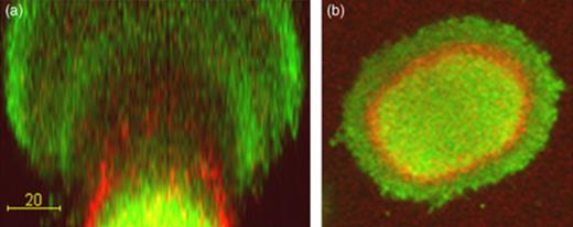
Confocal scanning laser microscopy micrographs acquired in a propidium iodide-stained biofilm formed by Gfp-tagged Pseudomonas aeruginosa PAO1. (a) Vertical section through a mushroom-shaped multicellular structure; (b) horizontal section through a mushroom-shaped multicellular structure. The bacteria appear green fluorescent and the eDNA appears red fluorescent. Size bar=20 µm. Reproduced from Allesen-Holm (2006).
The P. aeruginosa CupA, B, and C fimbria were shown to be involved in bacterial attachment and/or subsequent biofilm formation (Vallet et al., 2001; Kulasekara et al., 2005). Recently, a new Cup-fimbriae gene cluster, CupD, has been identified on the pathogenicity island, PAPI-I, present in P. aeruginosa strain PA14 (Mikkelsen et al., 2009). Overexpression of this gene cluster results in increased biofilm formation and decreased motility as was observed for overexpression of the other Cup-fimbrial gene clusters (Mikkelsen et al., 2009). Cup fimbriae are regulated by c-di-GMP (Kulasekara et al., 2005; Rao et al., 2008; Mikkelsen et al., 2009) as are many of the other P. aeruginosa matrix components. Expression of cupA was shown to be dependent on proteins with diguanylate cyclase/c-di-GMP-binding domains (WspR, MorA, and PA1120) (D'Argenio et al., 2002; Meissner et al., 2007), whereas expression of cupB and cupC was shown to be regulated by the RocR phosphodiesterase protein (Kulasekara et al., 2005; Rao et al., 2008), and expression of cupD was regulated by the PvrR phosphodiesterase protein (Mikkelsen et al., 2009). Overexpression of the PvrR protein has previously been shown to reduce CupA fimbria expression likely as a consequence of a reduction in the intracellular c-di-GMP level (Meissner et al., 2007). CupA was recently shown to be involved in SDS-induced autoaggregation dependent on increased intracellular levels of c-di-GMP, and two newly characterized proteins: SiaA and SiaD (Klebensberger et al., 2009).
Evidence has been provided that the lectin LecB binds to specific carbohydrate ligands located on the surface of P. aeruginosa cells, and a P. aeruginosa lecB mutant was shown to be impaired in biofilm formation in comparison with the wild-type strain, suggesting an important role for LecB in biofilm development (Tielker et al., 2005).
Quorum sensing
Quorum sensing in P. aeruginosa is mediated through three interconnected systems: the Las system that senses 3-oxo-C12-homoserine lactone, the Rhl system that senses C4-homoserine lactone, and the Pqs system that senses 2-heptyl-3-hydroxy-4-quinolone referred to as PQS (Juhas et al., 2005). Microarray analysis has suggested that several hundred genes in P. aeruginosa are quorum-sensing regulated (Schuster et al., 2003; Wagner et al., 2003; Hentzer et al., 2005).
Shrout (2006) provided evidence that the impact of quorum sensing on motility in P. aeruginosa biofilms is nutrition dependent, which may explain the different results regarding the role of quorum sensing in P. aeruginosa biofilm formation. Davies (1998) found that a P. aeruginosa wild type formed structured biofilms with large mushroom-shaped structures in flow chambers irrigated with a glucose medium, while the corresponding lasI quorum-sensing mutant formed flat and undifferentiated biofilms. The flat biofilms formed by the lasI mutant were susceptible to treatment with the detergent SDS, while the structured biofilms formed by the wild type were tolerant. Heydorn (2002) reported that a P. aeruginosa wild type and lasI mutant both formed flat biofilms in flow chambers irrigated with citrate medium. Purevdorj (2002) showed that in flow chambers irrigated with dilute Luria–Bertani (LB) under high-flow conditions, both the P. aeruginosa wild type and the lasI mutant formed biofilms containing large aggregates, although the biofilms differed slightly in microscopic appearance. Hentzer (2002) demonstrated that homoserine lactone signal analogues called furanones, known to inhibit P. aeruginosa quorum sensing, affected biofilm development when added to the growth medium consisting of diluted LB. Patriquin (2008) showed that the P. aeruginosa wild type formed mushroom-shaped structures in flow chambers irrigated with dilute tryptic soy broth, whereas an rhlI quorum-sensing mutant formed microcolonies lacking the mushroom caps. Similarly, Yang (2009) showed that the P. aeruginosa wild type formed mushroom-shaped structures in flow chambers irrigated with glucose medium, whereas a pqsA quorum-sensing mutant could only form small microcolonies lacking the mushroom cap. In addition, evidence was provided that chemical inhibition of the Pqs system caused P. aeruginosa to form microcolonies on which mushroom caps were not subsequently formed (Yang et al., 2007, 2009).
The above-mentioned studies indicate that quorum sensing is necessary for the formation of the cap portion of the mushroom-shaped structures in P. aeruginosa biofilms. Interestingly, quorum sensing is, however, shown to occur mainly in the stalk portion of the mushroom-shaped structures in P. aeruginosa biofilms (de Kievit et al., 2001; Yang et al., 2009). Pseudomonas aeruginosa uses quorum sensing to regulate numerous factors including the production of rhamnolipid (Ochsner & Reiser, 1995) and eDNA (Allesen-Holm et al., 2006). As mentioned in the previous section, Pamp & Tolker-Nielsen (2007) provided evidence that rhamnolipid plays a role in mushroom cap formation by promoting motility occurring in the later phase of P. aeruginosa biofilm formation. As described in more detail in the following section, evidence was recently presented that PQS quorum sensing in the initial microcolonies leads to the production of eDNA, which plays an important role in the subsequent formation of the mushroom caps (Allesen-Holm et al., 2006; Barken et al., 2008; Yang et al., 2009).
In addition to regulating the production of rhamnolipid and eDNA, quorum sensing evidently regulates a number of other factors involved in P. aeruginosa biofilm formation. Sakuragi & Kolter (2007) presented evidence that transcription of the pel operon is considerably reduced in P. aeruginosa lasI and rhlI mutants, suggesting that quorum-sensing signaling regulates the production of Pel polysaccharide during P. aeruginosa biofilm formation. On the contrary, however, Ueda & Wood (2009) recently reported that Las quorum-sensing represses Pel production in P. aeruginosa. The tyrosine phosphatase TpbA was shown to be positively regulated by Las quorum sensing, and evidence was presented that TpbA activity results in decreased levels of c-di-GMP and thereby a reduction in Pel production (Ueda & Wood, 2009). Transcriptome analysis has indicated that the cupA3 and cupB5 genes are subject to quorum-sensing control in P. aeruginosa biofilms (Hentzer et al., 2004), suggesting that the expression of CupA and CupB fimbria may be quorum sensing regulated during P. aeruginosa biofilm formation. In addition, expression of the lectins LecA and LecB was shown to be regulated via quorum sensing (Winzer et al., 2000). The production of both lectins was found to be directly dependent on the rhl locus, while, in a lasR mutant, the onset of lectin synthesis was delayed, but not abolished.
Unlike the previous examples, the last example of an effect of quorum sensing on P. aeruginosa biofilm development is related to the central metabolism in the bacteria. Evidence has been presented that anaerobic nitrate respiration may play an important role in P. aeruginosa biofilm development in clinical settings, and that the rhlRI system is necessary to prevent accumulation of toxic nitric oxide during the process (Worlitzsch et al., 2002; Yoon et al., 2002). In agreement, the nirCMSQ and napEF genes, which are required for respiratory nitrate reduction, were found to be strongly upregulated in P. aeruginosa during biofilm growth (Hentzer et al., 2005).
Subpopulation interactions
Pseudomonas aeruginosa bacteria release several compounds into their surrounding environment, for example iron siderophores, biosurfactants, and EPS. A common feature of these compounds is that they are costly to synthesize and able to benefit the entire population and therefore can be regarded as ‘public goods’ (West et al., 2007). Many extracellular public goods play a role in P. aeruginosa biofilm formation. For example, the iron siderophore pyoverdine is necessary for the formation of structured P. aeruginosa biofilms (Banin et al., 2005). The biosurfactant rhamnolipid plays multiple roles in P. aeruginosa biofilm formation, and is, among other things, required for biofilm channel maintenance as well as for migration of motile subpopulations (Davey et al., 2003; Pamp & Tolker-Nielsen, 2007). eDNA released via quorum sensing plays a role as EPS material required for the formation of structured P. aeruginosa biofilms (Allesen-Holm et al., 2006).
The bacteria in P. aeruginosa biofilms exist in various physiological states dependent on their spatial localization. Pseudomonas aeruginosa biofilms contain subpopulations that produce public goods and subpopulations that do not produce public goods. For example the pyoverdine synthesis genes, rhamnolipid synthesis genes, and quorum-sensing genes were all reported to be expressed specifically in the stalk portion of the mushroom-shaped structures in P. aeruginosa biofilms (De Kievit et al., 2001; Lequette & Greenberg, 2005; Kaneko et al., 2007; Yang et al., 2007, 2009).
Interactions between producers and nonproducers of public goods may play a role in P. aeruginosa biofilm formation. Yang (2009) studied subpopulation interactions in mixed P. aeruginosa biofilms containing a nonmotile subpopulation (pilA mutants) and a motile subpopulation (wild type). In this model system, the nonmotile bacteria form the stalk portion and the motile bacteria form the cap portion of mushroom-shaped biofilm structures. In the study, the role of pyoverdine in subpopulation interactions during P. aeruginosa biofilm formation was investigated. Using a pvdA∷gfp fluorescent reporter, the pyoverdine synthesis genes were found to be expressed specifically in the pilA stalk-forming subpopulation of the mushroom-shaped structures formed in pilA/wild-type mixed biofilms. In agreement with the study of Banin (2005), it was found that a P. aeruginosa pvdA mutant could form small microcolonies, but was unable to form mushroom-shaped structures (see Fig. 2a), suggesting that pyoverdine production is necessary for mushroom structure formation. In pilA/pvdA mixed biofilms, the pvdA mutant was able to associate with stalks formed by the pilA mutant and form the cap portion of the mushroom-shaped biofilm structures (Fig. 2b), indicating that pyoverdine production in the cap is not necessary for mushroom-structure formation. In contrast, in pilApvdA/wild-type mixed biofilms, the wild type was not able to associate with the pilApvdA microcolonies (Fig. 2c), suggesting that pyoverdine synthesis in the stalk is necessary for the cap to be formed. Additional experiments provided evidence that even though the cap-forming subpopulation in the mushroom-shaped structures did not synthesize pyoverdine; it used the major pyoverdine receptor FpvA for uptake of pyoverdine synthesized by the stalk-forming subpopulation. Yang (2009) also studied the role of PQS-mediated DNA release in subpopulation interactions during P. aeruginosa biofilm formation. Use of a pqsA∷gfp fluorescent reporter provided evidence that the pqs genes are expressed specifically in the pilA stalk-forming subpopulation of the mushroom-shaped structures formed in pilA/wild-type mixed biofilms. A P. aeruginosa pqsA mutant could form small microcolonies, but was unable to form mushroom-shaped structures, suggesting that pqs expression is necessary for mushroom structure formation. In a pilA/pqsA mixed biofilm, the pqsA mutant was able to associate with the pilA stalks and form the caps of the mushroom-shaped structures, indicating that pqs expression in the cap is not necessary for mushroom-structure formation. In contrast, in a pilApqsA/wild-type mixed biofilm, the wild type did not associate with the pilApvdA microcolonies, but formed independent mushroom-shaped structures, suggesting that pqs expression in the stalk is necessary for the cap to be formed. Evidence was presented that the stalk-forming subpopulation produces eDNA via PQS quorum-sensing, and that the motile bacteria bind to the eDNA using type IV pili and thereby initiate cap formation. The PQS quorum-sensing system is probably expressed in the stalk microcolonies in P. aeruginosa biofilms due to the high cell concentration in this location. Because PQS chelates iron and can thereby induce the synthesis of pyoverdin and its receptors (Bredenbruch et al., 2006; Diggle et al., 2007), it is possible that the observed distributed expression of the Pvd system in P. aeruginosa biofilms is linked to the distributed expression of the PQS system.
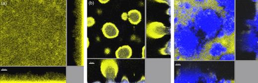
Confocal scanning laser microscopy (CSLM) micrographs acquired in biofilms formed by Pseudomonas aeruginosa pvdA (a), a pilA/pvdA mixture (b), and a pilApvdA/wild-type mixture (c). The wild type and pvdA mutant were tagged with Yfp (yellow), whereas the pilA mutant and the pilApvdA mutant were tagged with Cfp (blue). The central pictures show horizontal CLSM optical sections, and the flanking pictures show vertical CLSM optical sections. Size bars=20 µm. Reproduced from Yang (2009).
The study by Yang (2009) is one of the first to indicate that the formation of heterogeneous biofilms by P. aeruginosa might occur through mechanisms that involve complex interactions between subpopulations. The pilA, pvdA, and pqsA mutants, which alone are deficient in the formation of mushroom-shaped biofilm structures, are shown in this study to interact with each other and together build mature mushroom-shaped biofilm structures.
Tolerance
One of the most important features of microbial biofilms is their tolerance to antibiotics and components of the host immune system. Although antimicrobial agents may decrease the number of bacteria in biofilms, they will not completely eradicate the bacteria, which may have important clinical consequences in the form of relapses of infections. Tolerance to antimicrobial agents is a physiological condition that does not involve mutation and allows the bacteria to survive, but not necessarily grow, in the presence of the antimicrobial agent. Investigations of P. aeruginosa biofilms have revealed that biofilm tolerance is multifactorial. The mechanisms that contribute to tolerance include restricted antimicrobial diffusion, differential physiological activity, induction of specific tolerance mechanisms, and persister cell formation.
The available evidence suggests that biofilm matrices in general do not inhibit diffusion of antibiotics, but penetration of some antimicrobial compounds appears to be delayed. Penetration of ciprofloxacin and levofloxacin through P. aeruginosa flow-chamber biofilms was found not to be significantly delayed (Vrany et al., 1997). In support of this, Walters (2003) found that penetration of ciprofloxacin was also not significantly delayed in a P. aeruginosa colony biofilm; however, penetration of tobramycin was somewhat retarded, but eventually penetrated the biofilm completely. Whereas most antimicrobials might diffuse readily through biofilms formed by wild-type P. aeruginosa strains, it appears that alginate produced by mucoid P. aeruginosa strains can retard the diffusion of some antimicrobials (e.g. piperacillin, amikacin, gentamicin), whereas others penetrate readily (e.g. ciprofloxacin, levofloxacin, sparfloxacin, ofloxacin) (Hoyle et al., 1992; Shigeta et al., 1997). Recently, evidence was provided that the activity of an antibiotic on mucoid P. aeruginosa biofilms can be significantly enhanced by addition of alginate lyase, and DNase, suggesting that alginate and eDNA can function as an antibiotic barrier (Alipour et al., 2009). Cochran (2000) found that wild-type P. aeruginosa cells attached to alginate beads were significantly less susceptible to disinfection by hydrogen peroxide than planktonic cells of the same microorganism, although diffusion of hydrogen peroxide was not significantly delayed, indicating that mechanisms other than diffusion barriers contribute to the tolerance.
Biofilms contain distinct subpopulations of cells that exhibit differential physiological states. An increasing body of evidence suggests that the prevailing physiological states of biofilm cell subpopulations directly relate to their susceptibility and tolerance phenotypes dependent on the antimicrobial compound used. Evidence has been provided that P. aeruginosa flow-chamber biofilms, as well as colony biofilms and biofilms established in drip flow reactors, and capillary glass tubes, are composed of at least two distinct physiological subpopulations: a cell subpopulation close to the substratum (e.g. the stalk portion in biofilms with mushroom-shaped structures) that exhibits low metabolic activity and a cell subpopulation on top (e.g. the cap portion in biofilms with mushroom-shaped structures) that exhibits high metabolic activity (see Fig. 3a) (Xu et al., 1998; Walters et al., 2003; Werner et al., 2004; Pamp et al., 2008). This spatial distribution of bacteria with low and high metabolic activity is prevailing due to microscale chemical gradients that are generated by the relative diffusion and consumption rates of chemicals as can be revealed by microelectrode measurements (e.g. Xu et al., 1998; Werner et al., 2004). For example, oxygen and nutrient concentrations are high in the bulk liquid and top layer of the biofilms, whereas concentrations are low in the deeper layers of the biofilm. Conventional antimicrobial agents that are known to interfere with fundamental physiological processes of bacterial cells, such as replication (e.g. ciprofloxacin), or translation (e.g. tetracycline, tobramycin, gentamicin), were found to specifically kill the metabolically active cells in the top layer of biofilms, whereas cells of low metabolic activity survived the treatment (see Fig. 3c and d) (Hentzer et al., 2003; Walters et al., 2003; Banin et al., 2006; Pamp et al., 2008). In contrast, antimicrobial agents that interfere with bacterial membrane structure/function, such as colistin, EDTA, and SDS, were found to kill the cells in the deeper layer, whereas cells of high metabolic activity in the top layer survived the treatment (see Fig. 3b) (Banin et al., 2006; Haagensen et al., 2007; Pamp et al., 2008). In addition, DFO-gallium (a post-transition metal in complex with a siderophore), which is known to interfere with cellular iron metabolism, was found to preferentially kill bacteria in the deeper layers of P. aeruginosa biofilm (Kaneko et al., 2007; Banin et al., 2008). Systematic combined antimicrobial treatments simultaneously targeting physiologically distinct subpopulations, for example using ciprofloxacin+colistin (see Fig. 3e), tetracycline+colistin (see Fig. 3f), or gentamicin+DFO-gallium, enables killing of almost all the bacteria in P. aeruginosa biofilms (Banin et al., 2008; Pamp et al., 2008).
![Metabolic activity and antibiotic tolerance in Pseudomonas aeruginosa biofilms. (a) Visualization of metabolically active cells in P. aeruginosa biofilms. Cells appear green when they synthesize the unstable variant of the green fluorescent protein Gfp[AGA], which is expressed under the control of a ribosomal promoter. (b–f) Mature P. aeruginosa Gfp biofilms exposed to either 25 µg mL−1 colistin (b), 60 µg mL−1 ciprofloxacin (c), 200 µg mL−1 tetracycline (d), 25 µg mL−1 colistin +60 µg mL−1 ciprofloxacin (e), or 200 µg mL−1 tetracycline+25 µg mL−1 colistin (f). Live cells appear green due to the expression of Gfp, and dead cells appear red as a result of staining with the dead cell indicator propidium iodide. (g) The number of cells that survived the antimicrobial treatment were determined by plate counting of cells harvested from biofilms. The size bars are 50 µm. Reproduced from Pamp (2008).](https://oup.silverchair-cdn.com/oup/backfile/Content_public/Journal/femspd/59/3/10.1111_j.1574-695X.2010.00690.x/1/m_FIM_690_f3.jpeg?Expires=1716365681&Signature=tnwXkz9dA4lO8kw9K4M2~Tkr1e7XqvSjyBJu4fBm7-9TpZ5WvDs4EdfB4N403GHEPBZeS5IivPqLW3yoLSj8GDyBujbvdGICF35IqSMwYjthHZDsGB4-kC9Kv2usUMz1Ft~CmSkEeXlCoDpBDr0biEO2K0Kngn2SnDwnp-OrQfnVWuOhVzfcN0ljQ6HjwtVk3U9WCyEaN7wM4vgq5bIkhCaaacS02AZJOO5ir77gt~JTlJobomaX-~kD~4MtPJkDw28UM-naAHcasJbud2Ks5ZxRTEjZWi~3472~2xGbnctoRjBjF1x9Gt5m5r6-TA9jLB-T1DiFaz9nmwzmNF27Jw__&Key-Pair-Id=APKAIE5G5CRDK6RD3PGA)
Metabolic activity and antibiotic tolerance in Pseudomonas aeruginosa biofilms. (a) Visualization of metabolically active cells in P. aeruginosa biofilms. Cells appear green when they synthesize the unstable variant of the green fluorescent protein Gfp[AGA], which is expressed under the control of a ribosomal promoter. (b–f) Mature P. aeruginosa Gfp biofilms exposed to either 25 µg mL−1 colistin (b), 60 µg mL−1 ciprofloxacin (c), 200 µg mL−1 tetracycline (d), 25 µg mL−1 colistin +60 µg mL−1 ciprofloxacin (e), or 200 µg mL−1 tetracycline+25 µg mL−1 colistin (f). Live cells appear green due to the expression of Gfp, and dead cells appear red as a result of staining with the dead cell indicator propidium iodide. (g) The number of cells that survived the antimicrobial treatment were determined by plate counting of cells harvested from biofilms. The size bars are 50 µm. Reproduced from Pamp (2008).
In addition to tolerance caused by the mechanisms described above, certain gene products that are produced specifically in biofilms may have unique functions that enhance the antibiotic tolerance of the biofilm. One example of a biofilm-specific factor is the synthesis of periplasmic glucans that bind tobramycin and prevents cell death most likely by sequestering the antibiotic (Mah et al., 2003). Synthesis of the periplasmic glucans requires the ndvB gene in P. aeruginosa PA14, and biofilms formed by a P. aeruginosa ndvB mutant were found to be much more sensitive to tobramycin than wild-type biofilms. In contrast, the ndvB mutant and wild type showed no difference in tobramycin sensitivity when grown in planktonic culture. Reverse transcriptase PCR provided evidence that the ndvB gene was expressed specifically in P. aeruginosa PA14 biofilms and not in planktonic cells (Mah et al., 2003). However, microarray analysis has provided evidence that ndvB is expressed at the same level in biofilm and planktonic cells of P. aeruginosa PAO1 (Hentzer et al., 2005), and therefore the ndvB-mediated mechanism appears to be restricted to specific P. aeruginosa strains.
Upon antimicrobial exposure, some biofilm cells are able to actively adapt by inducing the expression of specific genes that confer tolerance. As described above, only the active cells in the top layer of P. aeruginosa flow-chamber biofilms survive colistin treatment, whereas the inactive cells in the deeper layers are killed by colistin (Fig. 3b). Upon exposure to colistin, the cells in the top layer induce the expression of the pmr-LPS-modification system as well as the MexAB-OprM efflux pump (Pamp et al., 2008). In contrast, the cells in the deeper layers are unable to induce the pmr operon and mexAB-oprM genes, and are hence killed by the action of colistin (Pamp et al., 2008). In contrast to the findings in biofilms, it was found that planktonic exponential-phase cells (exhibiting high metabolic activity) and planktonic stationary-phase cells (exhibiting low metabolic activity) were equally sensitive to colistin, suggesting that the mechanisms resulting in the observed colistin tolerance are biofilm specific (Pamp et al., 2008). The efflux-pump genes mexAB-oprM and mexCD-oprJ genes were also found to be induced in a stack reactor P. aeruginosa biofilm in the presence of azithromycin and were required for tolerance development, but the spatial distribution of the tolerant cells was not investigated (Gillis et al., 2005). As an active adaptation response to β-lactam antibiotics such as imipinem and ceftazidime, the cells in the top layer of the P. aeruginosa flow-chamber biofilms were found to specifically induce the chromosomally encoded AmpC β-lactamase (Bagge et al., 2004). Based on average transcriptional measurements on biofilm cell populations in comparison with planktonic cell populations, expression of a potential novel efflux system (encoded by PA1874–1877) appeared to be increased in biofilm cells and was described to be involved in the tolerance toward tobramycin, gentamicin, and ciprofloxacin (Zhang & Mah, 2008). In addition, planktonic P. aeruginosa cells appear to use an adaptation strategy, which involves upregulation of c-di-GMP-dependent genes that increase protective biofilm formation in the presence of subinhibitory concentrations of antimicrobial agents (Hoffman et al., 2005).
Mulcahy (2008) presented evidence that eDNA may create a cation-limited environment in P. aeruginosa biofilms that result in induction of the pmr-LPS-modification system conferring tolerance toward antimicrobial peptides and aminoglycosides. However, Pamp (2008) did not observe induction of the pmr genes in regions of P. aeruginosa flow-chamber biofilms shown by Allesen-Holm (2006) to have high concentrations of eDNA.
From the observations described above, it appears that the majority of biofilm cells can be killed by combined antimicrobial treatment targeting the major physiologically distinct cell subpopulations. However, it has been reported that even in combined treatments involving two antimicrobials, a small number of bacteria did survive (see Fig. 3g) (Pamp et al., 2008). Offspring of the bacteria that survived the combined antimicrobial treatment did not exhibit increased resistance to the antimicrobial agents used (Pamp et al., 2008). It might be speculated that these few surviving cells represent so-called persister cells. Persister cells are dormant or slowly dividing bacteria that are less vulnerable to antibiotics than the majority of the cell population (Brooun et al., 2000; Lewis, 2001; De Groote et al., 2009). Mutant screens in P. aeruginosa have provided evidence that a number of genes (e.g. rpoS, spoT, relA, dksA, dinG, spuC, algR, pilH, ycgM, pheA) are involved in persister formation, suggesting that the persister phenotype can be reached through multiple pathways (Murakami et al., 2005; Viducic et al., 2006; De Groote et al., 2009). In addition to the various physiologically distinct cell subpopulations in biofilms, the generation of genetic variant cells within biofilms has been described. Rough/wrinkled and small colony variants in P. aeruginosa biofilms can emerge in the absence or presence of an antimicrobial agent, whereof some of the variants can exhibit reduced susceptibility toward antimicrobial compounds (e.g. H2O2) (e.g. Drenkard & Ausubel, 2002; Boles et al., 2004; Kirisits et al., 2005).
Recent reports have demonstrated that rhamnolipid production by P. aeruginosa plays a role in the tolerance of P. aeruginosa biofilms toward immune cells (Bjarnsholt et al., 2005; Jensen et al., 2007; Alhede et al., 2009). Purified P. aeruginosa rhamnolipids were shown to be able to destroy polymorphonuclear neutrophilic leukocytes (PMNs) via necrosis (Jensen et al., 2007). Moreover, it was reported that biofilm cells of P. aeruginosa respond to the presence of PMNs by upregulating the synthesis of rhamnolipid (Alhede et al., 2009). The available evidence suggests that the rhamnolipids stick to the biofilm bacteria and function as a shield that eliminates immune cells upon contact (Alhede et al., 2009).
Dispersal
In addition to the mechanisms involved in biofilm formation, bacteria also possess mechanisms to disperse from biofilms. These mechanisms involve a reduction of bacterial adhesiveness, and breakdown or modulation of the biofilm matrix. Emigration of cells from biofilm communities is necessary to spawn novel communities at new locations, and it may be induced if the biofilm cells face unfavorable conditions (e.g. Tolker-Nielsen et al., 2000; Gjermansen et al., 2005).
In P. aeruginosa biofilms grown in flow chambers irrigated with LB medium, local dispersal was observed as a hollowing out of some microcolonies (Purevdorj-Gage et al., 2005). In the initial phase of the dispersal process, a wall-forming subpopulation of nonmotile cells constituted the outer part of the microcolonies, whereas a motile rapidly moving subpopulation was present inside the microcolonies. The motile subpopulation eventually found its way out of the microcolony, which resulted in microcolonies with a central void. This dispersal phenomenon was shown to be dependent on the microcolonies reaching a critical size.
Dispersal of P. aeruginosa biofilms in response to shifts in carbon availability was reported by Sauer (2004). Pseudomonas aeruginosa biofilms grown in flow chambers on glutamate medium responded to an abrupt upshift in carbon availability by initiating a dispersion process that led to the majority of the biomass being released from the biofilm. The extent of dispersion was dependent on the carbon source and was associated with increased expression of flagella and downregulation of twitching motility. In a subsequent study by Morgan (2006), a gene product involved in sensing environmental cues that trigger P. aeruginosa biofilm dispersal was identified. The gene encoding this sensor was denoted bdlA for biofilm dispersion locus, and through sequence analysis and phenotypic comparison of the P. aeruginosa wild type and bdlA mutant, the BdlA protein was suggested to be a chemotaxis regulator that affects the intracellular level of c-di-GMP. As described in a previous section, evidence is accruing that the production of matrix components such as Pel/Psl polysaccharides and Cup fimbriae is regulated via proteins that contain diguanylate cyclase or phosphodiesterase activities and control the intracellular level of c-di-GMP (Hickman et al., 2005; Gjermansen et al., 2006; Meissner et al., 2007). It appears that in general, high intracellular c-di-GMP levels upregulate matrix production and biofilm formation, whereas low intracellular c-di-GMP levels downregulate matrix production and induce a planktonic lifestyle. Carbon starvation and nitric oxide signaling were shown to induce the dispersal of P. aeruginosa biofilms via the induction of phosphodiesterase activity, causing decreased intracellular c-di-GMP levels (Barraud et al., 2009; Schleheck et al., 2009). Evidence was presented that the above-mentioned BdlA chemotaxis regulator is involved in nitric oxide-mediated biofilm dispersal (Barraud et al., 2009).
As described in a previous section, rhamnolipid appears to play multiple roles in the P. aeruginosa biofilm development cycle, one of them being that the production of large amounts may lead to the dispersal of cells from the biofilms (Schooling et al., 2004; Boles et al., 2005). Ryan (2009) recently presented evidence that rhamnolipid-mediated dispersal in P. aeruginosa biofilms may involve c-di-GMP. Evidence was provided that the PA2572 protein has a degenerate inactive c-di-GMP phosphodiesterase domain that may play a regulatory role, and a P. aeruginosa PA2572 mutant was found to overproduce rhamnolipid.
Recent work by Davies & Marques (2009) provides evidence that the compound cis-2-decenoic acid produced by P. aeruginosa is capable of inducing the dispersal of established biofilms and of inhibiting biofilm development. When added exogenously to P. aeruginosa biofilms at a native concentration, cis-2-decenoic acid was shown to induce the dispersal of biofilm microcolonies. This molecule was also shown to induce the dispersal of biofilms, formed by Escherichia coli, Klebsiella pneumoniae, Proteus mirabilis, Streptococcus pyogenes, Bacillus subtilis, Staphylococcus aureus, and the yeast Candida albicans. The authors suggested that cis-2-decenoic acid is produced continuously by P. aeruginosa during growth in biofilms, and that small microcolonies do not disperse because cis-2-decenoic acid is removed through diffusive and advective transport; however, dispersal from larger microcolonies occurs because the rate of production of cis-2-decenoic acid exceeds the rate of diffusion.
A recent publication by Gjermansen (2010) describes a mechanism involved in the dispersal of Pseudomonas putida biofilms that may also be of relevance for P. aeruginosa biofilms. In P. putida, the large adhesive outer-membrane protein, LapA, mediates attachment to surfaces and to matrix components. Release of LapA from the cell surface results in biofilm dispersal and is mediated through the activity of the periplasmic protease LapG. The activity of the LapG protease is controlled by the transmembrane protein LapD, which contains a c-di-GMP-binding domain, and represses LapG when the concentration of c-di-GMP is high, but derepressses LapG when the concentration of c-di-GMP is low. The available evidence (Hinsa et al., 2003; Hinsa & O'Toole, 2006; Monds et al., 2007; Newell et al., 2009) suggests that a similar system is operating in Pseudomonas fluorescens. Pseudomonas aeruginosa encodes a number of large adhesive proteins, but it does not have a homolog of lapA. However, P. aeruginosa does have homologs of lapD and lapG, and therefore a mechanism similar to the P. putida mechanism could potentially be involved in P. aeruginosa biofilm dispersal.
Final remarks
Recent work has provided knowledge about the environmental cues, genetic elements, and molecular mechanism involved in biofilm formation, tolerance, and dispersal. Based on this research, potential antibiofilm strategies can be envisioned, for example enzymatic digestion of matrix components, blocking of c-di-GMP regulated matrix synthesis, treatment with multiple antibiotics that target different subpopulations, and blocking of quorum sensing. However, the molecular understanding of the biofilm lifestyle obtained from in vitro studies should be used in future in vivo studies using animal models that mimic the complex interactions between the biofilm and the host. A more detailed understanding of the multicellular nature of microbial life will ultimately enable us to develop efficient treatments against biofilm-related infections.
Acknowledgement
This work was supported by grants from the Danish Council for Independent Research.
References
Author notes
Editor: Gianfranco Donelli
