
A 84-year-old male with severe symptomatic aortic stenosis (valve area 0.66 cm2; transvalvular gradient 48 mmHg) declined for surgical aortic valve replacement due to severe chronic obstructive pulmonary disease underwent transcatheter implantation of a 26 mm Cribier-Edwards aortic bioprosthesis (Edwards Lifesciences Inc, Irvine, CA, USA). The procedure was performed using the retrograde approach through the right femoral artery. The prosthetic heart valve made of three leaflets of bovine pericardium sutured within a balloon expandable stainless steel stent was implanted under rapid pacing. After implantation, the echocardiographic transvalvular gradient decreased to 9 mmHg and the valve area increased to 1.9 cm2. The hospital course was uneventful and the patient was discharged home 1 week after implantation.
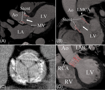
A 64-multislice computed tomography (MSCT) was performed 6 days after implantation (VCT General Electric HealthCare, Milwaukee, USA). The accurate positioning of the bioprosthesis within the calcific native valve just below the coronary ostia and above the mitral valve could be clearly observed (Figure).
This case illustrates the potential interest of 64-MSCT for evaluating the correct positioning of the new Cribier-Edwards transcutaneous heart valve. This technology might be particularly helpful in patients with technically limited approach by echocardiography.
Panel A. Contrast-enhanced ECG-gated 64-MSCT three chambers view reconstruction. The stent (in red) can be seen within the native valve and is clearly above the mitral valve. The cusps of the bioprosthesis (large arrow) are clearly delineated. LV, left ventricle; Ao, aorta; MV, mitral valve; LA, left atrium.
Panel B. MSCT reconstruction in coronal left ventricle outflow tract plane The stent is well below the ostium of the left main coronary artery (LMCA). The large arrow indicates the cusp of the bioprosthesis.
Panel C. The three cusps are well visualized inside the stent which is surrounded by the calcifications of the native aortic valve.
Panel D. Left ventricle outflow tract plane MSCT reconstruction demonstrating the correct positioning of the stent (in red) below the ostia of the coronary arteries. RCA, right coronary artery; RV, right ventricle.