
Abstract
We had recently reported unique random laser action such as quasi-single-mode and low-threshold lasing from a submicrometre-sized spherical ZnO nanoparticle film with polymer particles as defects. The present study demonstrates a novel approach to realize single-mode random lasing by adjusting the sizes of the defect particles. From the dependence of random lasing properties on defect size, we find that the average number of lasing peaks can be modified by the defect size, while other lasing properties such as lasing wavelengths and thresholds remain unchanged. These results suggest that lasing wavelengths and thresholds are determined by the resonant properties of the surrounding scatterers, while the defect size stochastically determines the number of lasing peaks. Therefore, if we optimize the sizes of the defects and scatterers, we can intentionally induce single-mode lasing even in a random structure (Fujiwara et al 2013 Appl. Phys. Lett. 102 061110).
Export citation and abstract BibTeX RIS

Original content from this work may be used under the terms of the Creative Commons Attribution 3.0 licence. Any further distribution of this work must maintain attribution to the author(s) and the title of the work, journal citation and DOI.
1. Introduction
Random lasers have recently attracted much attention as novel light sources because of their unique characteristics [1–11]. In coherent random lasers, multimode lasing superposed on amplified spontaneous emission at wavelengths and locations corresponding to the maximum gain is typically observed because interference from multiple light scattering provides randomly distributed feedback. Therefore, these lasing modes are difficult to control in frequency and spatial domains. Among several approaches for controlling random lasing modes [12–20], we numerically and experimentally proposed a singular structure [21–25] composed of mono-dispersive scatterers and defect regions. In this structure, we numerically demonstrated that adjusting the resonances of individual scatterers and intentionally introducing defect sites enable us to control the resonant frequency and localization position [23, 24]. To experimentally verify the proposed method, we prepared a random laser from mono-dispersive spherical ZnO nanoparticle film with polystyrene particles as defect sites [21, 22], whose features were clearly distinct from those of conventional ZnO random lasers. In particular, we found superior random lasing properties such as suppression of the number of lasing modes, low threshold, wavelength tunability and limiting lasing locations (figure 1) compared with conventional random lasers. Our proposed method for manipulating localized modes in a random structure has two key points. The first is the use of resonant scatterers in which the lasing wavelength (frequency band) is controlled due to the resonance of individual scatterers and photon confinement is improved (thereby lowering their threshold). The second is the use of defect particles. The defect site provides a localized region for resonant light as we confirmed that such unique random lasers could always be initiated at defect sites. However, it was still unclear what effects the defects had on random lasing properties such as lasing wavelengths and thresholds. In typical microcavity structures, the defect size defines the cavity length, which sets the resonant wavelength and mode spacing (the number of lasing peaks). Therefore, we conjectured that the defect size could also affect the random lasing properties and that a study of the dependence of random lasing properties on defect size might provide insights into the achievements of single-mode random lasing.
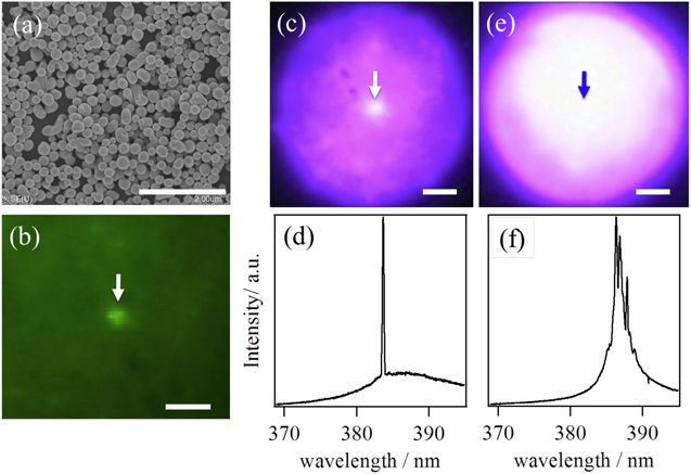
Figure 1. (a) Scanning electron micrograph of submicrometre-sized spherical ZnO particles after laser-induced melting. (b) Green fluorescence image of a ZnO particle film including a point defect of 900 nm. White bars in (a) and (b) indicate the scale of 2 μm. The white arrow indicates the position of the polymer particle. Emission spectra and images of UV random lasing emission from ZnO nanoparticles are shown after ((c) and (d)) and before ((e) and (f)) laser-induced melting, including a defect of 900 nm at the centre of each image (indicated by an arrow). The diameter of the excitation spot is ∼65 μm and the white bars in (c) and (e) indicate the scale of 10 μm.
Download figure:
Standard image High-resolution imageIn this study, to improve the controllability, especially, to clarify the roles of defect particles and the origins why the single mode lasing occurred in our proposed random lasers, we experimentally studied the dependence of random lasing properties on defect size in a structure of mono-dispersive spherical ZnO nanoparticles with polymer nanoparticles acting as the defects. Comparing random lasing properties with different defect diameters, we found that the number of lasing peaks strongly depends on the defect size, whereas the thresholds and wavelengths of the lasing peaks are independent of the defect size. From these results, we concluded that in our proposed random lasers, the lasing wavelengths are determined by the resonant characteristics of the surrounding mono-dispersive spherical scatterers, while the number of lasing peaks is determined by the defect size, where the probability of stochastically forming lasing modes at a particular defect site depends on the defect size. These results suggest a novel technique for manipulating resonant and lasing modes in random structures and for realizing single-mode random lasing.
2. Experiment
In the experiments, we used quasi-mono-dispersive ZnO nanoparticles as scatterers and gain materials. These nanoparticles were fabricated from commercially available ZnO nanoparticles with a mean diameter of 100 nm using a laser-induced melting method [26, 27]. We attempted to fabricate mono-dispersive spherical ZnO nanoparticles of ∼200 nm, the optimal size for resonance with the emission wavelength of ZnO, which is estimated based on Mie scattering theory. After fabrication by a laser-induced melting method [26, 27], spherical ZnO nanoparticles ∼212 nm in diameter were obtained (figure 1(a)); these were almost mono-dispersive and spherical, in contrast to the commercially available, irregularly shaped poly-dispersive ZnO nanoparticles. After dispersing these ZnO nanoparticles in water, we added commercially available green fluorescent polystyrene nanoparticles with diameters of 300, 900 and 2000 nm into the solution as point defects. A drop of the solution was placed on a cover glass and dried at room temperature. Note that the green emission from the polymer nanoparticles did not contribute to the results of random lasing measurements, because the emission from the green fluorescent nanoparticles was very weak and easily degraded by the irradiation of the intense pulsed laser light within several seconds.
The experimental setup was the same as that used in our previous study [21]. Pulses from a Q-switched pulsed laser (wavelength 355 nm, repetition rate 1 kHz, pulse duration 300 ps) were irradiated on the sample (spot size ∼65 μm), which was fixed on a confocal microscope stage by an objective lens (100×, NA = 0.9), and the emission from the ZnO nanoparticles was collected using the same objective. Then, emission passing through a pinhole (∼1 μm on the sample plane) was coupled to a spectroscope equipped with a cooled charge-coupled device (CCD). Before measurements of the random lasing properties were performed, the positions of the defects were determined by taking a green fluorescence image of the polystyrene nanoparticles. Figure 1(b) shows the green fluorescence image with a white arrow indicating a defect. After confirming the locations of the defects, we measured the emission spectra by changing the excitation intensity.
3. Result and discussion
Figures 1(c) and (d) show a CCD image of random lasing and emission spectra from a sample composed of spherical ZnO nanoparticles (mean diameter 212 nm) and a defect polymer particle (indicated by an arrow, diameter 900 nm). For reference, in figures 1(e) and (f), a CCD image of random lasing and emission spectra from a sample composed of commercially available ZnO nanoparticles and a defect polymer particle are shown. In both cases, the excitation intensity was set to a value two times higher than the respective thresholds. Comparing the results of figures 1(c) and (e), we found that regardless of the presence of a defect particle, random lasing was homogeneously induced in the commercially available ZnO nanoparticle film, while the lasing emission from the mono-dispersive spherical ZnO nanoparticle film was clearly concentrated in the defect region. This means that the emission from the spherical ZnO nanoparticles was efficiently confined at wavelengths corresponding to the resonance bands of individual spherical ZnO nanoparticles and enhanced the resonant lights around the defect region as opposed to the defect-free site. Furthermore, from the spectra (figure 1(f)) of a commercial ZnO nanoparticle film, multiple sharp peaks superposed on a collapsed broad emission spectrum were observed at wavelengths of ∼387 nm with a threshold of ∼50 MW cm−2, showing characteristics similar to those of a conventional random laser. On the other hand, from the spectra (figure 1(d)) of the spherical ZnO nanoparticle film, a single sharp peak was observed at ∼383 nm with a threshold of ∼5 MW cm−2, which is blue-shifted relative to conventional random laser wavelengths and always occurs ∼380 nm. From these results, our proposed random laser exhibits unique lasing properties such as suppression of the number of lasing modes, low threshold, lasing wavelength control and limiting lasing locations, as reported in our previous study [21].
In figures 2(a)–(c), the emission spectra for defects of different sizes are shown when the excitation intensity was set two times higher than each threshold (∼10 MW cm−2). For reference, in figure 2(d), the emission spectra at defect-free sites have multiple sharp peaks superposed on a collapsed broad emission spectrum, similar to those for typical random lasers. Regardless of the defect size, a few discrete sharp lasing peaks were observed at each defect, and their characteristics were clearly different from those observed for typical random lasers. Repeating the measurements with other defects, we confirmed that similar characteristics were observed, and we have summarized the data in figure 3. In this figure, scatter plots of lasing wavelengths and thresholds for cases with different defect sizes (triangles: 300 nm, circles: 900 nm, squares: 2000 nm, diamonds: without defect) are shown. The thresholds at the defect sites were about 10 times smaller than those at the defect-free sites, and the wavelengths of the lasing peaks were concentrated around 380 nm, while the wavelengths for defect-free sites occurred at around 388 nm, as typically observed for conventional random lasers in ZnO nanoparticle film. The difference in the thresholds agrees well with the CCD image of the random lasing emission shown in figure 1(c), where intense spots were observed only at the defect sites but not at the defect-free sites. In addition, these thresholds and lasing wavelengths were independent of the defect size. These results suggest that the lasing wavelength was determined by the resonance properties of the scattering nanoparticles and not by the defect size.

Figure 2. Typical lasing emission spectra at different defect sites. The defect particle sizes are (a) 300, (b) 900 and (c) 2000 nm, respectively. (d) Emission spectra at defect-free sites. Each spectrum was normalized by its maximum intensity.
Download figure:
Standard image High-resolution image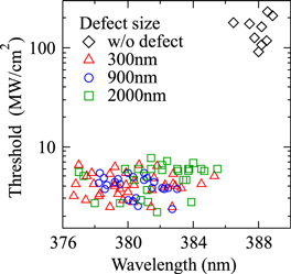
Figure 3. Scatter plots of thresholds and lasing wavelengths for different defect sizes (triangles: 300 nm, circles: 900 nm, squares: 2000 nm, diamonds: without defect).
Download figure:
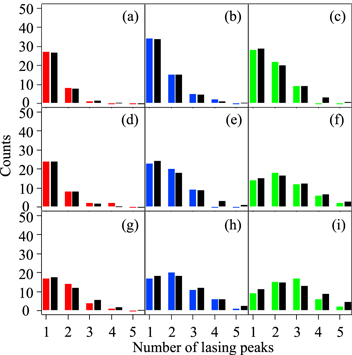
Standard image High-resolution imageTo verify the role of the defects, we made histograms of the number of lasing peaks against the defect size at excitation intensities of 1.0, 2.0 and 3.0 times each threshold (figure 4), in which about 40–50 lasing spectra at different defects were measured for each defect size and excitation intensity. In the figure, the coloured bars represent the experimental data and the black bars indicate that the data could be fitted well to a Poisson distribution (black bars in figure 4). This means that lasing modes were randomly induced around a defect at wavelengths within the resonant frequency band and that the number of lasing modes was stochastically determined. From the fit, the mean values of the Poisson distributions were estimated to be 0.6, 0.9 and 1.4 for defect sizes of 300, 900 and 2000 nm, respectively, when the excitation intensity was set to each threshold. Increasing the excitation intensity to two (three) times each threshold, the mean values further increased to 0.7 (1.4), 1.5 (2.0) and 2.1 (2.6) for each defect size. Because the average number of lasing peaks decreased (increased) with the decreasing (increasing) defect size, the probability of forming lasing modes around the defect would be greatly altered depending on the defect size.
Figure 4. Histograms of the number of lasing peaks. Defect particle sizes are (a), (d), (g) 300; (b), (e), (h) 900 and (c), (f), (i) 2000 nm. The excitation intensities are (a)–(c) 1.0; (d)–(f) 2.0 and (g)–(i) 3.0 times each threshold. Coloured and black bars indicate the experimental data and fitting results assuming Poisson distributions.
Download figure:
Standard image High-resolution image
In typical microcavity structures, the defect size is related to the resonant frequency and mode spacing (number of lasing peaks). In our proposed random laser, when the defect size changes, not the lasing wavelength, but the average number of lasing peaks changed, as shown in figures 3 and 4. This would suggest that the lasing properties was not simply determined by the defect size like typical lasers, because the feedback was provided by the multiple light scattering at the resonant frequency band of surrounding individual ZnO nanoparticles (around 380 nm) [23]. Considering that ZnO nanoparticles are used as scatterers and gain media, the gain for lasing is provided from the localized fields extended into surrounding ZnO nanoparticles around the defect. Therefore, when the defect size increases, the number of surrounding ZnO nanoparticles that would contribute to induce laser oscillation could be increased, resulting in the increase in the probability of different lasing modes being induced. With regards to the excitation intensity dependence, the number of lasing peaks increases by increasing the excitation intensity. The mechanism for increasing the number of lasing modes in our proposed random laser is basically similar to a conventional random laser [1–4]. However, in our proposed random laser, the localized modes are stochastically formed only around the defect and the number of ZnO nanoparticles that provides the gain depends on the defect size. Because the spatial overlap between the localized modes and the gain media is dependent on the defect size, the number of possible lasing modes is limited by the defect. Therefore, we think that the number of lasing modes does not rapidly increase like a conventional random laser. In contrast, regardless of the defect size, random lasing always began at wavelengths of around 380 nm and exhibited an almost constant threshold, as shown in figure 3. In the experiments, because the same spherical ZnO nanoparticles were used in each case, the resonant scattering of ZnO nanoparticles provides strong feedback and determines the lasing wavelength, independent of the defect size. Thus, the lasing wavelength was determined by the resonant properties of individual scatterers, independent of the defect size, while the probability of the number of lasing peaks was dependent on the defect size.
4. Conclusion
In summary, we experimentally studied the effect of defect size on the lasing properties of our proposed random laser composed of mono-dispersive spherical ZnO nanoparticles with polymer nanoparticles as defects. Comparing the random lasing properties at defects with different diameters, we found that the average number of lasing peaks decreased (increased) with decreasing (increasing) defect size, while the wavelengths of lasing peaks always appeared around 380 nm and their lasing thresholds were unchanged (∼4 MW cm−2) independent of the defect size. These results suggest that lasing wavelengths are determined by the resonant properties of the surrounding scatterers, while the number of lasing peaks depends on the defect size. Thus, by adjusting the defect size, the average number of lasing peaks could be roughly controlled, resulting in the realization of single-mode lasing even in a random structure. From the viewpoint of physical understanding and technological applications, this method provides important insights into the manipulation of randomly appearing lasing properties in spatial and frequency domains.
Acknowledgments
This work was supported by JSPS KAKENHI (Grant Numbers 22681011, 24651111 and 15J01516), the PRESTO program of the Japan Science and Technology Agency, the Murata Science Foundation, the Hokkaido University Clark Memorial Foundation and the Cooperative Research Program of 'Network Joint Research Center for Materials and Devices'.