
Key Points
-
Most white lesions in the mouth are inconsequential and caused by friction or trauma.
-
However, cancer and some systemic diseases such as lichen planus and candidosis may present in this way.
-
Biopsy may be indicated.
Key Points
Oral medicine
-
Aphthous and other common ulcers
-
Mouth ulcers of more serious connotation
-
Dry mouth and disorders of salivation
-
Oral malodour
-
Oral white patches
-
Oral red and hyperpigmented patches
-
Orofacial sensation and movement
-
Orofacial swellings and lumps
-
Oral cancer
-
Orofacial pain
Abstract
This series provides an overview of current thinking in the more relevant areas of oral medicine for primary care practitioners, written by the authors while they were holding the Presidencies of the European Association for Oral Medicine and the British Society for Oral Medicine, respectively. A book containing additional material will be published. The series gives the detail necessary to assist the primary dental clinical team caring for patients with oral complaints that may be seen in general dental practice. Space precludes inclusion of illustrations of uncommon or rare disorders, or discussion of disorders affecting the hard tissues. Approaching the subject mainly by the symptomatic approach — as it largely relates to the presenting complaint — was considered to be a more helpful approach for GDPs rather than taking a diagnostic category approach. The clinical aspects of the relevant disorders are discussed, including a brief overview of the aetiology, detail on the clinical features and how the diagnosis is made. Guidance on management and when to refer is also provided, along with relevant websites which offer further detail.
White lesions
Truly white oral lesions may consist of collections of debris (materia alba), or necrotic epithelium (such as after a burn), or fungi – such as candidosis. These can typically be wiped off the mucosa with a gauze.
Other lesions which cannot be wiped off, appear white usually because they are composed of thickened keratin, which looks white when wet (Fig. 1). A few rare conditions that are congenital, such as white sponge naevus (Fig. 2) present in this way but most such white lesions are acquired and many were formerly known as 'leukoplakia', a term causing misunderstanding and confusion. The World Health Organisation originally defined leukoplakia as a 'white patch or plaque that cannot be characterised clinically or pathologically as any other disease', therefore specifically excluding defined clinicopathologic entities such as candidosis, lichen planus (LP) and white sponge naevus, but still incorporating white lesions caused by friction or other trauma, and offering no comment on the presence of dysplasia. A subsequent seminar defined leukoplakia more precisely, as 'a whitish patch or plaque that cannot be characterised clinically or pathologically as any other disease and which is not associated with any physical or chemical causative agent except the use of tobacco'.

Leukoplakia, ventral tongue

White sponge naevus
There are a range of causes of white lesions (Table 1). Morphological features may give a guide to the diagnosis. For example, focal lesions are often caused by keratoses. Multifocal lesions are common in thrush (pseudomembranous candidosis) and in LP. Striated lesions are typical of LP, and diffuse white areas are seen in the buccal mucosa in leukoedema and some LP, in the palate in stomatitis nicotina and at any site in keratoses. White lesions are usually painless but this may not be the case in burns, candidosis, LP, or lupus erythematosus.
Local causes of white lesions
Debris, burns (from heat, radiation, chemicals such as mouthwashes), grafts and scars may appear pale or white. Materia alba can usually easily be wiped off with a gauze.
Furred tongue
Tongue coating is common, particularly in edentulous adults on a soft, non-abrasive diet, people with poor oral hygiene, and those who are fasting or have febrile diseases. The coating appears more obvious in xerostomia. The coating consists of epithelial, food and microbial debris and the tongue is the main reservoir of some micro-organisms such as Candida albicans and some Streptococci, and the various anaerobes implicated in oral malodour (see article four).
Diagnosis
The history is important to exclude a congenital or hereditary cause of a white lesion. The clinical appearances usually strongly suggest the diagnosis. Biopsy is only required if the white lesion cannot be rubbed off from the mucosa with a gauze.
Management
Treatment is of the underlying cause where this can be identified.
Congenital causes of white lesions
Fordyce spots
Some common whitish conditions, notably Fordyce granules (ectopic sebaceous glands) are really yellowish, but may cause diagnostic confusion (Fig. 3). This condition is entirely benign and does not require any further intervention.

Fordyce spots
Leukoedema
Leukoedema is a common benign congenital whitish-grey filmy appearance of the mucosa, seen especially in the buccal mucosae bilaterally in persons of African or Asian descent. Diagnosis is clinical — the white appearance disappears if the mucosa is stretched. No treatment is available or required.
Inherited dyskeratoses
Inherited disorders of keratin are rare, but may be diagnosed from a family history or other features associated, such as lesions on other mucosae, or skin appendages such as the nails.
White sponge naevus, the commonest of the inherited dyskeratoses, is an autosomal dominant condition characterised by thickened, folded white patches most commonly affecting the buccal mucosae (Fig. 2). Other mucosal sites in the mouth may be involved and some patients may have similar lesions affecting genital and rectal mucosa. Since other dyskeratoses may have wider implications and in particular the risk of malignant transformation, specialist care is indicated.
Inflammatory causes of white lesions
Infections
White lesions which can result from infections include candidosis (Fig. 4), hairy leukoplakia (caused by Epstein-Barr virus), warts and papillomas (caused by human papillomaviruses) (Fig. 5) and the mucous patches and leukoplakia of syphilis. Specialist care is usually indicated.

Pseudomembranous candidosis

Condyloma acuminatum (genital wart)
Candidosis (candidiasis; moniliasis)
The importance of Candida has increased greatly, particularly as the HIV pandemic extends. This common commensal can become opportunistic if local ecology changes, or the host immune defences fail. Candida albicans is the common cause but occasionally other species may be implicated; in decreasing order of frequency these are:
-
C. tropicalis
-
C. glabrata
-
C. parapsilosis
-
C. krusei
-
Other Candida species and other genera.
Some 50% of the normal healthy population harbour (carry) C. albicans as a normal oral commensal particularly on the posterior dorsum of tongue, and are termed Candida carriers.


Candidosis is the state when C. albicans causes lesions and these can be mainly white lesions; (thrush particularly; Fig. 4) or candidal leukoplakia (Fig. 6) in which hyphal forms are common, or red lesions (denture-related stomatitis, median rhomboid glossitis, erythematous candidosis) — in which yeast forms predominate, and which may be symptomless though antibiotic stomatitis and angular cheilitis can cause soreness (Article six).

Candidal leukoplakia, right buccal mucosa
Circumstances that cause susceptibility to candidosis include local factors influencing oral immunity or ecology, or systemic immune defects, or a combination of more than one factor (Table 2).
Diagnosis
The diagnosis of candidosis is clinical usually but a Gram-stained smear (hyphae) or oral rinse may help.
Management
Possible predisposing causes should be looked for and dealt with, if possible. Polyene antifungals such as nystatin of amphotericin, or imidazoles such as miconazole or fluconazole are often indicated.
Non-infective causes
Lichen planus (LP) is a very common cause of oral white lesions. Most dental practitioners will have patients afflicted with LP. It is the main skin disease that can present with oral white lesions but lupus erythematosus and keratoses can present similarly.
Lichen planus
Lichen planus (LP) usually affects persons between the ages of 30 to 65 and there is a slight female predisposition.
Aetiopathogenesis
LP is an inflammatory autoimmune type of disease but it differs from classic autoimmune disorders in having no defined autoantibodies, and only rarely being associated with other autoimmune diseases. There is also no definitive immunogenetic basis yet established for LP and familial cases are rare.
Many patients afflicted with LP have a conscientious type of personality with obsessive-compulsive traits and suffer mild chronic anxiety, suggesting neuro-immunological mechanisms may be at play. Stress has been held to be important in LP: patients have a tendency to be anxious and depressed, but of course the chronic discomfort may partially explain some cases in which this association has been documented.

Pathologically, there is a local cell-mediated immunological response characterised by a dense T lymphocyte inflammatory cell infiltrate in the upper lamina propria causing cell death (apoptosis) in the basal epithelium, probably caused by the production of cytokines such as tumour-necrosis factor alpha (TNF∝) and interferon gamma (IFN-γ).
The antigen responsible for this immune response is unclear but lesions very similar to LP – termed lichenoid lesions – are sometimes caused by:
-
Dental restorative materials (mainly amalgam and gold)
-
Drugs (non- steroidal anti-inflammatory agents, antihypertensive agents antimalarials, and many other drugs)
-
Chronic graft-versus-host disease seen in bone marrow (haemopoietic stem cell) transplant patients
-
Infection with hepatitis C virus (HCV) in some populations such as those from southern Europe and Japan
-
A variety of other systemic disorders such as hypertension and diabetes — probably a reaction to the drugs used.
Clinical features
LP can affect stratified squamous epithelium of the skin, the oral mucosa and genitalia.
Oral LP may present a number of different clinical pictures (Figs 7,8,9,10,11,12), including:
-
Papular LP — white papules (Fig. 7)
-
Reticular LP — a network of raised white lines or striae (reticular pattern) (Figs 8 and 9)
-
Plaque-like LP — simulating leukoplakia
-
Atrophic red atrophic areas — simulating erythroplasia (Fig. 10; mixed atrophic/erosive form): lichen planus is one of the most common cause of desquamative gingivitis.
-
Erosive erosions — less common, but persistent, irregular, and painful, with a yellowish slough ().

Papular lichen planus

Reticular lichen planus

Reticular lichen planus, dorsum of tongue

Erosive lichen planus, buccal mucosa

Erosive lichen planus, dorsum of tongue

Lichenoid reaction in buccal mucosa, reaction to amalgam contact
White lesions of LP are often asymptomatic, but there may be soreness if there are atrophic areas or erosions.
LP typically results in lesions in the posterior buccal mucosa bilaterally but the tongue or gingivae are other sites commonly affected.
On the skin, lichen planus frequently presents as a flat-topped purple polygonal and pruritic papular rash most often seen on the front (flexor surface) of the wrists (Fig. 13) in which lesions are often are crossed by fine white lines (Wickham's striae; Fig. 14). Oral LP may be accompanied by vulvovaginal lesions (the vulvovaginal-gingival syndrome).

Lichen planus, skin

Cutaneous lichen planus
Prognosis
Often the onset of LP is slow, taking months to reach its peak. It may clear from the skin within 18 months but in a few people persists for many years. Oral lesions often persist. There is no sign or test to indicate which patients will develop only oral, or oral and extraoral lesions of LP.
Non-reticular oral LP in particular has a small premalignant potential – probably of the order of 1%. There is no test to reliably predict this.
Diagnosis
LP is often fairly obviously diagnosed from the clinical features but, since it can closely simulate other conditions such as:
-
Lupus erythematosus,
-
Chronic ulcerative stomatitis,
-
Keratosis, or even
-
Carcinoma,
biopsy and histopathological examination of lesional tissue, occasionally aided by direct immunostaining, are often indicated.
Management
Treatment of LP is not always necessary, unless there are symptoms. Predisposing factors should be corrected:
-
It may be wise to consider removal of dental amalgams if the lesions are closely related to these, or unilateral, but tests such as patch tests will not reliably indicate which patients will benefit from this. Accordingly, empirical replacement of amalgam restorations may be indicated.
-
If drugs are implicated, the physician should be consulted as to the possibility of changing drug therapy.
-
If there is HCV infection, this should be managed by a general physician.
-
Improvement in oral hygiene may result in some subjective benefit; chlorhexidine or triclosan mouthwashes may help. Symptoms can often be controlled, usually with topical corticosteroids or sometimes with tacrolimus.
-
If there is severe or extensive oral involvement, if LP fails to respond to topical medications, or if there are extraoral lesions, specialist referral may be indicated.
-
Patients with non-reticular lichen planus should be monitored to exclude development of carcinoma. Tobacco and alcohol use should be minimised.
Changes that might suggest a tumour is developing could include any of the following persisting more than three weeks:
-
A sore on the lip or in the mouth that does not heal
-
A lump on the lip or in the mouth or throat
-
A white or red patch on the gums, tongue, or lining of the mouth
-
Unusual bleeding, pain, or numbness in the mouth
-
A sore throat that does not go away, or a feeling that something is caught in the throat
-
Difficulty or pain with chewing or swallowing
-
Swelling of the jaw that causes dentures to fit poorly or become uncomfortable
-
Pain in the ear
-
Enlargement of a neck lymph gland.
Websites and patient information
http://www.tambcd.edu/lichen/ http://www.aad.org/pamphlets/lichen.html
Keratoses and leukoplakias
Frictional keratosis
Frictional keratosis is quite common. It is caused particularly by friction from the teeth seen mainly at the occlusal line in the buccal mucosae, particularly in adult females – especially in those with temporomandibular pain-dysfunction syndrome. Patients with missing teeth may develop keratosis on the alveolar ridge (Figs 15 and 16).

Frictional keratosis, lateral tongue

Frictional keratosis, retromolar pad
Malignant change is rare but any sharp edges of teeth or appliances should be removed and the patient counselled about the habits.
Tobacco-induced keratoses
Tobacco is a common cause of keratosis, seen especially in males. The teeth are usually nicotine-stained and there may be mucosal smoker's melanosis but malignant change is uncommon in most forms (Table 3).
Idiopathic keratoses
Many leukoplakias are uncommon and arise in the absence of any identifiable predisposing factors and most – up to 70% in large series – are benign without any evidence of dysplasia. However, the remaining 10–30% may be, or may become, either dysplastic or invasive carcinomas. Overall the rate of malignant transformation of all keratoses and leukoplakias is of some 3–6% over 10 years.
The lesions of greatest malignant potential are those leukoplakias which are:
-
speckled, nodular or verrucous lesions (Figs 17 and 18)
Figure 17 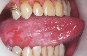
Erythroleukoplakia
Figure 18 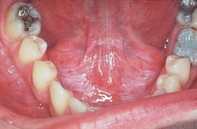
Leukoplakia, floor of mouth
-
in at-risk sites (lateral tongue, ventral tongue, floor of mouth and soft palate complex) (Figs 19 and 20)
Figure 19 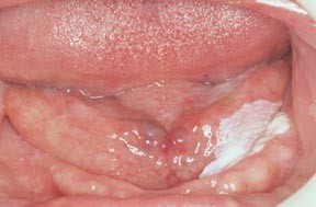
Sublingual keratosis
Figure 20 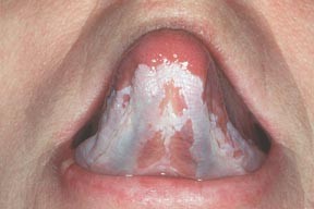
Leukoplakia, ventral tongue, floor of mouth
-
associated with Candida (Fig. 6).


In these, rates of malignant transformation up to 30% have been reported in some series.
Diagnosis
The nature of white lesions can often only be established after further investigation.
Biopsy is usually indicated, particularly where there is a high risk of malignant transformation, such as in lesions with:
-
Any suggestion of malignancy
-
Admixture with red lesions (speckled leukoplakia or erythroleukoplakia)
-
A raised lesion (nodular or verrucous leukoplakia)
-
Candidal leukoplakia
-
floor of mouth leukoplakia (sublingual keratosis)
-
a rapid increase in size
-
change in colour
-
ulceration
-
pain
-
regional lymph node enlargement.
Prognosis
The finding by the pathologist of epithelial dysplasia may be predictive of malignant potential but this is not invariable, and there can be considerable inter- and intra-examiner variation in the diagnosis of dysplasia.
Thus there has been a search for molecular markers to predict exactly which lesions are truly of malignant potential and may develop into oral squamous cell carcinoma (OSCC).

The most predictive of the molecular or cellular markers thus far assessed for OSCC development apart from dysplasia, include chromosomal polysomy, the tumour suppressor p53 protein expression, and loss of heterozygosity (LOH) at chromosome 3p or 9p. Routine use of these is, however, hampered by their complexity and lack of facilities in many pathology laboratories.
As a surrogate for individual molecular markers, measurement of gross genomic damage (DNA ploidy) may be a realistic option, and is now available in some oral pathology laboratories. See Fig. 21.

Leukoplakia, floor of mouth
Management
The dilemma in managing patients with potentially malignant oral lesions and field change has been of deciding which mucosal lesions or areas will progress to carcinoma. Specialist referral is indicated.
Cessation of dangerous habits such as tobacco and/or betel use (Figs 22 and 23), and the removal of lesions is probably the best course of action, particularly if they are the high-risk lesions or in a high risk group for carcinoma (see article nine).

Betel chewing keratosis

Tooth staining from betel chewing
Perhaps surprisingly, management of leukoplakias is very controversial, since there are no randomised controlled double blind studies that prove the best type of treatment. Thus specialists may still offer care which ranges from 'watchful waiting' to removal of the lesion (by laser, scalpel or other means) (Fig. 24).

Management of leukoplakias
Useful websites and patient information

http://www.cochrane.org/cochrane/revabstr/ab001829.htm http://www.emedicine.com/ent/topic731.htm http://www.mayoclinic.com/invoke.cfm?id=DS00458

Author information
Authors and Affiliations
Corresponding author
Additional information
Refereed Paper
Rights and permissions
About this article
Cite this article
Scully, C., Felix, D. Oral Medicine — Update for the dental practitioner: Oral white patches. Br Dent J 199, 565–572 (2005). https://doi.org/10.1038/sj.bdj.4812901
Published:
Issue Date:
DOI: https://doi.org/10.1038/sj.bdj.4812901
This article is cited by
-
Toluidine blue color perception in identification of oral mucosal lesions
Clinical Oral Investigations (2011)
-
Nicotine, smoke and patches
British Dental Journal (2006)
