
Abstract
Thoracoscopic repair is feasible and safe for congenital diaphragmatic hernia (CDH). The operation can be performed with three trocars using carbon dioxide insufflations at a pressure of 4–6 mmHG. From January 2001 to July 2012, we performed thoracoscopic repair for 311 children with CDH including 152 newborns and 159 infants and toddlers. Mean operative time was 75 ± 27 min. HFOV was used in 24 patients. Direct closure of two rims of diaphragmatic hernia was carried out in 175 patients. Closure of two rims of diaphragmatic hernia with the thoracic wall was performed in 136 patients. Prosthetic patches were required in 54 patients. Conversion to open surgery was required in 38 patients (12.2 %). There were no intraoperative deaths. 38 patients died postoperatively (13.5 %).
Introduction
Congenital diaphragmatic hernia (CDH) is a rare and life-threatening condition in children. The reported incidence of CDH is 1 in 2,107 births [1]. Although many advanced technologies have been applied, the mortality rate of CDH is still high.
The conventional surgery has been performed through laparotomy until recently when these were replaced by laparoscopic or thoracoscopic techniques [2–15].
The herniation is located in the left side in 80 % and in the right side in 20 %. Bilateral hernia is rare [16]. In left diaphragmatic hernia, the hernia usually contains small intestine and colon. The spleen or the stomach or both may be shifted into the hemithorax. The left lobe of the liver and the left kidney are rarely seen in the thorax. In right CDH, a part of the right lobe of the liver is always found in the thorax. Small intestine is occasionally seen in the thorax also.
Treatment
Preoperative care
Preoperative care is an essential step in management of CDH. All efforts aim to stabilize the cardiorespiratory system while avoiding even minimal iatrogenic injury. The resuscitation should begin with endotracheal intubation and mechanical ventilation. Pulmonary hypertension is treated by medicaments, high frequency oscillatory ventilation, nitrous oxide, and/or ECMO.
Timing of surgery
Congenital diaphragmatic hernia is no longer considered an emergency condition. Early surgical intervention is now replaced by delayed surgery. How long is a proper delay? A 100-h delay is recommended by some authors. It could be shorter to reduce the risk of ventilation-associated infection in situation where cardiovascular parameters and blood gaz measurements are relatively stable.
Anesthesia
Anesthesia with bilateral ventilation is used for newborns, and one-lung ventilation is applied for infant and toddlers. Conventional ventilators have been used. High frequency respirators have been used recently for newborns to provide better oxygen [17, 18].
Thoracoscopic repair for CDH
Indications most patients with CDH can be operated by a thoracoscopic approach.
In the past, contraindication for thoracoscopic repair included (1) stomach located in the thoracic cavity, (2) associated cardiac anomalies, and (3) requirement of prosthetic patch.
Recent report no longer considers those conditions contraindications for thoracoscopic repair [6, 13, 19–21].
Positioning
The patient is placed in the lateral decubitus position with the head elevated. The upper arm is left free.
The surgeon stands at the patient’s head, with the monitor positioned at the patient’s feet (Fig. 1).

Patient’s position
Procedure
-
Trocar placement A 5-mm skin incision is made over the third intercostal space at the mid-axillary line. A small clamp is introduced through the intercostal muscle, which is spread out to allow air to the thoracic cavity. A 5-mm trocar is introduced at this site for a thoracoscope. The second trocar is placed in the fourth intercostal space anteriorly, and the third trocar is inserted in the fourth intercostal space behind the scapular tip (Fig. 2).
Fig. 2 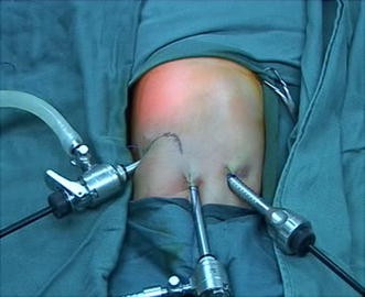
Trocar placement
-
Carbon dioxide insufflation is maintained at a pressure of 2–4 mmHg at a flow rate of 1 L/min in the thoracic cavity. Pressure is increased up to 6 mmHg if reduction of herniated organs is difficult.
-
Inspection Identify the herniated organs through thoracoscope and try to identify the orifice of the diaphragmatic defect if possible.
-
Reduction of herniated organs is the most difficult step, especially with much small intestine in the thoracic cavity in a low birth weight newborn. The small intestine is gently reduced into the abdominal cavity using blunt graspers. The direction of the reduction is important. It is better if the surgeon can identify the diaphragmatic defect before reducing. The reduction should start with the small intestine, then the colon. Reduction of the spleen is the last step and should be performed carefully. One grasper holds the gastrosplenic mesentery and pushes it to the abdominal cavity. The other grasper is used to push the spleen very gently at the same time.
Reduction of herniated organs is difficult when the orifice of the diaphragm is small. Inspecting and widening the orifice facilitate the reduction.

-
Closure of diaphragm defect If the defect is small, direct closure of the two diaphragmatic rims is carried out. The hernia defect is repaired using nonabsorbable interrupted sutures (Ethibon 2/0 or 3/0) encompassing two borders of the diaphragmatic defect with extracorporeal knots.
The first stitch is placed at the middle of the hernia defect (Fig. 3), and then the CO2 insufflation is turned off. Subsequent stitches could be done without insufflation to minimize cardiovascular disturbances.

First stich
-
Closure of lateral corner of the defect It is difficult to close the lateral corner of the diaphragmatic hernia completely when it is large (when the distance between the two diaphragmatic rims is larger than 2 intercostal spaces). This site is a location for recurrent herniation if it is not well managed.
A skin incision is made on the thoracic wall. A needle is passed through the wound to the thoracic cavity, capturing the anterior rim and then the posterior rim and looping back through the thoracic wall. Knot is made extracorporeal, and then the skin is closed over the knots (Fig. 4).

Closure of the two diaphragmatic defects with thoracic wall
-
Close the diaphragm defect with a patch If the direct closure of the diaphragmatic hernia is under tension or impossible, a prosthetic mesh is required. Different kinds of prosthetic mesh could be used; however, an impermeable one should be chosen to prevent fluid from the abdominal cavity entering the thoracic cavity due to negative pressure.
One trocar is withdrawn. The mesh was introduced into the thoracic cavity through the trocar wound after it has been appropriately trimmed. The mesh is fastened close to the diaphragmatic rims using intra- or extracorporeal sutures. Closure of the mesh to the thoracic wall is carried out as previously described.
-
Drainage Placement of a chest drain is still debated. Many surgeons do not place a chest drain to avoid mediastinal swing. However, we have seen pleural effusion in our patients without a chest drain. We now place a thoracic drain and perform intermittent manual suction every 3 h. The drain is removed after 48 h if the chest X-ray is satisfactory.
-
Surgery in the neonatal intensive care unit and under high frequency oscillatory ventilator (HFOV) To avoid the stress of transportation, we perform thoracoscopic repair for patients whose cardiovascular system is not very stable or under HFOV. We do not have any special difficulty in performing the operation in this situation.
-
Postoperative management Ventilation with high frequency and low peak airway pressure is continued. Cardiac echography is routinely obtained to assess pulmonary hypertension, shunt flow, and ventricular performance. HFOV, NO or ECMO is required when pulmonary arterial hypertension is present.
Thoracoscopic repair can be carried out successfully in toddlers, infants, and newborns. However in some reports, the conversion rate was from 11 to 42 % [5, 11, 19]. Reasons for conversion included difficulty in reducing herniated organs, oxygen desaturation, cardiovascular system instability, and requirement of prosthetic patch.
We overcome difficulties in reducing herniated organs by some different measures: (1) placing trocars as high as possible to gain enough working space in the thoracic cavity, (2) increasing temporarily CO2 insufflation pressure, (3) widening the orifice of the diaphragmatic hernia when it is narrow, and (4) using maximal dosage of relaxant medicament.
Oxygen desaturation is a great concern for surgeons who plan to perform thoracoscopic repair for CDH. To ensure oxygen saturation and cardiovascular stability, we now use HFOV instead of conventional ventilator during the operation for newborns.
Thoracoscopic repair is safe. No intraoperative death has been reported in the literature. Injury of intestine or spleen could occur during the reduction of herniated organs. However, the complication rate was low.
Our results
From January 2001 to July 2012, we performed thoracoscopic repair for 311 children with CDH including 152 newborns and 159 infants and toddlers. Mean operative time was 75 ± 27 min. HFOV was used in 24 patients. Direct closure of two rims of diaphragmatic hernia was carried out in 175 patients. Closure of two rims of diaphragmatic hernia with the thoracic wall was performed in 136 patients. Prosthetic patches were required in 54 patients. Conversion to open surgery was required in 38 patients (12.2 %). There were no intraoperative deaths. 38 patients died postoperatively (13.5 %).
Laparoscopic repair for CDH
Laparoscopic repair is difficult to perform in newborns. However, it could be done in infants and toddlers [22–24]. The rate of conversion in laparoscopic repair is higher than in the thoracoscopic approach. One multicenter study in Europe revealed that the rate of conversion was 41 % in laparoscopic repair versus 27 % in thoracoscopic repair [11].
Conclusion
Thoracoscopic repair is feasible and safe for CDH in children.
References
Puri P, Gorman WA (1987) Natural history of congenital diaphragmatic hernia: implication for management. Pediatr Surg Inst 2:327–330
Silen ML, Canvasser DA, Kurkchubasche AG et al (1995) Video-assisted thoracic surgical repair of a foramen of Bochdaleck hernia. Ann Thorac Surg 60:448–450
Becmeur F, Jamali RR, Moog R et al (2001) Thoracoscopic treatment for delayed presentation of congenital diaphragmatic hernia in the infant. A report of three cases. Surg Endosc 15:1163–1166
Liem NT (2003) Thoracoscopic surgery for congenital diaphragmatic hernia: a report of nine cases. Asian J Surg 26:210–212
Arca MJ, Barnhart DC, Lelli JL Jr et al (2003) Early experience with minimally invasive repair of congenital diaphragmatic hernias: results and lessons learned. J Pediatr Surg 38:1563–1568
Yang EY, Allmendinger N, Johnson SM et al (2005) Neonatal thoracoscopic repair of congenital diaphragmatic hernia: selection criteria for successful outcome. J Pediatr Surg 40:1369–1375
Liem NT, Le AD (2006) Thoracoscopic repair for congenital diaphragmatic hernia: lessons from 45 cases. J Pediatr Surg 41:1713–1715
Shalaby R, Gabr K, Al-Saied G et al (2008) Thoracoscopic repair of diaphragmatic hernia in neonates and children: a new simplified technique. Pediatr Surg Int 24:543–547
Guner YS, Chokshi N, Aranda A et al (2008) Thoracoscopic repair of neonatal diaphragmatic hernia. J Laparoendosc Adv Surg Tech A 18:875–880
Liem NT, le Dung A, Nhat LQ et al (2008) Thoracoscopic repair for right congenital diaphragmatic hernia. J Laparoendosc Adv Surg Tech A 18:661–663
Gomes Ferreira C, Reinberg O, Becmeur F et al (2009) Neonatal minimally invasive surgery for congenital diaphragmatic hernias: a multicenter study using thoracoscopy or laparoscopy. Surg Endosc 23:1650–1659
Gourlay DM, Cassidy LD, Sato TT et al (2009) Beyond feasibility: a comparison of newborns undergoing thoracoscopic and open repair of congenital diaphragmatic hernias. J Pediatr Surg 44:1702–1707
Liem NT, Nhat LQ, Tuan TM et al (2011) Thoracoscopic repair for congenital diaphragmatic hernia: experience with 139 cases. J Laparoendosc Adv Surg tech A 21:267–270
Jancelewicz T, Langer JC, Chiang M et al (2013) Thoracoscopic repair of neonatal congenital diaphragmatic hernia (CDH): outcomes after a systematic quality improvement process. J Pediatr Surg 48(2):321–325
Taskin M, Zengin K, Unal E et al (2011) Laparoscopic repair of congenital diaphragmatic hernias. J Laparoendosc Adv Surg Tech A 21:877–879
Neville HL, Jaksic T, Wilson JM et al (2003) Congenital diaphragmatic hernia study group: bilateral congenital diaphragmatic hernia. J Pediatr Surg 38:522–524
Liem NT, Dien TM, Ung NQ (2010) Thoracoscopic repair in the neonatal intensive care unit for congenital diaphragmatic hernia during high-frequency oscillatory ventilation. J Laparoendosc Adv Surg Tech A 20:111–114
Mortellaro VE, Fike FB, Adibe OO et al (2011) The use of high-frequency oscillating ventilation to facilitate stability during neonatal thoracoscopic operations. J Laparoendosc Adv Surg Tech A 21:877–879
Kim AC, Bryner BS, Akay B et al (2009) Thoracoscopic repair of congenital diaphragmatic hernia in neonates: lessons learned. J Laparoendosc Adv Surg Tech A 19:575–580
Holcomb GW 3rd, Ostlie DJ, Miller KA (2005) Laparoscopic patch repair of diaphragmatic hernias with Surgisis. J Pediatr Surg 40:E1–E5
Keijzer R, van de Ven C, Vlot J et al (2010) Thoracoscopic repair in congenital diaphragmatic hernia: patching is safe and reduces the recurrence rate. J Pediatr Surg 45:953–957
Pettiford-Cunningham J, Adibe OO, Gasior AC et al (2012) Laparoscopic repair of incarcerated congenital diaphragmatic hernias presenting beyond the newborn period. J Laparoendosc Adv Surg Tech A 22:1014–1016
Lima M, Lauro V, Dòmini M et al (2001) Laparoscopic surgery of diaphragmatic diseases in children: our experience with five cases. Eur J Pediatr Surg 11:377–381
Krishna A, Zargar N (2002) Laparoscopic repair of a congenital diaphragmatic hernia. Pediatr Surg Int 18:491–493
Author information
Authors and Affiliations
Corresponding author
Rights and permissions
About this article
Cite this article
Liem, N.T. Thoracoscopic approach in management of congenital diaphragmatic hernia. Pediatr Surg Int 29, 1061–1064 (2013). https://doi.org/10.1007/s00383-013-3394-5
Published:
Issue Date:
DOI: https://doi.org/10.1007/s00383-013-3394-5