
Abstract
Purpose
The ratio of femoral width and distance from medial epicondyle to joint line helps estimate the femoral joint line position from femoral width. The approximately radial position of the medial epicondyle on femoral condyle spheres is probably responsible for this relationship, The adductor tubercle approximately lies diametrically opposite to the joint line on condylar sphere. Then, a linear correlation could also exist between the femoral width and distance of adductor tubercle to joint line and is the purpose of the current study.
Methods
Femoral width, along with the distance to joint line from the medial epicondyle, the adductor tubercle and fibular pole, was measured on 110 standard antero-posterior knee radiographs. Correlation between femoral width and these measurements was evaluated. The individual ratio of FW with adductor tubercle joint line, medial epicondyle joint line and fibula joint line was calculated using linear regression analysis. Intra-observer and inter-observer reliability was assessed.
Results
Linear correlation was found between femoral width and distance of adductor tubercle to joint line (r = 0.83). It was more reliable than the correlation between femoral width and distance from medial epicondyle to joint line (r = 0.52). Inter-observer repeatability was better for distance from adductor tubercle to joint line than for distance from medial epicondyle to joint line.
Conclusions
We conclude that adductor tubercle can be used as a morphologic landmark to determine the knee joint line position, because a linear correlation between femoral width and distance from the adductor tubercle to the joint line was found.
Level of evidence
Case series, Level IV.
Introduction
Restoration of joint line in primary and revision total knee arthroplasty (TKA) is very important. In fact, alterations in the joint line may lead to instability, to an increased incidence of anterior knee pain and to decreased ROM [4, 9, 10]. There is no standard anatomical measuring system to correctly determine the joint line on radiographs and no consensus on the radiologic view to be used. The level of the joint line is usually ascertained by the absolute distance between a reference bone landmark and the tangent to the joint line. The most commonly used bony landmarks include the epicondyles, the tip of fibular head (FH), the tibial tubercle (TT) [4, 5, 7, 10, 14, 15]. Using these absolute values, however, has limited utility due to the large individual variation in size, as it has been reported in these studies. Therefore, some authors proposed to use the ratio of the distance between medial (or lateral) epicondyle and joint line tangent, to the trans-epicondylar width of the femur [5, 13, 14]. A ratio based on the femoral width (FW) allows the appropriate value to be calculated for each individual, independent of size. However, it is not always easy to identify the epicondyles on a radiograph. The aim of our study is to consider the adductor tubercle (AT), a well-defined anatomical landmark that is routinely used in medial patellofemoral ligament (MPFL) reconstruction with adductor magnus [1, 3, 8, 11].
The hypothesis was based on the assumption that the diameter of the medial and lateral femoral condyle spheres approximately equals to femoral width. It follows that probably the medial epicondyle is approximately one radial distance to the joint line on the medial femoral condyle sphere. Then, the distance from medial epicondyle to joint line should be roughly one-quarter of the femoral width. On the other hand, the adductor tubercle approximately lies diametrically opposite to the joint line on the femur condylar sphere. Therefore, distance from the adductor tubercle to joint line should be approximately one-half of femoral width.
The hypothesis was that a linear correlation existed between the FW and the distance from the adductor tubercle to the joint line (ATJL). If so, it could be more repeatable and reliable than the medial epicondyle. Consequently, the ratio of ATJL/FW would be useful to derive the true joint line position from a FW measurement compared to other methods.
Materials and methods
One hundred and ten standard antero-posterior knee radiographs of patients (55 males, 55 females), with a median age of 31 years, (range 27–38 years) treated in our department for meniscal or ACL lesions were included in this study. Radiographs were taken with the patient in the supine position with the knee in full extension and patella in neutral position, with beam centred on knee joint. Radiographs demonstrating osteoarthritis or previous knee surgery were excluded.
All measurements were performed on A-P radiographs by three different surgeons. Each surgeon measured the radiographs in sequence three times, on three different days, the following axes:
-
1.
FW: Femoral width as the line joining the medial and lateral epicondyles at their most prominent points.
-
2.
JL: The joint line was defined as a tangent to the most distal points of the medial and lateral femoral condyles.
-
3.
ATJL: Perpendicular distance between the adductor tubercle as the distal point on the medial supracondylar slope of the femur and the joint line.
-
4.
MEJL: Perpendicular distance between the upper edge of the sulcus on the medial epicondyle and the joint line.
-
5.
FJL: Perpendicular distance between the superior pole of fibula and the joint line (Fig. 1).
Fig. 1 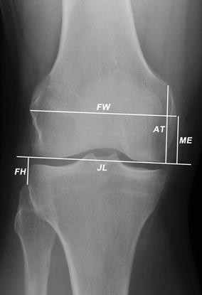
An antero-posterior plain radiograph of the knee showing technique of joint line measurement [ATJL (adductor tubercle to joint line) FW (femur width) FJL (fibula to joint line) JL (joint line) MEJL (medial epicondyle to joint line)]

Statistical analysis
Inter-observer and intra-observer reliabilities were analysed using two-way ANOVA, with average of repetition of each rater and subject as fators for inter-rater reliability.
After evaluating the repeatability of each measurement, correlation between FW, ATJL, MEJL and FJL was evaluated with Pearson correlation test (r values), in order to define a relation between the femoral width and the distance of joint line to medial epicondyle, adductor tubercle and fibular pole.
Linear regression analysis was also used to identify the individual ratios between FW to each of ATJL, MEJL and FJL.
After the ratio was found, it has been used to estimate the distance of joint line to medial epicondyle, adductor tubercle and fibular head. The estimation was then compared to the acquired data to verify the reliability of the method. An error lower than 5 mm was considered acceptable [2, 12].
In order to determine whether there was a difference in the measurements between males and females, student’s t test was used to compare the difference of the results between the two cohorts.
If a difference was found, the estimation for the two cohorts was compared to the acquired data, using a different ratio for males and females.
Since the study was a radiographic study and did not require any extra radiographs, no IRB approval was sought.
Results
Intra-tester and inter-observer reliability for ATJL was 0.96 and 0.86, respectively (Table 1).
The average FW was 89.7 ± 8.4 mm, with statistically significant difference (p < 0.0001) between males 95.0 ± 5.7 mm and females 82.6 ± 6.1 mm. Average MEJL was 30.7 ± 3.9 mm, ATJL was 48.7 ± 4.8 mm and FJL was 16.7 ± 4.0 mm with no statistical difference between males and females.
The correlation between FW and ATJL gives the best result with an excellent correlation (r = 0.83) (Fig. 2). Also, FW and MEJL resulted correlated but with a lower coefficient (r = 0.52) (Fig. 3). FW and FJL had a poor correlation (r = 0.21) even if statistically significant (Fig. 4).

Graph for correlation between FW and ATJL [ATJL (adductor tubercle to joint line) FW (femur width)]

Graph for correlation between FW and MEJL [MEJL (medial epicondyle to joint line) FW (femur width)]

Graph for correlation between FW and FJL [FJL (fibula to joint line) FW (femur width)]
From the linear regression analysis, the ratio between FW and MEJL was 0.343, and between FW and ATJL was 0.543. The ratio of FJL was not calculated because it had poor correlation with FW.
The average differences between the measured and estimated MEJL and ATJL, calculated using the corresponding ratio, were 0.0 ± 3.4–0.0 ± 2.7 mm.
Discussion
The most important finding of the present study was the evidence of a linear correlation between the FW and the distance from adductor tubercle to the joint line tangent, with a non-gender-specific coefficient, close to 0.83. In comparison, the linear correlation between the FW and the distance of ME from the joint line tangent, with a non-gender-specific coefficient was close to 0.52. This finding supports the initial hypothesis of this study that there is a relation between the distance between the adductor tubercle and joint line that could be more reliable than medial epicondyle to joint line.
There are various anatomical measures to identify the knee joint line on radiographs. Some surgeons use the AP view with the epicondyles or fibular head (FH) as a Ref. [6, 9, 10], whereas others use the distance from the tip of FH or tibial tubercle to the proximal tibial surface on a lateral radiographic view [4, 6]. Radiographs are used in this study because they are commonly used for the diagnosis of knee disease, for pre-operative planning of the surgical procedure and for evaluation of post-operative results. Joint line level is usually determined by measuring the distance from one of above mentioned anatomical landmarks and the joint line tangent. In fact, absolute values from ME and FH reported by these authors are very similar to the data reported in this study [5, 13, 14].
Such absolute values, however, are of limited utility because of large individual variations in physical dimensions [4, 5, 14, 15]. As suggested by literature, the relationship of FW to these measurements is more useful to control individual variations while calculating the joint line position [5, 13, 14]. The MEJL/FW ratio reported in an earlier study was 0.395 [13], while in the current study, it was 0.34. The inter-observer variability in that study was 0.97 for FW and 0.85 for MEJL [13], while in our study it was 0.95 for FW and 0.82 for MEJL. Furthermore, we found that the constant derived from AT/FW ratio was 0.543, without gender specificity and has a lower standard deviation than the ME/FW ratio. In predictive terms, the constant derived from AT–FW ratio should yield a more reliable joint line position.
Therefore, even though the estimated error in our study using medial epicondyle or adductor tubercle was similar, the distance from adductor tubercle to joint line ATJL was more precise and repeatable.
In addition, from the data about FH in this study, we report an important variability of the FH-JL tangent distance (Fig. 4). Conversion of these measurements to a ratio showed a large standard deviation that was independent of individual size. Therefore, we agree with other authors [6, 14] that the FH is not a reliable landmark to measure the joint line level even when measured with high accuracy, as demonstrated by low intra-observer and inter-observer variations (Table 1).
We think this study may have an important clinical application in case of revision knee arthroplasty because the surgeon can plan the joint line level on pre-operative radiographs and can use the information during the surgical procedure. We can determine the joint line position on pre-operative radiographs before a revision TKA when radiographs before the primary TKA are not available or the contralateral knee has also been replaced. Since the distance from the adductor tubercle to the joint line is linearly related to the femoral width, the ratio of this distance to the femoral width derives a constant, which can be multiplied to the measured FW to calculate the appropriate joint line level (ATJL = 0.543 × FW) (Fig. 1). Intra-operatively, with the exposed knee joint in flexion, a calliper is used to measure the FW. If it corresponds to the FW measured on the radiograph, we can use the planned distance from the adductor tubercle to implant the femoral component and restore the joint line position. If not, the new intra-operative value of FW can be multiplied by the constant to calculate the correct joint line position. By using a ratio instead of absolute values and being able to measure the FW intra-operatively, we do not need to calculate the radiographic magnification on every radiographs.
Some limitations of the current study would need to be highlighted. The current study was conducted in young patients without osteoarthritis. The knee flexion contracture seen in osteoarthritis may make it difficult to obtain accurate radiographs to determine joint line position. An intra-operative study to validate it intra-operatively in primary TKA for osteoarthritic knees is underway. The method, of course, will be limited by any surgeon dependent error to identify the landmarks and measurements. However, as demonstrated in this study, these may be reduced by using the adductor tubercle instead of the medial epicondyle.
This method will help the surgeon to choose the correct joint line in R-TKA. We did not mention the clinical use of this method in this paper because we are studying it in our R-TKA, and it will be the topic of another paper.
Conclusion
The method presented here, which uses a ratio of femoral width, ATJL/FW, was found to be more reliable than the MEJL/FW ratio to determine the joint line on AP X-rays because a linear correlation exists between FW and ATJL and the localization of adductor tubercle on radiographs is easier and more reliable than medial epicondyle. Furthermore, the planned joint line level may be helpful during a revision TKA because the femoral width can be measured and the adductor tubercle remains easily palpable.
References
Barnett AJ, Howells NR, Burston BJ, Ansari A, Clark D, Eldridge JD (2012) Radiographic landmarks for tunnel placement in reconstruction of the medial patellofemoral ligament. Knee Surg Sports Traumatol Arthrosc. doi:10.1007/s00167-011-1871-8
Bellemans J (2004) Restoring the joint line in revision TKA: does it matter? Knee 11:3–5
Christiansen SE, Jacobsen BW, Lund B, Lind M (2008) Reconstruction of the medial patellofemoral ligament with gracilis tendon autograft in transverse patellar drill holes. Arthroscopy 24:82–87
Figgie HE 3rd, Goldberg VM, Heiple KG, Moller HS III, Gordon NH (1986) The influence of tibial-patellofemoral location on function of the knee in patients with the posterior stabilized condylar knee prosthesis. J Bone Joint Surg Am 68:1035–1040
Griffin FM, Math K, Scuderi GR, Insall JN, Poilvache PL (2000) Anatomy of the epicondyles of the distal femur: MRI analysis of normal knees. J Arthroplasty 15:354–359
Havet E, Gabrion A, Leiber-Wackenheim F, Vernois J, Olory B, Mertl P (2007) Radiological study of the knee joint line position measured from the fibular head and proximal tibial landmarks. Surg Radiol Anat 29:285–289
Hoeffel DP, Rubash HE (2000) Revision total knee arthroplasty: current rationale and techniques for femoral component revision. Clin Orthop Relat Res 380:116–132
Jacobi M, Reischl N, Bergmann M, Bouaicha S, Djonov V, Magnussen RA (2012) Reconstruction of the medial patellofemoral ligament using the adductor magnus tendon: an anatomic study. Arthroscopy 28:105–109
Laskin RS (1998) Management of the patella during revision total knee replacement arthroplasty. Orthop Clin North Am 29:355–360
Laskin RS (2002) Joint line position restoration during revision total knee replacement. Clin Orthop Relat Res 404:169–171
Lind M, Jakobsen BW, Lund B, Christiansen SE (2008) Reconstruction of the medial patellofemoral ligament for treatment of patellar instability. Acta Orthop 79:354–360
Partington PF, Sawhney J, Rorabeck CH, Barrack RL, Moore J (1999) Joint line restoration after revision total knee arthroplasty. Clin Orthop Relat Res 367:165–171
Romero J, Seifert B, Reinhardt O, Ziegler O, Kessler O (2010) A useful radiologic method for preoperative joint-line determination in revision total knee arthroplasty. Clin Orthop Relat Res 468:1279–1283
Servien E, Viskontas D, Giuffrè BM, Coolican MRJ, Parker DA (2008) Reliability of bony landmarks for restoration of the joint line in revision knee arthroplasty. Knee Surg Sports Traumatol Arthrosc 16:263–269
Stiehl JB, Abbott BD (1995) Morphology of the transepicondylar axis and its application in primary and revision total knee arthroplasty. J Arthroplasty 10:785–789
Acknowledgments
There was no external funding for this study or for any of the authors in relation to this study.
Author information
Authors and Affiliations
Corresponding author
Rights and permissions
About this article
Cite this article
Iacono, F., Lo Presti, M., Bruni, D. et al. The adductor tubercle: a reliable landmark for analysing the level of the femorotibial joint line. Knee Surg Sports Traumatol Arthrosc 21, 2725–2729 (2013). https://doi.org/10.1007/s00167-012-2113-4
Received:
Accepted:
Published:
Issue Date:
DOI: https://doi.org/10.1007/s00167-012-2113-4