Article

Preservation Mechanism of Chitosan-Based Coating with Cinnamon Oil for Fruits Storage Based on Sensor Data

1
Sichuan Province Key Laboratory of Grain and Oil Processing and Food Safety, Food and Bioengineering College, Xihua University, Chengdu 610039, China
2
School of Engineering, University of Guelph, Guelph, ON N1G 2W1, Canada
3
Key Laboratory of Physiological and Storage of Agricultural Products after Harvest in the Ministry of Agriculture, National Engineering Technology Research Center for Preservation of Agricultural Products, Tianjin 300384, China
4
Food Engineering and Biotechnology College, Tianjin University of Science & Technology, Tianjin 300457, China
*
Authors to whom correspondence should be addressed.
Sensors 2016, 16(7), 1111; https://doi.org/10.3390/s16071111
Submission received: 15 April 2016 / Revised: 13 July 2016 / Accepted: 14 July 2016 / Published: 18 July 2016
(This article belongs to the Special Issue Sensors for Agriculture)

Abstract

:
The chitosan-based coating with antimicrobial agent has been developed recently to control the decay of fruits. However, its fresh keeping and antimicrobial mechanism is still not very clear. The preservation mechanism of chitosan coating with cinnamon oil for fruits storage is investigated in this paper. Results in the atomic force microscopy sensor images show that many micropores exist in the chitosan coating film. The roughness of coating film is affected by the concentration of chitosan. The antifungal activity of cinnamon oil should be mainly due to its main consistent trans-cinnamaldehyde, which is proportional to the trans-cinnamaldehyde concentration and improves with increasing the attachment time of oil. The exosmosis ratios of Penicillium citrinum and Aspergillus flavus could be enhanced by increasing the concentration of cinnamon oil. Morphological observation indicates that, compared to the normal cell, the wizened mycelium of A. flavus is observed around the inhibition zone, and the growth of spores is also inhibited. Moreover, the analysis of gas sensors indicate that the chitosan-oil coating could decrease the level of O2 and increase the level of CO2 in the package of cherry fruits, which also control the fruit decay. These results indicate that its preservation mechanism might be partly due to the micropores structure of coating film as a barrier for gas and a carrier for oil, and partly due to the activity of cinnamon oil on the cell disruption.

Graphical Abstract

1. Introduction

Consumption of fruits with good quality might decrease the development risk of chronic diseases in human body and provide benefits for body health [1,2]. However, the losses of nutritional ingredients in fruits always occur because of its decay and physiological changes, which could be induced by the microbial infection during the storage time [3,4]. Recently, many researchers have developed semipermeable edible coatings as a carrier for antimicrobial agents in order to control the nutrient loss and maintain the quality of fruits [5,6,7,8]. This is because the edible coating could keep textural properties, sensory appearance and control respiration rate of fruits by functioning as a semipermeable vapor and gas barrier to regulate the moisture and gases in the package [5,9,10]. Moreover, the antimicrobial activity of edible coating could be significantly improved by incorporating different antimicrobial agents [3,4]. Thus, there is increasing interest in developing a bio-based coating with high antimicrobial activity for fruits storage, such as a chitosan coating with cinnamon oil [4,11,12].
Chitosan is recognized as safe and has been widely applied as a successful carrier for incorporating with the antimicrobial agents because of its adventurous biocompatibility and film-forming ability [2,6,12,13,14,15,16]. The preservation effect of chitosan-based coatings might be partly due to its property of slowing the respiration rate and controlling the decay of fruits, and partly due to its function of preventing the formation of off odors and retarding lipid oxidation in fruits [9,12,17,18]. Velickova et al. (2013) found that the chitosan-beeswax coating could slow down the respiration rate and reduce the senescence of strawberries [19]. As reported by Waewthongrak et al. (2015), the decay of citrus fruit could be significantly reduced by the combined application of chitosan and the crude extract from the culture medium of Bacillus subtilis [20]. Kaya et al. (2016) expressed that the application of chitosan and acetic acid could extend the shelf life of kiwifruit berries [21]. Moreover, Duran et al. (2016) indicated that the quality of strawberries could be maintained by chitosan coatings with nisin, natamycin, pomegranate and grape seed extract [22]. The incorporation of essential oils as the antimicrobial agent into the coating carrier is also considered as the successful technology to control the decay and maintain the quality of fruits during the storage time [4,23].
In the last few years, the addition of essential oil into the chitosan-based coating have been developed to enhance its antimicrobial property and act an active agent in the film coated on the surface of fruits [3,12,13,24,25]. Cinnamon oil as “Generally Recognized as Safe” by the Food and Drug Administration (FDA) has been recognized as the better antimicrobial activity because it is rich in cinnamaldehyde and/or trans-cinnamaldehyde [5,26,27,28]. As reported by several researchers, the after-harvested use of chitosan-based coating incorporating with cinnamon oil might control the postharvest fungi in pepper, prevent the growth of microorganisms in fresh cut pear and melon, and enhance the quality of jujube fruits [4,6,9,12,29,30,31]. According to the investigation of Carvalho et al. (2016), they reported the chitosan coating enriched with trans-cinnamaldehyde could improve the quality characteristics and extend the shelf life of melon slices to 15 days [29]. The enhancement on the antimicrobial activity of chitosan-based coating might be due to the addition of cinnamon oil or trans-cinnamaldehyde with good killing effect against bacteria and fungi [9,26].
For the antimicrobial mechanism of chitosan, its killing actions might be due to the interaction between chitosan molecules with positive charged and microbial cell membranes with negative charged and also due to the inhibited effectiveness on the microorganism growth and the toxins production [3,32,33,34,35]. However, the fresh keeping of chitosan-based coating and antifungal mechanism of cinnamon oil are still not very clear. Zhang et al. (2016) have investigated the mechanism of antibacterial action of cinnamon oil against Escherichia coli and Staphylococcus aureus by observing the changes of cell microstructure [36]. The investigation of Clemente et al. (2016) also investigated the antimicrobial properties and mode of action of cinnamon oil against foodborne bacteria [37]. In the series of papers published by our authors previously, the antimicrobial property and the application effectiveness of chitosan-based coating and cinnamon oil were mainly reported. Xing et al. (2010b) conducted the investigation on the in vitro and in vivo activities of cinnamon oil against three kinds of fungi [26]. Xing et al. (2010a) have extended the shelf life of lotus root slices with the chitosan-based solution [18]. Moreover, as conducted by Xing et al. (2011a), the in vivo control effects of chitosan-oil coating on the disease caused by P. citrinum of Lingwu long jujube fruits were evaluated [3]. Xu et al. (2013) have analyzed the effect of cinnamon oil fumigation and chitosan coating on the microbial safety of pear slices [6]. Xing et al. (2011b) and Xing et al. (2015) have investigated the effects of chitosan-oil coating on the activities of defense-related enzymes and the quality attributes of pepper and jujube fruits, respectively [4,9]. Nevertheless, less literature has been published by researchers about the keeping-fresh mechanism of chitosan-based coatings and the antifungal mechanism of cinnamon oil for the application effectiveness on the quality of fruits.
Therefore, the objective in this article is to understand the preservation mechanism of chitosan-based coating and cinnamon oil based on sensor data. Firstly, the morphology and roughness analysis of coating film with different concentration of chitosan were conducted by AFM (atomic force microscopy) sensor. This section is mainly to understand its microstructure and ability as the modification of gas and as the carrier of antimicrobial agents. Secondly, the antifungal mechanism of cinnamon oil is investigated through the activity comparison between trans-cinnamaldehyde and oil, the effect of attachment time on the antifungal activity and the influence of oil concentration on the exosmosis rate of cells. This work is mainly to clarify the control and kill mechanism of cinnamon oil on the growth and cell integrity of fungi. Finally, the evaluations on the concentration changes of O2 and CO2, the decay and the activity of PPO (Polyphenylene Oxide) for the storage of sweet cherry are conducted in order to further validate the preservation mechanism of chitosan-oil coating.

2. Materials and Methods

In this section, several types of sensor instruments, such as AFM, SEM (scanning electron microscope), GC-MS (gas chromatography-mass spectrometer) and gas analyzer, are used to clarify the preservation mechanism of chitosan film and cinnamon oil. AFM and SEM are two appropriate methods used to investigate the surface analysis of coating film. GC-MS is used to analyze the components in cinnamon oil. The methods including the antifungal activity transfer experiment and the comparison in exosmosis ratio of fungi cell are described. Finally, the analysis technologies for the O2 and CO2 concentrations, the decay rate and PPO activity are introduced.

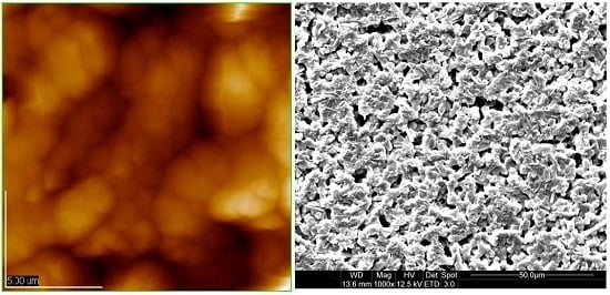
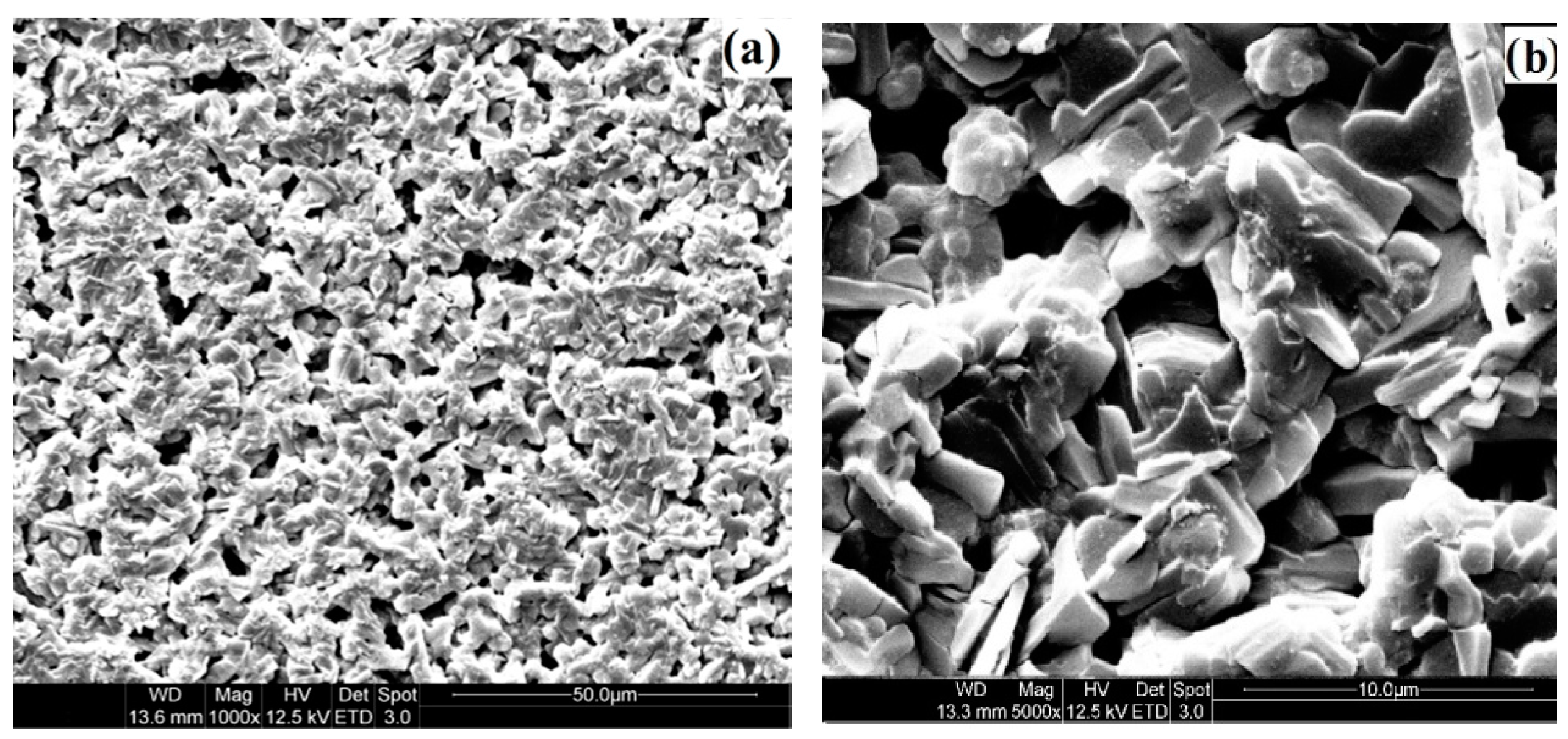
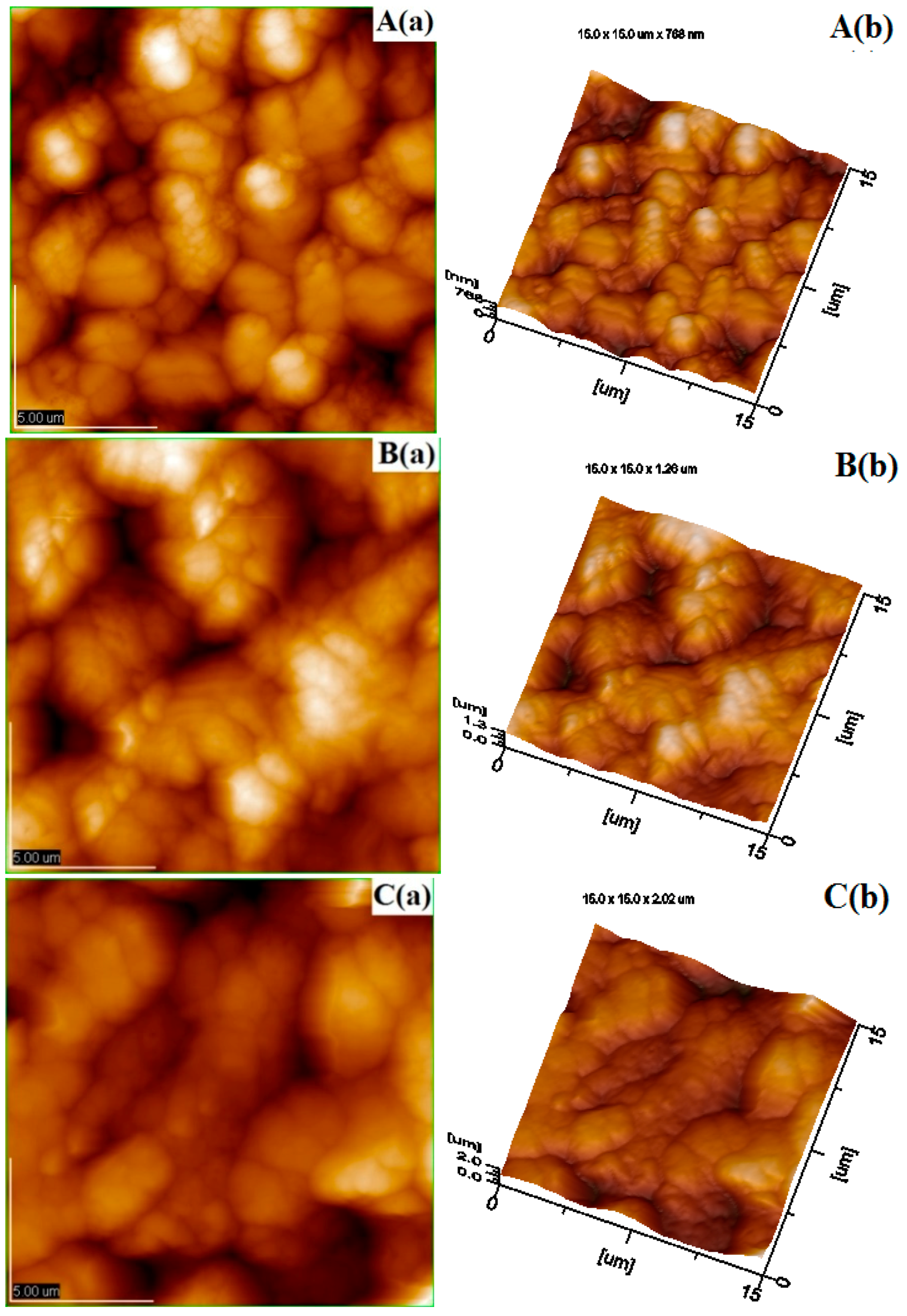
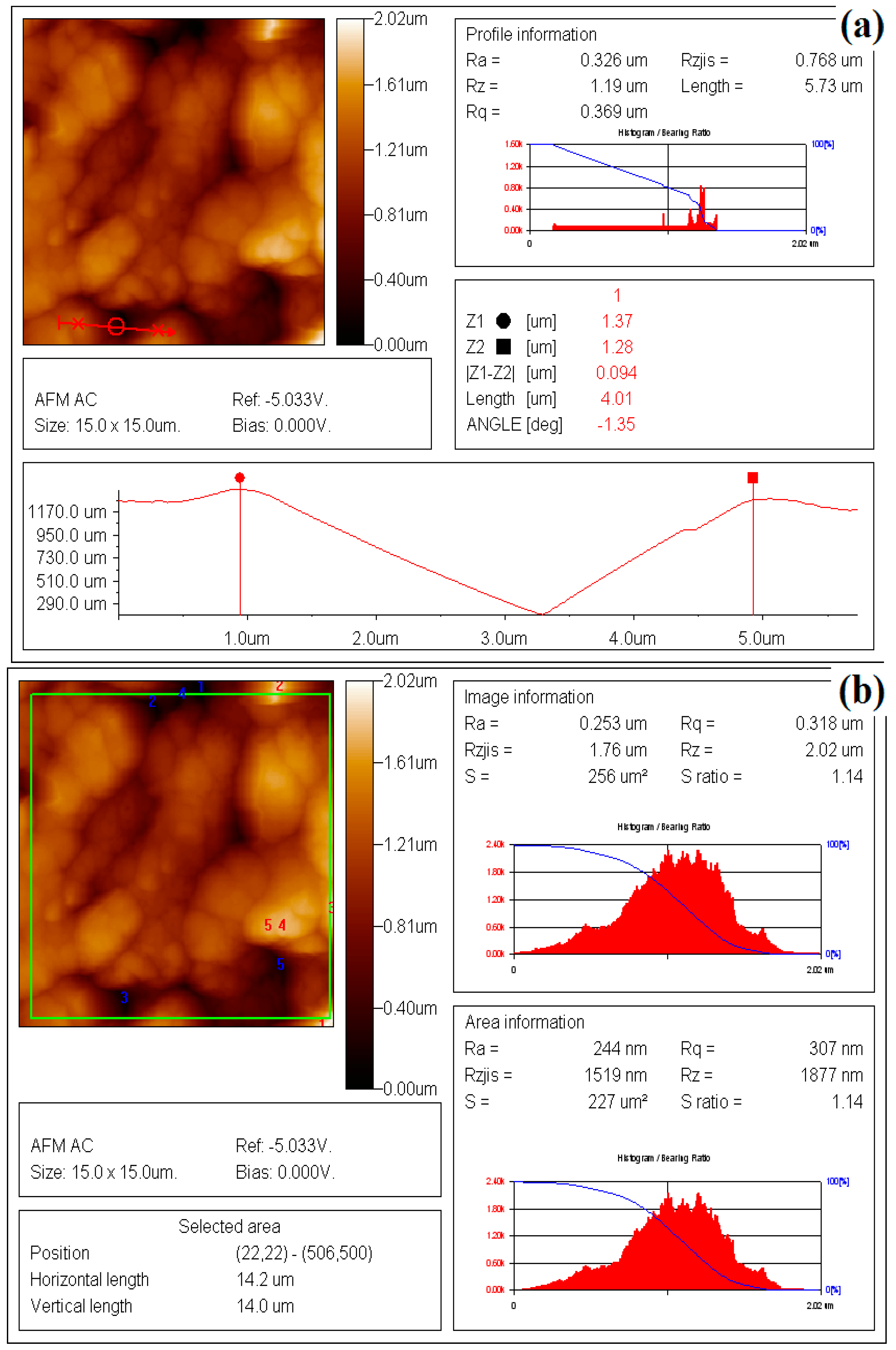
2.1. Preparation, AFM and SEM Observation of Chitosan Coating Film

In order to conduct the observation of the surface of the coating by AFM and SEM, the chitosan coating film was prepared according to the method reported by Xing et al. (2011a), Xing et al. (2015) and Sun et al. (2014) [3,4,38]. Different amounts of chitosan (0.25 g, 0.50 g and 1.0 g; Deacetylated ≥ 95%, viscosity ≤ 30 mPa·s; Ji’nan Haidebei Marline Bioengineering Co. Ltd., Ji’nan, China) were added into 100 mL distilled water with stirring by a magnetic stirrer. Then, acetic acid (0.50% v/v) and glycol (0.25% v/v) were added and stirred for 1 h in order to dissolve chitosan powder. After homogenized under aseptic conditions and stood for 2 h, 90 mL of the obtained chitosan solution was spread on a glass plate (20 cm × 20 cm). After being dried at room temperature for 4–5 days, they were peeled from the plate and conditioned at 4 °C prior to further observation by SEM and AFM [36].
The chitosan coating film was observed by SEM according to the method reported by Xing et al. (2015) [4]. The coating film with 1.0% chitosan was cut into thin pieces firstly and stuck on the sample stub. After Pt sputtering, the film piece was observed by SEM with the voltage of 5 kV acceleration.
The coating film with different concentration of chitosan was observed by AFM according to the method reported by Xing et al. (2011a,b) and Zdunek and Kurenda (2013) [3,9,39]. First, the chitosan coating film was cut into thin pieces (10 mm × 10 mm) using a small sharp knife and stuck on the stage. Ten pieces of film per treatment were scanned using the Tapping mode using a JSPM-5200 AFM (JEOL, Tokyo, Japan). In order to understand the roughness values Ra and Rq, the images were analyzed with Nanoscope software (Version 5.12, Tokyo, Japan). Ra and Rq are calculated as the following [40]:
R a = 1 L x L y 0 L y 0 L x | f ( x , y ) | d x d y
where f(x,y) is the surface relative to the center plane and Lx and Ly indicate the surface dimensions.
R q = ( Z i Z a v e ) 2 N
where Zave means the Z values average within the chosen area, Zi indicates the current Z value and N is the point number within the chosen area of samples. On the other hand, the thicknesses of chitosan coating films and the samples for AFM observation were also measured with a 0–25 mm dial thickness gauge with an accuracy of 0.01 mm in five random locations for one piece (n = 10 for each treatment). The thicknesses of film samples for AFM observation were 0.056 mm, 0.061 mm and 0.069 mm for films with the concentrations of chitosan at 0.25%, 0.50% and 1.0%, respectively.

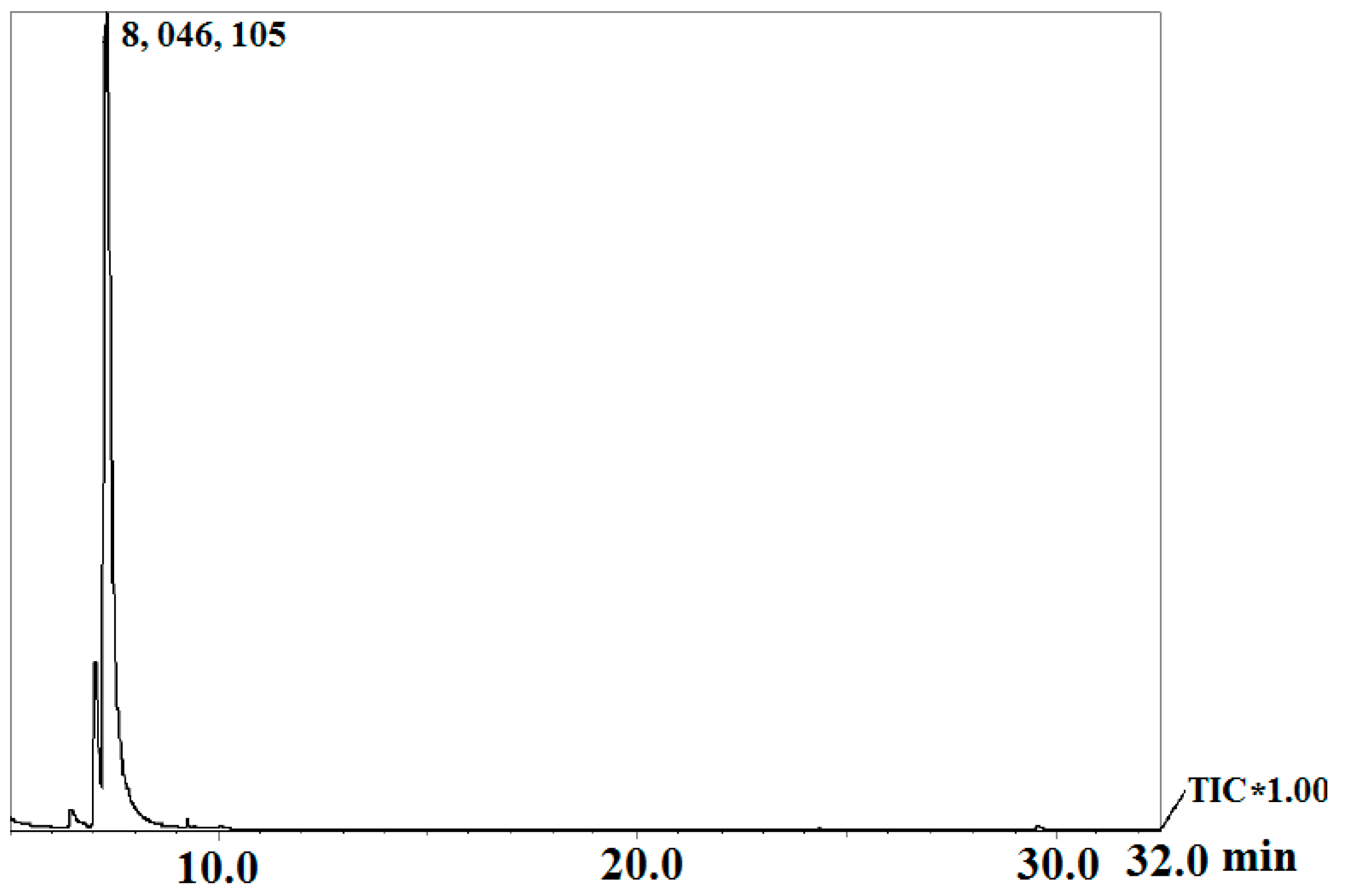
2.2. GC-MS Analysis of Cinnamon Oil

The GC-MS analysis technique for cinnamon oil was used according to method developed by Xing et al. (2010b) and Li et al. (2013) [26,41]. The GC-MS system used in this investigation has two components, which are Varian GC/MS 4000 equipped with a VF-5ms MS capillary column (30 m × 0.25 mm i.d. × 0.25 μm film; Scientific Instrument Services Inc., Varian, CA, USA), and an electron ionization system with ionization energy of 70 eV. He at the flow rate of 1 mL/min was used as the carrier gas during the analysis process. Firstly, the temperatures were set at 220 °C for injector and 280 °C for MS transfer line, respectively. Then, the temperature of the programmer was raised to 60–180 °C with a rate of 8 °C/min, which was held isothermal for 10 min and finally raised to 300 °C at the rate of 10 °C/min. Then, 1.0 μL diluted oil sample with hexane (1/10,000, v/v) was injected handy with the splitless mode. After the analysis procedure was completed, the identification of components in oil sample was conducted using literature data, the standard of NIST 05 by comparing their mass spectra and relative retention time.

2.3. Antifungal Activity of Trans-Cinnamaldehyde and Cinnamon Oil

The comparison on the antifungal activity between trans-cinnamaldehyde and cinnamon oil was determined against fungi including P. citrinum and A. flavus using the disk diffusion method as developed by Xing et al. (2011a) and Xing et al. (2012) [3,42]. First, the surfaces of potato dextrose-agar (PDA) plate were spread with 100 μL of inoculums containing 104–105 conidia/mL of P. citrinum and A. flavus. Then, the discs (D = 10 mm) impregnated with 10 μL trans-cinnamaldehyde and cinnamon oil with the concentrations of 100, 200, 300 and 400 μL/L were put on the center of PDA medium prepared above. Each treatment was performed in triplicate. The diameter of inhibition zones were measured after the plates incubated at 28 °C for 3–5 days.

2.4. Antifungal Activity Transfer Experiments of Cinnamon Oil

The method on the transfer experiments of antifungal activity for cinnamon oil against P. citrinum and A. flavus was developed from the investigation of Feng et al. (2006), Xing et al. (2011a) and Wang et al. (2012) with some modifications [3,43,44]. Certain volumes of cinnamon oil were added to PDA medium at the temperature of 40 °C in order to adjust the oil concentrations to 100, 200, 300 or 400 μL/L. The obtained mediums were shaken sufficiently in order to mix well and then prepared for use. Meanwhile, the PDA plates cultured for 7 days (D = 6 mm) with fungi P. citrinum and A. flavus were picked up with a sterile hole puncher and then put into the center of PDA plates containing cinnamon oil as prepared above. After contact for 0, 1, 3, 5, or 7 days, the PDA plate (D = 6 mm) was transferred to the center of newly PDA plates without cinnamon oil. The treated samples were performed in quintuplicate and cultured for 7 days into a 28 °C incubator. The diameters of colonies were measured and expressed in millimeters.

2.5. Effect of Different Oil Concentration on the Exosmosis Rate of Fungi Cell

Effects of different oil concentration on the exosmosis rate of fungi cell were conducted with the method developed from the investigation of Feng (2006) and Diao et al. (2014) with modifications [44,45]. First, cinnamon oil was added into the solution containing 0.05% Tween-20 and emulsified for 1 h before use. Then, the spores of P. citrinum and A. flavus were drawn from a slant PDA medium with inoculation loop and transferred to PDB medium. The cell concentrations in the medium were adjusted to about 5.0 × 10⁷ conidia/mL. The obtained PDB mediums containing P. citrinum and A. flavus spores were dispensed into flask and then the cinnamon oil emulsion prepared above was added. The final concentrations of cinnamon oil in the PDB mediums were adjusted to 0, 100, 200, 300 and 400 μL/L. The obtained flasks were put into a constant temperature shaker at 28 °C and 180 r/min.
The flasks were picked up and centrifuged at 4 °C and 4000 g for 15 min after being cultured for 14 days. The obtained precipitate was washed with deionized water 1–2 times and was divided into two groups after being weighed. The samples were weighed and divided into a small beaker. The 20 mL deionized water was added in order to immerse the sample. The samples of Group A were placed in a vacuum oven with a vacuum gas drainage repeated 3–4 times. Then, the pressure was controlled at 450–500 mm Hg and the vacuum infiltration was restored to normal pressure after 30 min. The treated samples were shaken at 25–30 °C temperature for another 2–3 h. The samples of Group B were placed in a boiling water bath and heated for 30 min in order to destroy the plasma membrane completely. Finally, the extravasation fluids for these two treatments were poured into a small beaker and the conductivity was measured with electric conductivity meter. Each treatment was performed in quintuplicate. The relative electrolyte leakage rate = (the extravasation fluid conductivity of Group A/the extravasation fluid conductivity of Group B) × 100%.

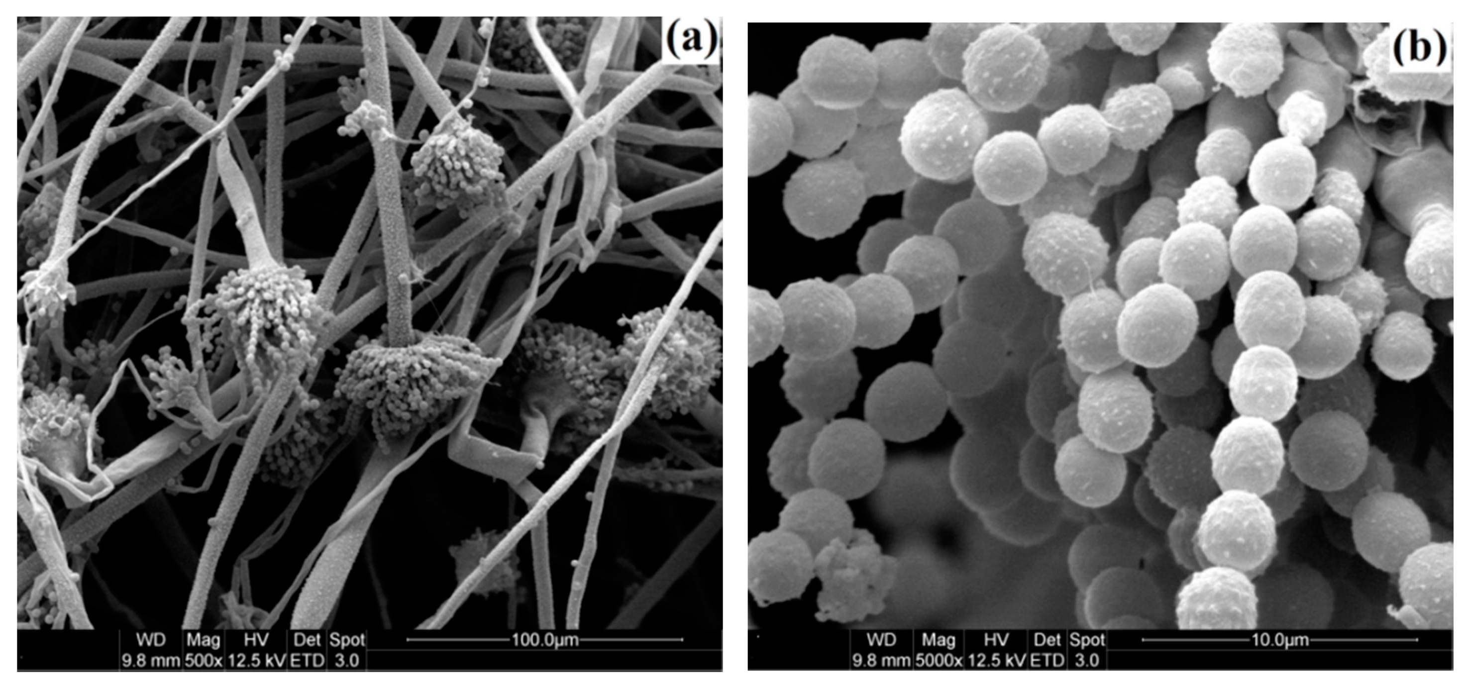
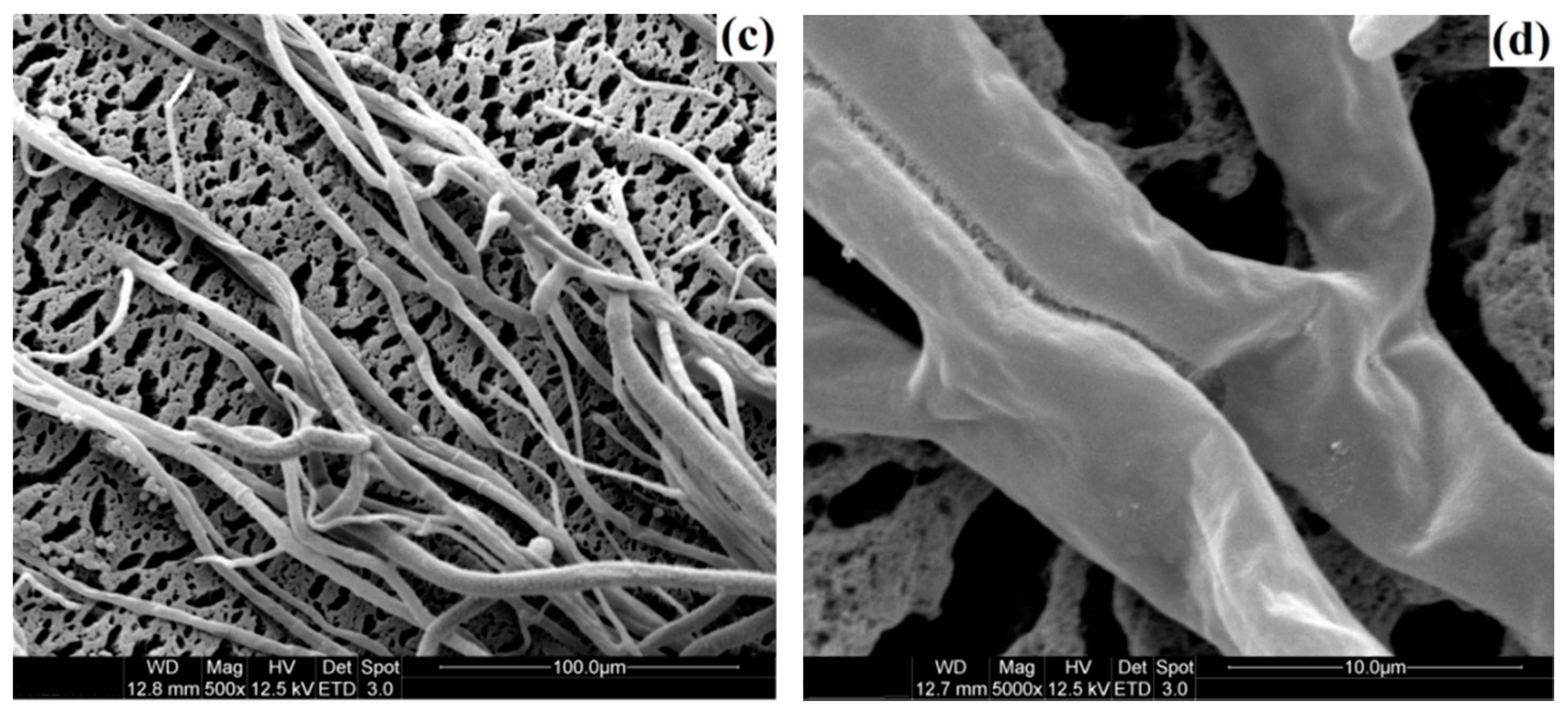
2.6. Morphology of Normal Cell and Cell at the Edge of Inhibition Zone of A. flavus Observed by SEM

Fungi cells of A. flavus on the edge of inhibition zone and on the control plate were picked up with the sterile hole puncher for observation. First, the fungi cell samples were fixed with 2% (v/v) glutaraldehyde at 4 °C for 12–18 h. After washing three times with phosphate buffer saline (PBS), the cells were post-fixed with 1% (w/v) osmium tetroxide prepared with 0.1 M PBS and dehydrated in a graded ethanol series from 30%, 50%, 70%, and 95% to absolute ethanol. Then, the dehydrated cells were subjected to critical point drying with liquid CO2. After Pt sputtering, the samples were observed by SEM equipped with a LEO Gemini 1530 field emission at a voltage of 5 kV acceleration [3,43].

2.7. Coating and Fruit Samples Preparation, O2 and CO2 Concentrations and Fruit Decay Analysis

The chitosan-oil coating was prepared according to the method developed from the investigation of Xing et al. (2011a) and Xing et al. (2015) [3,4]. One gram of chitosan powder was added into 100 mL distilled water during stirring with a magnetic stirrer. The substances of acetic acid (0.50% v/v) and glycol (0.25% v/v) were also added and stirred for 1 h in order to dissolve chitosan in the water. After that, the compounds of cinnamon oil (0 and 0.10%) (Xianghui Bio-technology Co. Ltd., Shanghai, China) and Tween 80 (0.15%) were added and stirred for another 1 h. The final solution was homogenized under aseptic conditions and let stand for 2 h before use.
“Brooks” sweet cherry fruits (Prunus avium L.) were harvested at the maturity stage from fruits base of MaoXian in Aba Tibetan and Qiang Autonomous Prefecture, China. Fruits were visually inspected as free of blemishes and defects, and then were precooled at 8 °C for 16–24 h. After precooling, fruits were divided into four groups with 200 fruits for each treatment. Four groups included the treatments of the chitosan (1.0%) + oil (0.10%) coating, the chitosan coating (1.0%), the cinnamon oil (0.10%) and the distilled water (control). Fruits were dipped into the different solutions for 5 min. The treated fruits were packaged with a polypropylene film (800 mm × 1000 mm) after kept over a plastic sieve for 120 min. All treatments were performed in triplicate and stored at 8 °C with the relative humidity of 85%–95% for analysis [3,4,9].
Concentrations of O2 and CO2 inside the packages were determined using an Oxygen and Carbon Dioxide gas analyzer (Shenzhen Empaer Technology Co. Ltd., Shenzhen, China) [9,46]. The decay rate of fruits in the packages was examined and calculated as the percentage of the weight of decay fruits to the total weight of fruits. PPO activity was assayed spectrophotometrically by the method based on Xing et al. (2015). Five grams of fruit tissue was homogenized with 10 mL extraction buffer (4 °C, 0.2 mol/L, pH 6.8 sodium phosphate buffer) in an external ice bath for 3 min including 0.1% polyvinylpolypyrrolidone. The homogenates were centrifuged for 12 min at 12,000 g and the supernatants were collected. The reaction solution was consisted of 0.1 mL crude extract and 2.9 mL substrate solution (0.02 mol/L catechol in 0.05 mol/L phosphate buffer, pH 6.5). An enzyme activity unit was defined as an increase of 0.0001 in absorbance per minute. The rates of catechol oxidation were examined at 420 nm for 2 min for all the treatment.

2.8. Statistical Analysis

Experimental data were analyzed by SPSS 16.0 software (SPSS Inc.) and reported as the mean ± S.D. The analysis of thickness and AFM for chitosan coating film was performed in decuplicate (10 pieces for each treatment). Effects of attachment time on the antifungal activity and the oil influence on the exosmosis rate of cells were conducted in quintuplicate. Other treatments were conducted in triplicate for each. The significant differences among the treatments were determined by the one way analysis of variance method followed by Student-Newman-Keul test.

3. Results and Discussion

In this section, the surface microstructure and roughness of chitosan coting is observed by AFM in order to clear its mechanism of slowing the respiration rate of fruits and its capacity as the carrier for cinnamon oil. Moreover, research work also needs to be conducted in order to clarify whether the antifungal activity of oil might be due to its main component. The effect of oil on the morphology change of cell is also discussed by comparing the exosmosis ratio of tested fungi cell before and after oil treatment and SEM observation. Finally, the functions on slowing the respiration rate, controlling the decay and PPO activity of fruits are investigated by in vivo tests and discussed in order to verify the preservation mechanism.

3.1. Morphological Observation of Chitosan Coating by SEM and AFM

In order to understand the mechanism and the oil loading capacity of edible coatings, morphology of chitosan coating film was observed using SEM and AFM. Many micropores are found on the chitosan film (1.0%) from the results of SEM observation in Figure 1. The plane and the three-dimensional profiles of coating film with different concentration of chitosan are shown in Figure 2. The morphology of AFM also illustrates that many micropores are observed on the chitosan coating film. Based on the AFM analysis on the 10 pieces of chitosan film for each concentration, the total number of micropores (10 pieces together) on the surface of selected area film decreases from 86 to 53 with increasing the concentration of chitosan from 0.25% to 1.0% (data are not listed in table form). The micropores on the surface of coating film with chitosan at 1.0% could also be validated from SEM findings, as shown in Figure 1. The distributions of micropores on the surface of film were not consistent among those prepared with different concentration of chitosan, which could also affect the smoothness and roughness of the coating surface. Thus, the parameters of surface roughness for chitosan coating membrane were also analyzed by AFM and expressed in terms of Ra and Rq of chitosan membrane surface. These two roughness parameters, Ra and Rq, indicated the mean roughness and the root mean square of Z data, respectively, are calculated from sample mathematical expressions and illustrated in Table 1. The sample images of analysis on the Ra and Rq of coating films with chitosan at 1.0% are shown in Figure 3a,b. As shown in Table 1, in the micropores profile information, the Ra and Rq of coating film with chitosan at 0.25% are 0.244 μm and 0.289 μm, respectively, which increase to 0.399 μm and 0.449 μm for the coating film with chitosan at 1.0%. Furthermore, in the image information analysis, the Ra and Rq of coating film with chitosan at 0.25% are 0.131 μm and 0.159 μm, which increase to 0.247 μm and 0.303 μm for the film containing chitosan at the concentration of 1.0%, respectively. The roughness of chitosan coating films could be improved by increasing the concentration of chitosan. Significance differences among the roughness of coating film surfaces are observed from the results among different concentrations of chitosan films. On the other hand, the thicknesses of films are measured and shown in Table 1. The thickness of films varied from 0.056 mm to 0.068 mm with increasing the chitosan concentration from 0.25% to 1.0%. However, no significant difference is found between films prepared with 0.25% and 0.50% chitosan. Differences in the thickness of coating film could be attributed to the different concentrations of chitosan added in the film-form solution.
The roughness of coating film is an inherent property of chitosan-based polymer in which chitosan forms the coating film with micro-perforated structure and influences the smoothness of fruit surfaces. Results in AFM images indicate that the chitosan coating membrane with many micropores could be used as the carrier of active compounds, which might fill the empty spaces of surface micropores [3,9,47]. Moreover, the chitosan coating on the surface of fruits might induce the increase in the concentration of CO2 and the decrease in the levels of O2 in the package, which could influence the respiration rate of fruits [6,9]. Moreover, it has the potential for inducing phenolic contents and defense-related enzymes such as PPO in fruit products [9,14]. These results are consisted with the investigations conducted by Xing et al. (2010a), Xing et al. (2011a) and Xing et al. (2015) [3,4,18]. As reported by Xing et al. (2010a), the optimum concentrations of O2 and CO2 in packages of fresh-cut lotus root were created by the application of chitosan coating. Furthermore, the inhomogeneous distribution of micropores is found on the surface of film with different concentration of chitosan. The number of micropores in the film surface might decrease with increasing the addition of chitosan in the coating solution system. This might be due the interaction among the water, emulsifier, acetic acid, glycol and the increased chitosan in the system. Moreover, the inhomogeneous distribution of micropores, meaning the uneven distribution of the peaks and valleys in the chitosan coating, could significantly affect the Ra and Rq values of film surface [48,49,50,51,52,53,54]. The surfaces with fewer micropores could become less sharp and show a smoother appearance, which might also be influenced by the added concentration of chitosan. The surface of film becomes smoother with the higher addition of chitosan. The good uniform microstructure of chitosan coating films are observed from SEM images, which indicates that the mixtures of chitosan, acetic acid and glycerol are homogenous in the coating film. These results could be explained by the interaction of the higher amount chitosan with other components in the prepared system, the adsorbed action of surface tension for coating liquid and the different number of micropores formed on the film surface [49,50,51,52]. Xing et al. (2011b) also reported that the roughness value of control group was higher than the chitosan–oil coated group fruits [9]. However, as reported by Segota et al. (2012), the small amount addition of polyethylene glycol to TiO2 film could drastically influence the roughness parameters. This might be due to the different property of added substances and based film. Furthermore, the thickness of films could be affected by the addition of chitosan, the type of chitosan used and the temperature of drying [38,55]. The addition of a relatively lower dose of chitosan exhibited the lower thickness. As reported by Sun et al. (2014), differences in the thickness of chitosan-Gallic acid films might be due to the addition and interaction of chitosan and Gallic acid [38]. The roughness and thickness of film surfaces are also influenced by the different chosen area, the analysis method and the film materials. Thus, chitosan-based solution coated on fruits might exhibit the difference surface characteristics compared to the film prepared from the dried solution. The influence of chitosan addition on the surface characteristics of films could be directly observed from the analysis results of film obtained from the dried solution, which is the in vitro analysis method used in many similar investigations [38,56,57,58]. After being coated on fruits, the ordered microstructure of film may be disrupted by the different types of fruit surfaces, which also affects the number/size and disruption of micropores and further influence the thickness and gas permeability of coating film covered on the surface of fruits. The optimum concentrations of O2 and CO2 in the packages could be created by the application of coating with the suitable thickness and chitosan concentration [18]. However, a coating with higher thickness or more chitosan might induce anaerobic respiration and quicken the senescence of fruits. Water vapor permeability and oxygen permeability of coating film might also be influenced by the addition and interaction of cinnamon oil and surfactant in the film-forming dispersion [38,56,57,58]. A coating film with a suitable number/size of micropores could form a protective barrier to reduce decay, water loss and respiration rate of fruit, which might also influence the resistance of tissue, the antioxidant enzyme activities and the senescence process of fruits [4,9,58,59]. These synergetic functions and mechanisms still need to be further investigated and clearly defined.

3.2. Chemical Composition of Cinnamon Oil Analyzed by GC-MS

In order to clear the main components, the analysis of the chemical composition of cinnamon oil was conducted by GC-MS. The chemical consistent of cinnamon oil is listed in Table 2. The total ion current of GC-MS analysis can be seen in Figure 4. As shown in Table 2, a total of 17 components are identified in the cinnamon oil sample. Results indicated that the most abundant component is trans-cinnamaldehyde, which is 85.64% of the relative percentage in cinnamon oil. In addition, cinnamon oil also contains cinnamaldehyde (1.37%) and benzenepropanol, a-methyl- (11.43%). The area percentages of other volatile compounds identified by GC-MS are less than 1.0%. The volatile compounds are comprised of two aldehydes, six ethers, two alcohols and seven others. From the analysis result of GC-MS, it could be confirmed that trans-cinnamaldehyde is the major component with the highest area percentage in the tested cinnamon oil.
Natural active compounds such as cinnamon oil should be developed because that the exceeded residue of chemical fungicides could cause an increased risk of diseases and increased probability of affecting the human health [4,25,26,27,28,41,60]. The component identification is very important for the application of cinnamon oil, which is a high quality natural antimicrobial agent and widely used in the storage of fruits. The result in this work indicates that the compound trans-cinnamaldehyde should be the major component in cinnamon oil. Li et al. (2013) also indicated that trans-cinnamaldehyde was the main compound of nine cinnamon bark oils using GC-MS analysis [41]. However, differences in the main compounds of cinnamon oil have been observed by other researchers. The investigations of Wang et al. (2005), Xing et al. (2010b) and Kim et al. (2015) indicated that the major compound in cinnamon oil was cinnamaldehyde [26,27,61]. The component of cinnamaldehyde is also identified in this research, as only 1.37% of the relative percentage in cinnamon oil. Moreover, Singh et al. (2007) have reported that eugenol is the major volatile compound (87.3%) and no trans-cinnamaldehyde was detected in the oil of C. zeylanicum leaf [62]. Goñi et al. (2009) expressed that the major component of cinnamon essential oils is trans-cinnamaldehyde or cinnamaldehyde [63]. Todd et al. (2013) and Kaskatepe et al. (2016) indicated that the major substance of Cinnamon was cinnamaldehyde [64,65]. This difference might be due to the spices of cinnamon bark, the environmental factors, the origin and parts of cinnamon, the analysis method and other factors [25,26,27,66,67,68]. More importantly, according to the results reported by Wang et al. (2005), Xing et al. (2010b) and Xing et al. (2011a), cinnamon oil containing cinnamaldehyde and/or trans-cinnamaldehyde exhibits an excellent antimicrobial property against fruits pathogens including bacterial and fungi [3,26,61]. Thus, it is suspected that the main components of trans-cinnamaldehyde and/or cinnamaldehyde are the principal antifungal agent in cinnamon oil. Further research on this point is discussed in the next section.

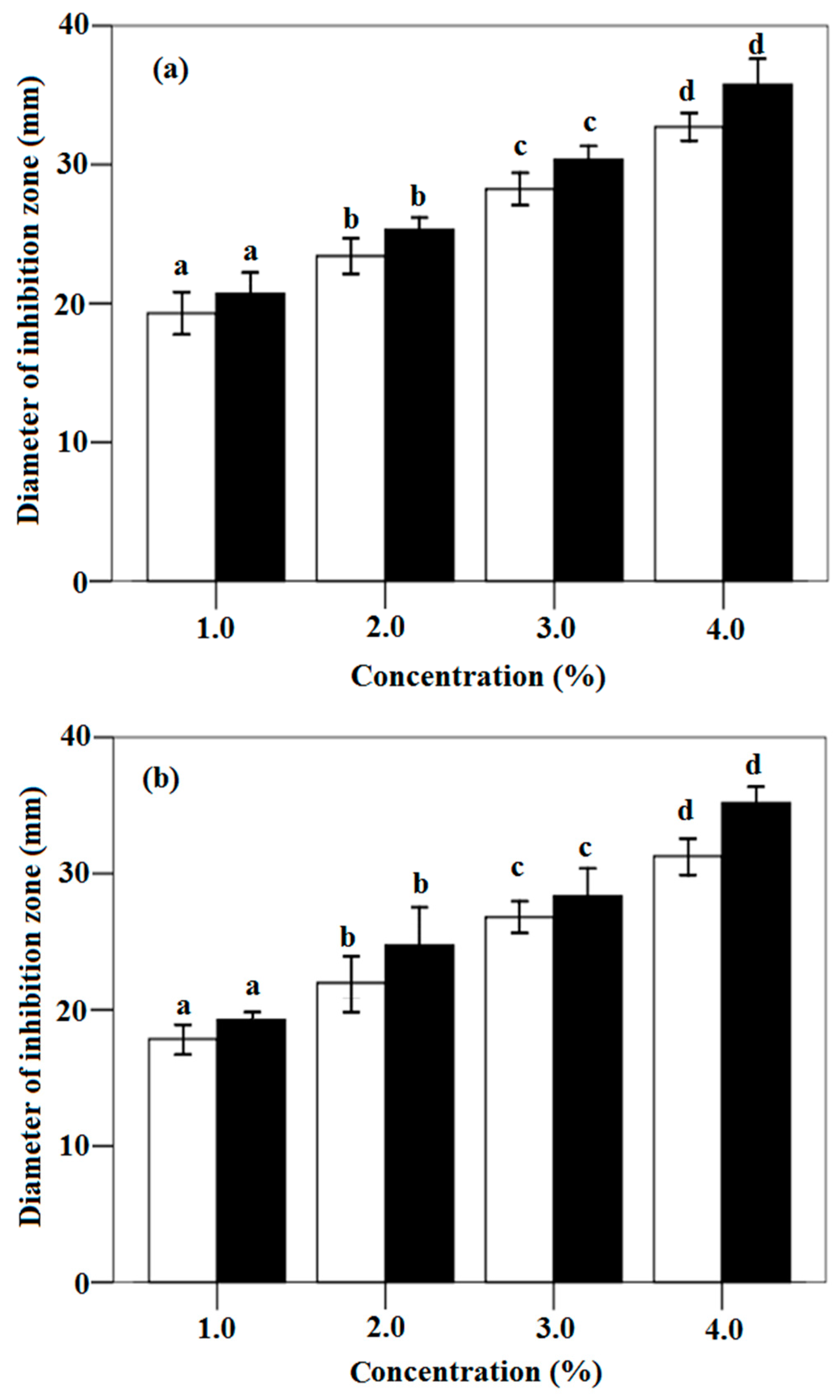
3.3. In vitro Antifungal Activity of Cinnamon Oil and Trans-Cinnamaldehyde against P. citrinum and A. flavus

In order to validate whether the antifungal property of cinnamon oil is due to its main component, the in vitro activities against two fungi, P. citrinum and A. flavus, were compared between trans-cinnamaldehyde and cinnamon oil. Results in Figure 5a indicate that the diameters of inhibition zones for trans-cinnamaldehyde and cinnamon oil against P. citrinum improve from 19.17 mm to 33.17 mm and from 20.81 mm to 36.23 mm with increasing the concentration from 1.0% to 4.0%, respectively. A similar result is also observed against A. flavus in Figure 5b: the zone diameters improve from 17.93 mm and 19.07 mm to 31.12 mm and 35.28 mm for the increase of the concentration of trans-cinnamaldehyde and cinnamon oil from 1.0% to 4.0%, respectively. However, significant differences are found in the antifungal activity of cinnamon oil and trans-cinnamaldehyde among different concentrations from the analysis result in Figure 5a,b. This means the inhibitory effects of trans-cinnamaldehyde and cinnamon oil are proportional to the concentration of trans-cinnamaldehyde. Excellent antifungal activities of oil and trans-cinnamaldehyde are found. Their antifungal activities against the growth of P. citrinum are higher than that against A. flavus.
These results shows that the killing activity of oil samples against fungi should be mainly due to its main component trans-cinnamaldehyde [26,27,43,61]. The antifungal activity of chitosan-based coating could enhance by the addition of cinnamon oil. These results were also reported by Kanatt et al. (2008), Xing et al. (2010b) and Xing et al. (2011a) [3,13,26]. Xing et al. (2010b) had observed that the strong inhibitory effect of cinnamon oil against all the tested microorganisms should be due to its main component cinnamaldehyde [9,26,27]. On the other hand, result in this work indicates that significant improvement in the inhibition on fungi growth was observed, which might be due to the increased levels of trans-cinnamaldehyde or cinnamon oil used. The antifungal activity of trans-cinnamaldehyde is only a little lower than that of cinnamon oil. It can be found that trans-cinnamaldehyde is the most effective components in cinnamon oil for inhibiting the fungi growth. More importantly, the higher antifungal activity of cinnamon oil should be due to the synergistic action of trans-cinnamaldehyde, cinnamaldehyde, and other components (such as benzenepropanolb, a-methyl-; propanoic acid, 2-methyl-, and 3-phenylpropyl ester). The inhibition of trans-cinnamaldehyde and cinnamaldehyde on the synthesizing enzymes of fungal cell was also found [3,26,37,61,64]. Cinnamaldehyde with a strong antifungal activity was reported by Wang et al. (2005) [61]. According to the investigation of Lambert et al. (2001) and Todd et al. (2013), the chemical constituents in oils could accumulate in the lipid-rich environments of cell membrane structures and cause its functional damage [64,68]. Clemente et al. (2016) demonstrated that the main bacterial target of cinnamaldehyde as the major component of cinnamon oil could cross the cell wall and act by the interaction of cinnamaldehyde carbonyl group and proteins [37,64]. It could be concluded that the antifungal activity might be due to the major and minor components in cinnamon oil, which means synergies of all components in cinnamon oil [26,41,43,47]. Burt (2004) has reported that the minor components might be critical to the activity and could provide a synergistic effect by the interactions among active substances in oil [69,70,71]. These findings emphasize the killing effectiveness of cinnamon oil and its principle component trans-cinnamaldehyde, which might induce the irreversible deleterious structural and morphological alterations of fungi cells [43,47].

3.4. Antifungal Activity of Cinnamon Oil for Different Attachment Time against P. citrinum and A. flavus

In order to investigate the mechanism for antifungal activity, the colony diameters of fungi growth after treated by cinnamon oil for different attachment time were examined. The index indicates impaired growth, and is used to describe the antimicrobial activity of cinnamon oil on the growth of P. citrinum and A. flavus on the PDA after the original fungi cell exposed to the active cinnamon oil with different time. As shown in Figure 6a, for the inhibition activity of oil against P. citrinum, the colony diameters decreased from 75.14 mm to 27.18 mm and from 74.01 mm to 4.49 mm with increasing the attachment time from 0 to 7 days at the concentration of cinnamon oil 100 μL/L and 400 μL/L, respectively. On the other hand, as can be seen in Figure 6b, with the attachment time of cinnamon oil on the fungi cells from 0 to 7 days, the colony diameters of A. flavus also decreased from 74.24 mm to 28.64 mm and from 75.22 mm to 4.04 mm with the oil concentration at 100 μL/L and 400 μL/L, respectively. Cinnamon oil almost inhibited the mycelial and spore growth of P. citrinum and A. flavus with the attached treatment of cinnamon oil at the concentration of 400 μL/L for 7 days. Furthermore, as can be seen in Figure 6, differences in the antifungal activity were observed among different attachment time for 0 to 7 days of cinnamon oil at the same oil concentration against P. citrinum and A. flavus. For the antifungal activity of cinnamon oil with the same attachment time from 1 to 7 days, there were differences among the concentrations of oil from 100 μL/L to 400 μL/L. However, no difference could be seen in the attachment time of 0 day among different oil concentration.
These results illustrate that the inhibition on the growth of P. citrinum and A. flavus could be affected by the attachment time and the concentration of cinnamon oil [3,26,43]. Cinnamon oil with the lower concentration could only cause the delay of conidia germination [3,43,71,72]. The longer attachment time and higher concentration of cinnamon oil could exhibit the higher inhibition on the growth and the higher kill activity of these two tested fungi. The most fungi cells could be inhibited or killed with the oil concentration at 400 μL/L for 7 days of the attachment time. The shorter attachment time and the lower concentration of active compounds could affect the initial development of molds, and produce a delay in the growth [3,67,71]. These results suggest a fungicidal effect at higher concentrations (400 μL/L) and an inhibition effect of cinnamon oil at lower concentrations (100 μL/L). For the durability of antifungal activity, the inhibition could be maintained for 7 days, especially for the higher concentration of oil. This might be due to these two reasons: the incomplete formation of conidia and the damage to the conidia caused by oil after the conidiophore formed [3,26,66,71]. The active paper disc created an inhibition area where the growth of fungi was inhibited, a clear zone where only white mycelium was presented and a strong inhibition in sporulation was noticed. These evident morphological changes can be appreciated due to the exposure to the vapor phase of oil during the attachment period. Volatile compounds in oil could induce the abnormalities in the cell wall structure, the disruption of hyphae, the loss of cytoplasm, the decrease in the conidia number and the conidiophores development, thereby disrupting various metabolic activities [2,71,72,73,74,75]. On the other hand, differences in the antifungal activity among different attachment time of oil with the same oil concentration might be due to the sustained release and action of oil vapor phase from the liquid phase [36,37,63]. The difference of the antifungal activity among different oil concentrations with the same attachment time might be due to the different concentration of oil vapor phase released from liquid phase and thereby inducing different killing or inhibiting actions [36,37,63].

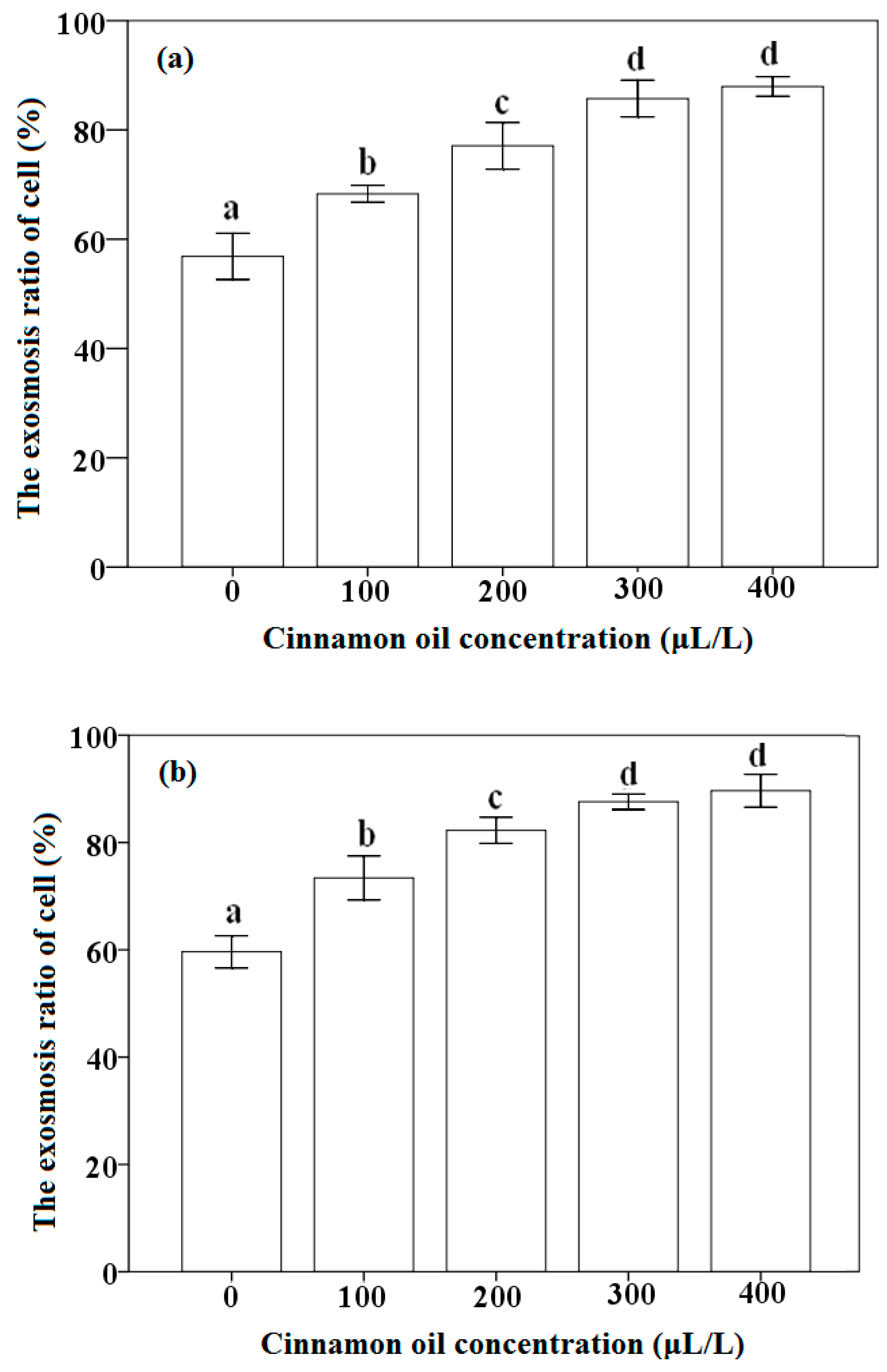
3.5. Effect of Cinnamon Oil on the Exosmosis Ratio of P. citrinum and A. flavus

The effects of different concentration of cinnamon oil on the exosmosis ratio of P. citrinum and A. flavus were investigated. Results in Figure 7a,b show that the exosmosis ratios of fungal cells with the concentration of cinnamon oil tests exhibit a close agreement with those above results of antifungal activity. The exosmosis ratios for the cell of P. citrinum and A. flavus as the control are 56.85% and 59.61%, respectively. However, the exosmosis ratios improve from 68.31% and 73.39% to 87.95% and 89.65% for P. citrinum and A. flavus with increasing the concentration from 100 μL/L to 400 μL/L, respectively. An increase in the exosmosis ratio of fungal cell is observed with improving the cinnamon oil concentration. Moreover, in order to investigate the disruption mechanism of cinnamon oil on the morphological structure of fungi cell, using A. flavus as an example, the morphological observation of cell around the inhibition zone and the normal fungi cell were conducted by SEM. As can be seen in Figure 8a,b, the normal cell of A. flavus on PDA shows a normal morphology with formed spores and linearly shaped hyphae. The hyphae and spore of normal A. flavus appear regular and homogeneous with the smooth walls. However, results in Figure 8c,d indicate that the mycelium of A. flavus with wizened, irregular and atrophied shapes is observed in the cells around the inhibition zone. From the images of SEM, it could be found that the phenomena of hyphae with shrank, slender and winding and the loss of the linearity on the hyphal surface were observed. The spore growth of A. flavus was inhibited completely by cinnamon oil. The disruption and control action of cinnamon oil might occur on the development of the spore and hypha of fungi cells, which should be due to the strong antimicrobial action of components in cinnamon oil, such as trans-cinnamaldehyde and cinnamaldehyde.
The investigations on the exosmosis ratio of fungi cells treated with different oil concentration and on the morphological observation of fungi cell could helpful to understand the mechanism of antifungal activity. The enhanced exosmosis ratio of fungi cells is related to the action and the increasing concentration of cinnamon oil. The disruption mechanism should be because most of the hypha cell walls became irregular and the contents either disappeared or were denatured. These also suggest that the oil might penetrate through the cell walls and membranes, causing mechanical damage and disrupting the cell metabolism [41,71]. SEM analysis also indicates that after the oil acted on A. flavus cells, the growth of spores and mycelium were destroyed and inhibited [26]. These processes could further damage the organelles and cell nucleus, and allow the contents to escape from the cells, eventually resulting in a cell burst and then death [36,37,41,71]. The investigation of Tzortzakis (2009) also indicated that the effects of cinnamon oil reduced the germination of spore and the growth of germ tube in Colletotrichum coccodes, Botrytis cinerea and Rhizopus stolonifer was mainly dependent on its concentration used [76]. The result reported by other workers also demonstrated that cinnamaldehyde could pass the cytoplasm and translocate to the cell nucleus of fungi, and then interfere with the synthesis of protein and RNA in the cell [66,77]. It is also mentioned that the hydroxyl groups in active components could affect the biosynthesis of mycotoxins by forming the hydrogen bonds with active enzymes and thereby resulting in deactivation [3,26,56,78].

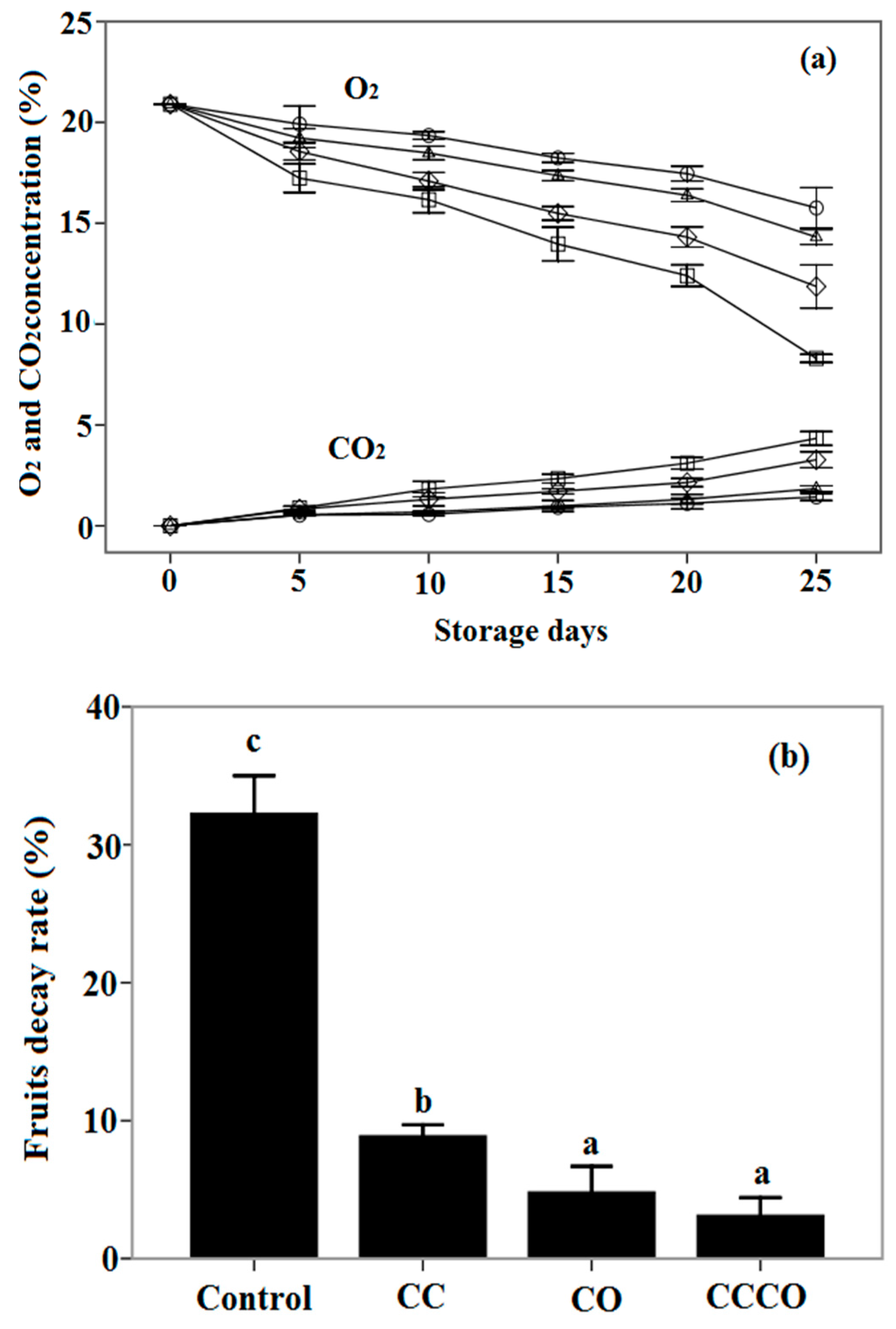
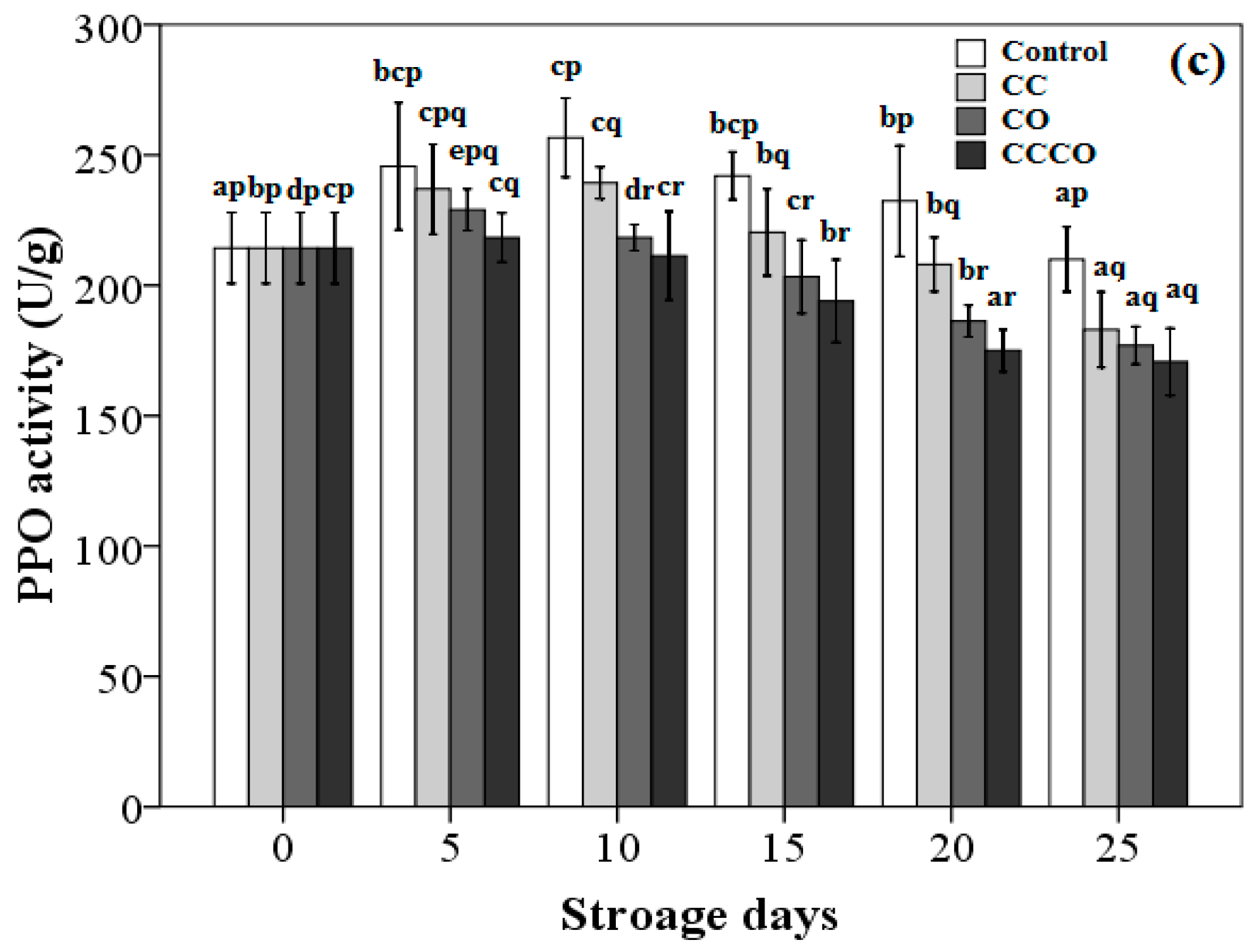
3.6. Gas Composition in Packages and Fruits Decay Treated by Chitosan-Oil Coating

Chitosan-oil coating as a barrier of gas and a carrier of the antimicrobial agent could create an internal modified atmosphere in the package and control the decay of fruits during the storage period [18]. The changes in the concentration of O2 and CO2 in the packages of sweet cherry fruits with different treatments were analyzed with gas sensors. As can be seen in Figure 9a, the O2 concentrations in the package for the samples treated by the chitosan coating with cinnamon oil and the chitosan coating are 15.76% and 14.32% after 25 days of storage, respectively. The O2 concentration inside the packages of control fruits was reduced significantly, and was only 8.3% for the control samples. On the other hand, the CO2 concentration in the packages for the treatments of chitosan-oil coating and control are 4.33% and 1.43%, respectively. The production of CO2 in the package for the chitosan-oil treatment are lower compared to that of fruits coated with only chitosan or only oil treatment. Moreover, during the storage time, the cheery fruits could be infected by molds bringing the fruits from the field, which could be strongly delayed by the treatment of chitosan-oil coating. As the results in Figure 9b show, the higher decay (32.37%) rate of uncoated cheery fruits is observed at the end of storage. However, cherry fruits coated with the chitosan-oil composite film display the lowest decay rate throughout the 25-day storage period, at 3.13%, which is much lower than that of control fruits. The decay rate of fruits treated by cinnamon oil is 4.83% after 25-day storage. The PPO activity of fruits treated with different coatings is shown in Figure 9c; the lowest activity of PPO was observed in the fruits treated with the chitosan-oil coating, which was 170.7 U/g on the 25th day of storage. However, the control fruits exhibited the highest activity for all of the storages. PPO activity is 210.0 U/g at the end of storage. It could be inhibited by the chitosan-oil coating during the storage period. Furthermore, as can be seen in Figure 9c, differences in PPO activity were observed among different storage days for 5 to 25 days of both treatments of only oil treatments and the chitosan-oil coating. Differences in the PPO activity of fruits were also found among the different treatment at the same storage days, from 5 to 25 days. The coating of chitosan with cinnamon oil could be used in maintaining the quality of cherry fruits.
These results indicate that the respiration rate of sweet fruits reflected by the concentrations of O2 and CO2 was markedly affected by the treatments of chitosan-based coating during storage [4,9,10,18]. Results of the decrease in the levels of O2 and the increase in the concentration of CO2 in the packages denote that chitosan-based coating is beneficial for reducing the respiration rate of fruits in the package [2,9,10,18]. The chitosan-based coating with/without cinnamon oil plays an important role in the modification of gas concentration. Incorporation of cinnamon oil in the chitosan coating could reduce the O2 consumption of fruits and inhibit the activity of PPO compared with that of other treatments, especially for the control samples. This is possibly because of the antioxidant activity of cinnamon oil and the interaction between chitosan and cinnamon oil as the active substance, which is different from both the only chitosan coating and the only oil treatment [3,4,9,23,79]. Significant differences regarding the consumption of O2 and the production of CO2 are also found between the coated fruits and control samples at the end of storage. Our results are in agreement with the findings of other researchers: the coating treatment could reduce the consumption of O2 for strawberries [26,80,81,82,83]. Perdones et al. (2012) have reported that the gas permeability was influenced by the chitosan coatings on the fruits surface [58]. The equilibrium O2 and CO2 levels might be created by the barrier of chitosan-based films, which might benefit the storage quality of cherry fruits [84,85]. On the other hand, the control of postharvest decay in cherry fruits has been of interest because it could significantly affect the quality safety of fruits during storage [86,87,88,89,90,91]. Fruits coated by this composite coating exhibit lower percentages of decay than those of control samples, which might be because of the higher inhibition of the pathogen growth of cinnamon oil in the coating and also due to the higher levels of CO2 in the packages [3,9]. The antioxidant property of some compounds in cinnamon oil could also inhibit the PPO activity in fruits and be helpful for keeping the quality of produce [3,4,6,9]. The chitosan-based coating on the surface of cherry fruits could control the pathogen infection by reducing the diffusion of oil vapor and slow the physiological processes in fruits [3,4,6]. The mechanism of the induction of chitosan-oil coating on the defense-related enzymes, the permeability and integrity of fruit cell membrane, and odors in fruits should be further investigated [3,4,6,9,92,93].

4. Conclusions

Preservation mechanism of chitosan coating and cinnamon oil for fruits storage is very important for understanding its application effect. The analysis of AFM indicates that many micropores exist in the chitosan based coating. The antifungal property of cinnamon oil should be mainly due to its main constituent, trans-cinnamaldehyde. It is also proportional to the concentration of oil and the attachment time. Compared to the normal cell, the wizened mycelium of A. flavus around the inhibition zone is observed, and the growth of spores could be inhibited completely. These indicate that the preservation mechanism of chitosan-oil coating should be due to the micropores structure of chitosan coating as the carrier and the antifungal activity of oil, which could slow the respiration rate and control the decay of fruits during the storage period. The antioxidant mechanism of chitosan-based coating and cinnamon oil, such as the in vitro and in vivo antioxidant activity and mechanism of cinnamon oil, the influence mechanism of chitosan-oil coating on the defense-related enzymes, free radical scavenging activity, and the permeability and integrity of fruit cell membrane in fruits. These effects will need to be researched further and will be published in the future.

Acknowledgments

This work is supported by the Innovation Team Construction Program of Sichuan Education Department (15TD0017), Key Laboratory Open Research Fund of Grain and Oil Processing and Food Safety of Xihua University (szjj2015-003, szjj2014-003 and szjj2014-002), Key Laboratory Open Research Fund of Physiological and Storage of Agricultural Products of Agriculture Ministry (KF008), National Natural Science Foundation of China (31501547), National High Technology Research and Development Program of China (“863” program, 2012AA101606), Science and technology support program of Sichuan (2016FZ0019) and Young Scholars Training Plan of Xihua University (01201413).

Author Contributions

Y.X. and X.L. conceived and designed the experiments; Y.X. and Q.X. performed the experiments; Y.X., Q.X. and L.Z. analyzed the data; X.L., C.C., S.Y. and Z.C. contributed reagents/materials/analysis tools; Y.X., S.S., Y.T., and S.Y. wrote the paper.

Conflicts of Interest

The authors declare no conflict of interest.

References

  1. Krikorian, R.; Boespflug, E.L.; Fleck, D.E.; Stein, A.L.; Wightman, J.D.; Shidler, M.D.; Sadat-Hossieny, S. Concord grape juice supplementation and neurocognitive function in human aging. J. Agric. Food Chem. 2012, 60, 5736–5742. [Google Scholar] [CrossRef] [PubMed]
  2. Guerra, I.I.C.D.; Oliveira, P.D.L.M.; Santos, M.F.; Lúcio, A.S.S.C.; Tavares, J.F.; Barbosa-Filho, J.M.; Madruga, M.S.; de Souz, E.L. The effects of composite coatings containing chitosan and Mentha (piperita L. or x villosa Huds) essential oil on postharvest mold occurrence and quality of table grape cv. Isabella. Innov. Food Sci. Emerg. 2016, 34, 112–121. [Google Scholar] [CrossRef]
  3. Xing, Y.; Xu, Q.; Che, Z.; Li, X.; Li, W. Effects of chitosan-oil coating on blue mold disease and quality attributes of jujube fruits. Food Funct. 2011, 2, 466–474. [Google Scholar] [CrossRef] [PubMed]
  4. Xing, Y.; Lin, H.; Cao, D.; Xu, Q.; Han, W.; Wang, R.; Che, Z.; Li, X. Effect of chitosan coating with cinnamon oil on the quality and physiological attributes of China jujube fruits. BioMed Res. Int. 2015, 2015, 835151. [Google Scholar] [CrossRef] [PubMed]
  5. Ojagh, S.M.; Rezaei, M.; Razavi, S.H.; Hosseini, S.M.H. Effect of chitosan coatings enriched with cinnamon oil on the quality of refrigerated rainbow trout. Food Chem. 2010, 120, 193–198. [Google Scholar] [CrossRef]
  6. Xu, Q.; Xing, Y.; Che, Z.; Guan, T.; Zhang, L.; Bai, Y.; Gong, L. Effect of chitosan coating and oil fumigation on the microbiological and quality safety of fresh-cut pear. J. Food Saf. 2013, 33, 179–189. [Google Scholar] [CrossRef]
  7. Carneiro, J.; Tedim, J.; Fernandes, S.C.M.; Freire, C.S.R.; Gandini, A.; Ferreira, M.G.S.; Zheludkevich, M.L. Functionalized chitosan-based coatings for active corrosion protection. Surf. Coat. Technol. 2013, 226, 51–59. [Google Scholar] [CrossRef]
  8. Azevedo, A.N.; Buarque, P.R.; Cruz, E.M.O.; Blank, A.F.; Alves, P.B.; Nunes, M.L.; Santan, L.C.L.A. Response surface methodology for optimisation of edible chitosan coating formulations incorporating essential oil against several foodborne pathogenic bacteria. Food Control 2014, 43, 1–9. [Google Scholar] [CrossRef]
  9. Xing, Y.; Li, X.; Xu, Q.; Yun, J.; Lu, Y.; Tang, Y. Effects of chitosan coating enriched with cinnamon oil on qualitative properties of sweet pepper (Capsicum annuum L.). Food Chem. 2011, 124, 1443–1450. [Google Scholar] [CrossRef]
  10. Vieira, J.M.; Flores-López, M.L.; de Rodríguez, D.J.; Sousa, M.C.; Vicente, A.A.; Martins, J.T. Effect of chitosan-Aloe vera coating on postharvest quality of blueberry (Vaccinium corymbosum) fruit. Postharvest Biol. Technol. 2016, 116, 88–97. [Google Scholar] [CrossRef] [Green Version]
  11. Vasconez, M.B.; Flores, S.K.; Campos, C.A.; Alvarado, J.; Gerschenson, L.N. Antimicrobial activity and physical properties of chitosanetapioca starch based edible films and coatings. Food Res. Int. 2009, 42, 762–769. [Google Scholar] [CrossRef]
  12. Arancibia, M.Y.; Lopez-Caballero, M.E.; Gomez-Guillen, M.C.; Montero, P. Chitosan coatings enriched with active shrimp waste for shrimp preservation. Food Control 2015, 54, 259–266. [Google Scholar] [CrossRef]
  13. Kanatt, S.R.; Chander, R.; Sharma, A. Chitosan and mint mixture: A new preservative for meat and meat products. Food Chem. 2008, 107, 845–852. [Google Scholar] [CrossRef]
  14. Badawy, M.E.I.; Rabea, E.I. Potential of the biopolymer chitosan with different molecular weights to control postharvest gray mold of tomato fruit. Postharvest Biol. Technol. 2009, 51, 110–117. [Google Scholar] [CrossRef]
  15. Kong, M.; Chen, X.G.; Xing, K.; Park, H.J. Antimicrobial properties of chitosan and mode of action: A state of the art review. Int. J. Food Microbiol. 2010, 144, 51–63. [Google Scholar] [CrossRef] [PubMed]
  16. European Union. Establishing a list of permitted health claims made on foods, other than those referring to the reduction of disease risk and to children’s development and health. Off. J. Eur. Union. 2012, 136, 1–40. [Google Scholar]
  17. Ce, N.; Norena, C.P.Z.; Brandelli, A. Antimicrobial activity of chitosan films containing nisin, peptide P34, and natamycin. CyTA J. Food 2012, 10, 21–26. [Google Scholar] [CrossRef]
  18. Xing, Y.; Li, X.; Xu, Q.; Jiang, Y.; Yun, J.; Li, W. Effects of chitosan-based coating and modified atmosphere packaging (MAP) on browning and shelf life of fresh-cut lotus root (Nelumbo nucifera Gaerth). Innov. Food Sci. Emerg. 2010, 11, 684–689. [Google Scholar] [CrossRef]
  19. Velickova, E.; Winkelhausen, E.; Kuzmanova, S.; Alves, V.D.; Moldão-Martins, M. Impact of chitosan-beeswax edible coatings on the quality of fresh strawberries (Fragariaananassa cv Camarosa) under commercial storage conditions. LWT Food Sci. Technol. 2013, 52, 80–92. [Google Scholar] [CrossRef]
  20. Waewthongrak, W.; Pisuchpen, S.; Leelasuphakula, W. Effect of Bacillus subtilis and chitosan applications on green mold (Penicilium digitatum Sacc.) decay in citrus fruit. Postharvest Biol. Technol. 2015, 99, 44–49. [Google Scholar] [CrossRef]
  21. Kaya, M.; Asan-Ozusaglam, M.; Erdogan, S. Comparison of antimicrobial activities of newly obtained low molecular weight scorpion chitosan and medium molecular weight commercial chitosan. J. Biosci. Bioeng. 2016, 121, 678–684. [Google Scholar] [CrossRef] [PubMed]
  22. Durana, M.; Adaya, M.S.; Zorba, N.N.D.; Temizkana, R.; Büyükcanb, M.B.; Canera, C. Potential of antimicrobial active packaging ‘containing natamycin, nisin, pomegranate and grape seed extract in chitosan coating’ to extend shelf life of fresh strawberry. Food Bioprod. Process. 2016, 98, 354–363. [Google Scholar] [CrossRef]
  23. Sun, X.; Narciso, J.; Wang, Z.; Ference, C.; Bai, J.; Zhou, K. Effects of chitosan-essential oil coatings on safety and quality of fresh blueberries. J. Food Sci. 2014, 79, 955–960. [Google Scholar] [CrossRef] [PubMed]
  24. Win, N.K.K.; Jitareerat, P.; Kanlayanarat, S.; Sangchote, S. Effects of cinnamon extract, chitosan coating, hot water treatment and their combinations on crown rot disease and quality of banana fruit. Postharvest Biol. Technol. 2007, 45, 333–340. [Google Scholar] [CrossRef]
  25. Li, Y.; Kong, D.; Wu, H. Analysis and evaluation of essential oil components of cinnamon barks using GC-MS and FTIR spectroscopy. Ind. Crop. Prod. 2013, 41, 269–278. [Google Scholar] [CrossRef]
  26. Xing, Y.; Li, X.; Xu, Q.; Yun, J.; Lu, Y. Antifungal activities of cinnamon oil against Rhizopus nigricans, Aspergillus flavus and Penicillium expansum in vitro and in vivo fruit test. Int. J. Food Sci. Technol. 2010, 45, 1837–1842. [Google Scholar] [CrossRef]
  27. Kim, Y.G.; Lee, J.H.; Kim, S.I.; Baek, K.H.; Lee, J. Cinnamon bark oil and its components inhibit biofilm formation and toxin production. Int. J. Food Microbiol. 2015, 195, 30–39. [Google Scholar] [CrossRef] [PubMed]
  28. Xing, Y.; Xu, Q.; Lin, H.; Li, X.; Zhang, D.; Yang, S.X.; Meng, Y.; Li, X.; Liu, X. Preparation, properties and in vivo antimicrobial activity in yacon roots of microencapsulation containing cinnamon oil. Mater. Technol. 2016. [Google Scholar] [CrossRef]
  29. Carvalho, W.M.; Brasil, I.M.; Gallão, M.I.; Moura, C.F.H.; Lopes, M.M.A.; de Miranda, M.R.A.; Carvalho, R.L.; Cabral, M.F.; Germano, T.A. Chitosan coating with trans-cinnamaldehyde improves structural integrity and antioxidant metabolism of fresh-cut melon. Postharvest Biol. Technol. 2016, 113, 29–39. [Google Scholar] [CrossRef]
  30. Liu, J.; Tian, S.; Meng, X.; Xu, Y. Effects of chitosan on control of postharvest diseases and physiological responses of tomato fruit. Postharvest Biol. Technol. 2007, 44, 300–306. [Google Scholar] [CrossRef]
  31. Suseno, N.; Savitri, E.; Sapei, L.; Padmawijaya, K.S. Improving shelf-life of cavendish banana using chitosan edible coating. Procedia Chem. 2014, 9, 112–113. [Google Scholar] [CrossRef]
  32. Rabea, E.I.; Badawy, M.E.T.; Stevens, C.V.; Smagghe, G.; Steurbaut, W. Chitosan as antimicrobial agent: Applications and mode of action. Biomacromolecules 2003, 4, 1457–1465. [Google Scholar] [CrossRef] [PubMed]
  33. Liu, H.; Du, Y.; Wang, X.; Sun, L. Chitosan kills bacteria through cell membrane damage. Int. J. Food Microbiol. 2004, 95, 147–155. [Google Scholar] [CrossRef] [PubMed]
  34. Kartal, S.; Aday, M.S.; Caner, C. Use of microperforated films and oxygen scavengers to maintain storage stability of fresh strawberries. Postharvest Biol. Technol. 2012, 71, 32–40. [Google Scholar] [CrossRef]
  35. Wang, L.; Wu, H.; Quin, G.; Meng, X. Chitosan disrupts Penicillium expansum and controls postharvest blue mold of jujube fruit. Food Control 2014, 41, 56–62. [Google Scholar] [CrossRef]
  36. Zhang, Y.; Liu, X.; Wang, Y.; Jiang, P.; Quek, S.Y. Antibacterial activity and mechanism of cinnamon essential oil against Escherichia coli and Staphylococcus aureus. Food Control 2016, 59, 282–289. [Google Scholar] [CrossRef]
  37. Clemente, I.; Aznar, M.; Silva, F.; Nerín, C. Antimicrobial properties and mode of action of mustard and cinnamon essential oils and their combination against foodborne bacteria. Innov. Food Sci. Emerg. 2016, 36, 26–33. [Google Scholar] [CrossRef]
  38. Sun, X.; Wang, Z.; Kadouh, H.; Zhou, K. The antimicrobial, mechanical, physical and structural properties of chitosanegallic acid films. LWT Food Sci. Technol. 2014, 57, 83–89. [Google Scholar] [CrossRef]
  39. Zdunek, A.; Kurenda, A. Determination of the elastic properties of tomato fruit cells with an atomic force microscope. Sensors 2013, 13, 12175–12191. [Google Scholar] [CrossRef] [PubMed]
  40. Kowal, A. Application of STM and AFM techniques for the investigation of corrosion processes and materials protection. Zaštita Materijala 2005, 46, 44–46. [Google Scholar]
  41. Li, T.; Li, J.; Hu, W.; Li, X. Quality enhancement in refrigerated red drum (Sciaenops ocellatus) fillets using chitosan coatings containing natural preservatives. Food Chem. 2013, 138, 821–826. [Google Scholar] [CrossRef] [PubMed]
  42. Xing, Y.; Li, X.; Yun, J.; Lu, Y. Extending the shelf-life of fresh-cut lotus root with antibrowning agents, cinnamon oil fumigation, and moderate vacuum packaging (MVP). J. Food Process Eng. 2012, 35, 505–521. [Google Scholar] [CrossRef]
  43. Wang, G.; Deng, J.; Ma, Y.; Shi, M.; Li, B. Mechanisms, clinically curative effects, and antifungal activities of cinnamon oil and pogostemon oil complex against three species of Candida. J. Tradit. Chin. Med. 2012, 32, 19–24. [Google Scholar] [CrossRef]
  44. Feng, W. The Study of Control and Mechanism of Action on Postharvest Diseases of Fruit and Vegetables by Essential Oil. Ph.D. Thesis, Zhejiang University, Zhejiang, China, 2006; pp. 92–93. [Google Scholar]
  45. Diao, W.R.; Hu, Q.P.; Zhang, H.; Xu, J.G. Chemical composition, antibacterial activity and mechanism of action of essential oil from seeds of fennel (Foeniculum vulgare Mill.). Food Control 2014, 35, 109–116. [Google Scholar] [CrossRef]
  46. Løkke, M.M.; Seefeldt, H.F.; Edwards, G.; Green, O. Novel wireless sensor system for monitoring oxygen, temperature and respiration rate of horticultural crops post harvest. Sensors 2011, 11, 8456–8468. [Google Scholar] [CrossRef] [PubMed]
  47. Xing, F.; Hua, H.; Selvaraj, J.N.; Zhao, Y.; Zhou, L.; Liu, X.; Liu, Y. Growth inhibition and morphological alterations of Fusarium verticillioides by cinnamon oil and cinnamaldehyde. Food Control 2014, 46, 343–350. [Google Scholar] [CrossRef]
  48. Lindseth, I.; Bardal, A. Quantitative topography measurements of rolled aluminium surfaces by atomic force microscopy and optical methods. Surf. Coat. Technol. 1999, 111, 276–286. [Google Scholar] [CrossRef]
  49. Curkovic, L.; Ljubas, D.; Juretic, H. Photocatalytic decolourization kinetics of diazo dye Congo Red aqueous solution by UV/TiO2 nanoparticles. React. Kinet., Mech. Cat. 2010, 99, 201–208. [Google Scholar]
  50. Segota, S.; Curkovic, L.; Ljubas, D.; Svetlicic, V.; Houra, I.F.; Tomasic, N. Synthesis, characterization and photocatalytic properties of Sol-gel TiO2 films. Ceram. Int. 2011, 37, 1153–1160. [Google Scholar] [CrossRef]
  51. Li, Y.J.; Brndiar, J.; Naitoh, Y.; Sugawara, Y.; Štich, I. Atomic force microscopy identification of Al-sites on ultrathin aluminum oxide film on NiAl (110). Nanotechnology 2015, 26, 505704. [Google Scholar] [CrossRef] [PubMed]
  52. Darrort, V.; Troyon, M.; Ebothé, J.; Bissieux, C.; Nicollin, C. Quantitative study by atomic force microscopy and spectrophotometry of the roughness and brightness of electrodeposited nickel in the presence of additives. Thin Solid Films 1995, 265, 52–57. [Google Scholar] [CrossRef]
  53. Ruiz-Garcia, L.; Lunadei, L.; Barreiro, P.; Robla, J.I. A review of wireless sensor technologies and applications in agriculture and food industry: State of the art and current trends. Sensors 2009, 9, 4728–4750. [Google Scholar] [CrossRef] [PubMed] [Green Version]
  54. Bochtis, D.D.; Sørensen, C.G.; Green, O.; Bartzanas, T. A diagnostic system for improving biomass quality based on a sensor network. Sensors 2011, 11, 4990–5004. [Google Scholar] [CrossRef] [PubMed]
  55. Pereda, M.; Amica, G.; Marcovich, N.E. Development and characterization of edible chitosan/olive oil emulsion films. Carbohyd. Polym. 2012, 87, 1318–1325. [Google Scholar] [CrossRef]
  56. Ma, Q.; Zhang, Y.; Zhong, Q. Physical and antimicrobial properties of chitosan films incorporated with lauric arginate, cinnamon oil, and ethylenediaminetetraacetate. LWT Food Sci. Technol. 2016, 65, 173–179. [Google Scholar] [CrossRef]
  57. Villalobos-Carvajal, R.; Hernández-Munoz, P.; Albors, A.; Chiralt, A. Barrier and optical properties of edible hydroxypropyl methylcellulose coatings containing surfactants applied to fresh cut carrot slices. Food Hydrocolloids 2009, 23, 526–535. [Google Scholar] [CrossRef]
  58. Perdones, A.; Sánchez-González, L.; Chiralt, A.; Vargas, M. Effect of chitosan-lemon essential oil coatings on storage-keeping quality of strawberry. Postharvest Biol. Technol. 2012, 70, 32–41. [Google Scholar] [CrossRef]
  59. Rojas-Graüa, M.A.; Tapiab, M.S.; Martín-Bellosoa, O. Using polysaccharide-based edible coatings to maintain quality of fresh-cut Fuji apples. LWT Food Sci. Technol. 2008, 41, 139–147. [Google Scholar] [CrossRef]
  60. Golmohammad, F.; Eikani, M.H.; Maymandi, H.M. Cinnamon bark volatile oils separation and determination using solid-phase extraction and gas chromatography. Procedia Eng. 2012, 42, 247–260. [Google Scholar] [CrossRef]
  61. Wang, S.Y.; Chen, P.F.; Chang, S.T. Antifungal activities of essential oils and their constituents from indigenous cinnamon (Cinnamomum osmophloeum) leaves against wood decay fungi. Bioresour. Technol. 2005, 96, 813–818. [Google Scholar] [CrossRef] [PubMed]
  62. Singh, G.; Maurya, S.; DeLampasona, M.P.; Catalan, C.A. A comparison of chemical, antioxidant and antimicrobial studies of cinnamon leaf and bark volatile oils, oleoresins and their constituents. Food Chem. Toxicol. 2007, 45, 1650–1661. [Google Scholar] [CrossRef] [PubMed]
  63. Goñi, P.; López, P.; Sánchez, C.; Gómez-Lus, R.; Becerril, R.; Nerín, C. Antimicrobial activity in the vapour phase of a combination of cinnamon and clove essential oils. Food Chem. 2009, 116, 982–989. [Google Scholar] [CrossRef]
  64. Todd, J.; Friedman, M.; Patel, J.; Jaroni, D.; Ravishankar, S. The antimicrobial effects of cinnamon leaf oil against multi-drug resistant Salmonella Newport on organic leafy greens. Int. J. Food Microbiol. 2013, 166, 193–199. [Google Scholar] [CrossRef] [PubMed]
  65. Kaskatepe, B.; Kiymaci, M.E.; Suzuk, S.; Erdem, S.A.; Cesur, S.; Yildiz, S. Antibacterial effects of cinnamon oil against carbapenem resistantnosocomial Acinetobacter baumannii and Pseudomonas aeruginosaisolates. Ind. Crops Prod. 2016, 81, 191–194. [Google Scholar] [CrossRef]
  66. Ricci, D.; Fraternale, D.; Giamperi, L.; Bucchini, A.; Epifano, F.; Burini, G.; Curini, M. Chemical composition, antimicrobial and antioxidant activity of the essential oil of Teucrium marum (Lamiaceae). J. Ethnopharmacol. 2005, 98, 195–200. [Google Scholar] [CrossRef] [PubMed]
  67. Wang, R.; Wang, R.; Yang, B. Extraction of essential oils from five cinnamon leaves and identification of their volatile compound compositions. Innov. Food Sci. Emerg. 2009, 10, 289–292. [Google Scholar] [CrossRef]
  68. Lambert, R.J.W.; Skandamis, P.N.; Coote, P.J.; Nychas, G.J.E. A study of the minimum inhibitory concentration and mode of action of oregano essential oil, thymol and carvacrol. J. Appl. Microbiol. 2001, 91, 453–462. [Google Scholar] [CrossRef] [PubMed]
  69. Burt, S. Essential oils: Their antibacterial properties and potential applications in foods—A review. Int. J. Food Microbiol. 2004, 94, 223–253. [Google Scholar] [CrossRef] [PubMed]
  70. Cox, S.D.; Mann, C.M.; Markham, J.L. Interactions between components of the essential oil of Melaleuca alternifolia. J. Appl. Microbiol. 2001, 91, 492–497. [Google Scholar] [CrossRef] [PubMed]
  71. Manso, S.; Cacho-Nerin, F.; Becerril, R.; Nerín, C. Combined analytical and microbiological tools to study the effect on Aspergillus flavus of cinnamon essential oil contained in food packaging. Food Control 2013, 30, 370–378. [Google Scholar] [CrossRef]
  72. Lopez-Malo, A.; Alzamora, S.M.; Palou, E. Aspergillus flavus dose-response curves to selected natural and synthetic antimicrobials. Int. J. Food Microbiol. 2002, 73, 213–218. [Google Scholar] [CrossRef]
  73. Helal, G.A.; Sarhan, M.M.; Shahla, A.N.K.A.; El-Khair, E.K.A. Antimicrobial activity of some essential oils against microorganisms deteriorating fruit juices. Mycobiology 2006, 34, 219–229. [Google Scholar] [CrossRef] [PubMed]
  74. Tolouee, M.; Alinezhad, S.; Saberi, R.; Eslamifar, A.; Zad, S.J.; Jaimand, K.; Jaleh, T.; Mohammad-Bagher, R.; Masanobu, K.; Masoomeh, S.G.; et al. Effect of Matricaria chamomilla L. flower essential oil on the growth and ultrastructure of Aspergillus niger van Tieghem. Int. J. Food Microbiol. 2010, 139, 127–133. [Google Scholar] [CrossRef] [PubMed]
  75. Ait-Ouazzou, A.; Lorán, S.; Arakrak, A.; Laglaoui, A.; Rota, C.; Herrera, A.; Pagán, R.; Conchello, P. Evaluation of the chemical composition and antimicrobial activity of Mentha pulegium, Juniperus phoenicea, and Cyperus longus essential oils from Morocco. Food Res. Int. 2012, 45, 313–319. [Google Scholar] [CrossRef]
  76. Tzortzakis, N.G. Impact of cinnamon oil-enrichment on microbial spoilage of fresh produce. Innov. Food Sci. Emerg. Technol. 2009, 10, 97–102. [Google Scholar] [CrossRef]
  77. Palma-Guerrero, J.; Jansson, H.B.; Salinas, J.; Lopez-Llorca, L.V. Effect of chitosan on hyphal growth and spore germination of plant pathogenic and biocontrol fungi. J. Appl. Microbiol. 2008, 104, 541–553. [Google Scholar] [CrossRef] [PubMed]
  78. Juglal, S.; Govinden, R.; Odhav, B. Spice oils for the control of co-occurring mycotoxin-producing fungi. J. Food Prot. 2002, 65, 683–687. [Google Scholar] [PubMed]
  79. Mei, J.; Yuan, Y.; Guo, Q.; Wu, Y.; Li, Y.; Yu, H. Characterization and antimicrobial properties of waterchestnut starch-chitosan edible films. Int. J. Biol. Macromol. 2013, 61, 169–174. [Google Scholar] [CrossRef] [PubMed]
  80. Sabaghi, M.; Maghsoudlou, Y.; Khomeiri, M.; Ziaiifar, A.M. Active edible coating from chitosan incorporating green tea extract as an antioxidant and antifungal on fresh walnut kernel. Postharvest Biol. Technol. 2015, 110, 224–228. [Google Scholar] [CrossRef]
  81. Iqbal, M.A.; Kim, K.H.; Ahn, J.H. Monoterpenes Released from Fruit, Plant, and Vegetable Systems. Sensors 2014, 14, 18286–18301. [Google Scholar] [CrossRef] [PubMed]
  82. Kim, Y.H.; Kim, K.H.; Szulejko, J.; Parker, D. Quantitative analysis of fragrance and odorants released from fresh and decaying strawberries. Sensors 2013, 13, 7939–7978. [Google Scholar] [CrossRef] [PubMed]
  83. Sandhya. Modified atmosphere packaging of fresh produce: Current status and future needs. LWT Food Sci. Technol. 2010, 43, 381–392. [Google Scholar] [CrossRef]
  84. Bandaa, K.; Caleb, O.J.; Jacobsc, K.; Oparaa, U.L. Effect of active-modified atmosphere packaging on the respiration rate and quality of pomegranate arils (cv. Wonderful). Postharvest Biol. Technol. 2015, 109, 97–105. [Google Scholar] [CrossRef]
  85. Abbasi, N.A.; Iqbal, Z.; Maqbool, M.; Hafiz, I.A. Postharvest Quality of mango (Mangifera Indica L.) fruits as affected by coating. Pakistan J. Bot. 2009, 41, 343–357. [Google Scholar]
  86. Khaled, D.E.; Novas, N.; Gazquez, J.A.; Garcia, R.M.; Manzano-Agugliaro, F. Fruit and vegetable quality assessment via dielectric sensing. Sensors 2015, 15, 15363–15397. [Google Scholar] [CrossRef] [PubMed]
  87. Tang, K.T.; Chiu, S.W.; Pan, C.H.; Hsieh, H.Y.; Liang, Y.S.; Liu, S.C. Development of a portable electronic nose system for the detection and classification of fruity odors. Sensors 2010, 10, 9179–9193. [Google Scholar] [CrossRef] [PubMed]
  88. Fajardo, P.; Martins, J.T.; Fucinos, C.; Pastrana, L.; Teixeira, J.A.; Vicente, A.A. Evaluation of a chitosan-based edible filmas carrier of natamycin to improve the storability of Saloiocheese. J. Food Eng. 2010, 101, 349–356. [Google Scholar] [CrossRef] [Green Version]
  89. Yun, J.; Fan, X.; Li, X.; Jin, T.Z.; Jia, X.; Mattheis, J.P. Natural surface coating to inactivate Salmonella entericaserovar Typhimurium and maintain quality of cherry tomatoes. Int. J. Food Microbiol. 2015, 193, 59–67. [Google Scholar] [CrossRef] [PubMed]
  90. Gazdik, Z.; Zitka, O.; Petrlova, J.; Adam, V.; Zehnalek, J.; Horna, A.; Reznicek, V.; Beklova, M.; Kizek, R. Determination of vitamin C (ascorbic acid) using high rerformance liquid chromatography coupled with electrochemical detection. Sensors 2008, 8, 7097–7112. [Google Scholar] [CrossRef]
  91. Yun, J.; Fan, X.; Li, X. Inactivation of Salmonella entericaserovar Typhimurium and quality maintenance of cherry tomatoes treated with gaseous essential oils. J. Food Sci. 2013, 78, M458–M464. [Google Scholar] [CrossRef] [PubMed]
  92. Berna, A. Metal Oxide Sensors for Electronic Noses and Their Application to Food Analysis. Sensors 2010, 10, 3882–3910. [Google Scholar] [CrossRef] [PubMed]
  93. Friedman, M.; Juneja, V.K. Review of antimicrobial and antioxidative activities of chitosans in food. J. Food Prot. 2010, 73, 1737–1761. [Google Scholar] [PubMed]
Figure 1. Morphological observation of chitosan coating (1.0%) by SEM: (a) 1000×; and (b) 5000×.
Sensors 16 01111 g001
Figure 2. AFM analysis images for the morphology of chitosan coating (15 μm × 15 μm): (A) film with chitosan at 0.25%; (B) film with chitosan at 0.50%; and (C) film with chitosan at 1.0%, (a) AFM plane profile of chitosan film and (b) AFM three-dimensional profile of chitosan film.
Sensors 16 01111 g002
Figure 3. The sample images for the roughness of chitosan coating films analyzed by AFM: (a) AFM profile information of chitosan at 1.0%; and (b) AFM image information of chitosan at 1.0%.
Sensors 16 01111 g003
Figure 4. The total ion current for GC-MS analysis on the components of cinnamon oil.
Sensors 16 01111 g004
Figure 5. The antimicrobial activity of trans-cinnamaldehydes and cinnamon oil against: P. citrinum (a); and A. flavus (b). □: trans-cinnamaldehydes; ■: cinnamon oil; mean bars with different letters (a–d) differ significantly at p < 0.05.
Sensors 16 01111 g005
Figure 6. Antifungal activity of different attachment time of cinnamon oil with different concentrations against: P. citrinum (a); and A. flavus (b). Mean bars with different letters (a–d) indicate significant differences at p < 0.05 for the different concentration of cinnamon oil at the same attachment time. Mean bars with different letters (p–t) indicate significant differences at p < 0.05 for the different attachment time at the same concentration of cinnamon oil.
Sensors 16 01111 g006
Figure 7. Effect of the concentration of cinnamon oil on the exosmosis ratio of: P. citrinum (a); and A. flavus (b). Mean bars with different letters (a–d) differ significantly at p < 0.05.
Sensors 16 01111 g007
Figure 8. SEM images of normal cells ((a) 500×; and (b) 5000×)) and cells on the edge of inhibition zone ((c) 500×; and (d) 5000×) of A. flavus.
Sensors 16 01111 g008aSensors 16 01111 g008b
Figure 9. Effect of coating treatments on: the concentrations of O2 and CO2 in the packages (a); fruit decay (b); and PPO (Polyphenylene Oxide) activity of fruits (c). □: Control; △: Chitosan coating; ◇: 0.25% Cinnamon oil; ○: Chitosan coating containing cinnamon oil; CC: Chitosan Coating; CO: Cinnamon Oil, CCCO: Chitosan Coating containing Cinnamon oil. Mean bars with different letters (a–e) differ significantly among different storage days for the same treatment at p < 0.05. Mean bars with different letters (p–r) differ significantly among different treatments for the same storage days at p < 0.05.
Sensors 16 01111 g009aSensors 16 01111 g009b
Table 1. The arithmetic average roughness (Ra) and the root mean square roughness (Rq) of chitosan coating films analyzed by AFM. CC: Chitosan Concentration; MPCF: Micropore profile in chitosan film; IICF: Image information of chitosan film; CCFT: chitosan coating film thickness; Mean data with different letters (ad) differ significantly at p < 0.05.
CCSamplesRa (μm)Rq (μm)CCFT (mm)
0.25%MPCF0.244 ab ± 0.0420.289 b ± 0.0340.056 a ± 0.004
IICF0.131a ± 0.0180.159 a ± 0.019
0.50%MPCF0.295 b ± 0.0510.334 b ± 0.0500.061 a ± 0.004
IICF0.181ab ± 0.0290.228 ab ± 0.029
1.0%MPCF0.399 c ± 0.093 0.449 c ± 0.1060.068 b ± 0.006
IICF0.247 ab ± 0.050 0.303 b ± 0.017
Table 2. The relative contents of chemical consistent in cinnamon oil analyzed by GC-MS.
CompoundsRetention Time (min)Relative Percentage (%)
Cinnamaldehyde6.4891.37
Propanoic acid, 2-methyl-, 3-phenylpropyl ester6.7000.47
Benzenepropanol, a-methyl-7.05111.43
trans-Cinnamaldehyde7.32285.64
Cyclobutanone8.7670.02
1-Hexadecyl-2,3-dihydro-1H-indene8.8330.03
1-Chloropropane8.9920.02
Aziridine, 1-methyl-9.0250.01
2-Penten-1-ol,5-[(1R,3R,6S)-2,3-dimethyltricyclo[2.2.1.02,6]hept-3-yl]-2-methyl-, (2Z)-9.2580.24
zingiberene9.4770.05
1,2-Benzenedicarboxylic acid dimethyl ester10.0520.18
Propargyl propionate11.8490.01
Methyl pentanoate22.2920.04
1-(Aminooxy)-2-propene22.8920.03
Arachidic Acid Ethyl Ester24.3330.12
2-Cyclohexylethanol29.3750.05
9-Octadecenoic acid(9Z)-, ethyl ester29.5820.29

