T Cells: Ready and waiting to go
Some T cells that have been activated by a herpesvirus can also respond to SARS-CoV-2, even if the original herpesvirus infection happened before the COVID-19 pandemic.
- School of Cellular and Molecular Medicine, Faculty of Life Sciences, University of Bristol, United Kingdom
- Bristol Veterinary School, Faculty of Health Sciences, University of Bristol, United Kingdom
Cells called T cells play an important role in protecting the body against infection by removing pathogens that may cause harm. Two major types of T cell are involved in the response to a viral infection. Both become activated when their receptors recognize short peptides from viral proteins called ‘epitopes’: CD8 T cells directly attack infected cells, whereas CD4 T cells help other immune cells (called B cells) to produce antibodies. Once the infection has been eliminated, some of these CD8 and CD4 T cells survive in the body as long-lived memory T cells which can immediately respond if the virus invades again.
Previous studies found that some blood samples taken before the COVID-19 pandemic already contained T cells that could recognize the SARS-CoV-2 virus (Grifoni et al., 2020; Le Bert et al., 2020). However, researchers still do not fully understand how these T cells arose, or how they impact immunity and disease outcomes for COVID-19 patients.
One possibility is that these pre-existing T cells arose due to a phenomenon called heterologous immunity (Welsh et al., 2010). This is when CD4 and CD8 T cells activated by a specific pathogen ‘cross-react’ and respond to epitopes from a different virus (Mason, 1998). It was previously thought that coronaviruses already circulating in the population before the pandemic were responsible for the existence of some T cells that could recognise SARS-CoV-2 (Grifoni et al., 2020; Swadling et al., 2022). Now, in eLife, Cilia Pothast (Leiden University Medical Center), Mirjam Heemskerk (also at Leiden) and colleagues report that another group of viruses may have also been involved (Pothast et al., 2022).
The team hypothesised that some of the T cells specific to SARS-CoV-2 had been activated by a herpesvirus called human cytomegalovirus (HCMV). This pathogen is highly prevalent in the population and has also been linked to changes in the severity of COVID-19 symptoms (Alanio et al., 2022). To investigate, they stimulated pre-pandemic blood samples with different segments of SARS-CoV-2 proteins. This led them to discover a population of ‘cross-reactive’ CD4 and CD8 T cells that can recognize epitopes from both SARS-CoV-2 and HCMV (Figure 1).
Figure 1
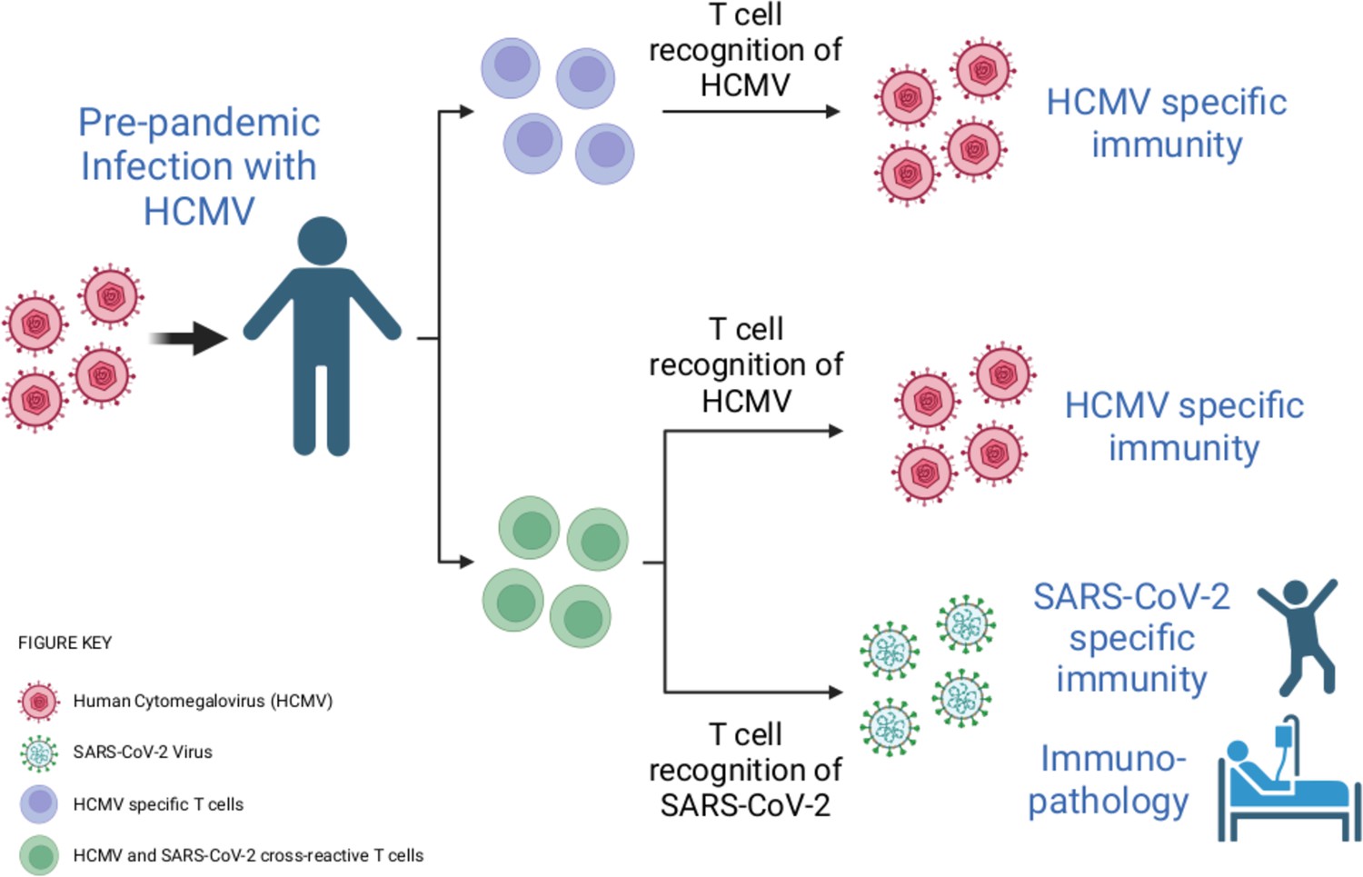
Infection with human cytomegalovirus (HCMV) can stimulate T cells that can recognise SARS-CoV-2.
When individuals are infected with HCMV (virus shown in pink), the population of T cells that can detect this virus expands (T cells shown here in purple). Cross-reactivity is a well-known feature of the immune response. Through this process, HCMV infection can activate T cells (shown here in green) that can recognise both HCMV and another pathogen – including the SARS-CoV-2 virus, even if the HCMV infection happened before the COVID-19 pandemic. These cross-reactive T cells may be able to contribute to the immunity of an individual to SARS-CoV-2, as well as to how COVID-19 affects their body.
Image credit: Created with BioRender.com.
Pothast et al. found that this cross-reactivity was due to a T cell receptor that is expressed in multiple individuals. However, there are very few similarities between the amino acid sequences of the SARS-CoV-2 and the HCMV epitopes, bringing into question how this T cell receptor can detect both viruses. It may be possible to explain the molecular basis for this observation by solving crystal structures of this T cell receptor in complex with either the presented HCMV or SARS-CoV-2 epitopes.
Further experiments then revealed that the cross-reactive T cells limit the replication of SARS-CoV-2 in vitro when the virus is present at low levels. However, the cross-reactive T cells did not appear to have an activated phenotype in patients with severe COVID-19. This might be because individuals included in this study were over 60 years of age, and HCMV-specific T cells do not work as well as people get older (Ouyang et al., 2004).
It has been suggested that heterologous immunity may play a beneficial role in protective immunity (Welsh et al., 2010). This is consistent with a recent study showing that T cells which cross-react with SARS-CoV-2 are associated with abortive infections (when the virus fails to spread to other cells) and asymptomatic cases of COVID-19 (Swadling et al., 2022). These pre-existing T cells may also enhance a person’s response to vaccines (Loyal et al., 2021). However, heterologous immunity is a double-edged sword, as it can also increase the severity of some viral infections. For example, in dengue infections, cross-reactive antibodies and T cells can result in an immune response that is harmful to the body (Welsh et al., 2010; Screaton et al., 2015).
Further studies are needed to establish whether other pathogens (including bacteria) can stimulate T cells capable of recognising epitopes from SARS-CoV-2. In addition, studies with larger cohorts of vaccinated individuals and patients with mild or severe COVID-19 are required to define the role that these cross-reactive T cells play in protective immunity, in response to vaccination, and in disease pathology.
References
-
Cytomegalovirus latent infection is associated with an increased risk of COVID-19-related hospitalizationThe Journal of Infectious Diseases 226:463–473.https://doi.org/10.1093/infdis/jiac020
-
Dysfunctional CMV-specific CD8(+) T cells accumulate in the elderlyExperimental Gerontology 39:607–613.https://doi.org/10.1016/j.exger.2003.11.016
-
New insights into the immunopathology and control of dengue virus infectionNature Reviews Immunology 15:745–759.https://doi.org/10.1038/nri3916
-
Heterologous immunity between virusesImmunological Reviews 235:244–266.https://doi.org/10.1111/j.0105-2896.2010.00897.x
Article and author information
Author details
Publication history
- Version of Record published: January 6, 2023 (version 1)
Copyright
© 2023, Rivino and Wooldridge
This article is distributed under the terms of the Creative Commons Attribution License, which permits unrestricted use and redistribution provided that the original author and source are credited.
Metrics
-
- 806
- views
-
- 69
- downloads
-
- 1
- citations
Views, downloads and citations are aggregated across all versions of this paper published by eLife.
Download links
A two-part list of links to download the article, or parts of the article, in various formats.
Downloads (link to download the article as PDF)
Open citations (links to open the citations from this article in various online reference manager services)
Cite this article (links to download the citations from this article in formats compatible with various reference manager tools)
T Cells: Ready and waiting to go
eLife 12:e85080.
https://doi.org/10.7554/eLife.85080
Further reading
-
- Developmental Biology
- Immunology and Inflammation
Cardiac macrophages are heterogenous in phenotype and functions, which has been associated with differences in their ontogeny. Despite extensive research, our understanding of the precise role of different subsets of macrophages in ischemia/reperfusion (I/R) injury remains incomplete. We here investigated macrophage lineages and ablated tissue macrophages in homeostasis and after I/R injury in a CSF1R-dependent manner. Genomic deletion of a fms-intronic regulatory element (FIRE) in the Csf1r locus resulted in specific absence of resident homeostatic and antigen-presenting macrophages, without affecting the recruitment of monocyte-derived macrophages to the infarcted heart. Specific absence of homeostatic, monocyte-independent macrophages altered the immune cell crosstalk in response to injury and induced proinflammatory neutrophil polarization, resulting in impaired cardiac remodeling without influencing infarct size. In contrast, continuous CSF1R inhibition led to depletion of both resident and recruited macrophage populations. This augmented adverse remodeling after I/R and led to an increased infarct size and deterioration of cardiac function. In summary, resident macrophages orchestrate inflammatory responses improving cardiac remodeling, while recruited macrophages determine infarct size after I/R injury. These findings attribute distinct beneficial effects to different macrophage populations in the context of myocardial infarction.
-
- Immunology and Inflammation
Osteoarthritis (OA) is a degenerative disease with a high prevalence in the elderly population, but our understanding of its mechanisms remains incomplete. Analysis of serum exosomal small RNA sequencing data from clinical patients and gene expression data from OA patient serum and cartilage obtained from the GEO database revealed a common dysregulated miRNA, miR-199b-5p. In vitro cell experiments demonstrated that miR-199b-5p inhibits chondrocyte vitality and promotes extracellular matrix degradation. Conversely, inhibition of miR-199b-5p under inflammatory conditions exhibited protective effects against damage. Local viral injection of miR-199b-5p into mice induced a decrease in pain threshold and OA-like changes. In an OA model, inhibition of miR-199b-5p alleviated the pathological progression of OA. Furthermore, bioinformatics analysis and experimental validation identified Gcnt2 and Fzd6 as potential target genes of MiR-199b-5p. Thus, these results indicated that MiR-199b-5p/Gcnt2 and Fzd6 axis might be a novel therapeutic target for the treatment of OA.


