Cardiac Optogenetics: A move in the light direction
Computer simulations show how low-intensity illumination can be used to terminate cardiac arrhythmias.
- Institute for Experimental Cardiovascular Medicine, University Heart Center Freiburg – Bad Krozingen, Germany
- Faculty of Medicine, University of Freiburg, Germany
For a human heart to pump blood through our circulatory system, billions of muscle cells, called cardiomyocytes, must contract in a well-orchestrated manner. Ordered contraction is achieved via sequential electrical excitation of cells. Cardiomyocytes are excited when a sufficiently strong electric stimulus causes them to depolarize; they then remain in their activated state for some time; and, lastly, they return to their resting state, ready to be activated again. For normal heartbeats, electrical excitation originates from specialized pacemaker cells in the sinus node of the heart, which depolarize automatically. This rhythmically generated electrical signal then propagates along cardiomyocytes throughout the heart. A short time after depolarization, cardiomyocytes contract. Thus, rhythmical electrical activity leads to a regular heartbeat.
Cardiac arrhythmias can be caused by various factors – such as diseased cells and scar tissue – and they are associated with serious clinical conditions, including myocardial infarction, heart failure, and sudden cardiac death. One specific mechanism responsible for cardiac arrhythmia is so-called re-entrant electrical activity: this happens when the electrical impulse that excites cardiomyocytes does not terminate after all cells have been activated. Instead, due to pathological changes in cell or tissue properties, the excitation wave finds a way to circle back and re-activate cells out-of-turn. Under certain conditions, a self-sustaining ‘rotor’ can form, where electrical activity keeps circling in the heart for extended periods of time. While rotating, it emits spiral waves of electrical excitation that override the normal heart rhythm (Pandit and Jalife, 2013; Figure 1A).
Figure 1
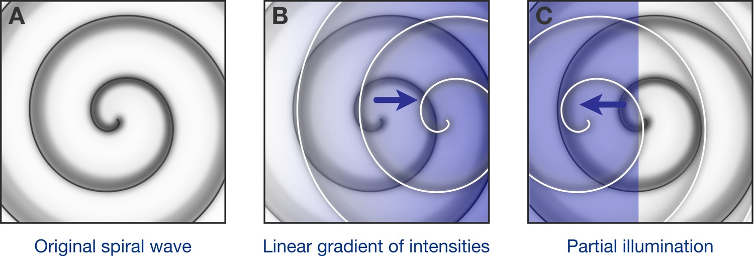
Optogenetic control of spiral waves.
(A) Under certain conditions in the heart, spiral waves of electrical excitation are emitted by a ‘rotor’ of self-sustained electrical excitation. Hussaini et al. have used computer simulations to explore how a rotor in a two-dimensional model of heart tissue responds to various patterns of low-intensity illumination. (B) Upon illumination with a spatial gradient (here, the intensity increases from left to right), the rotor moves in the direction of higher light intensity. (C) Partial illumination of the model tissue with constant sub-threshold intensity light also causes the rotor to move towards the illuminated area.
One elegant way to study mechanisms of cardiac arrhythmias is optogenetics. In cardiac optogenetics, light-sensitive proteins are expressed in heart cells and are used to monitor or steer their electrical properties and function (Schneider-Warme, 2018; Zgierski-Johnston and Schneider-Warme, 2021). For example, blue light can be used to activate an ion-channel protein called channelrhodopsin-2, which results in the depolarization of cells. By activating this protein in cardiomyocytes, researchers have already successfully generated optical pacemakers (Arrenberg et al., 2010; Bruegmann et al., 2010) and conducted optical defibrillation in animal models (Bruegmann et al., 2018; Bruegmann et al., 2016; Crocini et al., 2016; Nyns et al., 2017). Now, in eLife, Stefan Luther and colleagues – including Sayedeh Hussaini as first author – report that low-intensity light may be used to steer rotors (Hussaini et al., 2021). Using a combination of cardiac optogenetics and computational modelling, they describe guiding rotors towards locations where re-entrant electrical activity is no longer possible, thereby terminating cardiac arrhythmias.
Computational modelling has long been used by cardiac researchers to explore how cellular (and sub-cellular) mechanisms act together in healthy or diseased heart tissue (Loewe et al., 2018). Hussaini et al. started with an established mathematical model of cardiomyocyte electrical activity (Petkova-Kirova et al., 2012) and added differential equations that describe light-activated currents mediated by channelrhodopsin-2 (Williams et al., 2013). A reaction-diffusion equation (the so-called monodomain model) was then used to couple multiple model cells in a two-dimensional tissue model.
Utilizing this model, the researchers – who are based at the Max Planck Institute for Dynamics and Self-Organization in Göttingen, the University of Göttingen and other institutions in Germany, France, Italy and Canada – first investigated the effect of illumination with intensities so low that the cardiomyocytes were slightly depolarized from their resting state, but were not excited. They found that increasing the intensity of such ‘sub-threshold’ illumination has two effects: it decreases the velocity at which electrical excitation is relayed from cell to cell, and it decreases the dominant frequency of spiral waves. Both results are in keeping with classically expected single-cell behaviour, but Hussaini et al. confirmed them in real tissue for the first time by performing ex vivo experiments with intact mouse hearts.
Next, Hussaini et al. simulated what could happen when using sub-threshold illumination with linearly graded light intensity. They found that the rotor – which previously had been stable – tended to move towards the part of the tissue where the light intensity was higher (Figure 1B). Moreover, the steeper the gradient, the faster the rotor moved. In another simulation, one region of the tissue was exposed to light of constant, low intensity, while the remaining tissue was not illuminated: here the rotor moved to the illuminated region (Figure 1C). Finally, they predicted that successive partial illuminations of the 2D tissue could be used to steer the rotor to parts of the tissue where the rotor could not continue to self-sustain.
Hussaini and colleagues propose a novel approach for the termination of cardiac arrhythmias using sub-threshold illumination. In fact, the underlying mechanism may have played a role in previous breakthroughs in cardiac optogenetics, such as the demonstration of optical defibrillation in animal models mentioned above. Further work is needed to verify effects of interventions proposed based on computational modelling, as hearts are characterized not only by a complex geometry, but also by tissue heterogeneities that rotors might become anchored to. Looking to the future, the obvious question is whether optogenetic modulation of cardiac electrophysiology, as described here, may ultimately be used to terminate arrhythmias in humans. Answering this question will require further research – it will be necessary, for example, to overcome the challenges associated with the expression of light-gated channels in cardiomyocytes, and with targeted light delivery. The work reported in this paper is undoubtedly an important step in the light direction.
References
-
Optogenetic control of heart muscle in vitro and in vivoNature Methods 7:897–900.https://doi.org/10.1038/nmeth.1512
-
Optogenetic defibrillation terminates ventricular arrhythmia in mouse hearts and human simulationsJournal of Clinical Investigation 126:3894–3904.https://doi.org/10.1172/JCI88950
-
Optogenetic termination of atrial fibrillation in miceCardiovascular Research 114:713–723.https://doi.org/10.1093/cvr/cvx250
-
Cardiac ischemia—insights from computational modelsHerzschrittmachertherapie + Elektrophysiologie 29:48–56.https://doi.org/10.1007/s00399-017-0539-6
-
Rotors and the dynamics of cardiac fibrillationCirculation Research 112:849–862.https://doi.org/10.1161/CIRCRESAHA.111.300158
-
Mathematical modeling mechanisms of arrhythmias in transgenic mouse heart overexpressing TNF-αAmerican Journal of Physiology-Heart and Circulatory Physiology 302:H934–H952.https://doi.org/10.1152/ajpheart.00493.2011
-
The power of optogenetics: potential in cardiac experimental and clinical electrophysiologyHerzschrittmachertherapie + Elektrophysiologie 29:24–29.https://doi.org/10.1007/s00399-017-0545-8
-
Computational optogenetics: empirically-derived voltage- and light-sensitive channelrhodopsin-2 modelPLOS Computational Biology 9:e1003220.https://doi.org/10.1371/journal.pcbi.1003220
-
Observing and manipulating cell-specific cardiac function with lightAdvances in Experimental Medicine and Biology 1293:377–388.https://doi.org/10.1007/978-981-15-8763-4_24
Article and author information
Author details
Franziska Schneider-Warme

Publication history
- Version of Record published: January 27, 2021 (version 1)
Copyright
© 2021, Wülfers and Schneider-Warme
This article is distributed under the terms of the Creative Commons Attribution License, which permits unrestricted use and redistribution provided that the original author and source are credited.
Metrics
-
- 904
- views
-
- 90
- downloads
-
- 1
- citations
Views, downloads and citations are aggregated across all versions of this paper published by eLife.
Download links
A two-part list of links to download the article, or parts of the article, in various formats.
Downloads (link to download the article as PDF)
Open citations (links to open the citations from this article in various online reference manager services)
Cite this article (links to download the citations from this article in formats compatible with various reference manager tools)
Cardiac Optogenetics: A move in the light direction
eLife 10:e65360.
https://doi.org/10.7554/eLife.65360
Further reading
-
- Computational and Systems Biology
- Genetics and Genomics
Spatial transcriptomics (ST) technologies allow the profiling of the transcriptome of cells while keeping their spatial context. Since most commercial untargeted ST technologies do not yet operate at single-cell resolution, computational methods such as deconvolution are often used to infer the cell type composition of each sequenced spot. We benchmarked 11 deconvolution methods using 63 silver standards, 3 gold standards, and 2 case studies on liver and melanoma tissues. We developed a simulation engine called synthspot to generate silver standards from single-cell RNA-sequencing data, while gold standards are generated by pooling single cells from targeted ST data. We evaluated methods based on their performance, stability across different reference datasets, and scalability. We found that cell2location and RCTD are the top-performing methods, but surprisingly, a simple regression model outperforms almost half of the dedicated spatial deconvolution methods. Furthermore, we observe that the performance of all methods significantly decreased in datasets with highly abundant or rare cell types. Our results are reproducible in a Nextflow pipeline, which also allows users to generate synthetic data, run deconvolution methods and optionally benchmark them on their dataset (https://github.com/saeyslab/spotless-benchmark).
-
- Computational and Systems Biology
Transcriptomic profiling became a standard approach to quantify a cell state, which led to accumulation of huge amount of public gene expression datasets. However, both reuse of these datasets or analysis of newly generated ones requires significant technical expertise. Here we present Phantasus - a user-friendly web-application for interactive gene expression analysis which provides a streamlined access to more than 96000 public gene expression datasets, as well as allows analysis of user-uploaded datasets. Phantasus integrates an intuitive and highly interactive JavaScript-based heatmap interface with an ability to run sophisticated R-based analysis methods. Overall Phantasus allows users to go all the way from loading, normalizing and filtering data to doing differential gene expression and downstream analysis. Phantasus can be accessed on-line at https://alserglab.wustl.edu/phantasus or can be installed locally from Bioconductor (https://bioconductor.org/packages/phantasus). Phantasus source code is available at https://github.com/ctlab/phantasus under MIT license.


